Contents lists available atScienceDirect
Free Radical Biology and Medicine
journal homepage:www.elsevier.com/locate/freeradbiomed
Redox regulation of cell proliferation: Bioinformatics and redox proteomics
approaches to identify redox-sensitive cell cycle regulators
Christine H. Foyer
a,⁎, Michael H. Wilson
a, Megan H. Wright
baCentre for Plant Sciences, School of Biology, Faculty of Biological Sciences, University of Leeds, Leeds LS2 9JT, UK bThe Astbury Centre for Structural Molecular Biology, School of Chemistry, University of Leeds, Leeds LS2 9JT, UK
A R T I C L E I N F O
Keywords:
Cell cycle
Posttranslational modifications Reactive oxygen species Retinoblastoma protein Root apical meristem
A B S T R A C T
Plant stem cells are the foundation of plant growth and development. The balance of quiescence and division is highly regulated, while ensuring that proliferating cells are protected from the adverse effects of environment fluctuations that may damage the genome. Redox regulation is important in both the activation of proliferation and arrest of the cell cycle upon perception of environmental stress. Within this context, reactive oxygen species serve as‘pro-life’signals with positive roles in the regulation of the cell cycle and survival. However, very little is known about the metabolic mechanisms and redox-sensitive proteins that influence cell cycle progression. We have identified cysteine residues on known cell cycle regulators in Arabidopsis that are potentially accessible, and could play a role in redox regulation, based on secondary structure and solvent accessibility likelihoods for each protein. We propose that redox regulation may function alongside other known posttranslational mod-ifications to control the functions of core cell cycle regulators such as the retinoblastoma protein. Since our current understanding of how redox regulation is involved in cell cycle control is hindered by a lack of knowledge regarding both which residues are important and how modification of those residues alters protein function, we discuss how critical redox modifications can be mapped at the molecular level.
1. Introduction
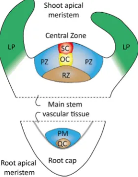
The plant redox signalling network constantly adjusts plant growth and development, as well as metabolism, to prevailing environmental conditions. Within this context reactive oxygen species (ROS) produc-tion controls numerous growth and developmental processes by mod-ifying enzyme activity and protein-protein interactions. The accumu-lation of ROS, either through increased production or regulated decreases in antioxidant capacity, shifts the redox regulatory network to a more oxidising state, in which thiols are oxidised to protein dis-ulfides, sulfenic or sulfinic acid derivatives, as well as glutathionylated and S-nitrosylated forms. ROS are well-known regulators of plant growth and cell fate, as well as stress signalling molecules in plants and animals[1–3]. For example, ROS production is important for tip growth in pollen tubes and root hairs, the respiratory burst oxidase homologue (rboh) NADPH oxidase homologue being required for elongation ac-tivities[1]. However, much less is known about the functions of ROS in plant stem cell niches. Plant growth and development is dependent upon the activity of two distinct populations of stem cells that are housed in separate niches, the shoot apical meristem (SAM) that gives rise to all the above ground plant structures and the root apical
meristem (RAM) that generates the underground root system, as illu-strated inFig. 1 [4,5].
Stem cells within these zones are in a process of continuous renewal. They also generate the precursor cells that subsequently divide and differentiate to form all the specialized cells within the plant. A cen-trally located group of organising stem cells (four in Arabidopsis) within the RAM form the quiescent centre (QC). As in animal stem cell niches, the multipotent stem cells of the plant root QC maintain the surrounding cells in an undifferentiated state[6]. These stem cell ‘in-itials’continuously produce daughter cells by asymmetric division. In roots, for example, asymmetric division of the multipotent stem cells generates the columella cells that form the root cap, which protects the RAM [7–9]. The columella stem cells are maintained in an un-differentiated state by the adjacent QC cells[7].
Upon leaving the meristematic stem cell niche, daughter cells of the multipotent initials undergo subsequent division, elongation, and dif-ferentiation to form the differentiated cells of the root, including the vasculature, epidermis and ground tissues [6]. Within the SAM, a central zone (CZ) of about 35 stem cells is localized above the rib-meristem region that provides signals for stem cell maintenance[7,8]. Progeny of CZ cells move outwards into peripheral zone (PZ) where
https://doi.org/10.1016/j.freeradbiomed.2018.03.047
Received 25 January 2018; Received in revised form 16 March 2018; Accepted 27 March 2018
⁎Corresponding author.
E-mail address:c.foyer@leeds.ac.uk(C.H. Foyer).
Available online 29 March 2018
0891-5849/ Crown Copyright © 2018 Published by Elsevier Inc. This is an open access article under the CC BY license (http://creativecommons.org/licenses/BY/4.0/).
they undergo division and initiate differentiation into leaf orflower primordia, or downwards into the RM region to differentiate into the stem[8,10].
The mitochondria in the SAM cells are organized in a perinuclear network suggesting a requirement for mitochondrial functions to drive the cell cycle[11]. The mitochondrial NTPase called APP1 is required for Complex I activity in Arabidopsis. Loss of APP1 functions led to lower levels of ROS accumulation, but increased proliferation of the QC cells and promoted differentiation of distal stem cells[12]. Similarly, the loss of function of an ATP-dependent mitochondrial protease (FTSH4) increased ROS accumulation in the SAM, leading to meristem termination at mildly elevated temperatures [13]. The cellular redox environment required for the cell-to-cell communication that is essen-tial for SAM maintenance can be controlled by regulation of plasmo-desmatal, for example by regulation of callose deposition via a plasti-dial thioredoxin (TRX)m3[14].
In addition to ROS signalling and regulation of cellular redox state, stem cell identity, cell proliferation and fate are regulated in mamma-lian cells by a tight control of oxygen availability. The stem cell niche in mammals is maintained at low oxygen partial pressures, physiological hypoxia being central to the integrity of the stem cell niche[15]. Si-milarly, it is probable that oxidative metabolism is controlled in the SAM and RAM by physiological hypoxia[16]. Hypoxia plays an im-portant role inthe identity and fate of mammalian stem cells [17]. Several lines of evidence suggest that cells in the QC are also main-tained in a hypoxic state, and that this may help to preserve genome integrity and pluripotency[18,19].
In addition to control of oxygen gradients, oxidants such as hy-drogen peroxide prime stem cell differentiation in animals and in plants [20–26]. Cell type-specific transcript profiling has shown that reactive oxygen species (ROS) and ROS-associated genes are expressed in spe-cific zones of the SAM and RAM[27]. ROS and redox components in-teract with phytohormone signalling to regulate SAM and RAM activ-ities, and different forms of reactive oxygen species (ROS) have been shown to have antagonistic roles in plant stem cell regulation[28]. The UPBEAT1 (UPB1) transcription factor mediates the balance between superoxide and hydrogen peroxide in the root in a way that influences the transition from cell proliferation to cell expansion and diff erentia-tion [29]. Accumulation of the superoxide anion in stem cells was shown to activate WUSCHEL, which is the key regulator of plant stem cell maintenance. In contrast, hydrogen peroxide is more abundant in the differentiating peripheral zone, where it promotes stem cell
differentiation [29]. Moreover, mutation of an ATP-dependent mi-tochondrial protease, AtFTSH4, caused increased oxidation of the SAM at high temperatures leading to altered mitochondrial morphology and SAM functions[13]. The regulation of the spatiotemporal patterns of ROS-metabolizing enzymes appears to be important in the orchestration of the balance between superoxide and hydrogen peroxide [26,29]. Differential and sometimes antagonistic effects of superoxide and hy-drogen peroxide in the RAM and SAM have been reported. For example, superoxide was linked to increased expression of the WUSHEL (WUS) transcription factor, which together with CLAVATA peptides de-termines SAM activity. In contrast, hydrogen peroxide, which accu-mulated mainly in the peripheral zone of cell differentiation, inhibited WUS expression[26]. Suchfindings are interesting and intriguing be-cause neither superoxide nor hydrogen peroxide show strong reactivity with other bio-molecules [30]. However, since superoxide and hy-drogen peroxide have divergent effects of cell death verses survival signalling in animals, superoxide having an inhibitory effect on cell death pathways and apoptotic signalling[31], it may be that super-oxide also promotes cell survival in plants.
Superoxide is a one-electron reduction product of molecular oxygen with no reactivity to most biological molecules. It can, however, in-teract with nitric oxide (NO) and oxidise ascorbic acid,as well as in-activate several enzymes that are important in energy production and amino acid metabolism[30]. Perhaps the most physiology-relevant of these interactions is with the tricarboxylic acid cycle enzyme aconitase. This enzyme is a sensitive target in the mitochondrial matrix. The su-peroxide-dependent inactivation of this enzyme leads to enhanced glycolysis relative to oxidative phosphorylation in cellular energy generation. While mice lacking Cu, Zn superoxide dismutase have little phenotype, they develop neurological problems and cancers in later life [30]. Thisfinding suggests that superoxide accumulation causes pro-blems only in certain tissues and only at defined stages of animal de-velopment [30]. The importance of superoxide accumulation in de-veloping root tips may reside in its effects on intracellular pH rather than its reactivity per se. A decrease in cellular superoxide levels and an increase in hydrogen peroxide results in a shift in the cytosolic pH, making the cellular environment more acid[31].
In plants, extracellular ROS interact with receptor-like kinases (RLKs) in the communication of information perceived in the cellular environment to the interior of the cell. The concurrent initiation of ROS-dependent and ROS-independent signalling linked to RLKs might also be critical in establishing overall cell fate and responses, the RLK-dependent modulation of apoplastic and intracellular conditions being important in regulating ROS perception and signalling[32].
[image:2.595.93.234.59.240.2]ROS have multi-faceted effects on cell fate. In animals, the concept that ROS are‘pro-life’signals serving as critical mediators of cytokine signalling with positive roles in survival is supported by a large body of evidence[31]. For example, superoxide and hydrogen peroxide fulfil important roles in proliferative signalling, as well as in triggering pro-grammed cell death, in animals. A burst of ROS stimulate mitogenic pathways in G1 that control CDK activity and the phosphorylation state of the retinoblastoma protein (pRB), thereby regulating S-phase entry. The enhanced oxidation promotes the expression of nuclear factor er-ythroid-2–related factor 2 (Nrf2). This bZIP transcription factor func-tions as a master regulator of the cellular response to oxidation[33]. Expression ofNrf2re-establishes a reduced intracellular redox state by inducing the expression of cytoprotective genes including those in-volved in the glutathione and thioredoxin systems. The ROS burst at G1 also triggers the expression of FOXO3, a transcription factor that plays essential roles in cell survival signalling with targets in the cell cycle such as the cyclin-dependent kinase inhibitor (CKI) p27 that is involved in cell cycle withdrawal, as well as defence against oxidative stress [34]. ROS also activate transcription factors such as AP-1 and NF-kB and control a number of early growth-related genes such asc-fosand c-jun as well as regulating the activities of protein kinases and phos-phatases[35,36]. ROS also have a direct stimulatory effect on tyrosine Fig. 1.Diagrammatic representation of the structures of the shoot and root
kinase activity mitogen activated protein kinases (MAPK) like JNK, p38MAPK, and ERK[31]. However, many of the mechanisms that allow ROS to support the pro-life’and survival signalling activities of ROS while also facilitating genetically programmed cell suicide pathways remain to be elucidated in plants and animals.
2. Redox regulation of the cell cycle
Cells use proliferative signalling pathways and stress surveillance systems to regulate entry and progress through the cell cycle[37–39]. Oxidative signalling is important in plants and animals for both the activation of proliferation and arrest of the cell cycle upon perception of environmental or metabolic stresses[40,41]. Cell cycle progression in animals is driven by an intrinsic redox cycle consisting of reductive and oxidative phases[42]. In this response, growth stimuli induce the cyclin D–CDK4/6 complex, which phosphorylates RB, releasing E2F tran-scription factors and facilitating the G1/S transition. The binding of growth factors, such as epidermal growth factor (EGF) to their receptors (such as EGFR) is promoted by oxidation resulting from ROS accumu-lation[42–44]. Redox processes have also been shown to be important in the regulation of RNA polymerase (Pol) III in mammals [45]. In vertebrates, the redox-sensing transcription factor TFIIB-related factor 2 (Brf2), which is a TFIIB-like core transcription factor family member, regulates the formation of a transcriptionally-active pre-initiation complex. This finding suggests direct redox-dependent control of a eukaryotic nuclear RNA polymerase regulates the transcription of genes encoding essential RNAs, a factor that might contribute to the ability of cancer cells to evade ROS-induced cell suicide programs[45].
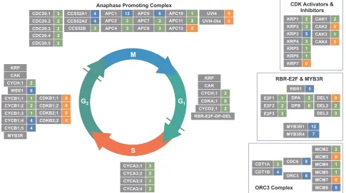
The A-type CDKs (CDKA) in plants regulate the G1/S and G2/M phase transitions and also function during the S phase, while the B7 1 type CDKs (CDKB) act on the G2/M transition and during the M phase [18,19]. The D-type CYCs (CYCD) function together with the CDKA during the G1/S transition. The A3 type CYCs (CYCA) operate during the S phase, and with B-type CYCs (CYCB) regulate the G2/M transition. The CYC B1 and CDK1 fractions of mammalian cells that are localised in the mitochondria phosphorylate Complex I subunits during the G2-to-M transition, enhancing mitochondrial respiration and ATP production to drive cell-cycle progression[46]. The concept of redox regulation of cell proliferation is also found in the plant literature[47–49]. The levels of CYC and CDK transcripts and their activities are changed by oxida-tive perturbations[27,35]. Moreover, the regulation of CYC transcrip-tion by TEOSINTE BRANCHED1-CYCLOIDEA-PROLIFERATING CELL FACTOR1 (TCP) transcription factors is inhibited by oxidation, possibly via effects on a conserved redox-sensitive cysteine residue that is re-quired for DNA binding[50,51].
A recent study using a redox-sensitive in vivoprobe provided the first evidence of a transient oxidation at G1 in the cytosol and nuclei of proliferating cells in the Arabidopsis embryonic root that is perturbed in mutants with low cellular antioxidant levels[52]. Thisfinding supports the concept that "oxidative stress”-sensitive checkpoints are important in the regulation of the cell cycle[38,39]. The complex redox control of the cell cycle is often explained very simply in terms of a given threshold ROS level required to generate cell proliferation or cell cycle arrest [44]. However, the outcomes of cellular oxidative signalling pathways depend on a number of parameters, principally the chemical nature of ROS form produced (i.e. superoxide, hydrogen peroxide or singlet oxygen) and the nature of the interacting partner (protein thiol, metabolite, lipid or DNA molecule), as well as cell identity. Moreover, the different types of oxidative protein modification (reversible and irreversible) also add a high level of sophistication and specificity to the redox signalling matrix that controls cell proliferation.
Many cellular functions are controlled by redox processes. Local changes in the redox environment mediate the spatio-temporal reg-ulation of protein functions and enzyme activities in a compartment-specific manner. At the molecular level, this is thought to be effected primarily via post translational modification (PTM) of cysteine residues
(as discussed below). Redox regulation serves as a crucial PTM and modulator of protein function that is as yet unexplored in relation to the plant cell cycle.
Cell cycle progression is regulated by the activity of cyclin depen-dent protein kinases (CDKs) and their regulatory partners, which are called cyclins (CYCs)[53], all of which are highly conserved in eu-karyotes. The activation of CDKs requires phosphorylation by CDK21 activating kinases (CAKs) and their inactivation involves cyclin de-pendent kinase inhibitors (CKIs), which are called as Kip-Related Pro-teins (KRPs) in plants. While the G1/S and G2/M transitions are the major regulatory check points for cell division, meristematic quies-cence, dormancy and terminal differentiation in plants are generally characterised by cell cycle arrest at G1 arrest[18]. The cohorts of genes operating at the G1/S- G2/ M phases in plants are regulated by the E2F and the MYB3R transcription factors, which are housed in the multi-protein RBR-MYB3R-E2F complexes that are thought to be related to the DREAM complex in animals[54]. Progression through the G1/S and G2/M phase transitions and S phase is regulated by A-type CDKs (CDKA). For example CDKA;1 is the major RETINOBLATOMA RE-LATED (RBR) kinase in plants [55]. D-type 2 CYCs (CYCD) operate together with CDKA to regulate the G1/S transition. A3 type CYCs (CYCA) function at S phase. B-type CYCs (CYCB) and CDKs (CDKB) function to regulate the G2/M transition and M. E3 ubiquitin ligases such as the Anaphase Promoting Complex/Cyclosome (APC/C) and Skp1/Cullin/F-box protein (SCF)- related complex, are also important regulators of the cell cycle progress functions to remove cell cycle regulators by proteolysis. The RB protein also shows E2F-independent functions through binding to other nuclear or extra-nuclear partners. In mammals, for example, RB cooperates with the MYOD or RUNX2 transcription factors to regulate cell differentiation in an E2F-in-dependent manner. Moreover, the direct binding of RB to SKP2 sup-presses the degradation of p27, attenuating cell cycle progression in an E2F-independent manner.
Mitogenic signals promote RBR phosphorylation in plants through the action of CDKs in association with D-type cyclins, particularly CYCLIN D3:1 (CYCD3:1). RBR1 is a signal-dependent scaffold protein and a conserved regulator of cell proliferation, differentiation, and stem cell niche maintenance in Arabidopsis[56]. It is regulated by phos-phorylation-dependent conformational changes that provide a range of interaction surfaces for diverse complexes and functions[57–59]. The hypophosphorylated forms of Rb in animals bind to E2F transcription factors during G1 leading to inhibition of cell cycle dependent, E2F-mediated gene expression[49]. Rb preferentially binds to the activating E2F transcription factors, E2F1, E2F2 and E2F3, with their dimerization partners DP1 and DP2 and represses their activity. While E2Fs are not required to drive cell proliferation [60,61], activating E2Fs promote expression of genes required for DNA synthesis during G1/S. The as-sociation between Rb and E2F transcription factors is weakened by sequential phosphorylation, initially by G1-, and then by G1/S- and finally mitosis-specific CDKs. In this way, Rb regulates cell proliferation by decreasing E2F-dependent transcription of cell cycle genes.
control of the cell cycle, in a manner similar to that observed in ani-mals.
3. Identification of putative redox-regulated cell cycle proteins in plants
Key questions therefore concern what cell cycle proteins are subject to redox regulation and how redox regulation functions alone or to-gether with other post translational modifications such as protein phosphorylation and ubiquitination to control the cell cycle in plants in response to metabolic, developmental and environmental cues. As a
first step to addressing this issue, we downloaded from the Gene Ontology all Arabidopsis gene annotations (http://www.geneontology. org/page/download-annotations) and then extracted the FASTA protein sequence for the representative gene model for each loci identified as cell-cycle related from Ensembl Plants (release-38). We subsequently used solvent accessibility prediction (NetSurfP v1.0[63]) to identify cysteine residues that are potentially accessible based on secondary structure and solvent accessibility likelihoods for each Arabidopsis protein (Supplemental Table, S1).
From the annotated genes, a restricted set of 108 core of cell cycle proteins[64]was created with some additions (Fig. 2). 22 either had no
cysteines or are predicted to have only buried cysteines. Of the re-maining 86, the majority (62) had between 1 and 3 accessible residues and the remaining 24 had between 4 and 12 potentially exposed cy-steines. This list includes RBR1, WEE1, the MYB3R transcription factors and several members of the cyclin A1, B1, B2 and D families that are key cell cycle regulators, but no CDK, CAK and only one CKI/KRP. Several families of cell-cycle components, including the KRPs the, cy-clin D, P and T families, the Anaphase Promoting Complex genes (APC/ C), the MCM genes of the ORC complex and the DP and DEL members of the E2F-DP complex, all contain members with several (up to 12) and no exposed cysteine residues, which may be indicative of a sensitive and insensitive population to modulate redox regulation (Fig. 3).
4. Mapping redox modifications at the molecular level
Our understanding of how redox regulation is involved in cell cycle control is hindered by a lack of knowledge regarding both which re-sidues are important and how modification of those rere-sidues alters protein function. Cysteine redox PTMs are generally formed non-en-zymatically via promiscuous reactive electrophilic species (RES) in-cluding ROS/RNS/RSS and, whilst widespread redox modification of the proteome, high RES and formation of irreversible oxidations are considered hallmarks of damaging oxidative stress, low levels of RES
are crucial for normal cellular function. There is growing appreciation that redox PTMs are site-specific, governed by the microenvironment of cysteine residues[65], and subject to temporal and spatial control, as discussed above.
Cysteine (-SH) is unique amongst amino acid residues in its ability to adopt oxidation states from -2 to + 6in vivo, and thus undergoes a very wide variety of redox-related PTMs[66]. Reversible PTMs include formation of disulfides via reaction with low molecular weight thiols such as glutathione (S-glutathionylation, -SSG), with other proteins (-SSR), or to formS-sulfhydrate (-SSH, also known as persulfide); re-action with ROS to generateS-sulfenic acid (-SOH), and reaction with RNS inS-nitrosylation (-SNO) (Fig. 4). Sulfenic acids (-SOH) can react further to give irreversibly oxidised species,S-sulfinic acid (-SO2H) and S-sulfonic acid (-SO3H). Historically this complex array of modifications was difficult to address in a global proteome context and with sufficient sensitivity to detect endogenous oxidative signalling PTMs. However, recent advances in redox proteomic technologies are delivering sig-nificant new insights into the scope and biology of redox regulation.
5. Tools and platforms for redox proteomics
[image:5.595.56.542.59.330.2]Mass spectrometry (MS)-based proteomics is unrivalled in its ability to identify and characterise large portions of the proteome at the level Fig. 3.The generalised plant cell cycle, showing the core cell cycle genes and complexes as described in the text, within the context of the cell cycle. Grey boxes, cell cycle gene names, coloured boxes number of potentially exposed cysteines: orange, 0 exposed; green, 1–3 exposed; blue, 4–12 exposed.
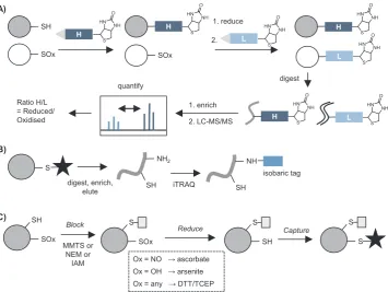
[image:5.595.138.458.628.727.2]of individual residues and PTMs. Shotgun proteomics requires sample processing steps including protein denaturation and digest to release peptides for separation and MS detection. Direct MS detection of cy-steine redox modifications is therefore not usually possible due to their low abundance and frequent instability. Most redox proteomics stra-tegies therefore exploit the intrinsic chemical reactivity of thiols or cysteine PTMs to introduce labels enabling enrichment (via biotinyla-tion or thiol-reactive resin). Quantificabiotinyla-tion, including of PTM site oc-cupancy, is becoming increasingly important and most redox proteomic approaches lend themselves well to isotope-based comparative quan-tification, for example exploiting labelling steps to introduce isotopic moieties. Methods can be broadly divided into two categories: those that exploit the intrinsic chemistry of redox PTMs to introduce a label directly at the modification site, and indirect methods that label free thiols then apply selective reduction to release specific cysteine PTM sites for differential labelling (Fig. 5A&B).
Here we review the most common and promising methods for profiling different redox PTMs and their applications in plant biology (summarised inSupplemental Table, S2). For more in depth discussion of how redox PTMs form, the methods used to detect them and con-siderations of sample preparation, the reader is referred to several ex-cellent recent reviews[67–69], including Akter et al.’s recent survey of ROS reactivity in plants [70]. The technical details of quantitative proteomic methods, including isobaric tagging (e.g. iTRAQ, TMT), have also been reviewed elsewhere[71].
6. Reagents for labelling free thiols
The high nucleophilicity of the free thiol or thiolate relative to other amino acid residues renders it susceptible to selective alkylation with electrophiles, the most commonly used of which are based on iodoa-cetamide (IAM) and maleimide (e.g. N-ethyl maleimide, NEM) (Fig. 5E). MMTS is another common thiol labelling reagent, which re-acts via disulfide exchange. Biotinylated versions of IAM and NEM enable direct labelling and enrichment of cysteine-containing proteins for MS analysis, and reagents that incorporate isotope tags have also been developed (see below). These reagents can be used to compara-tively label thiols across different conditions (e.g. in the presence and absence of oxidative stress;Fig. 5D). Provided changes in global protein expression are accounted for, this enables indirect determination of sites that are differentially oxidised i.e. decreased detection of a site indicates that it is being blocked by a redox PTM.
[image:6.595.117.481.54.421.2]and quantification. The recently reported isotope-incorporating IAA variant [74]should also prove useful in comparing across samples. Another limitation of biotin reagents is that they are not typically cell permeable, so the snapshot of thiols obtained is subject to perturbations that occur upon cell lysis. IAM-based probes are also toxic to cells. To circumvent the toxicity of IAA, versions have been developed which can be uncaged by UV light following cellular uptake[75]and could fa-cilitatein vivodetection of cysteine residues.
7. Detecting reversible oxidation: biotin-switch and related approaches
Labelling thiols and detecting a reduction in labelling due to oxi-dation is rather indirect. Differential alkylation, or tag-switch, ap-proaches have been widely applied to label sites of reversible cysteine oxidation (-SOH, -SNO, -SSH/R/G). In the tag-switch approach, alky-lation (or blocking) of free thiols is followed by reduction to release reversibly oxidised thiols and then a second labelling step is performed to introduce a means of detecting or enriching modified sites. In the biotin-switch method (BST) [76], biotin is introduced in the second labelling step, commonly via pyridylthiol-biotin (biotin-HPDP), a re-agent that forms a reversible disulfide with free thiols and thus enables release of the captured proteins or peptides following enrichment (Fig. 5E). Alternatively, thiols exposed by reduction can be captured on a resin (resin-assisted capture, RAC)[77], labelled with an alternative affinity reagent [78,79], or alkylated using a different label to that employed in thefirst blocking step but not enriched[80,81].
Although biotin is the most common affinity tag due to its extremely high binding affinity with streptavidin, enrichment introduces at least one additional step, resulting in quite lengthy protocols that reduce proteome coverage, and non-specific binding to the resin complicates analysis. To address these challenges, resin-based methods to capture thiols have been reported. Commercially available resin, thiopropyl Sepharose 6B, has been applied to directly capture free thiols via dis-ulfide exchange, followed by on-resin protein digestion and multiplexed
isobaric labelling [77] (via iTRAQ - an isotope labelling technique where amines on peptides are labelled with isobaric reagents that can later be distinguished via MS[71]).
A popular quantitative method using a tag-switch strategy is OxiCAT, which can be applied to determine PTM site occupancy in a single sample[82]. In OxiCAT both reduced and oxidised cysteines are sequentially captured using isotopically labelled, biotinylated thiol-re-active IAM-based reagents (Fig. 6A). Following digest, biotinylated cysteine-containing peptides are isolated and analysed by LC-MS/MS. Heavy and light cysteine-tagged peptides are distinguishable during MS, generating ratios of oxidised:reduced cysteines. OxiCAT recently revealed > 3800 H2O2-responsive residues in diatoms[83], including 4 with predicted roles in cell cycle control/division and 12 involved in ROS metabolism or redox signalling.
There are several other reported isotopic/isobaric labelling methods for introducing quantification into tag-switch workflows. Like OxiCAT, some introduce isotopic labels at the modification site: these include cysTMT[79]and iodoTMT[84,85], based on reversible disulfide and irreversible alkylation chemistry respectively. TMT-resin is available for affinity enrichment of labelled peptides. Other methods introduce labels elsewhere on the peptides (usually on free amines). The most widely used of these is OxiTRAQ, where isobaric iTRAQ reagents are applied to peptides after biotin or resin-based enrichment (Fig. 6B). OxiTRAQ has been successfully applied inArabidopsissuspension cells treated with various oxidants or other exogenous agents[86,87].
[image:7.595.121.477.54.321.2]This highlights the main limitation of tag-switch methods: their reliance on the selectivity of alkylating and reducing reagents, which is not complete. In a further example, a recent study investigating cross-re-activity between nitrosothiols and sulfinic acids found that MMTS may release methyl sulfinic acid that could react withS-nitrosylation sites and result in slower reduction to the free thiol, such that subsequent detection of -SNO is diminished [89]. Incomplete blocking is also a frequent challenge in tag-switch approaches. Similarly, in most proto-cols other cysteine PTMs such asS-acylation will also be reduced (and then the site alkylated), which can complicate interpretation of results. Nevertheless, these strategies have revealed thousands of potentially redox-sensitive cysteines across diverse proteomes.
8. Direct methods for profilingS-sulfenylation
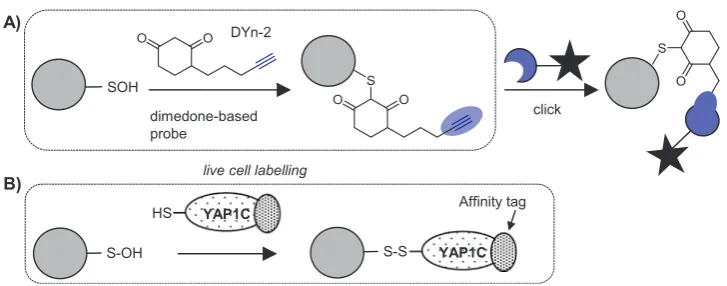
S-Sulfenylation (-SOH) is a transient PTM with signalling roles, for example in the regulation of protein tyrosine kinases and phosphatases [90,91], but is not stable under typical MS sample processing condi-tions[92]. -SOH also cross-reacts with the common thiol blocking re-agents, making it difficult to detect via indirect methods (such as the biotin-switch methods described above), although a novel blocking reagent that only reacts with free thiols was recently reported and may prove useful in circumventing this problem[93]. Several reagents for directly labelling -SOH have been developed, mostly inspired by the selective reaction of this PTM with β-dicarbonyls such as dimedone. Antibodies against the dimedone-sulfenic acid adduct are available and have been applied forS-sulfenylation profiling in plants[94], but most approaches use biotinylation[95–97]. Biotinylated reagents [95]are typically applied in cell lysates but cell permeable dimedone-based chemical probes incorporating clickable tags have recently been de-veloped to enable direct labelling of sulfenylated proteins in live cells [90,98,99](Fig. 7A). The small clickable tags allow labelled proteins to be subsequently ligated to biotin after cell lysis. Akter et al. used this method to demonstrateS-sulfenylation of 226 proteins in Arabidopsis cell suspensions treated with H2O2, over half of which had not been previously reported to be modified in plants[100]. Other recent studies have combined clickable dimedone probes with cleavable biotin re-agents to detect over 1000 endogenous sulfenylation sites in human cells [99], suggesting that with optimisation of protocols this tech-nology can achieve deep coverage of the endogenousS-sulfenome.
A potential limitation of dimedone-based capture is the slow speed of the reaction in comparison to other reactions of sulfenic acids, such as disulfide formation or further oxidation[92,101,102]. The Carroll group recently reported several new probes with accelerated reaction rates [101,103] and demonstrated that structurally different probes label surprisingly distinct sets of endogenous sulfenylated proteins in mammalian cells[103]. Whilst this data suggests that a general probe capable of targeting all sulfenic acids might be challenging to develop, it also means that different subsets of the S-sulfenylome, down to
specific proteins or protein families, might be targetable with different chemical probes[103].
Bicyclo[6.1.0]nonyne has been reported as alternative chemical probe to trap sulfenic acids, with reaction rates around 100-fold higher than dimedone[102]. Although the known cross-reactivity of thiols with such reagents[104]may present a problem for selective sulfenic acid proteome profiling, subsequent data suggests that this side reaction is not in fact occurring. Thus strained alkynes represent another po-tential tool forS-sulfenylation detection[130].
A completely complementary approach forS-sulfenic acid detection uses a cell-based genetic sensor for this PTM, based on the cysteine-rich domain of the yeast transcription factor YAP1, which forms disulfides with S-sulfenic acid modifications on its cognate signalling protein. Fusion of the cysteine-rich domain of YAP1 with an affinity tag creates a tool to capture and enrichS-sulfenylated proteins in vivo(Fig. 7B) [100,105–107]. This approach has identified∼100 sulfenylated pro-teins inArabidopsis thalianacells[105].
9. Direct probes forS-nitrosylation (-SNO)
S-Nitrosylation is another labile PTM thought to be mediated pri-marily by RNS, such as•NO formed by nitric oxide synthetases, and removed via transnitrosylation with small molecular cellular thiols [69,108]. Whilst the biotin-switch (ascorbate based) method described above[76]is by far the most commonly used, several approaches to directly label or enrich -SNO sites based on their chemistry have also been reported.
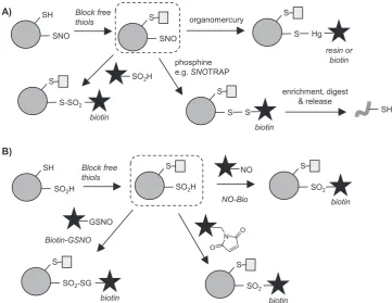
Phenylmercury compounds react with -SNO to generate an S-Hg bond, inspiring the development of mercury-functionalised resins and biotin reagents that can be applied to capture -SNO modifications after blocking of free thiols (Fig. 8A)[109]. Enriched proteins or peptides can then be released via treatment with performic acid, generating a sulfonic acid (-SO3H) at the site of modification, which is stable under further sample processing and MS analysis conditions[109]. Another approach used gold nanoparticles to enrich-SNO sites[110], although the selectivity for enrichment of -SNO over other thiol modifications such as disulfides is unclear, which may hinder the utility of this method in complex samples. Phosphine-based reagents (first reported in[111]) can also react with -SNO sites and have enabled -SNO imaging and identification of nitrosylation on select proteins (reviewed in[69]). This chemistry was developed into a global proteomic profiling ap-proach termedSNOTRAP (Fig. 8A): free thiols were blocked, then a biotin-incorporating triphenylphosphine thioester used to label -SNO sites, generating a disulfide linkage that could be cleaved following enrichment to release peptides [112]. Phosphine-based probes have known issues in biological samples, such as reaction with disulfides [69], and may require further development to be broadly applicable.
[image:8.595.116.480.54.197.2]Following blocking of free thiols, biotin-SO2H was used to label -SNO, generating a thiosulfonate (Fig. 8A) for enrichment and detection. This method successfully identified nearly 1000 candidate endogenous S -nitrosylated proteins in mammalian cells, pinpointing the site of mod-ification for around 100 of these. This study also analysed the relative occupancy of sites.
10. Direct probes forS-sulfinylation (-SO2H) andS-sulfonylation (-SO3H)
The more highly oxidised and (generally) irreversible redox PTMs of S-sulfinylation (-SO2H) andS-sulfonylation (-SO3H) have historically been considered mere markers of oxidative damage, rather than specific controlled signalling PTMs. However, increasing evidence suggests that this may be an over-simplification; for example, transient sulfinylation of peroxiredoxins was shown to be a conserved marker for circadian rhythms across all domains of life[113]. These PTMs cannot be readily analysed by tag-switch approaches because selective reagents for their reduction are not available, and identification of S-sulfonylation (-SO3H) is thus far limited to direct detection via MS[114] or prior enrichment on polyarginine resin (which also enriches sulfinylated peptides) [115]. However, several direct S-sulfinic acid approaches have been reported recently.
Carroll et al. developed novel aryl-nitroso probe NO-Bio to directly capture S-sulfinic acids in lysates, following blocking of free thiols (necessary to avoid cross-reactivity) (Fig. 8B)[116]. This represents the first direct labelling method for detecting this redox PTM and should be useful in global MS proteomics studies. Martin et al. have also em-ployed nitroso compounds to detect S-sulfinylation, in the reverse of their approach for detectingS-nitrosylation via sulfinylation (discussed above) [89]. They applied their Biotin-GSNO reagent (Fig. 8B) to identify endogenous sulfinic acid sites across the human proteome. One limitation of Biotin-GSNO is the relative instability of the reagent, which oxidises over time in aqueous solution [89]. Martin et al. therefore developed an alternative approach, in which, after alkylation of free thiols with IAM, maleimide (NEM-based) reagents react withS
-sulfinated cysteines to form a sulfone adduct that is stable under acidic conditions[117](Fig. 8B).
Combined with quantitative MS, these recently developed direct labelling approaches should promote investigation of these under-studied PTMs in different biological contexts.
11. Detection of sulfhydration/persulfides (-SSH)
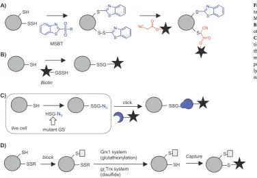
Identifying sites of sulfhydration (-SSH; also known as persulfides) is challenging due to the similar chemical properties and reactivity of this PTM compared to free thiols. Modified tag-switch approaches have had some success, but site identification is difficult and these methods lack specificity[69]. One approach designed to address some of these issues utilises MSBT to label both thiols and persulfides, followed by selective reduction of the disulfide formed from the latter to generate a free thiol for tagging with, for example, biotin[118]. One limitation of this ap-proach is an inability to discriminate between the newly formed dis-ulfide and other disdis-ulfide modifications. To address this and directly label sites ofS-sulfhydration, Zhang et al. developed chemistry to se-lectively displace the disulfide that is formed upon MSBT reaction with a CN-biotin reagent, enabling specific enrichment of these sites. (Fig. 9A)[119]. Another approach reacts both thiols and persulfides with IAM-biotin, enriches both sites and then selectively releases the disulfide at the site of persulfide modification from the resin via re-duction[120].
12. Detection of glutathionylation (-SSG) and disulfides
[image:9.595.116.478.55.334.2]address this, Ahn et al. developed a method where cells are transfected with a mutant glutathione synthetase able to generate in situa GSH analogue incorporating an azide bio-orthogonal tag, which is then metabolically incorporated into cellular proteins (Fig. 9C)[123]. The bio-orthogonal tag is then ligated to biotin using click chemistry. This approach was applied to profile native glutathionylation in cells in re-sponse to glucose starvation[124]and shows promise in circumventing the selectivity challenges faced by tag-switch approaches.
The final class of methods that have been developed to address disulfide-type redox PTMs exploit the activity of specific enzyme to classes, including thioredoxin (Trx), which reduces protein disulfides, and glutaredoxin (Grx), which reduces -SSG modifications. Qian, Thrall et al. used an enzymatic version of the tag-switch approach to detect glutathionylation: free thiols were first blocked and then a cocktail containing active Grx1 applied to selectively reduce glutathionylated proteins for subsequent capture on resin and identification (Fig. 9D) [77,125]. Quantification was introduced via on resin isobaric tagging with either iTRAQ or TMT reagents.
A similar approach has been applied to identify those disulfi de-containing proteins that are substrates for the enzyme Trx. Free cy-steines are blocked, and then addition of the Trx enzymatic system results in conversion of disulfide-linked proteins to free thiols, which are subsequently biotinylated (Fig. 9D)[126]. An alternative method reported in the same study is to immobilise a mutant Trx on resin and use this to covalently capture Trx substrates upon flow-through. The mutant enzyme lacks one of its two active site cysteines and thus sub-strate-enzyme intermediates remain trapped until eluted by DTT or another reductant. The “thioredoxome”of the unicellular green alga Chlamydomonas reinhardtiiwas recently extensively characterised using both Trx systems[127].
13. Conclusions and perspectives
Recent years have seen rapid evolution of methods to characterise the redox proteome. The key approaches are summarised, along with their advantages and limitations, in Supplemental Table S3. The se-lectivity of the reagents and enzymes is key to successfully diff er-entiating PTMs across the complex redox landscape, and the chemistry for analysing these continues to develop. This growing intensity of
research efforts is promising, as approaches that are sufficiently sensi-tive to map and (importantly) quantifyendogenous redox PTMs and changesat the residue level are absolutely crucial for analysing subtle changes such as those occurring at different cell cycle stages. Thus the best methods incorporate both quantification and a means of identi-fying the precise sites of PTM into their workflow. Integrating chemical and enzymatic tools with state-of-the-art mass spectrometry-based in-strumentation and optimised workflows also has a large impact. The potential increase in coverage is well-illustrated by the example of OxiCAT: the group who pioneered the method[82]reported quantifi -cation of redox status for a few hundred cysteines in the yeast pro-teome, which was a landmark study at that time[128]; a recent report in yeast identified ~ 4000 sites [129]. Interesting this latter study reached sufficient depth of coverage to quantify the percentage oxida-tion of cysteines on several cell cycle regulators. OxiCAT is probably currently the best method for obtaining an overview of total reversible oxidation levels across the proteome, as it incorporates quantification and site identification, uses commercially available reagents, and is readily applied across different sample types. However, this method must still be applied in lysates and thus is necessarily subject to artifi -cial changes in redox status that occur upon cell lysis. Alternative ap-proaches - such as methods to label thiols inside live cells - should prove useful in validating data fromin vitromethods, although few reported studies have directly compared multiple techniques.
In addition to technical developments that advance our ability to interrogate the proteome with existing tools, exciting approaches are emerging to address previously understudied redox PTMs. Examples discussed above include the dimedone-based and more recently devel-oped nucleophilic probes that are able to profile transient S-sulfenic acid modifications in live cells, and approaches to tackle PTMs that are difficult to distinguish from other cysteine redox states (e.g.S -sulfhy-dration). We anticipate that method development will also continue to underpin advances in redox biology.
[image:10.595.37.414.52.312.2]the redox regulation of cell cycle control a realistic possibility, at least at the outset in plant cell cultures, where the cell cycle can be syn-chronised and cell cycle progression can easily be determined, with appropriate amounts of materials harvested for analysis. It will be in-teresting to determine whether redox processes influence the properties and functions of key cell cycle regulators such as RB and E2Fs, which control DNA replication and mitosis as well as the G1/S transition during the cell cycle. The RB family proteins regulate the G1/S transi-tion in animals. Inactivatransi-tion of RB cause oxidative stress by increasing ROS production in mitochondria, where RB is localized. The RB–E2F complex directly suppresses the expression of oxidative metabolism-related enzymes and mitochondrial protein translation genes. It will be intriguing to see if similar mechanisms of redox regulation of cell proliferation are present in plant cells, as well as other redox networks that link cell cycle regulation to metabolism and environmentally sensing. A better understanding of these processes will not only identify the similarities in the redox footprints between plants and animals but also clarify the mechanisms whereby plant stem cells respond to the cellular redox environment. Understanding the functional regulation of redox reactive cysteine residues in cell cycle proteins, and their inter-actions with mitochondrial and plastid signalling networks, will lead to a step change in our appreciation of how cell metabolism uses the redox reactions that harness energy for life to control cell proliferation and survival in plants and animals.
Acknowledgements
We are indebted to Dr Michael J. Considine of the University of Western Australia for the provision ofFig. 1. CF thanks Biotechnology and Biological Sciences Research Council for financial support (BB/ M009130/1), M.H.Wright thanks the University of Leeds for a Uni-versity Academic Fellowship. Part of this work was undertaken on MARC1, part of the High Performance computing facilities at the Uni-versity of Leeds, UK.
Appendix A. Supporting information
Supplementary data associated with this article can be found in the online version athttp://dx.doi.org/10.1016/j.freeradbiomed.2018.03. 047.
References
[1] S. Swanson, S. Gilroy, ROS in plant development, Physiol. Plant 138 (2010) 384–392.
[2] C.H. Foyer, G. Noctor, Ascorbate and glutathione: the heart of the redox hub, Plant Physiol. 155 (2011) 2–18.
[3] M.J. Considine, C.H. Foyer, Redox regulation of plant development, Antioxid. Redox Signal. 21 (2014) 1305–1326.
[4] M.B. Singh, P.L. Bhalla, Plant stem cells carve their own niche, Trends Plant Sci. 11 (2006) 241–246.
[5] B. Scheres, Stem-cell niches: nursery rhymes across kingdoms, Nat. Rev. Mol. Cell Biol. 8 (2007) 345–354.
[6] C. van den Berg, V. Willemsen, G. Hendriks, P. Weisbeek, B. Scheres, Short-range control of cell differentiation in the Arabidopsis root meristem, Nature 390 (1997) 287–289.
[7] G.V. Reddy, E.M. Meyerowitz, Stem-cell homeostasis and growth dynamics can be uncoupled in the Arabidopsis shoot apex, Science 310 (2005) 663–667. [8] B. Peret, B. De Rybel, I. Casimiro, E. Benkova, R. Swarup, L. Laplaze, T. Beeckman,
M.J. Bennett, Arabidopsis lateral root development: an emerging story, Trends Plant Sci. 14 (2009) 399–408.
[9] K.F. Mayer, H. Schoof, A. Haecker, M. Lenhard, G. Jurgens, T. Laux, Role of WUSCHEL in regulating stem cell fate in the Arabidopsis shoot meristem, Cell 95 (1998) 805–815.
[10] J.R. Dinneny, P.N. Benfey, Plant stem cell niches: standing the test of time, Cell 132 (2008) 553–557.
[11] J.M. Segui-Simarro, M.J. Coronado, L.A. Staehelin, The mitochondrial cycle of Arabidopsis shoot apical meristem and leaf primordium meristematic cells is
de-fined by a perinuclear tentaculate/cage-like mitochondrion, Plant Physiol. 148 (2008) 1380–1393.
[12] Q. Yu, H. Tian, K. Yue, J. Liu, B. Zhang, X. Li, Z. Ding, A P-Loop NTPase regulates quiescent center cell division and distal stem cell identity through the regulation
of ROS homeostasis in arabidopsis root, PLOS Genet. 12 (2016) e1006175. [13] A. Dolzblasz, E. Smakowska, E.M. Gola, K. Sokolowska, M. Kicia, H. Janska, The
mitochondrial protease AtFTSH4 safeguards Arabidopsis shoot apical meristem function, Sci. Rep. 6 (2016) 28315.
[14] Y. Benitez-Alfonso, M. Cilia, A. San Roman, C. Thomas, A. Maule, S. Hearn, D. Jackson, Control of Arabidopsis meristem development by thioredoxin-depen-dent regulation of intercellular transport, Proc. Natl. Acad. Sci. USA 106 (2009) 3615–3620.
[15] A. Mohyeldin, T. Garzon-Muvdi, A. Quinones-Hinojosa, Oxygen in stem cell biology: a critical component of the stem cell niche, Cell Stem Cell 7 (2010) 150–161.
[16] M.J. Considine, P. Diaz-Vivancos, P. Kerchev, S. Signorelli, P. Agudelo-Romero, D.J. Gibbs, C.H. Foyer, Learning to breathe: developmental phase transitions in oxygen status, Trends Plant Sci. 22 (2017) 140–153.
[17] K.A. Webster, Hypoxia: life on the edge, Antioxid. Redox Signal. 9 (2007) 1303–1307.
[18] Y. Velappan, S. Signorelli, M.J. Considine, Cell cycle arrest in plants: what dis-tinguishes quiescence, dormancy and differentiated G1? Ann. Bot. 120 (2017) 495–509.
[19] K. Jiang, T. Zhu, Z. Diao, H. Huang, L.J. Feldman, The maize root stem cell niche: a partnership between two sister cell populations, Planta 231 (2010) 411–424. [20] R.K. Yadav, T. Girke, S. Pasala, M. Xie, G.V. Reddy, Gene expression map of the
Arabidopsis shoot apical meristem stem cell niche, Proc. Natl. Acad. Sci. USA 106 (2009) 4941–4946.
[21] E. Owusu-Ansah, U. Banerjee, Reactive oxygen species prime Drosophila haema-topoietic progenitors for differentiation, Nature 461 (2009) 537–541. [22] Y. Kanda, T. Hinata, S.W. Kang, Y. Watanabe, Reactive oxygen species mediate
adipocyte differentiation in mesenchymal stem cells, Life Sci. 89 (2011) 250–258. [23] R.B. Hamanaka, A. Glasauer, P. Hoover, S. Yang, H. Blatt, A.R. Mullen, S. Getsios,
C.J. Gottardi, R.J. DeBerardinis, R.M. Lavker, N.S. Chandel, Mitochondrial re-active oxygen species promote epidermal differentiation and hair follicle devel-opment, Sci. Signal. 6 (2013) ra8.
[24] A. Ludin, S. Gur-Cohen, K. Golan, K.B. Kaufmann, T. Itkin, C. Medaglia, X.J. Lu, G. Ledergor, O. Kollet, T. Lapidot, Reactive oxygen species regulate hematopoietic stem cell self-renewal, migration and development, as well as their bone marrow microenvironment, Antioxid. Redox Signal. 21 (2014) 1605–1619.
[25] C.R. Mantel, H.A. O'Leary, B.R. Chitteti, X. Huang, S. Cooper, G. Hangoc, N. Brustovetsky, E.F. Srour, M.R. Lee, S. Messina-Graham, D.M. Haas, N. Falah, R. Kapur, L.M. Pelus, N. Bardeesy, J. Fitamant, M. Ivan, K.S. Kim, H.E. Broxmeyer, Enhancing hematopoietic stem cell transplantation efficacy by mitigating oxygen shock, Cell 161 (2015) 1553–1565.
[26] J. Zeng, Z. Dong, H. Wu, Z. Tian, Z. Zhao, Redox regulation of plant stem cell fate, EMBO J. 36 (2017) 2844–2855.
[27] V.B. Tognetti, A. Bielach, M. Hrtyan, Redox regulation at the site of primary growth: auxin, cytokinin and ROS crosstalk, Plant Cell Environ. 40 (2017) 2586–2605.
[28] J.H. Schippers, C.H. Foyer, J.T. van Dongen, Redox regulation in shoot growth, SAM maintenance andflowering, Curr. Opin. Plant Biol. 29 (2016) 121–128. [29] H. Tsukagoshi, W. Busch, P.N. Benfey, Transcriptional regulation of ROS controls
transition from proliferation to differentiation in the root, Cell 143 (2010) 606–616.
[30] B. Halliwell, Reactive species and antioxidants. Redox biology is a fundamental theme of aerobic life, Plant Physiol. 141 (2006) 312–322.
[31] S. Pervaiz, M.V. Clement, Superoxide anion: oncogenic reactive oxygen species? Int. J. Biochem. Cell Biol. 39 (2007) 1297–1304.
[32] S. Kimura, C. Waszczak, K. Hunter, M. Wrzaczek, Bound by fate: the role of re-active oxygen species in receptor-like kinase signaling, Plant Cell 29 (2017) 638–654.
[33] T.W. Kensler, N. Wakabayashi, S. Biswal, Cell survival responses to environmental stresses via the Keap1-Nrf2-ARE pathway, Annu Rev. Pharmacol. Toxicol. 47 (2007) 89–116.
[34] A.R. Gomes, F. Zhao, E.W. Lam, Role and regulation of the forkhead transcription factors FOXO3a and FOXM1 in carcinogenesis and drug resistance, Chin. J. Cancer 32 (2013) 365–370.
[35] W.C. Burhans, N.H. Heintz, The cell cycle is a redox cycle: linking phase-specific targets to cell fate, Free Radic. Biol. Med. 47 (2009) 1282–1293.
[36] S.G. Menon, P.C. Goswami, A redox cycle within the cell cycle: ring in the old with the new, Oncogene 26 (2007) 1101–1109.
[37] G. Potters, T.P. Pasternak, Y. Guisez, M.A. Jansen, Different stresses, similar morphogenic responses: integrating a plethora of pathways, Plant Cell Environ. 32 (2009) 158–169.
[38] K. De Schutter, J. Joubes, T. Cools, A. Verkest, F. Corellou, E. Babiychuk, E. Van Der Schueren, T. Beeckman, S. Kushnir, D. Inze, L. De Veylder, Arabidopsis WEE1 kinase controls cell cycle arrest in response to activation of the DNA integrity checkpoint, Plant Cell 19 (2007) 211–225.
[39] J.P. Reichheld, T. Vernoux, F. Lardon, M. Van Montagu, D. Inze, Specific check-points regulate plant cell cycle progression in response to oxidative stress, Plant J. 17 (1999) 647–656.
[40] D. Zhou, L. Shao, D.R. Spitz, Reactive oxygen species in normal and tumor stem cells, Adv. Cancer Res. 122 (2014) 1–67.
[41] S. Varum, O. Momcilovic, C. Castro, A. Ben-Yehudah, J. Ramalho-Santos, C.S. Navara, Enhancement of human embryonic stem cell pluripotency through inhibition of the mitochondrial respiratory chain, Stem Cell Res. 3 (2009) 142–156.
[43] M. Schieber, N.S. Chandel, ROS function in redox signaling and oxidative stress, Curr. Biol. 24 (2014) R453–R462.
[44] S. Mangano, S.P. Juarez, J.M. Estevez, ROS regulation of polar growth in plant cells, Plant Physiol. 171 (2016) 1593–1605.
[45] J. Gouge, K. Satia, N. Guthertz, M. Widya, A.J. Thompson, P. Cousin, O. Dergai, N. Hernandez, A. Vannini, Redox signaling by the RNA polymerase III TFIIB-re-lated factor Brf2, Cell 163 (2015) 1375–1387.
[46] Z. Wang, M. Fan, D. Candas, T.Q. Zhang, L. Qin, A. Eldridge, S. Wachsmann-Hogiu, K.M. Ahmed, B.A. Chromy, D. Nantajit, N. Duru, F. He, M. Chen, T. Finkel, L.S. Weinstein, J.J. Li, Cyclin B1/Cdk1 coordinates mitochondrial respiration for cell-cycle G2/M progression, Dev. Cell 29 (2014) 217–232.
[47] P. Diaz Vivancos, T. Wolff, J. Markovic, F.V. Pallardo, C.H. Foyer, A nuclear glutathione cycle within the cell cycle, Biochem. J. 431 (2010) 169–178. [48] P. Diaz-Vivancos, A. de Simone, G. Kiddle, C.H. Foyer, Glutathione–linking cell
proliferation to oxidative stress, Free Radic. Biol. Med. 89 (2015) 1154–1164. [49] A. Feher, K. Otvos, T.P. Pasternak, A.P. Szandtner, The involvement of reactive
oxygen species (ROS) in the cell cycle activation (G(0)-to-G(1) transition) of plant cells, Plant Signal. Behav. 3 (2008) 823–826.
[50] Y. Kadota, T. Furuichi, T. Sano, H. Kaya, W. Gunji, Y. Murakami, S. Muto, S. Hasezawa, K. Kuchitsu, Cell-cycle-dependent regulation of oxidative stress re-sponses and Ca2+ permeable channels NtTPC1A/B in tobacco BY-2 cells, Biochem. Biophys. Res. Commun. 336 (2005) 1259–1267.
[51] I.L. Viola, L.N. Guttlein, D.H. Gonzalez, Redox modulation of plant developmental regulators from the class I TCP transcription factor family, Plant Physiol. 162 (2013) 1434–1447.
[52] A. de Simone, R. Hubbard, N.V. de la Torre, Y. Velappan, M. Wilson, M.J. Considine, W.J.J. Soppe, C.H. Foyer, Redox changes during the cell cycle in the embryonic root meristem of arabidopsis thaliana, Antioxid. Redox Signal. 27 (2017) 1505–1519.
[53] D. Inze, L. De Veylder, Cell cycle regulation in plant development, Annu Rev. Genet. 40 (2006) 77–105.
[54] Z. Magyar, L. Bogre, M. Ito, DREAMs make plant cells to cycle or to become quiescent, Curr. Opin. Plant Biol. 34 (2016) 100–106.
[55] M.K. Nowack, H. Harashima, N. Dissmeyer, X. Zhao, D. Bouyer, A.K. Weimer, F. De Winter, F. Yang, A. Schnittger, Genetic framework of cyclin-dependent ki-nase function in Arabidopsis, Dev. Cell 22 (2012) 1030–1040.
[56] F.A. Dick, S.M. Rubin, Molecular mechanisms underlying RB protein function, Nat. Rev. Mol. Cell Biol. 14 (2013) 297–306.
[57] H. Harashima, K. Sugimoto, Integration of developmental and environmental signals into cell proliferation and differentiation through RETINOBLASTOMA-RELATED 1, Curr. Opin. Plant Biol. 29 (2016) 95–103.
[58] S.M. Rubin, Deciphering the retinoblastoma protein phosphorylation code, Trends Biochem. Sci. 38 (2013) 12–19.
[59] B.M. Horvath, H. Kourova, S. Nagy, E. Nemeth, Z. Magyar, C. Papdi, Z. Ahmad, G.F. Sanchez-Perez, S. Perilli, I. Blilou, A. Pettko-Szandtner, Z. Darula, T. Meszaros, P. Binarova, L. Bogre, B. Scheres, Arabidopsis RETINOBLASTOMA RELATED directly regulates DNA damage responses through functions beyond cell cycle control, EMBO J. 36 (2017) 1261–1278.
[60] D. Chen, M. Pacal, P. Wenzel, P.S. Knoepfler, G. Leone, R. Bremner, Division and apoptosis of E2f-deficient retinal progenitors, Nature 462 (2009) 925–929. [61] S. Wang, Y. Gu, S.G. Zebell, L.K. Anderson, W. Wang, R. Mohan, X. Dong, A
noncanonical role for the CKI-RB-E2F cell-cycle signaling pathway in plant ef-fector-triggered immunity, Cell Host Microbe 16 (2014) 787–794.
[62] A.M. Narasimha, M. Kaulich, G.S. Shapiro, Y.J. Choi, P. Sicinski, S.F. Dowdy, Cyclin D activates the Rb tumor suppressor by mono-phosphorylation, Elife 3 (2014).
[63] B. Petersen, T.N. Petersen, P. Andersen, M. Nielsen, C. Lundegaard, A generic method for assignment of reliability scores applied to solvent accessibility pre-dictions, BMC Struct. Biol. 9 (2009) 51.
[64] K. Vandepoele, J. Raes, L. De Veylder, P. Rouze, S. Rombauts, D. Inze, Genome-wide analysis of core cell cycle genes in Arabidopsis, Plant Cell 14 (2002) 903–916.
[65] P. Trost, S. Fermani, M. Calvaresi, M. Zaffagnini, Biochemical basis of sulphe-nomics: how protein sulphenic acids may be stabilized by the protein micro-environment, Plant Cell Environ. 40 (2017) 483–490.
[66] M. Lo Conte, K.S. Carroll, The redox biochemistry of protein sulfenylation and sulfinylation, J. Biol. Chem. 288 (2013) 26480–26488.
[67] J. Yang, K.S. Carroll, D.C. Liebler, The expanding landscape of the thiol redox proteome, Mol. Cell Proteom. 15 (2016) 1–11.
[68] J. Duan, M.J. Gaffrey, W.J. Qian, Quantitative proteomic characterization of redox-dependent post-translational modifications on protein cysteines, Mol. Biosyst. 13 (2017) 816–829.
[69] L.J. Alcock, M.V. Perkins, J.M. Chalker, Chemical methods for mapping cysteine oxidation, Chem. Soc. Rev. 47 (2018) 231–268.
[70] S. Akter, J. Huang, C. Waszczak, S. Jacques, K. Gevaert, F. Van Breusegem, J. Messens, Cysteines under ROS attack in plants: a proteomics view, J. Exp. Bot. 66 (2015) 2935–2944.
[71] N. Rauniyar, J.R. Yates 3rd., Isobaric labeling-based relative quantification in shotgun proteomics, J. Proteome Res. 13 (2014) 5293–5309.
[72] E. Weerapana, C. Wang, G.M. Simon, F. Richter, S. Khare, M.B. Dillon, D.A. Bachovchin, K. Mowen, D. Baker, B.F. Cravatt, Quantitative reactivity
pro-filing predicts functional cysteines in proteomes, Nature 468 (2010) 790–795. [73] Y. Zhou, S.L. Wynia-Smith, S.M. Couvertier, K.S. Kalous, M.A. Marletta,
B.C. Smith, E. Weerapana, Chemoproteomic Strategy to quantitatively monitor transnitrosation uncovers functionally relevant s-nitrosation sites on cathepsin D and HADH2, Cell Chem. Biol. 23 (2016) 727–737.
[74] M. Abo, C. Li, E. Weerapana, Isotopically-labeled iodoacetamide-alkyne probes for quantitative cysteine-reactivity profiling, Mol. Pharm. (2017).
[75] M. Abo, E. Weerapana, A caged electrophilic probe for global analysis of cysteine reactivity in living cells, J. Am. Chem. Soc. 137 (2015) 7087–7090.
[76] S.R. Jaffrey, S.H. Snyder, The biotin switch method for the detection of S-ni-trosylated proteins, Sci. STKE 2001 (2001) pl1.
[77] D. Su, M.J. Gaffrey, J. Guo, K.E. Hatchell, R.K. Chu, T.R. Clauss, J.T. Aldrich, S. Wu, S. Purvine, D.G. Camp, R.D. Smith, B.D. Thrall, W.J. Qian, Proteomic identification and quantification of S-glutathionylation in mouse macrophages using resin-assisted enrichment and isobaric labeling, Free Radic. Biol. Med. 67 (2014) 460–470.
[78] J. Parker, N. Zhu, M. Zhu, S. Chen, Profiling thiol redox proteome using isotope tagging mass spectrometry, J. Vis. Exp. (2012).
[79] C.I. Murray, H. Uhrigshardt, R.N. O'Meally, R.N. Cole, J.E. Van Eyk, Identification and quantification of S-nitrosylation by cysteine reactive tandem mass tag switch assay, Mol. Cell Proteom. 11 (M111) (2012) 013441.
[80] J.M. Held, S.R. Danielson, J.B. Behring, C. Atsriku, D.J. Britton, R.L. Puckett, B. Schilling, J. Campisi, C.C. Benz, B.W. Gibson, Targeted quantitation of site-specific cysteine oxidation in endogenous proteins using a differential alkylation and multiple reaction monitoring mass spectrometry approach, Mol. Cell Proteom. 9 (2010) 1400–1410.
[81] B. Schilling, C.B. Yoo, C.J. Collins, B.W. Gibson, Determining cysteine oxidation status using differential alkylation, Int. J. Mass Spectrom. 236 (2004) 117–127. [82] L.I. Leichert, F. Gehrke, H.V. Gudiseva, T. Blackwell, M. Ilbert, A.K. Walker,
J.R. Strahler, P.C. Andrews, U. Jakob, Quantifying changes in the thiol redox proteome upon oxidative stress in vivo, Proc. Natl. Acad. Sci. USA 105 (2008) 8197–8202.
[83] S. Rosenwasser, S. Graffvan Creveld, D. Schatz, S. Malitsky, O. Tzfadia, A. Aharoni, Y. Levin, A. Gabashvili, E. Feldmesser, A. Vardi, Mapping the diatom redox-sensitive proteome provides insight into response to nitrogen stress in the marine environment, Proc. Natl. Acad. Sci. USA 111 (2014) 2740–2745. [84] Z. Qu, F. Meng, R.D. Bomgarden, R.I. Viner, J. Li, J.C. Rogers, J. Cheng,
C.M. Greenlief, J. Cui, D.B. Lubahn, G.Y. Sun, Z. Gu, Proteomic quantification and site-mapping of S-nitrosylated proteins using isobaric iodoTMT reagents, J. Proteome Res. 13 (2014) 3200–3211.
[85] Z. Yin, K. Balmant, S. Geng, N. Zhu, T. Zhang, C. Dufresne, S. Dai, S. Chen, Bicarbonate induced redox proteome changes in arabidopsis suspension cells, Front Plant Sci. 8 (2017) 58.
[86] P. Liu, H. Zhang, B. Yu, L. Xiong, Y. Xia, Proteomic identification of early sali-cylate- andflg22-responsive redox-sensitive proteins in Arabidopsis, Sci. Rep. 5 (2015) 8625.
[87] P. Liu, H. Zhang, H. Wang, Y. Xia, Identification of redox-sensitive cysteines in the Arabidopsis proteome using OxiTRAQ, a quantitative redox proteomics method, Proteomics 14 (2014) 750–762.
[88] A.T. Saurin, H. Neubert, J.P. Brennan, P. Eaton, Widespread sulfenic acid forma-tion in tissues in response to hydrogen peroxide, Proc. Natl. Acad. Sci. USA 101 (2004) 17982–17987.
[89] J.D. Majmudar, A.M. Konopko, K.J. Labby, C.T. Tom, J.E. Crellin, A. Prakash, B.R. Martin, Harnessing redox cross-reactivity to profile distinct cysteine mod-ifications, J. Am. Chem. Soc. 138 (2016) 1852–1859.
[90] C.E. Paulsen, T.H. Truong, F.J. Garcia, A. Homann, V. Gupta, S.E. Leonard, K.S. Carroll, Peroxide-dependent sulfenylation of the EGFR catalytic site enhances kinase activity, Nat. Chem. Biol. 8 (2011) 57–64.
[91] A. Salmeen, J.N. Andersen, M.P. Myers, T.C. Meng, J.A. Hinks, N.K. Tonks, D. Barford, Redox regulation of protein tyrosine phosphatase 1B involves a sul-phenyl-amide intermediate, Nature 423 (2003) 769–773.
[92] V. Gupta, K.S. Carroll, Sulfenic acid chemistry, detection and cellular lifetime, Biochim. Biophys. Acta 1840 (2014) 847–875.
[93] X. Chen, H. Wu, C.M. Park, T.H. Poole, G. Keceli, N.O. Devarie-Baez, A.W. Tsang, W.T. Lowther, L.B. Poole, S.B. King, M. Xian, C.M. Furdui, Discovery of hetero-aromatic sulfones as a new class of biologically compatible thiol-selective re-agents, ACS Chem. Biol. 12 (2017) 2201–2208.
[94] S. Akter, S. Carpentier, F. Van Breusegem, J. Messens, Identification of dimedone-trapped sulfenylated proteins in plants under stress, Biochem. Biophys. Rep. 9 (2017) 106–113.
[95] L.B. Poole, C. Klomsiri, S.A. Knaggs, C.M. Furdui, K.J. Nelson, M.J. Thomas, J.S. Fetrow, L.W. Daniel, S.B. King, Fluorescent and affinity-based tools to detect cysteine sulfenic acid formation in proteins, Bioconjugate Chem. 18 (2007) 2004–2017.
[96] L.B. Poole, B.B. Zeng, S.A. Knaggs, M. Yakubu, S.B. King, Synthesis of chemical probes to map sulfenic acid modifications on proteins, Bioconjugate Chem. 16 (2005) 1624–1628.
[97] R.L. Charles, E. Schroder, G. May, P. Free, P.R. Gaffney, R. Wait, S. Begum, R.J. Heads, P. Eaton, Protein sulfenation as a redox sensor: proteomics studies using a novel biotinylated dimedone analogue, Mol. Cell Proteom. 6 (2007) 1473–1484.
[98] J. Yang, V. Gupta, K.S. Carroll, D.C. Liebler, Site-specific mapping and quantifi -cation of protein S-sulphenylation in cells, Nat. Commun. 5 (2014) 4776. [99] J. Yang, V. Gupta, K.A. Tallman, N.A. Porter, K.S. Carroll, D.C. Liebler, Global, in
situ, site-specific analysis of protein S-sulfenylation, Nat. Protoc. 10 (2015) 1022–1037.
[100] S. Akter, J. Huang, N. Bodra, B. De Smet, K. Wahni, D. Rombaut, J. Pauwels, K. Gevaert, K. Carroll, F. Van Breusegem, J. Messens, DYn-2 Based identification of arabidopsis sulfenomes, Mol. Cell Proteom. 14 (2015) 1183–1200.
[102] T.H. Poole, J.A. Reisz, W. Zhao, L.B. Poole, C.M. Furdui, S.B. King, Strained cy-cloalkynes as new protein sulfenic acid traps, J. Am. Chem. Soc. 136 (2014) 6167–6170.
[103] V. Gupta, J. Yang, D.C. Liebler, K.S. Carroll, Diverse redoxome reactivity profiles of carbon nucleophiles, J. Am. Chem. Soc. 139 (2017) 5588–5595.
[104] R. van Geel, G.J. Pruijn, F.L. van Delft, W.C. Boelens, Preventing thiol-yne addition improves the specificity of strain-promoted azide-alkyne cycloaddition, Bioconjugate Chem. 23 (2012) 392–398.
[105] C. Waszczak, S. Akter, D. Eeckhout, G. Persiau, K. Wahni, N. Bodra, I. Van Molle, B. De Smet, D. Vertommen, K. Gevaert, G. De Jaeger, M. Van Montagu, J. Messens, F. Van Breusegem, Sulfenome mining in Arabidopsis thaliana, Proc. Natl. Acad. Sci. USA 111 (2014) 11545–11550.
[106] C.L. Takanishi, L.H. Ma, M.J. Wood, A genetically encoded probe for cysteine sulfenic acid protein modification in vivo, Biochemistry 46 (2007) 14725–14732. [107] C.L. Takanishi, M.J. Wood, A genetically encoded probe for the identification of proteins that form sulfenic acid in response to H2O2in Saccharomyces cerevisiae, J. Proteome Res. 10 (2011) 2715–2724.
[108] M. Zaffagnini, M. De Mia, S. Morisse, N. Di Giacinto, C.H. Marchand, A. Maes, S.D. Lemaire, P. Trost, Protein S-nitrosylation in photosynthetic organisms: a comprehensive overview with future perspectives, Biochim. Biophys. Acta 1864 (2016) 952–966.
[109] P.T. Doulias, J.L. Greene, T.M. Greco, M. Tenopoulou, S.H. Seeholzer, R.L. Dunbrack, H. Ischiropoulos, Structural profiling of endogenous S-ni-trosocysteine residues reveals unique features that accommodate diverse me-chanisms for protein S-nitrosylation, Proc. Natl. Acad. Sci. USA 107 (2010) 16958–16963.
[110] A. Faccenda, C.A. Bonham, P.O. Vacratsis, X. Zhang, B. Mutus, Gold nanoparticle enrichment method for identifying S-nitrosylation and S-glutathionylation sites in proteins, J. Am. Chem. Soc. 132 (2010) 11392–11394.
[111] H. Wang, M. Xian, Fast reductive ligation of S-nitrosothiols, Angew. Chem. Int. Ed. Engl. 47 (2008) 6598–6601.
[112] U. Seneviratne, A. Nott, V.B. Bhat, K.C. Ravindra, J.S. Wishnok, L.H. Tsai, S.R. Tannenbaum, S-nitrosation of proteins relevant to Alzheimer's disease during early stages of neurodegeneration, Proc. Natl. Acad. Sci. USA 113 (2016) 4152–4157.
[113] R.S. Edgar, E.W. Green, Y. Zhao, G. van Ooijen, M. Olmedo, X. Qin, Y. Xu, M. Pan, U.K. Valekunja, K.A. Feeney, E.S. Maywood, M.H. Hastings, N.S. Baliga, M. Merrow, A.J. Millar, C.H. Johnson, C.P. Kyriacou, J.S. O'Neill, A.B. Reddy, Peroxiredoxins are conserved markers of circadian rhythms, Nature 485 (2012) 459–464.
[114] E.S. Witze, W.M. Old, K.A. Resing, N.G. Ahn, Mapping protein post-translational modifications with mass spectrometry, Nat. Methods 4 (2007) 798–806. [115] J. Paulech, K.A. Liddy, K. Engholm-Keller, M.Y. White, S.J. Cordwell, Global
analysis of myocardial peptides containing cysteines with irreversible sulfinic and sulfonic acid post-translational modifications, Mol. Cell Proteom. 14 (2015) 609–620.
[116] M. Lo Conte, J. Lin, M.A. Wilson, K.S. Carroll, A chemical approach for the de-tection of protein sulfinylation, ACS Chem. Biol. 10 (2015) 1825–1830. [117] Y.H. Kuo, A.M. Konopko, N.B. Borotto, J.D. Majmudar, S.E. Haynes, B.R. Martin,
Profiling protein S-sulfination with maleimide-linked probes, ChemBioChem 18 (2017) 2028–2032.
[118] R. Wedmann, C. Onderka, S. Wei, I.A. Szijarto, J.L. Miljkovic, A. Mitrovic, M. Lange, S. Savitsky, P.K. Yadav, R. Torregrossa, E.G. Harrer, T. Harrer, I. Ishii, M. Gollasch, M.E. Wood, E. Galardon, M. Xian, M. Whiteman, R. Banerjee, M.R. Filipovic, Improved tag-switch method reveals that thioredoxin acts as de-persulfidase and controls the intracellular levels of protein persulfidation, Chem. Sci. 7 (2016) 3414–3426.
[119] D. Zhang, I. Macinkovic, N.O. Devarie-Baez, J. Pan, C.M. Park, K.S. Carroll, M.R. Filipovic, M. Xian, Detection of protein S-sulfhydration by a tag-switch technique, Angew. Chem. Int. Ed. Engl. 53 (2014) 575–581.
[120] E. Doka, I. Pader, A. Biro, K. Johansson, Q. Cheng, K. Ballago, J.R. Prigge, D. Pastor-Flores, T.P. Dick, E.E. Schmidt, E.S. Arner, P. Nagy, A novel persulfide detection method reveals protein persulfide- and polysulfide-reducing functions of thioredoxin and glutathione systems, Sci. Adv. 2 (2016) e1500968.
[121] D.M. Sullivan, N.B. Wehr, M.M. Fergusson, R.L. Levine, T. Finkel, Identification of oxidant-sensitive proteins: tnf-alpha induces protein glutathiolation, Biochemistry 39 (2000) 11121–11128.
[122] J.P. Brennan, J.I. Miller, W. Fuller, R. Wait, S. Begum, M.J. Dunn, P. Eaton, The utility of N,N-biotinyl glutathione disulfide in the study of protein S-glutathiola-tion, Mol. Cell Proteom. 5 (2006) 215–225.
[123] K.T. Samarasinghe, D.N. Munkanatta Godage, G.C. VanHecke, Y.H. Ahn, Metabolic synthesis of clickable glutathione for chemoselective detection of glu-tathionylation, J. Am. Chem. Soc. 136 (2014) 11566–11569.
[124] K.T. Samarasinghe, D.N. Munkanatta Godage, Y. Zhou, F.T. Ndombera, E. Weerapana, Y.H. Ahn, A clickable glutathione approach for identification of protein glutathionylation in response to glucose metabolism, Mol. Biosyst. 12 (2016) 2471–2480.
[125] J. Duan, V.K. Kodali, M.J. Gaffrey, J. Guo, R.K. Chu, D.G. Camp, R.D. Smith, B.D. Thrall, W.J. Qian, Quantitative profiling of protein s-glutathionylation reveals redox-dependent regulation of macrophage function during nanoparticle-induced oxidative stress, ACS Nano 10 (2016) 524–538.
[126] C. Marchand, P. Le Marechal, Y. Meyer, P. Decottignies, Comparative proteomic approaches for the isolation of proteins interacting with thioredoxin, Proteomics 6 (2006) 6528–6537.
[127] M.E. Perez-Perez, A. Mauries, A. Maes, N.J. Tourasse, M. Hamon, S.D. Lemaire, C.H. Marchand, The deep thioredoxome in Chlamydomonas reinhardtii: new in-sights into redox regulation, Mol. Plant (2017).
[128] N. Brandes, D. Reichmann, H. Tienson, L.I. Leichert, U. Jakob, Using quantitative redox proteomics to dissect the yeast redoxome, J. Biol. Chem. 286 (2011) 41893–41903.
[129] U. Topf, I. Suppanz, L. Samluk, L. Wrobel, A. Boser, P. Sakowska, B. Knapp, M.K. Pietrzyk, A. Chacinska, B. Warscheid, Quantitative proteomics identifies redox switches for global translation modulation by mitochondrially produced reactive oxygen species, Nat. Commun. 9 (2018) 324.