R E S E A R C H A R T I C L E
Open Access
Human breast cancer cells educate
macrophages toward the M2 activation status
Sofia Sousa
1*, Régis Brion
2,3,4, Minnamaija Lintunen
5, Pauliina Kronqvist
5, Jouko Sandholm
6, Jukka Mönkkönen
1,
Pirkko-Liisa Kellokumpu-Lehtinen
7, Susanna Lauttia
8, Olli Tynninen
9, Heikki Joensuu
8,10,
Dominique Heymann
2,3,4and Jorma A. Määttä
1,5Abstract
Introduction:The immune system plays a major role in cancer progression. In solid tumors, 5-40 % of the tumor mass consists of tumor-associated macrophages (TAMs) and there is usually a correlation between the number of TAMs and poor prognosis, depending on the tumor type. TAMs usually resemble M2 macrophages. Unlike M1-macrophages which have pro-inflammatory and anti-cancer functions, M2-macrophages are immunosuppressive, contribute to the matrix-remodeling, and hence favor tumor growth. The role of TAMs is not fully understood in breast cancer progression.
Methods:Macrophage infiltration (CD68) and activation status (HLA-DRIIα, CD163) were evaluated in a large cohort of human primary breast tumors (562 tissue microarray samples), by immunohistochemistry and scored by automated image analysis algorithms. Survival between groups was compared using the Kaplan-Meier life-table method and a Cox multivariate proportional hazards model. Macrophage education by breast cancer cells was assessed byex vivodifferentiation of peripheral blood mononuclear cells (PBMCs) in the presence or absence of breast cancer cell conditioned media (MDA-MB231, MCF-7 or T47D cell lines) and M1 or M2 inducing cytokines (respectively IFN-γ, IL-4 and IL-10). Obtained macrophages were analyzed by flow cytometry (CD14, CD16, CD64, CD86, CD200R and CD163), ELISA (IL-6, IL-8, IL-10, monocyte colony stimulating factor M-CSF) and zymography (matrix metalloproteinase 9, MMP-9).
Results:Clinically, we found that high numbers of CD163+M2-macrophages were strongly associated with fast proliferation, poor differentiation, estrogen receptor negativity and histological ductal type (p<0.001) in the studied cohort of human primary breast tumors. We demonstratedex vivothat breast cancer cell-secreted factors modulate macrophage differentiation toward the M2 phenotype. Furthermore, the more aggressive mesenchymal-like cell line MDA-MB231, which secretes high levels of M-CSF, skews macrophages toward the more immunosuppressive M2c subtype.
Conclusions:This study demonstrates that human breast cancer cells influence macrophage differentiation and that TAM differentiation status correlates with recurrence free survival, thus further emphasizing that TAMs can similarly affect therapy efficacy and patient outcome.
Introduction
Metastasis is often explained with the‘seed and soil’ the-ory. Conceptually, it implies that the cancer cell (seed) undergoes epithelial to mesenchymal transition (EMT), invades vessels, becomes a circulating tumor cell (CTC), migrates, extravasates, undergoes mesenchymal to
epithelial transition, and eventually colonizes distant sites as a disseminating tumor cell (DTC). ‘Soil’ relates to tumor microenvironment elements which contribute to these processes, making the distant sites permissive to colonization by CTCs or DTCs [1].
The immune system is a major player in the cancer cell/tumor microenvironment crosstalk. In solid tumors, 5−40 % of the tumor mass consists of tumor-associated macrophages (TAMs). Approximately 80 % of the publi-cations in this field report an association between TAMs * Correspondence:sofia.sousa@uef.fi
1
School of Pharmacy, Faculty of Health Sciences, University of Eastern Finland, Yliopistonranta 1C, P.O. Box 1627, FI-70211 Kuopio, Finland Full list of author information is available at the end of the article
and poor prognosis [2, 3]. In humans macrophage polarization is a continuum that spans two extremes from the classically activated M1 macrophages to the al-ternatively activated M2 macrophages. M1 macrophages derive from interferon γ (IFN-γ) or lipopolysaccharide (LPS) stimuli and secrete inflammatory cytokines (e.g., IL-6, IL-12, reactive oxygen species (ROS), reactive ni-trogen species (RN) and TNF-α). The validated surface-markers of human M1 macrophages include high levels of CD14 and CD16, CD64, CD86 and HLA-DRα[4, 5]. M2 macrophages, can be further divided into M2a, M2b and M2c macrophages. M2a macrophages arise from IL-4 or IL-13 stimuli and release matrix-remodeling cyto-kines. Elevated expression of CD200R and CD86 is a validated phenotypic marker of M2a macrophages [4, 5]. M2b macrophages result from the recognition of im-mune complexes in combination with IL-1β or LPS stimuli and like M2a macrophages, they are involved in wound healing. The immunosuppressive M2c-macrophages are the outcome of IL-10, TGF-β (transforming growth factor β), glucocorticoids or im-mune complex rich environments. M2c macrophages gen-erate further IL-10 and matrix-remodeling factors such as matrix metalloproteinases (MMPs) [4, 5]. Elevated CD163 expression is a validated marker of M2c polarization [5].
TAMs, a macrophage population recruited and edu-cated by tumor cells, which are therefore exposed to IL-10, TGF-β, M-CSF (monocyte colony stimulating factor) [6] and other immunosuppressive stimuli [7], are more closely related to the M2 type [8]. In the tumor micro-environment, TAMs will preferentially perform trophic and immunosuppressive rather than immune effector tasks [3, 9, 10]. Hence, TAMs promote epithelial out-growth and invasion, which are common features of de-velopment and cancer [3, 9]. Wickoff et al. have shown that mammary tumors exhibit a paracrine loop between TAMs and cancer cells. TAMs express monocyte colony stimulating factor receptor (M-CSFR, also known as CSF-1R or cFMS), which binds monocyte colony stimu-lating factor (M-CSF, also known as CSF-1) secreted by cancer cells. Conversely, TAMs secrete epidermal growth factor (EGF) and activate the EGF receptor (EGFR) on the cancer cells. This allows co-migration of the two cell types, thus, enhancing motility and subsequent invasion of healthy surrounding tissue and intravasation [11, 12]. Also, breast cancer cell leucocyte receptor, vascular cell adhesion molecule 1 (VCAM1) binding to TAM α 4-integrin explains the increased survival of VCAM1+ tumor cells in leucocyte-rich environments [13].
Like their phenotype and interactions with tumor cells, the location of TAMs in relation to hypoxic areas is a key parameter controlling tumor growth. In addition to the perivascular TAMs, which take part in cancer cell in-vasion [11, 12], TAMs are also recruited into hypoxic
areas [14]. Within these avascular areas TAMs alter their gene expression profile, favoring a pro-tumor M2 phenotype [15]. This may explain why in the early stages [16] of cancers of the lung [17], colon [18] and stomach [19], the macrophages in the normoxic milieu display an M1 phenotype and are associated with good prognosis.
Immunohistopathological breast carcinoma studies with restricted numbers of samples (n = 53 and 120, respectively) reveal a gradual increase in the amount of infiltrating macrophages (CD68+) from normal breast tissue to benign proliferative breast disease, ductal car-cinoma in situ (DCIS) and infiltrating ductal carcar-cinoma [20, 21]. Two larger studies (n = 1,322 and 168, respect-ively) confirmed that CD68+ macrophages were associ-ated with higher tumor grade, estrogen receptor (ER) and progesterone receptor (PR) negativity, human epi-thelial growth factor receptor 2 (HER-2) positivity and a basal phenotype, but led to the conclusion that CD68 expression was not an independent prognostic factor [22, 23]. Another breast cancer cohort study (n = 144), looking at total macrophage number (CD68+) and M2 macrophages (CD163+) found that CD163 was also asso-ciated with other prognostic markers [24]. It showed that CD68+ cells in the tumor stroma but not in the tumor nest were an independent prognostic factor for decreased cancer-specific survival, accounting for the localization of TAMs in the tumors more than their mere presence. Triple-negative/basal-like breast tumor stroma had more CD163+ and CD68+cells and a higher proportion of CD163 relative to CD68 when compared to the stroma of luminal A tumors. This indicates a pre-dominance of mature M2 macrophages and possibly im-mature myeloid-derived cells (MDCs, also CD163+) in triple-negative disease [24].
Several clinical studies have found an association between macrophage infiltration and angiogenesis in breast cancer [22, 25–28]. However, in relation to prognosis it is unani-mous that larger studies of macrophage subpopulations are needed. This study intends to fill that gap. Focusing on the expression of M1 and M2 markers in samples from a large cohort of patients with breast cancer (n = 562), we looked for possible associations with tumor progression. Addition-ally, by studying the ex vivo differentiation of human mac-rophages in the presence of breast cancer conditioned media (CM), we aimed to find possible mechanisms of TAM education. To achieve these aims, we revisited tissue microarrays from a large cohort [29] of early human breast tumors of different subtypes, grades and aggressiveness and used different breast cancer cell lines.
Methods Human samples
retrospectively. Clinicopathological characteristics of the sub-cohort are described in Table 1. Formalin-fixed, paraffin-embedded tumor samples were used for TMA. Blocks were made using a 1.0-mm tissue cylinder through a histologically representative area of each donor tumor block. From each donor block, 2–4 cores were cut and 15 TMA blocks were prepared, each containing
61–84 tumor samples plus 2–3 liver samples as positive controls.
Immunohistochemical analysis
[image:3.595.59.541.199.717.2]Sections (4-μm) of the TMA blocks were stained using standard immunohistochemical techniques for the expres-sion of CD68 (anti-SA2 antibody clone 3C6, Abcam,
Table 1Patient demographics and relevant clinical characteristics
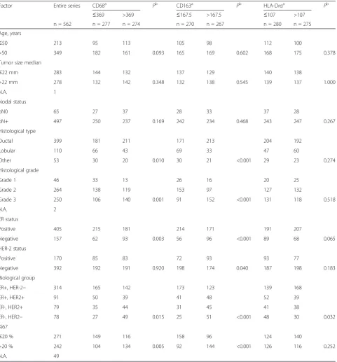
Factor Entire series CD68a Pb CD163a Pb HLA-Drαa Pb
≤369 >369 ≤167.5 >167.5 ≤107 >107
n = 562 n = 277 n = 274 n = 270 n = 267 n = 280 n = 275
Age, years
≤50 213 95 113 105 98 112 100
>50 349 182 161 0.093 165 169 0.602 168 175 0.378
Tumor size median
≤22 mm 283 144 132 137 129 140 138
>22 mm 278 132 142 0.348 132 138 0.545 139 137 1.000
N.A. 1
Nodal status
pN0 65 27 37 28 33 37 28
pN+ 497 250 237 0.169 242 234 0.468 243 247 0.267
Histological type
Ductal 399 181 211 171 213 204 192
Lobular 110 66 43 69 33 47 60
Other 53 30 20 0.010 30 21 <0.001 29 23 0.274
Histological grade
Grade 1 46 33 13 26 16 20 25
Grade 2 264 138 119 153 97 127 132
Grade 3 250 106 140 0.001 91 152 <0.001 131 118 0.518
N.A. 2
ER status
Positive 405 215 181 214 171 191 207
Negative 157 62 93 0.003 56 96 <0.001 89 68 0.065
HER-2 status
Positive 170 85 83 72 93 93 77
Negative 392 192 191 0.920 198 174 0.040 187 198 0.183
Biological group
ER+, HER-2− 314 165 142 173 123 139 168
ER+, HER2+ 91 50 39 41 48 52 39
ER-, HER2+ 79 35 44 31 45 41 38
ER-, HER2− 78 27 49 0.015 25 51 <0.001 48 30 0.032
Ki67
≤20 % 271 149 116 158 96 124 140
>20 % 242 104 134 0.005 92 144 <0.001 126 116 0.252
N.A. 49
Results are presented as number of patients.a
Cambridge, UK), CD163 (clone 10D6, Novocastra, New-castle, UK) and HLA-DRα (Dako, Glostrup, Denmark) [30, 31] (detailed information provided in Additional file 1). All the stained TMA slides were scanned using an Olympus virtual microscope equipped with Dotslide using the 10× objective (Olympus BX51, Olympus, Munich, Germany), and AxioCam camera (Zeiss, Jena, Germany). Positively stained cells were counted using Fiji equipment version 1.48s (Wayne Rasband, NIH). After color decon-volution for hematoxylin and 3,3’-Diaminobenzidine (DAB), the threshold was set for macrophage visualization. The size limit for particle analysis was carefully chosen to include only macrophages. Damaged samples were ex-cluded from the analysis. The data were analyzed in a double-blinded fashion. The investigators were blinded to the identity and clinical pathological characteristics of each sample while analyzing/scoring the macrophage content and differentiation status. The final numbers of positive cells per marker, per sample were passed on to hypothesis-naïve investigators who performed the statistical analysis of the cohort.
Cell culture
Human breast cancer cell lines MCF-7, MDA-MB231 and T47D, obtained from American Type Culture Collection (ATCC), were grown in Roswell Park Memorial Institure (RPMI)-1640 medium (Sigma-Aldrich, St Louis, MO, USA) supplemented with 10 % fetal bovine serum (FBS) (Gibco, Grand Island, NY, USA) and 100 IU/ml penicillin and streptomycin (Gibco, Bleiswijk, Netherlands) at 37 °C in a 5 % CO2atmosphere. After reaching confluence, cell culture
medium was changed to medium containing only 1 % FBS and kept in culture for 72 h. At the end of the culture period, the CM were collected from at least three independent cell line batches from each cell type. The CM were centrifuged for 5 minutes at 2,800 g, aliquoted and frozen at−20 °C. CM were used as 50 % supplement of the macrophage differenti-ation culture medium together with 10 % FBS. The cell lines were recently authenticated by STR (Short tandem repeat) profiling by a certified cell line authentication service (DDC Medical, Fisher Scientific, London, UK). Mycoplasma detec-tion was performed on a routine basis by 4,6-Diamidino-2-’ phenylindole (DAPI) staining of cultured cells.
Peripheral blood mononuclear cell (PBMC) isolation
PBMCs from five different donors were isolated by centri-fugation over Ficoll gradient (Sigma-Aldrich, St Louis, MO, USA). CD14+ cells were magnetically labeled with
α-CD14 microbeads and positively selected by MACS technology (Miltenyi Biotec, Cologne, Germany).
Macrophage differentiation
To obtain M1, M2a and M2c macrophages, CD14+ mono-cytes were cultured in MEM (Lonza, Basel, Switzerland)
supplemented with 10 % FBS (Gibco, Grand Island, NY, USA) (control, CTR), with IFN-γ(50 ng/ml; M1), or IL-4 (50 ng/ml; M2a), or IL-10 (50 ng/ml; M2c) for 5 days with replacement of half of the culture media at day 3 [32]. To assess the effect of breast cancer cell-line-secreted factors, the same differentiation protocol was carried out in the presence or absence of 50 % CM from MDA-MB231, MCF-7 or T47D cells. For activation status experiments (ELISA), cells were treated with LPS (10 ng/ml, Sigma-Aldrich, St Louis, MO, USA) for one additional day. Un-less otherwise stated, all the used cytokines were from R&D Systems (Minneapolis, MN, USA). Supernatants were collected, centrifuged for 5 minutes at 2,800 g, ali-quoted and stored at−20 °C until further analysis. Cells were harvested with Accutase (Invitrogen, Paisley, UK), debris were removed by centrifugation (5 minutes at 400 g), and cells were used for flow cytometry analysis. Supernatants were used for ELISA and zymography.
Flow cytometry
Ex vivo polarized macrophages were analyzed by validated flow cytometry methods [5], with the BD LSR II flow cytometer (BD Biosciences, Erembodegem-Dorp, Belgium). In brief, cells were washed with PBS 0.1 % BSA (Sigma-Aldrich, St Louis, MO, USA) and before staining, Fc receptors were blocked with FcR blocking reagent (BD Biosciences, Erembodegem-Dorp, Belgium): 0.2 × 10⁶cells were incubated with adequate antibody mixes and washed prior to analysis. Surface-marker expression was analyzed with flow cytometry using the following fluorochrome-labeled monoclonal antibodies: CD14-APC-Cy7 (clone61D3; eBioscience, Paris, France), CD16-PE-Cy7 (clone DJ130c; AbD Serotec, Kidlington, UK), CD64-AF488 (clone 10.1; BioLegend, San Diego, CA, USA), CD200R-PE (clone OX108; AbD Serotec, Kidlington, UK), CD163-AF647 (clone GHI/61; BD Pharmingen, Erembodegem-Dorp, Belgium), and CD86-AF488 (clone IT2.2, BD Pharmingen, Erembodegem-Dorp, Belgium). Equivalent amounts of isotype-matched control antibodies and unstained cells were included in all experiments as negative and autofluo-rescence controls. Data were analyzed with BD FACSDiva software, after gating on the myeloid population in the FSC/SSC plot. Values were expressed as the percent ratio of the median fluorescence intensity (MedFI) of the marker of interest over the MedFI of the unstained cells.
ELISA
Zymography
The potential proteolytic activity of MMPs in the superna-tants of the obtained macrophages was determined by zymography as previously described [33]. The stained polyacrylamide-gelatin gels were observed with the Image Quant RT ECL imager. Densitometry of the bands corre-sponding to pro-MMP-9 activity (92 kDa) was performed using Fiji equipment version 1.48s (Wayne Rasband, NIH). Presented values are the optical densities of pro-MMP-9-digested bands normalized to the total protein content of the corresponding total cell lysate compared with the dens-ity of the equivalent background area.
Statistics
TMA results were analyzed with SAS version 8.2 for Win-dows (SAS Institute, Cary, NC, USA) using the median values of the numbers of positive cells in the entire series as the cutoff value. Frequency tables were analyzed using the chi-square (χ2) test. Survival between groups was com-pared using the Kaplan-Meier life-table method and a Cox multivariate proportional hazards model. The log-rank test was used to confirm the robustness of the analysis. The subgroup analyses were performed including the macro-phage markers, the subgroup variable, and their interaction in the Cox model. The Mann-Whitney or Kruskal-Wallis tests were applied when suitable. All P values are two-sided and are not adjusted for multiple testing. Experimen-tal data were expressed as median ± SD, unless otherwise indicated. The Kruskal-Wallis test followed by Dunn’s post hoc test was employed to calculate statistically significant differences between the CTR and the various conditions, using GraphPad Prism software.
Study approval
Permission to use the tissues from the FinXX study for re-search purposes was provided by the Finnish Ministry of So-cial Affairs and Health. The ethics committee at the Helsinki University Central Hospital (Helsinki, Finland) approved the FinXX study and the current study (permission HUS 35/13/ 03/02/2015). Ethical approval for the use of peripheral blood from healthy donors was obtained from the Nantes Univer-sity Hospital Ethics Committee. Samples were obtained from the Établissement Français du Sang with informed consent (agreement reference NTS 2000–24, Avenant n°10).
Online supplemental material
A supplemental table (Additional file 1) and supplemental figures (Additional files 2, 3, 4, 5 and 6) are available online.
Results
Clinical significance of TAM numbers and differentiation status in breast cancer patients
To explore the clinical relevance of TAM differentiation in breast cancer patients, we evaluated total TAM number
(CD68), M1 TAM (HLA-DRα) and M2 TAM (CD163) in a large human breast cancer TMA cohort. There was hetero-geneity among the patients in the expression levels of the different macrophage markers (Fig. 1). M2 macrophage number, identified as the umber of CD163+ cells, was strongly associated with fast proliferation (Ki67 positivity >20 %), poor differentiation (grade 3), ER negativity and histological ductal type (Table 2). None of the individual markers (CD68, HLA-DRα or CD163) was on its own strongly correlated with prognosis, recurrence-free survival (RFS) or overall survival (Additional file 2). In the multivari-ate Cox model for RFS, markers such as Ki67 positivity >20 %, node positivity and primary tumor size >22 mm were strongly significant predictive factors (p <0.001 for tumor size and <0.01 for the other factors). In the same model CD163 was a significant factor (p = 0.011) together with other model covariates, such as ER negativity (Table 3).
Human breast cancer cells condition ex vivo differentiation and activation of human macrophages
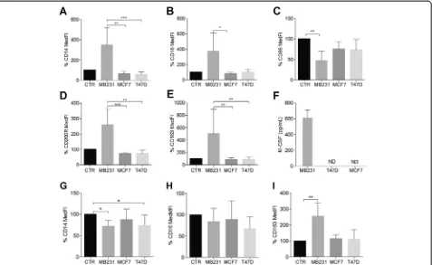
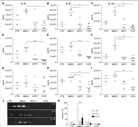
As a proof of concept, we showed that the isolated CD14+ cells could be differentiated to M1 (high CD64, high IL-6 secretion), M2a (high CD200R and CD86, low IL-6 and high IL-8 secretion) and M2c macrophages (high CD163, low IL-6 and high IL-10 secretion), respectively, using IFN-γ, IL-4 and IL-10, thus, demon-strating their proven [34] ex vivo plasticity (Figs. 2, 3 and 4 and Additional file 3). Considering M1 differenti-ation in the presence of IFN-γ, none of the CM affected the expression levels of M1 surface-markers (Additional file 4) nor the secretion profile (data not shown).
CD14+ cells differentiated in the presence of MDA-MB231 CM alone yielded an M2 macrophage population (Fig. 2a-e). This result was most obvious in terms of CD86, CD200R and CD163 expression levels (Fig. 2c-e). The differ-entiation only in the presence of other CM retained the con-trol phenotype features (Fig. 2a-e and Additional file 3).
Macrophages differentiated in the presence of MDA-MB231 CM produced higher amounts of IL-6, IL-8, IL-10 (Fig. 3a-c) and MMP-9 (Fig. 3j-k) than the CTR or other CM. That scenario remained true when the cells were con-comitantly treated with IL-4 (Fig. 3d-f) or IL-10 (Fig. 3g-i), except for IL-6 secretion in the latter treatment (Fig. 3g).
MDA-MB231 cells secrete large amounts of M-CSF, skewing macrophages to an M2c-like phenotype
The strong effects of the MDA-MB231 cells led us to in-spect all the CM for dissimilarly secreted macrophage-differentiating factors. We saw no differences in terms of IL-10, transforming growth factor (TGF)-βor IL-4 (data not shown), but only MDA-MB231 cells secreted high amounts of M-CSF (Fig. 2f ).
file 5) showed a decrease in CD14 expression levels when compared to cells differentiated only with IL-10 (CTR, Fig. 2g). MDA-MB231 CM significantly increased CD163 expression levels (Fig. 2i). The macrophages treated with IL-10 and MDA-MB231 CM secreted more IL-8, IL-10 and MMP-9 than the CTR IL-10-treated macrophages (Fig. 3g-k).
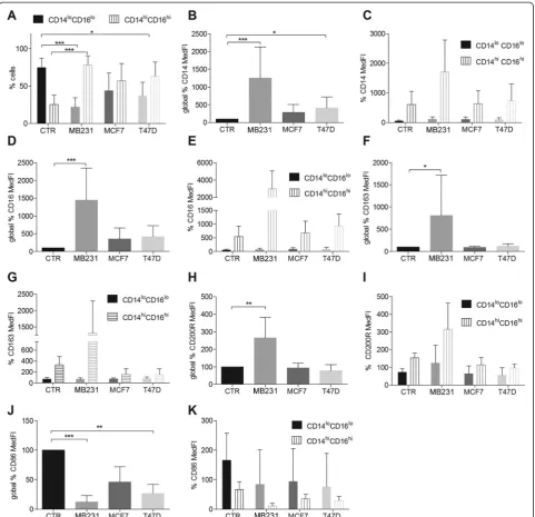
Human breast cancer cells affect M2a macrophage differentiation, rendering macrophages to a mixed M2a/M2c phenotype
When exposed to IL-4, two major macrophage subpopu-lations arose in all the conditions: CD14lo/C16lo and CD14hi/CD16hi(Fig. 4). In the presence of breast cancer cell line CM, the relative percentage of each of these subpopulations changed. Instead of a predominance of the CD14lo/C16lopopulation (CTR, Fig. 4a), there was a statistically significant inversion toward a predominance of the CD14hi/CD16hi population (Fig. 4a), especially in
the presence of MDA-MB231 (p<0.0005) or T47D CM (p<0.05). MCF-7 CM showed the same trend reaching equilibrium between the two subpopulations not signifi-cantly statistically different from the subpopulation dis-tribution in the CTR (IL-4 alone). The detailed analysis of the surface-markers from those subpopulations indi-cated that MDA-MB231 and T47D CM increase CD14 (Fig. 4b) and decrease CD86 overall expression (Fig. 4j). MDA-MB231 CM increases CD16 (Fig. 4d), CD163 (Fig. 4f ) and CD200R expression (Fig. 4h). These fluctu-ations are mostly due to the CD14hi/CD16hi population (Fig. 4c, e, g, i, k) and the results were statistically signifi-cant for the global expression (Fig. 4b, d, f, h, j;p<0.05). Although not statistically significant, MCF-7 CM in-duced the same trend as T47D CM (Fig. 4a-k).
[image:6.595.56.540.90.450.2]IL-8 (Fig. 3d, e) and MMP-9 secretion (Fig. 3j, k). If compared to MDA-MB231 CM alone, IL-4 combined with MDA-MB231 CM decreased the secretion of IL-6, IL-8 and MMP-9 and increased IL-10 secretion (Fig. 3a-c and Fig. 3j, k). Overall, MDA-MB231 CM in the presence of IL-4 produced a macrophage subpopulation with an intermediate/mixed M2a/M2c phenotype (Additional
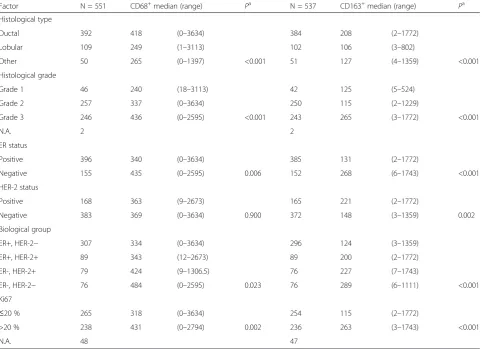
[image:7.595.58.542.101.450.2]file 6), with an abundant production of the immunosup-pressive M2c-inducing cytokine IL-10. These macrophages retain matrix-remodeling properties by secreting MMP-9. The possibility that the MCF-7 or T47D CM-induced CD14hi/CD16hi macrophage subpopulations also secrete different levels of cytokines should not be discarded. Those more subtle differences may be masked by the higher titers Table 2CD68+and CD163+cell number median values and relevant clinical characteristics
Factor N = 551 CD68+median (range) Pa N = 537 CD163+median (range) Pa
Histological type
Ductal 392 418 (0–3634) 384 208 (2–1772)
Lobular 109 249 (1–3113) 102 106 (3–802)
Other 50 265 (0–1397) <0.001 51 127 (4–1359) <0.001
Histological grade
Grade 1 46 240 (18–3113) 42 125 (5–524)
Grade 2 257 337 (0–3634) 250 115 (2–1229)
Grade 3 246 436 (0–2595) <0.001 243 265 (3–1772) <0.001
N.A. 2 2
ER status
Positive 396 340 (0–3634) 385 131 (2–1772)
Negative 155 435 (0–2595) 0.006 152 268 (6–1743) <0.001
HER-2 status
Positive 168 363 (9–2673) 165 221 (2–1772)
Negative 383 369 (0–3634) 0.900 372 148 (3–1359) 0.002
Biological group
ER+, HER-2− 307 334 (0–3634) 296 124 (3–1359)
ER+, HER-2+ 89 343 (12–2673) 89 200 (2–1772)
ER-, HER-2+ 79 424 (9–1306.5) 76 227 (7–1743)
ER-, HER-2− 76 484 (0–2595) 0.023 76 289 (6–1111) <0.001
Ki67
≤20 % 265 318 (0–3634) 254 115 (2–1772)
>20 % 238 431 (0–2794) 0.002 236 263 (3–1743) <0.001
N.A. 48 47
a
Mann-Whitney or Kruskal-Wallis test.ERestrogen receptor,HER-2human epidermal growth factor receptor 2,N.A.not available
Table 3Independent prognostic factors in Cox multivariate model for recurrence-free survival in years
Variables Regression
coefficent
Standard error
Regression coefficient/
standard error χ
2
P Exp (Coef) 95 % CI
Lower Upper
ER+ −0.612 0.252 −2.430 5.903 0.0151 0.542 0.331 0.888
HER-2+ 0.050 0.239 0.211 0.045 0.8326 1.052 0.659 1.679
Ki67 >20% −0.711 0.269 −2.642 6.981 0.0082 0.491 0.290 0.832
Node positivity −1.096 0.394 −2.780 7.730 0.0054 0.334 0.154 0.724
Size >22 mm −0.865 0.237 −3.657 13.372 0.0003 0.421 0.265 0.669
Histological grade 3 −0.071 0.268 −0.266 0.071 0.7899 0.931 0.550 1.575
CD163 >167.5 0.580 0.229 2.531 6.408 0.0114 1.786 1.140 2.798
[image:7.595.56.539.601.724.2]produced by the MDA-MB231-CM-induced CD14hi/ CD16hisubpopulations.
Discussion
Clinical significance of TAM numbers and differentiation status in breast cancer patients
In this study CD163+ cells in primary breast tumor tis-sue were brought up as a negative prognosis factor for RFS. However, we could not precisely determine which would be the additional interacting factors involved in this effect. It is clear that CD163 correlates with known factors to be associated with a bad prognosis, such as ER negativity, poor differentiation (grade 3) and ductal type (Tables 1 and 2). Previous studies have shown that higher tumor grade [22] and higher Ki67 index are asso-ciated with increased CD68+ macrophage infiltration in breast tumors [23, 35]. It was suggested that highly pro-liferative high-grade tumors elicit an active immune re-sponse that further supports angiogenesis and tumor growth. Further, these high-grade tumors may secrete higher levels of macrophage-recruiting/modulating cyto-kines such as M-CSF (ex vivo results, Fig. 2f ), IL-10 and/or TGF-β[6], which is in agreement with the high
number of CD163+ M2-macrophages. A study exploring stromal gene signatures in DCIS and invasive breast can-cer found that higher grade ER-negative and PR-negative tumors are associated with macrophage responses [36]. Macrophage infiltration was present early in the tumor progression at the DCIS stage, and the majority of cases remained positive in matched invasive breast cancer cases, accounting for early macrophage recruitment in breast cancer progression [36]. Although widely ac-cepted as a specific monocyte/macrophage marker, CD163 can also be expressed by immature MDCs, which include myeloid-derived suppressor cells (MDSCs), known to favor tumor progression [24]. Therefore, we cannot exclude the possibility that a percentage of the CD163+ cells detected may in fact be MDSCs, which could account for the poor prognostic role of CD163.
Differential ex vivo conditioning of human macrophage differentiation and activation by different breast cancer cell types
Levano et al. [7] explored the cytokine receptor profile of different breast cancer cell types and found that basal-like cells (e.g., MDA-MB231) express Fig. 2Flow cytometry analysis of CD14+cells differentiated for 5 days with or without 50 % conditioned media (CM).a-ePercentual variation of median fluorescence intensity (MedFI) of CD14, CD16, CD86, CD200R and CD163 compared to control (CTR), n = 5.fMonocyte colony stimulating factor (M-CSF) protein levels in breast cancer cell line CM (n = 3).g-iFlow cytometry analysis of CD14+cells differentiated in the presence of IL-10 for 5 days with or without 50 % breast cancer cell line CM. Percent variation of MedFI of CD14, CD16 and CD163 compared to CTR (IL-10 alone).
[image:8.595.62.539.89.382.2]preferentially granulocyte monocyte colony stimulat-ing factor (GM-CSF), hepatocyte growth factor receptor (HGFR, also known as c-MET), CD44, epithelial growth factor receptor (EGFR), transforming growth factor re-ceptor 2 (TGFR2) and oncostatin M rere-ceptor (OSMR). Luminal-type breast cancer cells (e.g., MCF-7 and T47D) express RET (a proto-oncogene which encodes for a re-ceptor tyrosine kinase for members of the glial cell line-derived neurotrophic factor) [7] and leukemia inhibitory factor (LIF) [37]. This suggests that TAMs have a differ-ent influence depending on the tumor subtype, as breast
[image:9.595.57.540.89.529.2]mammary gland were proven essential in supporting and activating mammary stem cells necessary for normal morphogenesis [39], branching [9] and in the postpartum-related influx of M2 macrophages [10]. All these developmental processes occur via mechanisms similar to molecular cancer mechanisms, such as vascular endothelial growth factor A (VEGF-A)-stimulated angio-genesis. Additionally, TAMs secrete EGF, TNF-α, VEGF and basic fibroblast growth factor (bFGF) and have
reduced antigen presenting ability. Also the release of IL-10 by both tumor cells and TAMs immunosuppresses cytotoxic T-lymphocytes (CTLs) [9].
A study of murine and human macrophage polarization profiles showed that M-CSF-differentiated human macro-phages are pro-M2, meaning that LPS or IFN-γ stimula-tion can still induce an M1 response. However, if stimulated with IL-4 or IL-10 they become more M2 type than the basal macrophages [40]. M-CSF induces CD163 Fig. 4Flow cytometry analysis of CD14+cells differentiated for 5 days in the presence of IL-4 with or without 50 % conditioned media (CM). aCD14loCD16loand CD14hiCD16hisubpopulation distribution.b,d,f,h,jOverall percent variation of CD14, CD16, CD163, CD200R and CD86 median fluorescence intensity (MedFI), compared with control (CTR) (IL-4 alone) (c,e,g,i,k) CD14 CD16, CD163, CD200R and CD86 MedFI percent variation in the subpopulations CD14loCD16loand CD14hiCD16hi.
[image:10.595.58.540.88.553.2]expression in macrophages which, when LPS-stimulated, secrete higher levels of IL-12p40, TNF-α and IL-6 [34]. Our MDA-MB231 cells produce copious amounts of M-CSF (Fig. 2f ), in levels similar to clinical samples and MDA-MB231 cells, as reported previously [6]. Similarly, the monocyte shift toward M2/CD163+ TAMs by increased levels of M-CSF has been seen in other tumor types such as glioma [41], clear cell renal carcin-oma [42], ovarian carcincarcin-oma [43] and a mouse model of osteosarcoma [44]. In those tumor types, the elevated M-CSF and CD163 expression correlates with higher tumor grade [41, 42].
CD163 is a monocyte/macrophage-restricted scavenger receptor. It clears hemoglobin/haptoglobin complexes, hence protecting tissues from hemoglobin-induced oxi-dative damage [45]. It was recently shown that breast cancer CD163+ TAMs correlate with Wnt5a expression, the latter factor being responsible for macrophage repro-gramming to an anti-inflammatory M2 status. The same group has reported that Wnt5a acts as a feedback antag-onist of toll-like receptor (TLR) signaling, inducing IL-10 secretion [26]. Our results fit this mechanism well, as MDA-MB231 CM induced CD163 expression, a feature of M2c TAMs. This could indicate a parallel in-crease in Wnt5a that inhibits TLR response and inin-creases IL-10 secretion upon LPS stimulation (Fig. 3c, f, i).
As previously discussed, the M2c-boosted differentiation by MDA-MB231-secreted products may have consequences in terms of microenvironment-aided tumor progression via immunosuppressive, matrix remodeling and scavenging TAM functions. These effects may impair an effective im-mune tumor rejection as our in vitro findings with murine macrophages and breast cancer cell line CM indicate [46].
The overall decrease of CD86 expression in M2a mac-rophages by breast cancer CM may contribute to im-munosuppressive, tumor-promoting behavior. CD86, also known as B7-2, is a type I transmembrane protein of the immunoglobulin superfamily. It is a co-stimulatory molecule expressed by antigen-presenting cells such as dendritic cells and macrophages. Its binding to CD28 on naïve T-cells is essential for Th2 differentiation, cytokine secretion and induction of effector function [47].
In our system, M2a macrophages differentiated in the presence of IL-4 and MDA-MB231 CM had the poten-tial for increased CD200R signaling which in vivo would indicate an immunosuppressive tumor-promoting envir-onment. CD200R is a myeloid receptor expressed on macrophages, granulocytes, dendritic cells and NK (natural killer) cells [48]. CD200R signaling is known to increase the immune activation threshold, being physio-logically relevant in restraining inflammation [48]. CD200 ligand interaction with its receptor CD200R on macrophages decreases TNF-αand IFN-γsecretion [49]. CD200 is expressed by cancer cells and other cell types
like mesenchymal stem cells, thymocytes, activated T cells, B cells and dendritic cells. Studies in different tumor types, including breast cancer, showed that CD200-CD200R interaction delivers an immunosup-pressive signal. This signal directly decreases inflamma-tory cytokine secretion by macrophages, and indirectly increases regulatory T cells (Treg) and decreases effector T-cell numbers, thereby promoting tumor progression by immune evasion [50].
Limitations of the study
In the TMA analysis the median of positive cells for each marker in all samples was used as a cutoff value and analysis of normal breast tissue - probably carrying rest-ing macrophages as a healthy baseline control - could not be included. However, our study brought up differ-ences in TAM activation status between patients rele-vant to the disease outcome. Larger studies are thus justified to ascertain the exact role of M2 macrophages in disease progression.
In the ex vivo macrophage differentiation studies, the extrapolation of the MDA-MB231 CM effects on macro-phage differentiation to the clinical situation of triple-negative breast cancer patients should be made with care. MDA-MB231 is an aggressive model cell line, rele-vant in the field as it is the parental cell line for several metastatic sub-clones widely used in experiments in vivo [51–53]. The MDA-MB231 cell line belongs to the mesenchymal-like subtype, while cancer cells from triple-negative breast tumors, which can be of seven dif-ferent subtypes, have phenotypic diversity from epithelial to mesenchymal characters [54]. However, we think our study remains relevant as it shows that breast cancer cells, regardless of their hormone receptor status and epithelial/mesenchymal nature, secrete factors that edu-cate macrophages toward M2 differentiation. The most aggressive one, MDA-MB231, did it most effectively. Further studies are needed to unveil the factors respon-sible for the effects seen. M-CSF appears to be a key factor in M2 TAM differentiation, as shown by others [6, 55], but as breast cancer cell lines (MCF-7 and T47D) that do not produce M-CSF also affected the M2 phenotype, inducing M2a differentiation, we think there are other relevant M2 skewing factors, which our work cannot address. The discovery of such factors is of utmost relevance, and calls for further studies.
Conclusions
to the multivariate Cox model. The presented ex vivo re-sults are to our knowledge the first demonstrating the modulation of macrophage differentiation solely by breast cancer cell-secreted factors, providing evidence for the mechanisms of breast cancer macrophage education be-hind clinical findings. Particularly, the mesenchymal-type cell line MDA-MB231 polarizes macrophages toward a mixed M2a/M2c status. It is therefore rational to venture that the screening of TAM activation in breast cancer pa-tients could be useful in predicting papa-tients with a high metastatic risk. The knowledge of TAM activation status may allow the therapeutic targeting of TAMs, once TAMs targeting/modulating agents pass clinical trials and become widely available. These include bisphosphonates [56]; M-CSF and M-M-CSFR inhibitors and targeting antibodies [57], NCT01316822, NCT01444404; anti-macrophage migra-tion inhibitory factor, NCT01765790 and L-MTP-PE, NCT00631631. There is a scarcity of therapeutic options for patients with triple-negative metastatic breast cancer, and growing resistance to the available options biased by a continuous focus on cancer cell targets, which are by na-ture genetically unstable and prone to mutations. Ap-proaches such as ours fuel a necessary paradigm change, contributing to the notion that the immunological tumor microenvironment should be taken into account in the de-velopment of new multi-target cancer therapies.
Additional files
Additional file 1:Immunohistochemical analysis conditions. (PDF 273 kb)
Additional file 2:Tissue microarray (TMA) analysis of recurrence-free survival and overall survival curves related to the macrophage markers A CD68 B CD163 and C HLA-DRIIα.(PDF 22622 kb)
Additional file 3:Flow cytometry analysis of CD14+cells differentiated for 5 days with or without 50 % conditioned media (CM), IFN-γ, IL-4 and IL-10 A mean fluorescence intensity (MFI) of CD14 B CD16 C CD64 D CD163 E CD86 and F CD200R, normalized to MFI of unstained cells; n = 3, ***p<0.0005.(PDF 1833 kb)
Additional file 4:Flow cytometry analysis of CD14+cells differentiated for 5 days in the presence of IFN-γwith or without 50 % conditioned media (CM) A mean fluorescence intensity (MFI) of CD14 B CDC16 and C CD64 normalized to MFI of unstained cells; n = 3.(PDF 2112 kb)
Additional file 5:Representative flow cytometry dot plot of IL-10 macrophages A human macrophages in the presence of IL-10 with or without breast cancer cell line conditioned media (CM), detected with antibodies against CD14, CD16 and CD163 B mean fluorescence intensity (MFI) of the analyzed surface-markers.(PDF 18647 kb)
Additional file 6:Representative flow cytometry dot plot of IL-4 macrophages A human macrophages in the presence of IL-4 with or without breast cancer cell line conditioned media (CM), detected with antibodies against CD14, CD16, CD163, CD200R and CD86 B mean fluorescence intensity (MFI) of the analyzed surface-markers. (PDF 2715 kb)
Abbreviations
ATCC:American Type Culture Collection; bFGF: basic fibroblast growth factor; BSA: bovine serum albumin; CM: conditioned media; CTC: circulating tumor
cell; CTLs: cytotoxic T-lymphocytes; CTR: control; DAB: 3,3’-Diaminobenzidine; DCIS: ductal carcinomain situ; DTC: disseminating tumor cell; EGF: epidermal growth factor; EGFR: epidermal growth factor receptor; ELISA: enzyme-linked immunosorbent assay; EMT: epithelial to mesenchymal transition; ER: estrogen receptor; FBS: fetal bovine serum; GM-CSF: granulocyte monocyte colony stimulating factor; HER-2: human epidermal growth factor receptor 2; HGFR: hepatocyte growth factor receptor; IFN-γ: interferonγ; kDa: kiloDaltons; LIF: leukemia inhibitory factor; LPS: lipopolysaccharide; M-CSF: monocyte colony stimulating factor; M-CSFR: monocyte colony stimulating factor receptor; MDCs: myeloid-derived cells; MDSCs: myeloid-derived suppressor cells; MedFI: median fluorescence intensity; MMP-9: matrix metalloproteinase 9; MMPs: matrix metalloproteinases; NK: natural killer; OSMR: oncostatin M receptor; PBMCs: peripheral blood mononuclear cells; PBS: phosphate-buffered saline; PR: progesterone receptor; RET: proto-oncogene, which encodes for a receptor tyrosine kinase for members of the glial cell line-derived neurotrophic factor; RFS: recurrence-free survival; RNS: reactive nitrogen species; ROS: reactive oxygen species; TAMs: tumor-associated macrophages; TGFR2: transforming growth factor receptor 2; TGF-β: transforming growth factorβ; TLR: toll-like receptor; TMA: tissue microarray; TNF-α: tumor necrosis factorα; Treg: regulatory T-cells; VCAM1: vascular cell adhesion molecule 1; VEGF-A: vascular endothelial growth factor A.
Competing interests
The authors declare that they have no competing interests.
Authors’contributions
SS conceived and designed the study, developed the methodology, acquired, analyzed and interpreted the data and wrote and revised the manuscript. RB acquired the flow cytometry data. ML, PK and JS provided technical support in the TMA analysis. SL and OT participated in the TMA study and performed its statistical analysis. HJ participated in the TMA study, acquiring, analyzing, and interpreting the data. DH participated in the study conception and design particularly in supervising the ex vivo macrophage differentiation studies. JuM supervised and coordinated the study. JM conceived, designed, supervised and participated in the manuscript writing and revision. All authors read, revised critically for important intellectual content and approved the final manuscript.
Acknowledgements
The current research was funded by the Seventh Framework Programme [FP7/2007-2013] under grant agreement no.264817–BONE-NET and by the Academy of Finland, decision numbers 132389 and 250917.
Author details
1School of Pharmacy, Faculty of Health Sciences, University of Eastern
Finland, Yliopistonranta 1C, P.O. Box 1627, FI-70211 Kuopio, Finland.2INSERM,
UMR957, Equipe LIGUE 2012, Nantes F-44035, France.3Université de Nantes,
Nantes atlantique universités, Laboratoire de Physiopathologie de la Résorption Osseuse et Thérapie des Tumeurs Osseuses Primitives, Nantes F-44035, France.4CHU de Nantes, Nantes F-44035, France.5Institute of
Biomedicine, Department of Cell Biology and Anatomy, University of Turku, Turku, Finland.6Cell Imaging Core, Turku Centre for Biotechnology, University
of Turku, and Åbo Akademi University, Turku, Finland.7Medical School,
University of Tampere and Department of Oncology Tampere University Hospital, Tampere, Finland.8Laboratory of Molecular Oncology, Biomedicum
Helsinki, University of Helsinki, Helsinki, Finland.9Department of Pathology,
Haartman Institute, University of Helsinki and HUSLAB, Helsinki, Finland.
10Comprehensive Cancer Center, Helsinki University Hospital, and
Department of Oncology, University of Helsinki, Helsinki, Finland.
Received: 24 April 2015 Accepted: 21 July 2015
References
1. Chambers AF, Groom AC, MacDonald IC. Dissemination and growth of cancer cells in metastatic sites. Nat Rev Cancer. 2002;2:563–72.
2. Bingle L, Brown NJ, Lewis CE. The role of tumour-associated macrophages in tumour progression: implications for new anticancer therapies. J Pathol. 2002;196:254–65.
4. Mosser DM, Edwards JP. Exploring the full spectrum of macrophage activation. Nat Rev Immunol. 2008;8:958–69.
5. Ambarus CA, Krausz S, van Eijk M, Hamann J, Radstake TR, Reedquist KA, et al. Systematic validation of specific phenotypic markers for in vitro polarized human macrophages. J Immunol Methods. 2012;375:196–206. 6. Grugan KD, McCabe FL, Kinder M, Greenplate AR, Harman BC, Ekert JE, et al.
Tumor-associated macrophages promote invasion while retaining Fc-dependent anti-tumor function. J Immunol. 2012;189:5457–66. 7. Levano KS, Jung EH, Kenny PA. Breast cancer subtypes express distinct
receptor repertoires for tumor-associated macrophage derived cytokines. Biochem Biophys Res Commun. 2011;411:107–10.
8. Mantovani A, Sozzani S, Locati M, Allavena P, Sica A. Macrophage polarization: Tumor-associated macrophages as a paradigm for polarized M2 mononuclear phagocytes. Trends Immunol. 2002;23:549–55. 9. Gyorki D, Asselin-Labat M, van Rooijen N, Lindeman G, Visvader J. Resident
macrophages influence stem cell activity in the mammary gland. Breast Cancer Res. 2009;11:R62.
10. O'Brien J, Martinson H, Durand-Rougely C, Schedin P. Macrophages are crucial for epithelial cell death and adipocyte repopulation during mammary gland involution. Development. 2012;139:269–75.
11. Qian B, Li J, Zhang H, Kitamura T, Zhang J, Campion LR, et al. CCL2 recruits inflammatory monocytes to facilitate breast-tumour metastasis. Nature. 2011;475:222–5.
12. Wyckoff J, Wang W, Lin EY, Wang Y, Pixley F, Stanley ER, et al. A paracrine loop between tumor cells and macrophages is required for tumor cell migration in mammary tumors. Cancer Res. 2004;64:7022–9.
13. Chen Q, Zhang XH, Massagué J. Macrophage binding to receptor VCAM-1 transmits survival signals in breast cancer cells that invade the lungs. Cancer Cell. 2011;20:538–49.
14. Casazza A, Laoui D, Wenes M, Rizzolio S, Bassani N, Mambretti M, et al. Impeding macrophage entry into hypoxic tumor areas by Sema3A/Nrp1 signaling blockade inhibits angiogenesis and restores antitumor immunity. Cancer Cell. 2013;24:695–709.
15. Movahedi K, Laoui D, Gysemans C, Baeten M, Stangé G, Van Bossche JD, et al. Different tumor microenvironments contain functionally distinct subsets of macrophages derived from Ly6C(high) monocytes. Cancer Res. 2010;70:5728–39.
16. Biswas SK, Sica A, Lewis CE. Plasticity of macrophage function during tumor progression: Regulation by distinct molecular mechanisms. J Immunol. 2008;180:2011–7.
17. Ohri CM, Shikotra A, Green RH, Waller DA, Bradding P. Macrophages within NSCLC tumour islets are predominantly of a cytotoxic M1 phenotype associated with extended survival. Eur Respir J. 2009;33:118–26. 18. Forssell J, Öberg Å, Henriksson ML, Stenling R, Jung A, Palmqvist R. High
macrophage infiltration along the tumor front correlates with improved survival in colon cancer. Clin Cancer Res. 2007;13:1472–9.
19. Ohno S, Inagawa H, Dhar DK, Fujii T, Ueda S, Tachibana M, et al. The degree of macrophage infiltration into the cancer cell nest is a significant predictor of survival in gastric cancer patients. Anticancer Res. 2003;23:5015–22.
20. Hussein MR, Hassan HI. Analysis of the mononuclear inflammatory cell infiltrate in the normal breast, benign proliferative breast disease, in situ and infiltrating ductal breast carcinomas: preliminary observations. J Clin Pathol. 2006;59:972–7.
21. Volodko N, Reiner A, Rudas M, Jakesz R. Tumour-associated macrophages in breast cancer and their prognostic correlations. Breast. 1998;7:99–105. 22. Mahmoud SMA, Lee AHS, Paish EC, Macmillan RD, Ellis IO, Green AR.
Tumour-infiltrating macrophages and clinical outcome in breast cancer. J Clin Pathol. 2012;65:159–63.
23. Al Murri AM, Hilmy M, Bell J, Wilson C, McNicol AM, Lannigan A, et al. The relationship between the systemic inflammatory response, tumour proliferative activity, T-lymphocytic and macrophage infiltration, microvessel density and survival in patients with primary operable breast cancer. Br J Cancer. 2008;99:1013–9.
24. Medrek C, Ponten F, Jirstrom K, Leandersson K. The presence of tumor associated macrophages in tumor stroma as a prognostic marker for breast cancer patients. BMC Cancer. 2012;12:306-2407-12-306.
25. Leek RD, Lewis CE, Whitehouse R, Greenall M, Clarke J, Harris AL. Association of macrophage infiltration with angiogenesis and prognosis in invasive breast carcinoma. Cancer Res. 1996;56:4625–9.
26. Bergenfelz C, Medrek C, Ekström E, Jirström K, Janols H, Wullt M, et al. Wnt5a induces a tolerogenic phenotype of macrophages in sepsis and breast cancer patients. J Immunol. 2012;188:5448–58.
27. Król M, Mucha J, Majchrzak K, Homa A, Bulkowska M, Majewska A, et al. Macrophages mediate a switch between canonical and non-canonical wnt pathways in canine mammary tumors. PLoS ONE. 2014;9:e83995. 28. Ojalvo LS, Whittaker CA, Condeelis JS, Pollard JW. Gene expression analysis
of macrophages that facilitate tumor invasion supports a role for Wnt-signaling in mediating their activity in primary mammary tumors. J Immunol. 2010;184:702–12.
29. Joensuu H, Kellokumpu-Lehtinen P, Huovinen R, Jukkola-Vuorinen A, Tanner M, Asola R, et al. Adjuvant capecitabine in combination with docetaxel and cyclophosphamide plus epirubicin for breast cancer: an open-label, randomised controlled trial. Lancet Oncol. 2009;10:1145–51. 30. Buddingh EP, Kuijjer ML, Duim RAJ, Bürger H, Agelopoulos K, Myklebost O,
et al. Tumor-infiltrating macrophages are associated with metastasis suppression in high-grade osteosarcoma: A rationale for treatment with macrophage activating agents. Clin Cancer Res. 2011;17:2110–9. 31. Ambarus CA, Santegoets KC, van Bon L, Wenink MH, Tak PP, Radstake TR,
et al. Soluble immune complexes shift the TLR-induced cytokine production of distinct polarized human macrophage subsets towards IL-10. PLoS One. 2012;7:e35994.
32. Guihard P, Danger Y, Brounais B, David E, Brion R, Delecrin J, et al. Induction of osteogenesis in mesenchymal stem cells by activated monocytes/macrophages depends on oncostatin M signaling. Stem Cells. 2012;30:762–72.
33. Damiens C, Fortun Y, Charrier C, Heymann D, Padrines M. Modulation by soluble factors of gelatinase activities released by osteoblastic cells. Cytokine. 2000;12:1727–31.
34. Vogel DYS, Glim JE, Stavenuiter AWD, Breur M, Heijnen P, Amor S, et al. Human macrophage polarization in vitro: Maturation and activation methods compared. Immunobiology. 2014;219:695–703.
35. Lin EY, Pollard JW. Tumor-associated macrophages press the angiogenic switch in breast cancer. Cancer Res. 2007;67:5064–6.
36. Sharma M, Beck AH, Webster JA, Espinosa I, Montgomery K, Varma S, et al. Analysis of stromal signatures in the tumor microenvironment of ductal carcinoma in situ. Breast Cancer Res Treat. 2010;123:397–404. 37. Estrov Z, Samal B, Lapushin R, Kellokumpu-Lehtinen P, Sahin AA,
Kurzrock R, et al. Leukemia inhibitory factor binds to human breast cancer cells and stimulates their proliferation. J Interferon Cytokine Res. 1995;15:905–13.
38. Su S, Liu Q, Chen J, Chen J, Chen F, He C, et al. A Positive feedback loop between mesenchymal-like cancer cells and macrophages is essential to breast cancer metastasis. Cancer Cell. 2014;25:605–20.
39. Kellokumpu-Lehtinen P, Johansson RM, Pelliniemi LJ. Ultrastructure of human fetal mammary gland. Anat Rec. 1987;218:66–72.
40. Jaguin M, Houlbert N, Fardel O, Lecureur V. Polarization profiles of human M-CSF-generated macrophages and comparison of M1-markers in classically activated macrophages from GM-CSF and M-CSF origin. Cell Immunol. 2013;281:51–61.
41. Komohara Y, Ohnishi K, Kuratsu J, Takeya M. Possible involvement of the M2 anti-inflammatory macrophage phenotype in growth of human gliomas. J Pathol. 2008;216:15–24.
42. Komohara Y, Hasita H, Ohnishi K, Fujiwara Y, Suzu S, Eto M, et al. Macrophage infiltration and its prognostic relevance in clear cell renal cell carcinoma. Cancer Sci. 2011;102:1424–31.
43. Duluc D, Delneste Y, Tan F, Moles MP, Grimaud L, Lenoir J, et al. Tumor-associated leukemia inhibitory factor and IL-6 skew monocyte differentiation into tumor-associated macrophage-like cells. Blood. 2007;110:4319–30.
44. Ségaliny AI, Mohamadi A, Dizier B, Lokajczyk A, Brion R, Lanel R, et al. Interleukin-34 promotes tumor progression and metastatic process in osteosarcoma through induction of angiogenesis and macrophage recruitment. Int J of Cancer. 2015;137:73–85.
45. Schaer CA, Vallelian F, Imhof A, Schoedon G, Schaer DJ. CD163-expressing monocytes constitute an endotoxin-sensitive Hb clearance compartment within the vascular system. J Leukoc Biol. 2007;82:106–10.
47. Santin AD, Hermonat PL, Ravaggi A, Chiriva-Internati M, Cannon MJ, Hiserodt JC, et al. Expression of surface antigens during the differentiation of human dendritic cells vs macrophages from blood monocytes in vitro. Immunobiology. 1999;200:187–204.
48. Rygiel TP, Meyaard L. CD200R signaling in tumor tolerance and inflammation: A tricky balance. Curr Opin Immunol. 2012;24:233–8.
49. Pietilä M, Lehtonen S, Tuovinen E, Lähteenmäki K, Laitinen S, Leskelä HV, et al. CD200 positive human mesenchymal stem cells suppress TNF-alpha secretion from CD200 receptor positive macrophage-like cells. PLoS ONE. 2012;7:e31671.
50. Gorczynski RM, Chen Z, Khatri I, Podnos A, Yu K. Cure of metastatic growth of EMT6 tumor cells in mice following manipulation of CD200:CD200R signaling. Breast Cancer Res Treat. 2013;142:271–82.
51. Peyruchaud O, Winding B, Pécheur I, Serre C, Delmas P, Clézardin P. Early detection of bone metastases in a murine model using fluorescent human breast cancer cells: application to the Use of the bisphosphonate zoledronic acid in the treatment of osteolytic lesions. J Bone Miner Res. 2001;16:2027–34.
52. Kang Y, Siegel PM, Shu W, Drobnjak M, Kakonen SM, Cordón-Cardo C, et al. A multigenic program mediating breast cancer metastasis to bone. Cancer Cell. 2003;3:537–49.
53. Minn AJ, Gupta GP, Siegel PM, Bos PD, Shu W, Giri DD, et al. Genes that mediate breast cancer metastasis to lung. Nature. 2005;436:518–24. 54. Lehmann BD, Bauer JA, Chen X, Sanders ME, Chakravarthy AB, Shyr Y.
Identification of human triple-negative breast cancer subtypes and preclinical models for selection of targeted therapies. J Clin Invest. 2011;121:2750–60.
55. Solinas G, Schiarea S, Liguori M, Fabbri M, Pesce S, Zammataro L, et al. Tumor-conditioned macrophages secrete migration-stimulating factor: A new marker for M2-polarization, influencing tumor cell motility. J Immunol. 2010;185:642–52.
56. Gnant M, Clézardin P. Direct and indirect anticancer activity of bisphosphonates: A brief review of published literature. Cancer Treat Rev. 2012;38:407–15.
57. Ségaliny AI, Tellez-Gabriel M, Heymann M, Heymann D. Receptor tyrosine kinases: Characterisation, mechanism of action and therapeutic interests for bone cancers. J Bone Oncol. 2015;4:1–12.
Submit your next manuscript to BioMed Central and take full advantage of:
• Convenient online submission
• Thorough peer review
• No space constraints or color figure charges
• Immediate publication on acceptance
• Inclusion in PubMed, CAS, Scopus and Google Scholar
• Research which is freely available for redistribution