POSTMORTEM SERUM THYROGLOBULIN LEVELS AS A MARKER TO DIAGNOSE NECK COMPRESSION
Dissertation submitted in partial fulfilment of the requirements for the degree
M.D. (Forensic Medicine) BRANCH - XIV
INSTITUTE OF FORENSIC MEDICINE MADRAS MEDICAL COLLEGE
CHENNAI – 600 003
THE TAMIL NADU
Dr. M.G.R. MEDICAL UNIVERSITY
CHENNAIBONAFIDE CERTIFICATE
This is to certify that the work embodied in this dissertation
entitled
“
POSTMORTEM SERUM THYROGLOBULIN LEVELS AS A MARKER TO DIAGNOSE NECK COMPRESSION”
has been carried
out by
Dr. R.Narendar, M.B.B.S,
a Post Graduate student under my
supervision and guidance for his study leading to Branch XIV M.D.
Degree in Forensic Medicine during the period of April - 2014 to
September - 2014.
DEAN
Director and Professor
Madras Medical College &
Institute of Forensic Medicine
Rajiv Gandhi Govt. General
Madras Medical College
Hospital
Chennai-3
Chennai-3
Date: Date:
DECLARATION
I, Dr.R.Narendar, solemnly declare that this dissertation titled
“
POSTMORTEM SERUM THYROGLOBULIN LEVELS AS AMARKER TO DIAGNOSE NECK COMPRESSION
”
is the bonafide work done by me under the expert guidance and supervision ofDr.R.Vallinayagam M.D., Director & Professor, Institute of Forensic Medicine, Madras Medical College, Chennai – 3. This dissertation is submitted
to The Tamil Nadu Dr. M.G.R Medical University towards partial fulfilment of
requirement for the award of M.D., Degree (
Branch XIV
) in ForensicMedicine.
Place: Dr. R.NARENDAR
ACKNOWLEDGEMENT
I am greatly obliged to the Dean, Dr.R.Vimala M.D., Madras Medical College and Rajiv Gandhi Govt. General hospital, Chennai -3 for allowing me to conduct this study.
I would like to express my heartfelt gratitude to my esteemed Professor & Director, Dr.R.Vallinayagam M.D., who has been a constant source of inspiration and encouragement.
I am especially thankful to my Associate Professor, Dr. M.N. Rajamani Bheem Rao M.D., and Assistant Professor, Dr. T. Vedanayagam M.D.,
Institute of Forensic Medicine, Madras Medical College, Chennai -3 for their interest and encouragement, in bringing out this dissertation for my MD exam.
I would like to express my heartfelt gratitude to my esteemed Professor & Former Director, Capt. Dr B. Santhakumar M.D., Dean, Madurai Medical College, who has been a constant source of inspiration and encouragement.
I immensely thankDr. Saraswathi M.D, Director and Professor, Institute of Pathology and Electron Microscopy, Madras Medical College, Chennai -3 for giving permission for histopathological examination of Thyroid specimens.
I am very grateful to Professor Dr.Geetha Devadoss M.D., Professor
Dr.Padmavathi M.D., Professor Dr.Sudha M.D., and Assistant Professor
Dr.Vanitha M.D., Institute of Pathology and Electron Microscopy, Madras Medical College, Chennai -3 for helping me in the interpretatation of Gross examination and Histologic examination of Thyroid specimens.
Medical college, Villupuram for helping me in the preparation of the analysis and resuts.
My very special thanks to Dr. P.Vinod Kumar M.D, Assistant Professor, Balaji Medical College, Chennai, Dr. A.Nagendra kumar,M.D, Assistant Professor, Govt. Vellore medical college, Vellore and Dr. K.Priyatharsini M.D., Assistant Professor, Stanley medical college, Chennai for helping me to complete my thesis.
My very special thanks to Dr.Vindhu M.D., Winpath Labs, Mylapore for analyzing the blood samples and promptly issuing me the reports.
INSTITUTIONAL ETHICAL COMMITTEE
INDEX
S.NO. DESCRIPTION
PAGE NO.
1. Introduction 1
2. Aims and Objectives 5
3. Review of Literature 6
4. Materials and Methods 67
5. Analysis and Results 72
6. Discussion 98
7. Conclusion 102
8. References 104
9. Annexure
A) Colour Plates
B) Master Chart
115
LIST OF TABLES
TABLE.
NO. TITLE
PAGE NO.
1. Normal levels of serum thyroglobulin 29 2. Number of cases in study group 72 3. Number of cases in control group 73 4. Sex distribution of cases in study group 74 5. Sex distribution of cases in control group 75 6. Age distribution of cases in study group 76 7. Age distribution of cases in control group 77 8. Cases in study group 78 9. Correlation of Serum Thyroglobulin levels with
Histopathological findings
79
10. Comparison of Serum Thyroglobulin levels in cases of Hanging, Manual strangulation and Ligature strangulation in the study group
84
LIST OF FIGURES
Figure
No. Title
Page No.
1. Triangles of the neck 12 2. Thyroid gland and its anatomic relations 16 3. Thyroid diverticulum which gives rise to the thyroid
gland
19
4. Normal Histology of the Thyroid gland 20 5. The synthesis of the thyroid hormones and the role of
Thyroglobulin
24
6. Regulation of thyroid hormones 27
7. Immulite Analyser 70
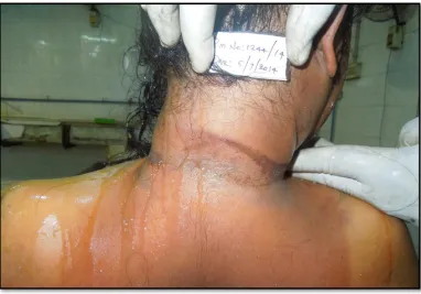
8. Ligature abrasion in a deceased died due to ligature strangulation
115
9. Ligature abrasion in left lateral view 115 10. Ligature abrasion at the back of the neck 116 11. Contusion in the muscles of the neck 116 12. Anteroposterior compression fracture of the superior
horn of the hyoid bone in case of ligature strangulation
117
13 Anteroposterior compression fracture with outwardmovement
117
14. Hanging of the deceased with heavy nylon rope in scene of crime
118
18. Ligature abrasion with the nylon rope 120 19. Ligature material – Nylon saree 121 20. Faint ligature abrasion with the nylon saree 121 21. Ligature material - Thupatta 122 22. Faint ligature abrasion using Thupatta 122 23. Dissection of neck in a case of death due to hanging 123 24 Dissected out thyroid gland in autopsy 123 25. Cyanosis in a case of death due to neck compression 124
26 Petechial haemorrhages on the anterior surface of lungs in case of death due to neck compression
124
27. Normal Histopathology of the thyroid gland 125 28. Hashimoto’s thyroiditis with lymphocytic infiltration 125 29. Encapsulated Papillary carcinoma of thyroid 126 30. Orphane anne nuclei –
characterististic of Papillary carcinoma of thyroid
LIST OF CHARTS
Chart
No. Title
Page No.
1. Number of cases in study group 72 2. Number of cases in control group 73 3. Sex distribution of cases in study group 74 4. Sex distribution of cases in control group 75 5. Age distribution of cases in study group 76 6. Age distribution of cases in control group 77 7. Comparison of Serum Thyroglobulin levels in cases of
Hanging, Manual strangulation and Ligature strangulation in the study group
85
ABBREVIATIONS
CLIA Chemi Luminiscence Immuno assay
DIT Di Iodo Thyronine
ELISA Enzyme linked immuno sorbent assay IRMA Immuno radio metric assay
T3 Tri Iodo Thyronine T4 Thyroxine
TRH Thyrotropic releasing hormone
TSH Thyroid stimulating hormone TG Thyroglobulin
ABSTRACT FOR THE DISSERTATION TO BE SUBMITTED TO
THE TAMIL NADU Dr. M.G.R. MEDICAL UNIVERSITY, CHENNAI
FOR APRIL 2015 .M.D. FOERNSIC MEDICINE EXAMINATIONS
TITLE: POSTMORTEM SERUM THYROGLOBULIN LEVELS
AS A MARKER TO DIAGNOSE NECK COMPRESSION
Abstract:
Asphyxia by neck compression occurs mainly in Hanging, Manual
strangulation, Ligature strangulation. Even though the signs like Cyanosis,
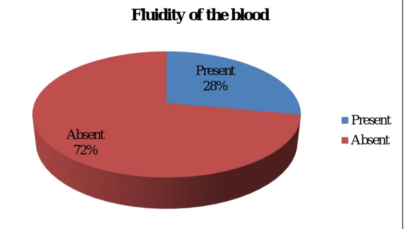
Petechial hemorrhages, Congestion, Fluidity of the blood are called as asphyxial
stigmata; these signs are not specific to asphyxia alone. These can also occur in
deaths due to other causes also. Sometimes ligature marks, nail mark abrasions,
neck muscle contusions can help in suggesting the death is due to neck
compression, these findings can be absent if the ligature material is soft and
broad or any material intervening between skin and ligature material in hanging
and ligature strangulation and usage of gloves by the assailant in manual
strangulation. This study suggests that newer scientific markers and methods
can be used in autopsies to help the forensic pathologist in such precarious
situations to arrive at the cause of death. In this prospective study, Serum
using Chemiluminescence Immuno assay. The results establish that levels of
serum thyroglobulin increases in death due to neck compression (1061.6 ±398.7
ng / ml) when compared with control group (21.8 ± 2.71 ng/ml). The level of
increase is marked in manual strangulation (5103.2 ± 539.6 ng/ml) and ligature
strangulation (4659.9 ± 2094 .2 ng/ml) than in hanging (257.4 ± 28.03 ng/ml).
The false positive increase in serum thyroglobulin as in cases of carcinoma
thyroid, Hashimoto’s Thyroiditis is ruled by Histopathological examination of
the thyroid gland taken during autopsy.
Keywords:
Neck compression, Hanging, Ligature Strangulation, Manual
strangulation, Thyroglobulin Chemiluminescence Immuno assay Carcinoma
INTRODUCTION
Etymologically the word ‘Asphyxia’ in Greek means ‘Lack of
Pulselessness’. But as time progressed the term has come up with the
meaning of Lack of oxygen. This may be due to the understanding of the
fact that air is necessary for the maintenance of life which is carried via the
blood.1
The other terms related to asphyxia are
Hypoxia – inadequate supply of oxygen to the tissues or an
impairment of the cell to utilise the oxygen
Hypoxemia – decreased oxygen in the arterial blood
Anaemia – decreased oxygen carrying capacity of the blood
Anoxia – lack of oxygen
From the medico legal point of view, asphyxia can be grouped into
Mechanical and Non-Mechanical causes.
Mechanical causes includes
1) Pressure to the exterior of the neck: Hanging, Strangulation, etc.
3) Obstruction of the airways from the interior: Gagging, Choking,
etc
4) Pressure upon the chest: Traumatic asphyxia
5) Occlusion of airways by fluid: Drowning
Non- Mechanical asphyxia can happen when the environmental oxygen is
replaced by other gases, Body cannot utilise the inhaled oxygen, the
environmental oxygen is insufficient
Ex: Carbon monoxide poisoning, Cyanide poisoning, etc.2
Neck compression is a common mode of death that can happen in
Hanging and Strangulation. However many anatomical and physiological
factors may operate in combinations leading to death. The important factors
are occlusion of the airway either by compression of larynx and trachea or
upward lifting of the tongue thereby blocking the pharynx, occlusion of the
blood vessels either jugular venous system or carotid arteries, Pressure on
the carotid sinuses leading to stimulation of vagus nerve thereby causing
death by bradycardia or cardiac arrest.
There are some signs which can be seen in most of the asphyxial
signs. These signs are called as ‘Classical signs of Asphyxia’. They are
2) Petechial Hemorrhages due to increased venous pressure and
increased permeability and rupture of venules
3) Cyanosis increase in the amount of reduced hemoglobin
4) Abnormal Fluidity of blood due to increased fibrinolytic activity
in the blood.
However these signs are nonspecific and can occur in deaths from
other causes also.3
So in Post-mortem Examination, The Autopsy surgeon is in need of
some specific signs to find out the exact method in which the fatal chain of
events was initiated. Some of the specific signs to look out for are
Ligature mark in deaths of Hanging and Ligature Strangulation.
Fingernail Abrasions in deaths of Manual Strangulation
Fluid medium in the air passages and stomach in deaths of
Drowning
Foreign body in the larynx in deaths of Choking, gagging 4
The ligature mark in the deaths of Hanging and ligature strangulation
depends on composition of the ligature material, Mode of application of
Ligature, Position of the knot, Period and Degree of Suspension, weight of
The Ligature mark may not be evident
- If any soft broad ligature material is used
- Any material like beard, collar, muffler, any ornament intervenes
between ligature material and the skin
- The Point of Suspension is low2
In these situations, The Forensic Pathologist is in a precarious
situation in coming to the cause of death. Even after thorough Post-mortem
examination, Visiting the Scene of Crime, Verbal autopsy with the relatives
could not provide the cause of death in some cases. Therefore The Forensic
Pathologist may follow some of the scientific investigation techniques in
these cases. One such scientific method is Estimation of the Serum
Thyroglobulin level in the heart blood which will be elevated in deaths due
to asphyxia caused by neck compression.
In this study, Serum Thyroglobulin was estimated in totally 25 cases
of death due to Neck compression due to Hanging, Ligature strangulation
and Manual Strangulation. Serum Thyroglobulin was also estimated in
totally 25 cases Natural Cause, Road Traffic Accident, Train Traffic
AIMS AND OBJECTIVES
1) To estimate Post-mortem Serum Thyroglobulin levels in heart
blood to diagnose Neck Compression.
2) To compare the levels of Post-mortem Serum Thyroglobulin levels
in various types of Neck Compression.
3) To rule out thyroid diseases causing Post-mortem elevation Serum
Thyroglobulin levels with Histopathological study of Thyroid
THYROGLOBULIN AND NECK COMPRESSION:
The idea of using Thyroglobulin to use it as a marker for neck
compression was first developed in 1971 by Yada et alin Japan where they found out Thyroglobulin in the serum of a strangled infant to be raised in a
significant level using Precipitation Electrophoresis.5, 61
Later in 1972, Yada et al demonstrated the presence of thyroglobulin in heart blood in many cases of death due to strangulation and confirmed
that the level of serum thyroglobulin raises in cases of strangulation6
In 1973, Yada et al demonstrated a comparative study of serum thyroglobulin levels in deaths due to strangulation and cut throat injuries.7
In all these studies Yada et al used Precipitation electrophoresis as a
tool to estimate serum thyroglobulin levels in heart blood. As the
precipitation electrophoresis had poor specificity and sensitivity the
researchers shifted on to further sophisticated techniques.5, 6, 7
In 1980, Katsumata et al compared the results of serum thyroglobulin levels obtained using Precipitation electrophoresis and Radio
immuno assay and found out that Radio immuno assay8 as a sensitive and
Later on Enzyme linked Immuno sorbent Assay (ELISA) superseded
the usage of Radio immunoassay (RIA) in the field of Forensic medicine.
This led to the shift of detecting the serum thyroglobulin using Enzyme
linked Immuno sorbent Assay (ELISA) rather than Radio immunoassay
(RIA). Many comparative studies were conducted by Katsumata et al
between the years 1980 -1985 to study the sensitivity and specificity of
Enzyme linked Immuno sorbent Assay (ELISA) and Radio immunoassay
(RIA). The sensitivity of the Enzyme linked Immuno sorbent Assay
(ELISA) was 5 ng /ml and the sensitivity of Radio immunoassay (RIA) was
4 ng / ml.
Tamaki et al in 1987 published a paper on the usage of Plasma thyroglobulin in the post-mortem diagnosis of mechanical asphyxia. They
found out that plasma thyroglobulin levels in 12 out of 14 victims died due
to neck compression was higher than 200 ng/ml while all of the victims died
due to other causes showed plasma thyroglobulin levels lesser than 200
ng/ml. The sensitivity of the Enzyme linked Immuno sorbent Assay
(ELISA) was 5 ng /ml. They concluded that there is a direct correlation
between the levels of plasma thyroglobulin levels and neck compression
however the diseases causing the raise in plasma thyroglobulin levels must
Tamaki et al extended their study on the usage of Plasma thyroglobulin in the post-mortem diagnosis of in 1990 at the Nagoya
University in Japan. Plasma thyroglobulin levels in 36 victims died due to
other causes other than neck compression showed less than 200ng/ml and
plasma thyroglobulin levels in 42 victims died due to neck compression
showed more than 200 ng /ml and the highest plasma thyroglobulin level
were 24600 ng/ml. The mean of Plasma thyroglobulin levels in Hanging
cases were 833ng/ml (n=3), ligature strangulation 2300 ng/ml (n=25),
Manual strangulation 2280 ng/ml (n=14) and of other cases 73.6 ng/ml
(n=36). They concluded that the thyroglobulin level will be raised in deaths
due to neck compression and however thyroid pathology is encountered rare
in autopsies the thyroid gland must be dissected out to look for any
pathology in thyroid gland by Histopathological examination.11
Muller et al in 1988 at Dresden University in Germany did a similar study concluding that there is a direct correlation with the increase of
Plasma thyroglobulin levels and Strangulation.15
Muller et al in 1990 correlated the increased levels of Thyroglobulin with the various types of neck compression like Hanging, Manual
Muller et al published a paper in Forensic science International titled Thyroglobulin and violent asphyxia with the conclusion that there is
significant raise in the serum thyroglobulin levels in deaths due to Hanging,
Manual strangulation and Ligature strangulation. There was a significant
rise in thyroglobulin levels in cases of manual strangulation as compared to
ligature strangulation and hanging. The mean values of thyroglobulin levels
in Throttling were 561.6 ng/ ml, Ligature strangulation was 193.1 ng /ml
and Hanging was 149.9 ng /ml. The mean values of thyroglobulin levels in
deaths due to other violent deaths were 17.3 ng /ml.12
Franke et al published a journal in 1995 published that the thyroglobulin can be used a tool in forensic medicine and research using
Immuno Radiometric assay16
Encyclopedia of Forensic Medicine also states that the serum Thyroglobulin levels can be used as to determine the mode of neck
compression whether it is Hanging, Manual strangulation and Ligature
strangulation.17
ANATOMY OF NECK STRUCTURES:
Neck forms an important part of the body connecting the head with
the other parts of the body. It extends from the base of the cranium and
vessels like Carotid arteries and Jugular veins, Vagus nerve, Larynx,
Trachea, Oesophagus, Thyroid gland, Parathyroid glands.
The layers of the neck consists of
1) Skin
2) Superficial fascia containing Platysma, Anterior Jugular veins.
3) Deep cervical fascia
4) Deep structures18
The skin of the neck is normally under tension and the direction in
which this is greatest varies according to the region.
Platysma is a sheet of superficial muscle arising from the fascia covering the
upper parts of pectoralis major and deltoid. The fibres of platysma cross the
clavicle and ascend medially. The anterior fibres interlace across the midline
with the fibres of the opposite muscle fibre, below and behind the
symphysis menti. The Other fibres attach to the lower border of the
mandible or to the lower lip or cross the mandible to attach to skin and
superficial layer of the face.
The deep cervical fascia of the neck or the fascia colli condenses to form
the following layers.
1) Investing layer
3) Prevertebral layer
4) Carotid sheath
5) Buccopharyngeal fascia
6) Pharyngobasilar fascia
The deep structures lying above the hyoid bone includes
1) Suprahyoid muscles
2) Superficial part of submandibular salivary gland
3) Mylohyoid nerve and vessels
4) Submental branch of facial artery
The deep structures lying below the hyoid bone are divided in three planes
1) Infrahyoid muscles
2) Pretracheal fascia and thyroid gland
Deep plane containing Larynx, Trachea and structures associated with
them.19
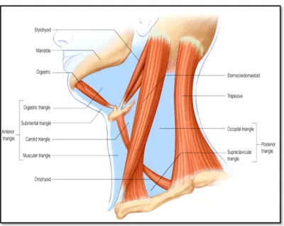
The neck is divided by the Sternocleidomastoid muscle into Anterior
Fig.1 : The triangles of the neck
The anterior triangle is subdivided by the digastric muscle and the
superior belly of omohyoid muscle into four triangles.
1) Submental triangle
2) Digastric triangle
3) Carotid triangle
4) Muscular triangle
The main contents of the Submental Triangle are Submental lymph
The main contents of the Digastric triangle are submandibular
salivary gland, submental artery, mylohyoid nerve and vessels, Internal
carotid artery, Internal jugular vein and the vagus nerve
The main contents of the Carotid triangle are The common carotid
artery and its termination, Internal & External carotid arteries, Internal
jugular vein, Common facial vein, Lingual vein, Vagus nerve, Superior
Laryngeal nerve, Hypoglossal nerve ,Spinal accessory nerve and
Sympathetic chain
The main contents of the Muscular triangle are the infrahyoid muscles
– Sternohyoid, Sternothyroid, Thyrohyoid and Omohyoid
The posterior triangle is further subdivided by the inferior belly of
omohyoid into two triangles
1) occipital triangle
2) supraclavicular triangle
The main contents of the occipital triangle are Occipital artery, Spinal
accessory nerve
The main contents of the supraclavicular triangle are the three trunks
suprascapular nerve, third part of subclavian artery, suprascapular artery and
vein and lower part of external jugular vein.
The other structures seen in the neck are the Thyroid Gland,
Submandibular Salivary Gland, Cervical portions of the Trachea and the
Oesophagus.20
ANATOMY OF THYROID GLAND
Thyroid (shield like) gland is one of the largest endocrine gland
located in the front of neck situated anterior and lateral to the junction of
larynx and trachea just inferior to the thyroid cartilage
It is a butterfly shaped gland with bilobed structure having two lateral
lobes connected by a midline isthmus. Occasionally there may be a
pyramidal lobe which represents the distal part of the thyroglossal duct
which can be seen from isthmus to as above as hyoid bone.
Each lateral lobe extends from the middle of thyroid cartilage to the
4th or 5th Tracheal ring opposite C 5, C 6, C 7 and T 1 Vertebrae.21
The size of the thyroid gland is variable and it depends on heredity,
environment and nutritional factors. The average weight is about 34 g (1.2
The gland is slightly heavier in females and enlarges during
menstruation an pregnancy. The Thyroid gland has a deep red coloration
because of the increased vascularity of the gland due to large number of
blood vessels supplying the gland.21
There are two capsules for the thyroid gland,
1) The true capsule formed by the peripheral condensation of the
connective tissue of the thyroid gland
2) The False capsule formed by the pretracheal layer of the deep
cervical fascia which is thin on the posterior border of the lateral
lobes and thick on the inner surface of the gland where it is called
as the ‘Suspensory ligament of Berry’ which connects the lateral
lobes to the cricoid cartilage.
The lateral lobes are conical in shape having an apex, base, three
Surfaces - Lateral, Medial and Postero- Lateral, Two Borders- Anterior and
Posterior.
The Isthmus has two surfacesanterior and posterior, two borders
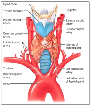
Fig.2 showing the Thyroid gland and its anatomic relations
BLOOD SUPPLY OF THE THYROID GLAND
The Arterial supply to the Thyroid gland is mainly by Two pairs of
Superior Thyroid artery and Inferior Thyroid artery.
The Superior Thyroid artery is a branch of the External Carotid artery
which arises just immediately above the bifurcation of the common carotid
pole. As the Superior Thyroid artery progresses medially, it is adjacent to
the external branch of the superior laryngeal nerve.
The Inferior Thyroid artery arises from the thyrocervical trunk. It
ascends into the neck behind the carotid sheath and then it arches medially
to enter the thyroid gland posteriorly usually near the ligament of Berry.
There is usually no direct arterial supply to the thyroid inferiorly. The
recurrent laryngeal nerve is usually directly adjacent to the inferior thyroid
artery in either anterior or posterior position. The Inferior thyroid artery
mostly supplies both the superior and inferior parathyroid glands
Occasionally a thyroidea ima artery may be present in less than 5% of
patients and it usually arises directly from the innominate artery or from the
aorta.
There are Three pairs of venous systems draining the thyroid gland.
Superior Thyroid veins are just immediately adjacent to the superior thyroid
arteries and they join the internal jugular vein at the level of the carotid
bifurcation. Middle Thyroid veins are present in more than half of
individuals and they course immediately laterally into the internal jugular
vein. The inferior Thyroid veins are usually two to three in number and they
descend directly from the lower pole of the thyroid gland into the
Kocher) may be present between middle and inferior thyroid veins which
may drain into the internal jugular vein.21
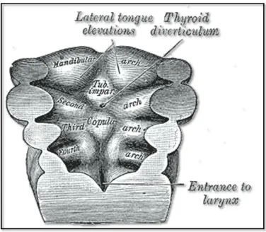
EMBRYOLOGY OF THYROID GLAND
The Thyroid gland arises initially as a tissue bud from a midline
diverticulum in the floor of the pharynx. The main portion of this structure
descends into the neck and develops into a butterfly shaped bilobar solid
organ. In the pharynx, the original attachment is the foramen cecum located
in the buccal cavity. This structure becomes the thyroglossal duct, which is
reabsorbed after 6 weeks of age. The very distal end of this duct may
occasionally be retained and form as a pyramidal lobe in the adult thyroid.22
Microscopic thyroid follicles first appear as the lateral lobes of the
thyroid gland develops. When the embryo is about 6 cm in length, these
follicles begins to develop colloid. In the third month the cuboidal follicular
cells first demonstrate iodine trapping, and thyroid hormone secretion
initially begins.23
Calcitonin-producing C cells arise from the fourth pharyngeal pouch
and they migrate from the neural crest into the lateral lobes of the thyroid.
These C cells migrate into the lateral and posterior upper two thirds of the
are present on only in the upper and middle areas of the thyroid gland,
[image:36.612.120.496.190.518.2]usually in the posterior and medial aspects.
Fig. 3: the thyroid diverticulum which gives rise to the thyroid gland
HISTOLOGY OF THYROID GLAND
The thyroid gland covered by a fibrous capsule is divided into lobules
by septa extending from the capsule. Each lobule is made up of aggregation
of follicles (100 to 300 micrometers). The portion of the thyroid gland
Fig.4 : Normal Histology of the Thyroid gland
Each spherical follicle is surrounded by a single layer of epithelial
cells and filled with pink stained proteinaceous material called colloid.
Colloid consists mainly of the thyroglobulin, a glycoprotein produced from
the endoplasmic reticulum and the golgi body of the follicular cells.
When the gland is inactive, the colloid content is abundant, the
follicles are large, and the follicular cells lining them are flat. When the
gland is active, the follicular cells are small, the cells are cuboid or
columnar, and areas where the colloid is being actively reabsorbed into the
thyrocytes are visible as "reabsorption lacunae"
Microvilli project into the colloid from the apexes of the thyroid cells
and canaliculi extend into them. The endoplasmic reticulum is prominent, a
feature common to most glandular cells, and secretory granules containing
thyroglobulin are seen. The individual thyroid cells rest on a basal lamina
that separates them from the adjacent capillaries. The capillaries are
fenestrated, similar to those of other endocrine glands25
PHYSIOLOGY OF THYROID GLAND:
Of the metabolically active hormones secreted by thyroid gland,
About 93 per cent is Thyroxine (T4), and 7 per cent Triiodothyronine (T3).
However, almost all the thyroxine is converted to Triiodothyronine in the
tissues. The functions of both these hormones are qualitatively the same.
They differ in rapidity and intensity of action. Triiodothyronine (T3) is four
times as potent as thyroxine. It is present in the blood in smaller quantities
and also persists for a much shorter time than thyroxine.26
Iodine is a key requirement in the formation of thyroid hormones and
a minimum of ingestion of 1mg/week is needed for the adequate formation
of the thyroid hormones. The steps in the formation of thyroid hormone
1) Iodide trapping
2) Oxidation of the iodide ion
3) Organification
4) Coupling27
Iodide trapping:
The first and foremost important step in the formation of thyroid
hormones is the transport of ingested iodides into the follicular epithelial
cells of the thyroid gland by the sodium and Iodide(Na+/I-) symporter in the
basolateral membrane of the thyrocytes. This is a energy dependant process
and is mainly regulated by the level of Thyroid stimulating hormone
(TSH).28, 30
Oxidation:
The trapped iodide ion must be transported across the apical
membrane to reach the thyroglobulin molecule. This is facilitated by the Cl
-/I- Exchanger in the apical membrane, also called as Pendrin. During this
exchange another enzyme Thyroid peroxidase converts the trapped iodide
Organification:
The Iodine molecule is attached to the Carbon 3 position of the
tyrosine residues in the thyroglobulin molecule in the colloid.29 This
reaction is called as organification and it is mediated by the enzyme
Iodinase. One tyrosine aminoacid attached to one Iodine constitutes Mono
Iodo Tyrosine (MIT). If two Iodine attaches to the Carbon 3 and 5 of the
Tyrosine, it is called as Di Iodo Tyrosine (DIT).
Coupling:
The two molecules of Di Iodo Tyrosine(DIT) combines to form
Thyroxine(T4) and one molecule of Di Iodo Tyrosine(DIT) comines with
one molecule of Mono Iodo Tyrosine(MIT) to form
Triiodothyronine(T3).This coupling reaction occurs within the thyroglobulin
molecule. In the normal thyroid, the average distribution of iodinated
compounds is 23% MIT, 33% DIT, 35% T4, and 7% T3.
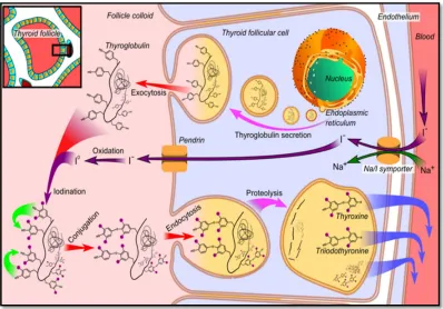
Release of Thyroid hormones:
On appropriate stimulation, the apical membrane extends pseudopods
to pinocytose Thyroglobulin molecules so that they are cleaved by the
intracellular lysosomes to release only Thyroxine and Triiodothyronine(T3)
Tyrosine(DIT) and Mono Iodo Tyrosine(MIT) are recycled back to the
[image:41.612.121.519.204.482.2]colloid.30, 31, 32
Fig. 5 : The synthesis of the thyroid hormone and the role of Thyroglobulin
NORMAL PHYSIOLOGICAL EFFECTS OF THYOID HORMONES
1) Basal metabolic rate (BMR)/ temperature regulation
Promotes normal oxygen use and BMR
Increases calorigenesis
2) Carbohydrate/ Lipid/protein metabolism
Promotes glucose catabolism
Mobilizes fats
Essential for protein synthesis
Enhances liver’s synthesis of cholesterol
3) Nervous system
Promotes normal development of nervous system in fetus and
infant
Promotes normal adult nervous system function
4) Cardiovascular system
Promotes normal functioning of the heart
5) Muscular system
Promotes normal muscular development and function
6) Skeletal system
7) Gastrointestinal system
Promotes normal GI motility and tone
Increases secretion of digestive juices
8) Reproductive system
Promotes normal female reproductive ability and lactation
9) Integumentary system
Promotes normal hydration and secretory activity of skin
REGULATION OF THYROID HORMONES:
Thyroid gland is under the negative feedback control by the Thyroid
Stimulating Hormone (TSH) which is produced by the anterior pituitary
gland. Thyroid Stimulating Hormone is under the negative feedback control
by the Thyrotropin releasing hormone (TRH) produced by the hypothalamus
When the body need thyroid hormones it is released by the thyroid
gland under the influence of Thyroid Stimulating Hormone. Thus in
hypothyroidism the thyroid hormone (T3 and T4) levels are low which can
be confirmed by the high levels of Thyroid Stimulating Hormone. In
Fig.6 showing regulation of thyroid hormones
THYROGLOBULIN – STRUCTURE & FUNCTIONS
Thyroglobulin is a large glycosylated protein synthesized from the
endoplasmic reticulum and the golgi apparatus of the thyroid follicular cells.
It has a molecular weight of 660 KDa with 5000 amino acids.33 Of the total
molecular weight , carbohydrate accounts for 8 – 10 % .Thyroglobulin is
made up of two large subunits. It contains 115 tyrosine residues each of the
The iodides account for 0.2 – 1 % and it depends on iodide quantity
in the diet. About 70% of the iodide in thyroglobulin exists in the inactive
precursors monoiodotyrosine (MIT) and diiodotyrosine (DIT) and 30% is
in the iodothyronyl residues Thyroxine(T4) and TriIodoThronine(T3).
When iodine in the diet is sufficient, the T4:T3 ratio is about 7:1. In iodine
deficiency, the ratio decreases as well as the DIT: MIT ratio.
As said earlier, Thyroglobulin is synthesized from the endoplasmic
reticulum and the golgi apparatus of the thyroid follicular cells near the
basal portion of the cell. It is transported across the membrane into the
colloid where it acts as a pre hormone in the formation of T3 and T4.The the
third and fourth stages of thyroid hormone synthesis namely organification
and coupling occurs in the thyroglobulin molecule.
Then the thyroglobulin molecule acts a storage form for thyroid
hormones .several weeks supply of thyroid hormones are stored in the
thyroglobulin so that when there is a stimulation by TSH, the whole
thyroglobulin molecule is endocytosed by the pseudopods from the apical
membrane of the thyrocytes.
Then by the phagolysosomal activity of the cell, the thyroglobulin
from the basal portion of the cells. Remaining Di Iodo Tyrosine(DIT) and
Mono Iodo Tyrosine(MIT) are recycled back to the colloid.33,34
Thus the Thyroglobulin molecule acts as a
1) Pre hormone for the synthesis of thyroid hormones
2) Site of synthesis of thyroid hormones
3) Storage form of thyroid hormones35
Usually for a normal thyroid gland, the reference range of
thyroglobulin values is taken as 3 - 40 ng/ml. The values depend on the age
of the individual.
Table 1 illustrating the normal levels of serum thyroglobulin Age Reference Interval
6 months - 3 years 7.4-48.7 ng/mL
4-7 years 4.1-40.5 ng/mL
8-17 years 0.8-29.4 ng/mL
USES OF THYROGLOBULIN:
Thyroglobulin is a glycosylated protein which is secreted only by the
thyroid gland alone and not anywhere else in the body. Usually it is not
secreted into the blood along with the thyroid hormones. But it can be found
in very minute amounts only. The reference range of thyroglobulin values is
taken as 3 - 40 ng/ml.36
In Thyroid Carcinomas Usually of Papillary and Follicular type, the
thyroglobulin levels are increased due to destruction of the follicles and
release of thyroglobulin from the colloid into the blood stream. Therefore
the thyroglobulin is used as a tumour marker in Thyroid Carcinoma and the
levels are studied in the Post operative period following Total
Thyroidectomy procedures for Thyroid Carcinomas to look for any
recurrence of the carcinoma or any remaining thyroid tissue after surgery.37
Factors which may affect the values of thyroglobulin levels:
The following are the factors affecting the Thyroglobulin levels.38
1. Thyroid carcinoma
2. Hashimoto’s Thyroiditis
3. Graves disease
Thyroid carcinoma:
Thyroid carcinomas are mainly of four types. They are
1. Papillary carcinoma
2. Follicular carcinoma
3. Medullary carcinoma
4. Anaplastic carcinoma
Some other carcinomas are Hurthle cell carcinoma, Mixed Papillary
and follicular carcinomas.
Papillary carcinoma is the most common type of thyroid carcinoma
most commonly occuring in the females of less than 40 years. They have an
excellent prognosis if detected and treated earlier. On Histopathological
examination, it shows a typical pattern of Orphan anne nuclei. They can be
classified into well differentiated and poorly differentiated carcinoma on
Histopathological examination.
Follicular carcinoma is the second most common thyroid carcinoma
occurring most commonly in older females. They have a good prognosis
with less mortality rate. It most commonly occurs in the functioning thyroid
Medullary carcinoma occurs in the Parafollicular C Cells and
Anaplastic Carcinoma occurs in the elderly individuals. Both these are less
common thyroid malignancies and have bad prognosis with an increased
mortality rate.
Papillary and Follicular Carcinoma can raise the serum thyroglobulin
levels due to the destruction of the follicles. They can interfere with the
values in the study, thus giving false positive and are promptly ruled out by
the Histopathological examination of the thyroid gland taken out during the
autopsy.39, 57, 58
Hashimoto’s Thyroiditis:
Hashimoto’s thyroiditis is an autoimmune disease characterised by
the presence of antibodies to normal thyroid tissue. The antibodies in
Hashimoto’s thyroiditis are mainly of three types. They are
1. Antibody to Thyroglobulin
2. Antibody to thyroid peroxidase and
3. Antibody to TSH Receptor
These antibodies mediate complement fixation and killing by Natural
killer cells (NK cells). Hashimoto’s thyroiditis is also mediated by CD 4 + T
Hashimoto’s thyroiditis is commonly seen in women and mostly
presents as a firm nodular gland with features of hypothyroidism. 5 % of the
patients may present with features of hypothyroidism. On gross examination
the thyroid gland is mildly enlarged throughout and has a granular, nodular
and firm surface.21
On microscopic examination the gland is diffusely infiltrated by small
lymphocytes and plasma cells .Thyroid follicles are smaller with reduced
amounts of colloid and increased interstitial tissue. The follicles are lined by
Hurthle or Askanazy cells which are characterized by abundant
eosinophilic, granular cytoplasm.
If present in cases of neck compression it produces false positive
results and hence it can be eliminated by histopathological study of the
thyroid gland.
Graves’s disease:
Graves’s disease or diffuse toxic goitre is an autoimmune disease
with more female preponderance characterised by diffuse goitre,
thyrotoxicosis, ophthalomopathy. The thyroid-stimulating antibodies
stimulate the thyroid epithelial cells to grow and synthesize excess thyroid
thyroid gland leading to false positive increase in serum thyroglobulin
levels.
On Gross the thyroid gland in patients with Graves' disease is
diffusely enlarged with increased vascularity. Microscopically the gland is
hyperplastic and the epithelium is columnar with minimal colloid. There
may be aggregates of lymphoid tissue and vascularity is markedly increased.
Graves’s disease produces false positive rise in serum thyroglobulin
levels and hence on post-mortem examination it can be ruled by
histopathological examination of the thyroid gland.37, 42
Toxic Multinodular goitre:
Toxic multinodular goitre most commonly arises in the long standing
non toxic multinodular goitre and has a female preponderance. This also
presents with destruction of the thyroid gland leading to false positive
increase in serum thyroglobulin levels and hence on post-mortem
examination it can be ruled by histopathological examination of the thyroid
gland.40, 43
ASPHYXIA:
As stated earlier, Asphyxia means ‘Pulselessness’. But as time
According to Adelson asphyxia is the physiologic and chemical state in a living organism in which acute lack of oxygen available for cell
metabolism is associated with inability to eliminate excess of carbon
dioxide.41
The word Anoxia originally means lack of oxygen.
Barcroft divided Anoxia into three types.
1) Anoxic anoxia – due to prevention of oxygen from reaching the
lungs
2) Anaemic anoxia – due to decreased oxygen carrying capacity of
the blood due to low hemoglobin
3) Stagnant anoxia – due to impaired circulation of blood 44
Peters and Van Slyke added the fourth type of anoxia
4) Histotoxic anoxia – due to non utilisation of the freely available
oxygen by the cells.
Histotoxic anoxia is further subdivided into
a) Extracellular – tissue oxygen enzyme system is inactivated as in
b) Pericellular – oxygen cannot permeate cells due to lipid soluble
anaesthetic agents as in chloroform, halothane, etc.
c) Substrate – there is inadequate food for cell’s metabolism
d) Metabolic – the end products of the cellular respiration cannot be
removed as in uraemia or carbon dioxide poisoning44
CLASSICAL SIGNS OF ASPHYXIA:
The so called classical signs of asphyxia are non specific and can also
occur in deaths due to other causes.
1. Cyanosis
2. Congestion
3. Fluidity of blood
4. Petechial hemorrhages45
CYANOSIS:
Cyanosis in Greek means ‘dark blue’. Cutaneous cyanosis depends
mostly on the absolute amount of reduced hemoglobin rather than the
amount of oxy hemoglobin. There must be at least 5 gm % of reduced
hemoglobin before cyanosis becomes evident irrespective of the amount of
In neck compression cyanosis follows congestion of face due to much
amount of reduced hemoglobin in venous blood and also partly due to the
compression of the airway leading to reduced oxygenation of the blood in
the lungs. This depends on the complete or partial obstruction of the airway.
When the deceased is examined within few hours of death the
presence of cyanosis is of some significance. The appearance of cyanosis
after 24 hours may be even due to the post-mortem changes. Absence of
cyanosis within few hours of death does not signify that cyanosis was
present at the time of death.
Cyanosis can also occur in other forms of death. So it is a non
specific sign of asphyxia.47
CONGESTION:
Congestion of viscera in asphyxia deaths is due to the capillo venous
dilatation which occurs due to the susceptibility of the capillaries to
hypoxia. It leads to dilatation of capillaries resulting in stasis of blood in the
capillaries and venules. This is not specific only for deaths due to
interference in respiration but also to deaths due to other causes also.
Congestion may be important finding in neck compression if the
Ligature strangulation. Usually in hanging the congestion above the ligature
mark is minimal when compared to strangulation.47, 48
PETECHIAL HEMORRHAGES:
Petechial hemorrhages otherwise called as Tardieu spots are pinpoint
hemorrhagic spots seen mostly in the visceral pleura and epicardium wee
first described a French Police surgeon Tardieu49 in 1866. He said that these
were pathognomic of deaths due to mechanical asphyxia. Later in 1944
Gordon and Lime found these kinds of petechial hemorrhages in many
forms of death.50, 51
Shapiro, in 1955 described that some of the petechial hemorrhages
were post mortem artefacts and disappeared in very short period.52 In 1981
Zaini and Knight confirmed that only one third of these petechial
hemorrhages were due to mechanical asphyxia and res two thirds were
false.53
These Tardieu spots develop due to the following factors
1. Increased stasis of blood in the venules causing congestion which
leads to the increased venous pressure causing rupture of the venules
FLUIDITY OF BLOOD:
The fluidity of the blood in deaths due to mechanical asphyxia may
be due to activation of the fibrinolytic system leading to increased levels of
fibrinolysins. However the fluidity and the amount of fibrinolysin mostly
depend on the rapidity of death rather than the nature of death.48
NECK COMPRESSION - TYPES
From the medico - legal point of view, asphyxia can be grouped into
Mechanical and Non-Mechanical causes.
Mechanical causes includes
1. Pressure to the exterior of the neck : Hanging, Strangulation, etc.
2. Obstruction of the airways from the exterior: suffocation,
Smothering, etc.
3. Obstruction of the airways from the interior: Gagging, Choking,
etc
4. Pressure upon the chest: Traumatic asphyxia
5. Occlusion of airways by fluid: Drowning
Neck compression can happen in many ways. The main occurrences
1. Hanging
2. Strangulation – Ligature Strangulation or Manual Strangulation or
Throttling
3. Other forms of Neck Compression – Mugging, Garotting,
bansdola, Strangulation in the bend of knee, Foot Strangulation 54
HANGING:
Hanging is one of the forms of neck compression causing asphyxia
which is caused by suspension of the body by a ligature encircling the neck
where the constricting force being the weight of the body2
Types of Hanging:
Hanging can be classified based on various categories.
1. Position of Knot: A. Typical hanging B. Atypical hanging 2. Position of feet:
A. Complete hanging B. Partial hanging 3. Manner of hanging:
4. Mode of hanging: A. Judicial B. Sexual C. Lynching
Complete hanging:
Complete hanging is a type of hanging where the body is fully
suspended in the air with the foot does not touching the ground and the
constricting force is the weight of the body2 ,3
Incomplete hanging:
Incomplete hanging is a type of hanging where the body is not fully
suspended in the air with the toes, foot, knees, buttocks does touch the
ground and the constricting force is the weight of the head
(about 5 -6 kg). 2,3
Typical Hanging:
Typical Hanging is a type of hanging where the position of the knot is
present over the occipital region and the ligature mark is prominent over the
front of the neck .2, 3
Atypical Hanging:
Suicidal Hanging:
Most commonly hanging is a mode of suicide. According to National
crime records bureau statistics of 2013 shows that 37 % of the suicidal
deaths in India are due to Hanging. Tamil Nadu tops the list of number of
suicides in the country and in hanging Tamil Nadu comes in the third
place.63, 64
Age of the victim can be in both extremes of life and both the sexes
are prone for this mode of death. Suicidal Hanging can be corroborated with
evidences like suicide notes in the deceased handwriting, secluded place of
occurrence, very easily approachable point of suspension and easily
accessible ligature material.47
Accidental Hanging:
Accidental hanging can occur while at work, during playing,
exhibiting hanging exercises or showing some performances in the circus,
auto erotic masochistic sexual exercises, etc. It can also occur when a
person’s neck gets compressed below the chin by getting suspended from a
steering wheel of a car, edge of a sofa or an arm of a chair.
While at work for example in a factory, a worker may fall from a
accidentally hanged if a restraining apparatus slips or a cloth tightened
around the neck while crawling.48
Homicidal Hanging:
Homicidal hanging is very rare but can occur ii the victim is a child,
incapacitated by drugs or alcohol, or if the victim is rendered unconscious
by a blow to the head. It is obvious that homicidal hanging to be
accomplished single handedly. There may be evidence of dragging or
pulling the victim as evidence of friction. The hands of the victim may show
some foreign materials like hair, button and piece of clothing. There may be
signs of struggle or presence of injuries on the body48
LYNCHING:
The term Lynch means to put a person to death by a mob action for
an alleged offence without a legal trial. It was named after Captain W Lynch
who practiced this mode of hanging in Virginia. At first the victim is beaten
with iron rods by the mob and after he falls unconscious he is dragged to the
village square to be hanged till death. 2,3
POSTMORTEM FINDINGS IN HANGING:
The post-mortem findings in hanging depends on the mechanisms of
EXTERNAL FINDINGS:
FACE:
The face in case of hanging may be pale if the death is due to vagal
inhibition or if there is any injury to the spinal cord. The face may appear
congested if the death to asphyxia and if its due to apoplexy it is markedly
congested.
If the hanging is complete there will be occlusion of carotid arteries
and the face may be pale with minimal petechial hemorrhages. If the
hanging is incomplete there may not be complete occlusion of the carotid
arteries and the face appears much congested with increased petechial
hemorrhages over face, conjunctivae, forehead, etc2,3
EYES:
The eyes may be closed or open. More often the eyes are protruded
out with visible petechial hemorrhages in the subconjuctival region. ‘La
Facies sympathique’ is a condition produced when there is a pressure in the
cervical sympathetic chain on one side the eye on the same side will remain
TONGUE:
The tongue is usually swollen and blue in colour with forcing of the
tongue between teeth. The tongue is also pushed upwards against the
posterior pharyngeal wall thereby occluding the airway. The protruding part
of the tongue is dark in colour with minimal petechial hemorrhages on its
surface
SALIVA:
Saliva may be found dribbling from the mouth opposite the side of
the knot. This may be produced due to the stimulation of salivary glands or
due to congestive hypoxia. Dried marks of saliva may be present in the
deceased suggesting that the hanging is ante mortem. But the absence of the
dried salivary stains will not suggest post-mortem hanging since it may be
absent in deaths due to vagal inhibition or injury to the spinal cord.47
HANDS:
Hands are usually found clenched and may some fibres of the ligature
material or any other material exchanged from the assailant in case of
GENITALS:
Penis may be found engorged due to hypostasis with emission of
semen at its tip sometimes. In females the vagina may be found with
turgescence with discharge of blood stained fluid sometimes. Urine and
faeces may sometimes escape through urethra and anus due to relaxation of
sphincters
POSTMORTEM STAINING:
If the body is suspended in upright position as per rule the
postmortem hypostasis will be found in the dependant part in this case lower
limbs, forearm, hands and genetalia. There may be evidence of petechial
hemorrhages in the skin of the legs. If the ligature is cut and the body is
transferred to supine position before the fixation of the postmortem
hypostasis secondary areas of hypostasis will be developed in the dependant
parts
CYANOSIS:
Cyanosis will be seen in lips, fingernails, tip of nose and ear lobules
and the cyanosis may be seen marked if the point of suspension is from a
LIGATURE MARK:
The ligature mark is the principle external sign in cases of hanging. It
is a type of pressure abrasion most commonly running obliquely around the
neck above the level of thyroid cartilage. The ligature mark appears in the
form of groove or furrow deepest in the opposite side of the knot. It is
usually yellowish or yellowish brown after death but gets dried up due to
exudation of tissue fluid assuming parchment like consistency.87
When the knot is in the nape of the neck as in typical hanging the
ligature mark is more pronounced at the anterior part of the neck. The
ligature mark mostly never completely encircles the neck due to intervening
hair in the nape of the neck and the firmness of the structures of back of the
neck. It assumes the shape of inverted ‘v’ with the apex of the v
corresponding to the site of the knot. Most commonly the knot is located in
the either side of the neck or in the nape of the neck. Very rarely it is
situated below the chin.62
The width of the ligature mark may be equal or less than the with of
the ligature material. The ligature mark may be patterned exhibiting the
nature of the ligature material. Sometimes the ligature mark may be faint in
confirm it. There are various factors influencing the appearance of the
ligature mark.2,3They are
1. Composition of ligature material 2. Mode of application of ligature 3. Position of the knot
4. Course of ligature around the neck 5. Period and degree of suspension 6. Slipping of the ligature
7. Weight of the body3
Composition of ligature material:
The ligature material may be any kind of material like rope made of
cotton, jute, nylon , dhotis, saree, bed sheets, thupattas, belts, brace, etc. In
cases of suicidal hanging any of the easily available materials can be used as
a ligature material.
The ligature mark is marked and deep if the ligature material is tough
and narrow. The ligature mark is less prominent if the ligature material is
soft and broad. In a folded cloth producing ligature mark the width and
appearance of the ligature mark may vary significantly. In cases of hanging
where nylon, Terylene, silk is used as a ligature material the ligature mark
Mode of application of ligature:
In most cases of hanging, fixed loop is applied where the ligature
mark is seen as obliquely above the level of thyroid cartilage opposite the
side of the knot. Ligature marks produced by running noose may have a
horizontal pattern mostly above the level of thyroid cartilage
Position of the knot:
Most commonly the position of the knot is to the right or left side of
the neck as atypical hanging is most common type of hanging. Next is the
knot at the nape of the neck. Knot below the chin is very rare in cases of
hanging.
Course of ligature around the neck:
Ligature mark in hanging is most commonly seen above the level of
thyroid cartilage and it runs obliquely around the neck in the form of
inverted ‘v’ with the apex of the v corresponds to the site of the knot. Some
studies showed that the ligature mark can be seen at the level of thyroid
cartilage in 15% of cases and below the level of thyroid cartilage in 5% of
Period and degree of suspension:
If the period of the suspension of the body is more the ligature mark is very likely to be prominent and have parchment like pattern. In cases of complete hanging the ligature mark is more prominent than the case of partial hanging
If the period of the suspension of the body is less the ligature mark is very likely to be less prominent. When the hanging is from the low point of suspension, the ligature mark may have a horizontal course at the level of thyroid cartilage with increased findings of venous congestion.
Slipping of the ligature:
The slipping of the ligature may prevent the formation of the more prominent ligature mark but may produce a wider ligature mark or abrasion due to frictional displacement of the ligature material
Weight of the body:
The ligature mark is more prominent if the weight of the body is more and vice versa
Ligature mark may not be well pronounced
1. If the ligature material used is soft and broad
INTERNAL FINDINGS:
The internal findings in the post-mortem of cases of death due to
hanging carries a significant importance as many artefacts need to be
differentiated from the normal neck findings in the hanging so that
strangulation is ruled out as a cause of death.
Schrader in 1940 recommended that the neck dissection has to be
done at last in cases of hanging after removing the brain and the heart so
that seepage of blood from the neck vessels is minimal while examining
neck in the postmortem of cases of hanging. Prinsloo and Gordon artefacts
are more common if the dissection of the neck is not done atlast in cases of
hanging.
As a rule there should not be any bruising in the subcutaneous tissue,
muscles of the neck in cases of hanging to differentiate it from the
strangulation. The subcutaneous tissue under the ligature mark is very dry,
white and glistening if the body is suspended for a long time. There may be
presence of petechial hemorrhages in the adjacent tissues above and below
the ligature mark.
Muscle fibres of the platysma and sternocleidomastoid (5- 10%) gets
Hemorrhages in the strap muscles may be present in about 25 % cases if the
force is considerable.
There may be presence of tears in the intimal layers of the carotid
arteries (5–10%) usually around the region of the carotid sinuses with
extravasation of the blood in cases of death due to hanging with long drop
According to Gordon et al, histopathological examination of the skin
showing ligature mark may show tissue reaction which will be seen after a
few hours.
Fracture of the thyroid cartilages, cricoid cartilage and trachea are
uncommon in cases of death due to hanging. Fracture of thyroid cartilage
can occur in 40% of deaths due to hanging above the age of 40 years.
Fracture of hyoid bone is also uncommon in cases of death due to
hanging. Incidence of Fracture of the hyoid bone in hanging varies in
different studies from 0-60%. Usually the average is 15-20%. Fractures of
the hyoid bone are rare below 40 years due to the elasticity of the cartilages
and the mobility of the joints. Fracture of the hyoid bone usually occurs in
the superior horns at the junction of inner two thirds and outer one third.
Fracture of hyoid bone is divided into three types. They are
2. Anteroposterior compression fractures most common in hanging
3. Traction or tug or avulsion fracture
In cases of hanging the hyoid bone is forced directly backwards
leading to greater divergence of the superior horns of the hyoid bone
resulting in the fracture and outward displacement of the posterior segment.
In these type of fracture the periosteum is torn on the inner side so that the
fragment moves easily outwards and the inward movement is possible only
upto normal position. When the pressure is more the fragment detaches
from the main hyoid bone and should be found out during the postmortem
examination
Usually the hyoid bone fracture is unilateral but can also occur
bilaterally. In some cases of bilateral fracture one side may have an inward
compression fracture and the other may show an outward fracture. This is
due to impingement of the posterior end of the hyoid bone getting struck
against a bony ridge or a vertebra.48
STRANGULATION:
Strangulation is a form of asphyxial death by neck compression
ways:-Application of ligature as in Ligature Strangulation
Application of human hands as in Manual Strangulation
Any other means – stranglehold, foot or by some solid substance
Strangulation is mainly classified based on
A. Material used:
1. Ligature strangulation
2. Manual strangulation
B. Manner of strangulation:
1. Suicidal strangulation
2. Homicidal strangulation
3. Accidental strangulation
Ligature Strangulation:
Asphyxia produced by constriction of the neck by a ligature without
suspension of the body is called as Ligature Strangulation.
Manual strangulation:
Asphyxia produced by the compression of the neck by human hands
Suicidal strangulation:
Suicide by strangulation is rare but possible. The signs of struggle and
resistance will be absent apart from the absence of injuries to the deeper
structures of the neck being insignificant. The signs of venous congestion
are markedly seen and the ligature is found in situ. Suicidal strangulation
can happen in the following ways. 2,3
Application of the ligature once or many times and finally tying
the free ends
Application of ligature by tourniquet mechanism i.e., applying the
ligature only once in the neck following which a small piece of a
rod or stick is passed through the ligature and twisted as a lever.
After the consciousness is lost, the stick gets struck under angle of
jaw thus maintaining the compression
Application of a running noose to the neck and passing the free
end several times around the right hand thereby strangulating
wherein weight of the hand and forearm acts as a constricting
force
Application of a running noose and attaching a weight to the free
Homicidal strangulation:
Strangulation is a common form of murder and strangulation is
always assumed to be homicidal until proved otherwise. Injuries to the neck
structures are more severe and extensive in cases of homicidal strangulation.
Evidence of signs of struggle is common unless the victim is child or
incapacitated by drugs or alcohol. Also in old age, females there may be less
signs of struggle.
Many of the victims may be adult women and frequently
strangulation may be associated with sexual assault. Usually there is a single
turn of ligature and if there is more than one ligature mark homicide is
almost certain. Fingernail marks may be seen if victims tries to resist the
ligature or by application of hands by assailant in manual strangulation. The
signs of venous congestion are markedly seen in cases of strangulation
noted by subconjuctival hemorrhages in the eyes, intense cyanosis of the
lips, nose and ear bleeding, marked congestion of the parts above the neck
compression.2,3
Accidental strangulation:
Accidental strangulation is very rare and can occur in some
Cord entanglement of the foetus before or during the process of
birth
Strangulation of children while playing2,3
Persons under the influence of alcohol, epileptics and old
individuals fall into a situation where they can’t get out and die of
strangulation
Workers getting accidentally strangled by a clothing caught in a
machinery
Accidental throttling may occur by sudden application of pressure
over neck of other person by hands though as a joke or as a
symbol of affection
POSTMORTEM FINDINGS IN LIGATURE STRANGULATION:
EXTERNAL FINDINGS:
The asphyxial findings in strangulation will be more prominent more
than hanging. The face may be swollen with petechial hemorrhages over the
eyelids, face, forehead and scalp. The eyes are fully suffused and bulged out
with dilated pupils. The tongue is swollen, protruding and caught between
the teeth. Frothy fluid may be seen exuding from the mouth and the nostrils.
The ligature material used in ligature strangulation leaves a ligature
mark whose depth is inversely proportional to the width of the constricting
material. If a material whose width is smaller like rope, twines, wires, etc
are used it produces a deep ligature mark. If a broad material like saree,
dhoti, thupattas, etc is used it produces shallow ligature mark. 2,3
Usually the ligature mark is yellowish or yellowish brown in colour
completely encircling the neck at the level or below the thyroid cartilage.
Sometimes the mark may be absent on the back due to increased
musculature in the back. Homicidal ligature strangulation may have a U
shaped ligature mark due to the attacking of the victim from the back by the
assailant.62
The ligature mark produced depends on the material used for ligature
strangulation. The pattern of the ligature mark can be seen well with the
help of oblique lighting and magnifying glass. The ligature material can be
compared with the ligature mark if its available during the postmortem
Occasionally there may be more than one ligature mark due to more
than one turns of the ligature material around the neck. The ligature mark
will be more marked depending on the duration and period of constriction of
The ligature mark may be absent if any material like clothing, hair,
victim’s fingers, etc intervenes between neck and ligature material. There
may be presence of fingernail abrasions caused either by victim or assailant,
congestion of the face, petechial hemorrhages around the ligature mark.
INTERNAL FINDINGS:
On dissection of the neck the presence of bruising of subcutaneous
tissues and the neck muscles are the hallmark of ligature strangulation
which helps in differentiating them from hanging. Bruising may be present
even if the ligature mark is not present due to incomplete pressure by the
ligature material.
Injuries to the blood vessels are rare in ligature strangulation. Injury
to the hyoid bone is also rare in cases of ligature strangulation as compared
to hanging since the level of compression is most commonly below the
hyoid bone. But anteroposterior fracture may appear if a broad ligature is
used or if the level of compression is at the level of hyoid. There may be
presence of petechial hemorrhages in the muscles of the neck47
Lungs may show petechial hemorrhages also called as Tardieu spots
along with the presence of emphysematous bulla at occasional places. All
POSTMORTEM FINDINGS IN MANUAL STRANGULATION:
The postmortem findings in cases of death due to manual
strangulation can be grouped into
Cutaneous abrasions