“A COMPREHENSIVE STUDY ON COMPLICATIONS OF LAPAROSCOPIC INGUINAL HERNIA REPAIR”
A DISSERTATION SUBMITTED TO
THE TAMILNADU DR.M.G.R MEDICAL UNIVERSITY
In partial fulfillment of the regulations for the award of the degree of MASTER OF SURGERY (GENERAL SURGERY)
BRANCH I: M.S (General Surgery)
DEPARTMENT OF GENERAL SURGERY
GOVERNMENT STANLEY MEDICAL COLLEGE AND HOSPITAL
THE TAMILNADU DR.M.G.R MEDICAL UNIVERSITY
CHENNAI
CERTIFICATE
This is to certify that the dissertation titled “A COMPREHENSIVE STUDY ON COMPLICATIONS OF LAPAROSCOPIC INGUINAL
HERNIA REPAIR ” is the bonafide work done by
DR.KRISHNABHARATH.S Post Graduate student (2012 – 2015) in the Department of General Surgery, Government Stanley Medical College and
Hospital, Chennai under my direct guidance and supervision, in partial fulfillment of the regulations of The Tamilnadu Dr. M.G.R. Medical University, Chennai for M.S., Degree (General Surgery) Branch - I, Examination to be held in April 2015.
Prof. S. VISWANATHAN, M.S.,
Professor and Head of the Department Dept. of General Surgery,
Stanley Medical College, Chennai -600 001.
PROF.A.L.MEENAKSHI SUNDARAM, M.D., D.A.,
The Dean,
DECLARATION
I, DR.KRISHNABHARATH.S solemnly declare that this dissertation titled “A COMPREHENSIVE STUDY ON COMPLICATIONS OF
LAPAROSCOPIC INGUINAL HERNIA REPAIR” is a bonafide work done by me in the Department of General Surgery, Government Stanley Medical College and Hospital, Chennai under the guidance and supervision of my unit chief.
Prof. S. VISWANATHAN, M.S
Professor and head of the department of Surgery
This dissertation is submitted to The Tamilnadu Dr. M.G.R. Medical University, Chennai in partial fulfillment of the university regulations for the award of M.S., Degree (General Surgery) Branch - I, Examination to be held in April 2015.
Place: Chennai.
ACKNOWLEDGEMENT
It gives me immense pleasure for me to thank everyone who has helped me during the course of my study and in preparing this dissertation.
My sincere thanks to Dr. A. L.MEENAKSHI SUNDARAM M.D, D.A., the Dean, Govt. Stanley Medical College for permitting me to conductthe study and use the resources of the College.
I am very thankful to the chairman of Ethical Committee and members of Ethical Committee, Government Stanley Medical College and hospital for their
guidance and help in getting the ethical clearance for this work. I consider it a privilege to have done this study under the supervision of my
beloved Professor and Head of the Department Prof. S.VISWANATHAN, M.S, who has been a source of constant inspiration and encouragement to accomplish this work.
I express my deepest sense of thankfulness to my Assistant Professors Dr. T. BABU ANTONY, M.S, Dr. C. MANIMEGALAI, M.S, DGO,
DR. S. THIRUMURUGANAND, M.S, DR. D.S. KUMARESAN, M.S, MRCS,
DR. M. KARTHIKEYAN, M.S, for theirvaluable inputs and constant
I express my sincere gratitude to my mentors Prof. P.DARWIN former Professor and Head of the Department of General Surgery.
I am particularly thankful to my fellow postgraduate colleagues
Dr. S. Shankar and Dr.J.Pandiaraja and other fellow postgraduates for their valuable support in the time of need throughout the study.
I thank my Seniors Dr.Vishnu varathan, Dr. Ramamoorthi,
Dr. Sadasivam, Dr.Venkatesh, Dr.Murugan, Dr. Robin Prabhu, my junior PG’s Dr.S.Chelladurai,Dr.Mruthul mathew,Dr. T.Jayalakshmi, Dr. S.Vijaya sarathy, Dr. S. Mathan shanker,Dr.N. Fazil Navabjan, Dr. K. Anbarasan who supported me in completing the dissertation.
It is my earnest duty to thank my dear parents, sisters and brothers without whom accomplishing this task would have been impossible. I am extremely
thankful to my patients who consented and participated to make this study possible
TABLE OF CONTENTS
Sl. No Contents Page No.
1 INTRODUCTION 1
2 AIMS AND OBJECTIVES 2
3 REVIEW OF LITERATURE 3
4 METHODOLOGY 87
5 OBSERVATIONS 89
6 DISCUSSION 106
7 SUMMARY 114
8 CONCLUSION 116
9 BIBLIOGRAPHY
10 ANNEXURES
a. Proforma
ABSTRACT
AIM OF THE STUDY:
The aim of this study is to understand the intraoperative and
postoperative complications of laparoscopic inguinal hernia repair (TEP,TAPP) and formulate methods to prevent them.
MATERIALS AND METHODS:
This is a observational study consists of50 patients who underwent laparoscopic inguinal hernia repair(TEP,TAPP) in our institution during 2012 to 2014.With the patient consent all their intra operative and postoperative
complications were documented. Exclusion criteria includes other abdominal wall hernias, femoral hernia and patients who require emergency exploration.
OBSERVATIONS:
The following factors were taken into account,
1. Type of hernia
2. Type of hernia Vs complications
4. Operation time Vs complications.
5. Postoperative hospital stay.
Our observations were,
Overall 50 patients were operated-5 u/l direct,29 u/l indirect, 7 b/l direct and 7b/l indirect.
All were operated under general anesthesia.
The mean operative time was 66.8 minutes
There was no major complications
Minor complications rate were 46% (23 patients)
There was 4 minor complications namely, 1.surgical emphysema 2.groin pain, 3.shoulder pain, 4.scrotal edema
Surgical emphysema(21patients,42%) depends upon operation
time
Groin pain(11 patients-22% all are indirect) depends upon type of hernia mainly in indirect type, because of the dissection carried
Shoulder pain (6 patients-12%) is directly proportional to the time of surgery (all were >90 minutes)probably due to retention of CO2
which lead to diaphragmatic intervention.
Scrotal edema(6 patients-12%) depends upon the type of hernia as it occurred only in indirect hernias due to the dissection for
indirect sac
All these minor complications were subsided with supportive care without any surgical intervention.
Mean operation time in our study is 66.8 minutes.
Mean Post operative hospital stay- 2.6 days.
Laparoscopic hernia repair has a steep learning curve and time consuming in its initial phase but in later stage it can be done with
shorter duration without any complications and with early
postoperative recovery.
CONCLUSION:
Laparoscopic hernia repair has a steep learning curve and fearsome complications but once mastered , it is the safest and efficacious technique with reduced operative time and early postoperative recovery. .In our study (TEP) we encountered only minor complications all those complications were managed conservatively.
INTRODUCTION
Hernios means budding in greek. Earliest written records dealing with inguinal hernia dates back to 1500 b.c . After so many ups and downs by the first decade of the 19th century, giants such as Astley cooper ,Franz hesselbach and Antonio
scarpa facilitated the development of modern hernia repairs.later Ger and his colleagues revolutionized the concept of open repair into so called laparoscopic repair.
Of all abdominal hernias 75% occur in inguinal region.
Groin hernias can be performed conventionally i.e open repair or by laparoscopically
Laparoscopic inguinal herniorrhaphy is technically more challenging than tension free repair,thus has a long learning curve but it underwent numerous
modifications after its invention like Trans abdominal pre peritoneal repair(TAPP) Trans extra peritoneal repair(TEP) but these revolutionary concepts did not
drastically reduce the complications.
OBJECTIVES
# To understand the complications of laproscopic inguinal hernia repair both intraoperatively and postoperatively.
#To know the limitations of laproscopic inguinal hernia repair
#To formulate the methods to prevent the complications of laproscopic
hernia repair .
.
REVIEW OF LITERATURE
The earliest written records dealing with inguinal hernias date back to approximately 1500 bc Early operations involved ligation of the sac and cord at the level of the external ring with excision of the sac, cord, and testis. Notable figures such as Herophilus, Erasistratus, Heliodorus and Galen, influenced by Hippocrates, “the father of medicine,” and Aristotle, “the philosopher,” performed and wrote about hernia surgery
The Renaissance (15th through mid-17th centuries) heralded many improvements for society, and surgery was no exception. Ambroise Pare is considered by many to be the father of modern surgery. The use of anesthesia was reinstated for inguinal hernia surgery, and preserving the testicle became an essential part of the operation as described by Casper Stromayr in 1559. The 18th
.
century surgeon/anatomists were the first to publish treatises with illustrations based on detailed anatomic dissections. Sir Percivall Pott's refuted the older theories concerning the cause of hernias and methods of treatment. While being the first to describe congenital hernias, he also gave a classical description of the operative repair of some the complicated hernias. Richter, a German surgeon, described the partial enterocele strangulation. A French contemporary, Alexis Littre, described herniation of a Meckel diverticulum. Jean Louis Petit
recommended surgical repair of strangulated hernias only and described an external herniotomy without entering the sac, an operation that is eponymously linked to him. He also described the inferior lumbar triangle formed by the latissimus dorsi muscle, external oblique muscle, and iliac crest. It was John Hunter who renamed the lacunar ligament as Gimbernat's ligament after the Spanish anatomist described his technique of incision of the lacunar ligament for reduction of femoral hernia contents. Camper, a physician and philosopher, was the first to describe the processus vaginalis and the superficial fascia laying over the subcutaneous tissue.
fascia and closing the internal ring when repairing an inguinal hernia. Furthermore, he emphasized the need for antisepsis. Edoardo Bassini, another pupil of Lister, described his technique of dissecting and ligating the sac high in the retroperitoneal space after dividing the transversalis fascia and emphasized the importance of including the transversalis fascia in his posterior wall buttress, which involved suturing the internal oblique and transversus abdominis with the upper layer of the transversalis fascia in one layer (Bassini's famous triple layer!) to the lower leaf of the transversalis fascia and the inguinal ligament with interrupted silk sutures. These phenomenal results have earned him the title of Father of Modern Herniorrhaphy..
Proponents of prosthetic material began to express the opinion that these materials might be the solution for achieving the holy grail of a “tension-free” repair as early as the 1950s. However,Modern hernia specialists such as Lichtenstein in 1986 and Gilbert in 1987 reported their techniques of “tensionless and sutureless” repairs, which involved placing a synthetic polypropylene mesh either deep to or in front of the repaired transversalis fascia in addition to using a rolled-up strip of mesh to plug wide hernial defects.
The procesus vaginalis is a diverticulum ofperitoneal layer in the development of wall of abdomen that crosses the inguinal canal; in males it forms the tunica vaginalis testis. In the eighth week of fetal life, the processus vaginalis is open into the inguinal canal with an extraperitoneal gubernaculm, a column of tissue mesenchymal origin that connects the fetal testis and the scrotal axis and plays a role in testicular descent. The primitive testis and metanephros lie close together near the pelvic brim. As the trunk of the fetus elongates, the kidney migrates upward and the testis follows its anchoring gubernaculum downward. By the third trimester, it is located behind the processus vaginalis. At birth, 60% of infants still have an open processus. This figure drops by half after the first month. Although a persistent processus vaginalis is associated with an indirect inguinal hernia, it is important to realize that the processus vaginalis remains open in 25% of adult men, in most of whom an inguinal hernia never develops.A persistent processus vaginalis in females is known as the canal of Nuck.
ETIOLOGY, BIOCHEMICAL BASIS, AND MECHANICAL STRESS
inguinal hernias. Russel proposed the so-called saccular theory based on the presence of a patent procesus vaginalis as the cause of an inguinal hernia indirect in origin. Increased intra-abdominal pressure and relative weakness of the posterior inguinal wall are thought to be important in the development of direct inguinal hernias.. Fruchaud's concept states that the basic concept of inguinal hernias is due to failure of these fascioperitoneal reflections.
Familial predisposition and the role of connective tissue diseases in hernia development have received considerable attention in recent years. Various connective tissue disorders, such as osteogenesis imperfecta, Marfan's syndrome, Ehlers-Danlos syndrome, and congenital dislocation of hip, are associated with hernias.
ANATOMY
A surgeon who is attempting to repair a hernia with an open technique as opposed to one using a laparoscopic approach views the abdominal wall anatomy differently. The abdominal wall spans the space between the lower ribs and the pelvis.
Anterior Abdominal Wall
External Oblique Muscle and Associated Ligaments
The inguinal ligament is important because of its role as both a landmark and an integral component of many groin hernia repairs. It is the incurved free edge of the external oblique aponeurosis between its origin on the iliac crest and its insertion at the pubis. The ligament has a caudally directed convexity as a consequence of its connection to the fascia lata of the thigh. The ligament bridges the muscular and vascular structures that leave the pelvis inferiorly. This area deep to and above the inguinal ligament, including Hasselbach's triangle (see later), is called the myopectineal orifice. At its insertion to the pubic tubercle, the fibers of the inguinal ligament flare out in a fan-like fashion and fuse with the anterior rectus sheath and fibers from the opposite inguinal ligament along the upper border of the pubic bone to form the superior pubic ligament. The inguinal ligament continues downward to the superior pubic ramus to form the lacunar (Gimbernat's) ligament and courses laterally along the pectineal line as Cooper's ligamet
Laparoscopic Anatomical description of the Inguinal Region
Deep Aspects of the Anterior Abdominal Wall, Peritoneal Folds, and
Associated Structures
to the urinary bladder and covers the fibrous remnant of the allantois, the urachus. The medial umbilical fold, on either side, is formed by the underlying obliterated portion of the fetal umbilical artery, a branch of the internal iliac artery divides anteriorly. The patent proximal portion of this artery supplies the superior vesical artery to the bladder. The lateral umbilical fold covers the inferior epigastric arteries as they course toward the posterior rectus sheath, which they enter approximately at the level of the arcuate line. The supravesical fossa is the depression found between the medial and median umbilical ligaments. This is also the site for hernias of the same name. The medial fossa is the space between the medial and lateral ligaments and is the site of direct inguinal hernias. The lateral fossa is less well delineated than the others. The lateral umbilical ligament and the rectus abdominis form the medial border of the fossa. This fossa does not have a lateral border; rather, the concavity slowly attenuates and is the site of congenital or indirect inguinal hernias.
Transversalis Fascia and Its Derivatives
Harrison in 1922 was the first to stress the importance of the fascia transversalis in the pathology and repair of inguinal hernias. The transversalis fascia is a layer which continous and spreads in extraperitoneal region ,it covers illiopsoas muscles and transverse abdominis and few region of the periosteum.
space of Bogros lies behind the posterior lamina. It is important that in any preperitoneal approach the prosthesis be placed deep to the posterior lamina of the transversalis fascia, but superficial to the vas deferens and the parietalized
spermatic vessels lying in the extraperitoneal fat.
which results in a valvular action that helps prevent the formation of an indirect hernia. With the increasing use of laparoscopy the iliopubic tract has become a more important surgical landmark. It is the thickened band of transversalis fascia formed at the zone of transition between the deep surfaces of the iliac and transversus abdominis muscles.
Hesselbach's Triangle and the Spermatic Cord
The inguinal (Hesselbach's) triangle is formed by the rectus abdominis medially, the inferior epigastric vessels superolaterally, and the inguinal ligament at the base. It is the site of herniation ofdirect inguinal hernia. Only the peritoneum and transversalis fascia cover the triangle in this area. The aponeurotic arch, which is formed from the transversus abdominis muscle, crosses the apex of this triangle and reinforces this area of weakness when one strains. A high arch may predispose to the formation of direct inguinal hernias by offering less reinforcement. The cord structures include the ductus deferens, the pampiniform venous plexus, the testicular artery, and the genital branch of the genitofemoral nerve, a branch of the lumbar plexus.
subsequently enters the inguinal canal. The iliohypogastric nerve supplies the skin of the lower part of the anterior abdominal wall, and the ilioinguinal nerve passes through the inguinal canal to supply the skin of the groin and the scrotum or labium majus. The lateral cutaneous nerve crosses the iliac fossa under the iliac fascia and pierces the inguinal ligament to enter the thigh. The femoral nerve lies immediately below the lateral aspect of the psoas muscle and is not routinely encountered in laparoscopic surgery, although there are some reports of injury to this nerve.
they course along the iliopubic tract, they can be inadvertently stapled or otherwise injured during laparoscopic herniorrhaphy.
Laparoscopic Inguinal Hernia Repair
Laparoscopic techniques and procedures were introduced into mainstream general surgery in the 1980s with the development of laparoscopic cholecystectomy. Since then, the laparoscopic approach has been adapted for numerous conventional general surgical operations, and many ingenious surgeons have devised new operations using videoscopic principles. Inguinal hernia surgery is no exception. The two most commonly performed laparoscopic inguinal hernia repairs, the transabdominal preperitoneal (TAPP) repair and the totally extraperitoneal (TEP) repair, have been modeled after the conventional open preperitoneal inguinal hernia repairs. The intraperitoneal onlay mesh (IPOM) repair, however, is a novel laparoscopic approach and is the only truly minimally invasive laparoscopic herniorrhaphy because radical dissection of the preperitoneal space is avoided. .
APPLIED ANATOMY OF THE REGION
laparoscopic inguinal hernia repair. Mastery of this knowledge is especially important because the region contains a number of major blood vessels and nerves that may be exposed to injury..
Peritoneal Folds and Fascia Transversalis
fibrous remnant of the fetal allantois, and extends from the urinary bladder to the umbilicus. The urachus may be patent for a variable length along its course, usually close to the urinary bladder in adults and close to the umbilicus in children. The paired medial umbilical folds are created by the obliterated fetal umbilical arteries. The artery, like the urachus, may be patent in its proximal course and may contribute to the superior vesical artery. The paired lateral umbilical folds are created by the peritoneal coverings over the inferior epigastric vessels. The inferior epigastric artery arises from the external iliac artery and supplies the anterior abdominal wall. It enters the rectus sheath at about the level of the arcuate line. Injury to this vessel may occur during accessory trocar placement. The fossa lying between the median and medial umbilical folds is called the supravesical fossa. The fossa formed between the medial and lateral ligaments is the medial fossa and is the site of direct inguinal hernias. The lateral fossa extends lateral to the lateral umbilical fold and is the site of indirect inguinal hernias.
indirect hernias.
SYMPTOMS AND DIAGNOSIS
Physical examination is the best way to determine the presence or absence of an inguinal hernia. The diagnosis may be obvious by simple inspection when a visible bulge is present. Nonvisible hernias require digital examination of the inguinal canal, which is best done in both the lying and standing positions. This invagination test helps distinguish a true hernia from a normal expansile bulge of muscle. Classic teaching is that an indirect hernia will push against the fingertip whereas a direct hernia will push against the pulp of the finger. The ring occlusion test is based on the premise that fingertip pressure over the midinguinal point will prevent an indirect hernia from protruding but will not be able to control a direct hernia.
reduction might occur, which is defined as displacement of a hernia mass without relief of incarceration or strangulation secondary to a constricting fibrous ring. Strangulation is a life-threatening condition. The irreducible hernia is tense and tender, and the overlying skin may be discolored with a reddish or bluish tinge. The patient is often febrile, dehydrated, and toxic. Laboratory investigations often reveal metabolic acidosis and leukocytosis with a left shift.
Radiologic investigations are sometimes warranted to correctly diagnose the cause of groin pain. Herniography, though invasive, helps avoid unnecessary surgical exploration. Ultrasound is useful, especially in acute manifestations of groin swelling, to distinguish incarcerated bowel from acute lymphadenitis. It is, however, operator dependent.
Cross-sectional imaging techniques such as magnetic resonance imaging (MRI) and computed axial tomography are increasingly being used for the investigation of groin pain and swelling. Hernias are visualized as anteroposterior ballooning of the inguinal canal with simultaneous protrusion of fat or bowel.
CLASSIFICATION
concept of direct and indirect, it was Hesselbach who used the inferior epigastric vessels as the defining boundary between these two areas. With the advent of a new generation of herniorrhaphies in the 1950s there arose importance in devising a classification which isscientifically accepted for groin hernias. Harkins developed a grading system to classify groin hernias. Grade I consists of indirect infant hernias, whereas grade 2 represents simple indirect hernias in older children and healthy young adults. Grade 3 hernias are “intermediate” types of hernia (larger indirect hernias, inguinal hernias in young adults or small hernias in older patients with strong tissue, or direct inguinal hernias in older patients with strong tissue or narrow necks). Grade 4 hernias include recurrent, femoral, direct, and indirect hernias not specifically falling within the earlier grades.
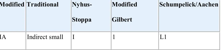
[image:39.612.74.545.585.692.2]The prime importance of classifying hernias are to compare them according to severity so that various treatment modalities can be achieved but with lotof surgical approaches available none of the classifications were accepted universally.
Table 45-1 -- Inguinal Hernia Classification Systems Modified Traditional
Nyhus-Stoppa
Modified
Gilbert
Schumpelick/Aachen
Modified Traditional
Nyhus-Stoppa
Modified
Gilbert
Schumpelick/Aachen
IB Indirect medium
II 2 L2
IC Indirect large IIIB 3 L3
IIA Direct small IIIA 5 M1
IIB Direct medium IIIA — M2
IIC Direct large — 4 M3
III Combined IIIB 6 Mc
IV Femoral IIIC 7 F
0 Other — — —
R Recurrent IV A, B, C, D — —
SURGERY
Indications and Alternatives
Preoperative Preparation
replacement of hernia contents into the abdominal cavity during herniorrhaphy could be followed by respiratory embarrassment or abdominal compartment syndrome, or both. The term “loss of domain” refers to this clinical scenario and can be addressed by establishment of pneumoperitoneum in preparation for hernia surgery. A CT scan allows the surgeon to determine the extent of domain loss and make a final decision about the need for pneumoperitoneum. The objective of pneumoperitoneum, which is applied in successive sessions, is to increase the amount of room in the peritoneal cavity. Many techniques have been described, including daily needle puncture, placement of an indwelling catheter by a percutaneous system or minilaparotomy, or a completely implanted system involving a tunneled peritoneal catheter and a venous access reservoir. Room air is inflated into the abdominal cavity on a once- or twice-daily timetable to patient tolerance as determined by abdominal discomfort or shortness of breath. Usually, 1 to 2 L is insufflated at each session. Upright chest roentgenography is useful because the level of the diaphragm is a measurable objective monitor.
higher risk for thromboembolic complications. Deep venous thrombosis prophylaxis is prudent when one is considering this approach.
Anesthesia
Although general anesthesia is almost always recommended for laparoscopic hernia repairs, the choice of anesthesia for open inguinal herniorrhaphy depends on the personal preference of the surgeon. Local anesthesia, when used in adequate doses and far enough in advance, proves very effective, especially in combination with short-acting amnesic and anxiolytic agents such as propofol. The local anesthetic should be injected before preparing and draping the patient for best results. One of the biggest advantages of local anesthesia is that the patient can be aroused from sedation at intervals to perform Valsalva maneuvers and test the repair. Regional anesthesia can also used by some experienced individuals in a successful manner. Local anesthesia can be used at the end of general anesthesia to lessen the postoperative pain.
Choice of Prosthetic Material
unequivocally proved the superiority of prosthetic repairs over pure tissue repairs in terms of recurrence. Tissue repairs are associated with an irreducible recurrence rate of 5% to 10%. The modern era of hernia repair has seen a progressive decrease in recurrence rates because of improvement in surgical technique and prosthetics. Materials that have emerged as suitable for routine use in hernia surgery and fulfill Cumberland's classic ideal characteristics include polypropylene, either monofilament (Marlex, Prolene) or polyfilament (Surgipro), Dacron (Mersilene), and expanded polytetrafluoroethylene (ePTFE) (Gore-Tex). An absorbable prosthesis has no role in groin hernia surgery. The newer biologic prostheses made of human cadaver skin, porcine cross-linked dermal collagen, or porcine small intestinal submucosa are more expensive and have no proven advantage over synthetic prostheses in uncomplicated groin hernia surgery. However, they can be useful in infected groin hernia wounds. Recently, the development of prostheses that modulate ECM expression by incorporating basic fibroblast growth factor has attracted the attention of investigators.
Not modified physically by tissue fluid
Chemically inert
Not carcinogenic
Does not cause an allergic or hypersensitivity response
Resistant to mechanical strain
Pliable and therefore moldable
Easily sterilized
one of the major arguments for the routine use of mesh in inguinal hernia surgery is to preserve fertility.
Approaches to Repair of Groin Hernias
Groin hernia repairs can be performed conventionally (anterior or preperitoneal) or laparoscopically. For conventional operations one can use a prosthesis or a pure tissue technique for repair. Whereas prosthetic approaches are by definition tension-free, avoidance of tension in nonprosthetic repairs is accomplished by relaxing incisions.
Commonly Recognized Conventional Inguinal Hernia Repairs
Anterior Preperitoneal Combined
Nonprosthetic Marcy Original Nyhus-Condon (historical interest only now)
Bassini
Moloney darn
Shouldice
Anterior Preperitoneal Combined
Miscellaneous
Prosthetic Lichtenstein tension-free
Anterior approach
Posterior approach
Bilayer repair
Hernioplasty Read-Rives GPRVS
Mesh plug and patch Kugel
Nyhus-Condon
GPRVS, great prosthesis for reinforcement of the visceral sac.
Conventional Anterior, Nonprosthetic
form the superficial inguinal ring tight enough to avoid a so-called industrial hernia, but slightly loose to avoid strangulation of the cord structures. The term industrial hernia refers to the presence of a dilated external ring that an inexperienced examiner confuses with a hernia.
The Bassini Repair
The Moloney Darn
The Moloney darn and its variant the Abramson darn use nonabsorbable suture to form a meshwork over the inguinal floor. The interstices of this meshwork fill with fibrous connective tissue that buttresses the weakened area of the inguinal canal. The initial layer consists of a continuous nylon suture to appose the transversalis fascia and the transversus abdominis, rectus, and internal oblique muscles medially to the reflected portion of the inguinal ligament laterally, similar to a Bassini repair. A difference is that the first stitch taken into cord muscle and continued in and out around the cord finally tied to the lateralside of internal ring including inguinal ligament. on the lateral side of the internal ring. The darn is a second layer with sutures applied in a crisscross fashion through muscular tissue medially to the inguinal ligament. Abramson stresses the importance of leaving the suture loose and not forcing the edges of the repair together during the darn, thereby allowing a “tension-free” repair and maintaining the meshwork structure. The darn must be carried well over the medial edge of the inguinal canal onto the anterior rectus sheath.
The Shouldice Technique
initial approach is similar to the Bassini repair, much of the importance given to release cord from the surrounding adhesions and dissection of cremaster and sac ligation highly. Steel wire is used and woven continuously for the floor to ensure even distribution of tension and avoid the defects that could potentially occur between interrupted sutures.the first stitch is taken from the pubictubercle and in continous fashion it was carried out laterally upto the internal ring approximatingthe superior muscle layer with the illiopubic tract and then it was carried out in reverse fashion towards pubic tubercle.next layer isapproximating the superior muscle layer in view of an artificial inguinal ligament like support. The fourth layer is organized in a similar technique above the third layer,inorder to get hold of external oblique muscle cribriform fascia can be incised.. When performed by experienced surgeons at the Shouldice Clinic, the operation has a recurrence rate of less than 1% and was the other techniques were compared to this standard technique. The major criticisms are that it is difficult to teach and it is hard for surgeons to understand what is really being sewn to what. This is further compounded by the fact that modifications outside the Shouldice Clinic have resulted in different versions.
Conventional Anterior, Prosthetic
The Lichtenstein Clinic is dedicated to hernia repairs. The herniorrhaphy is performed under local anesthesia with sedation. The former steps are similar to those of the Bassini repair. After the external oblique aponeurosis is opened,the inferior muscle layers is separated in aavascular plane.then the cord structures which contains cord with testicular vessel and nerves was held up by fingers . The effect is to create a large space for eventual placement of the prosthesis and at the same time provide excellent visualization of the nerves.
High ligation is performed by dissecting the sac from the surrounding cord structures after incising the cremaster muscle longitudinally. Direct hernias are separated from the nearby adhesions and the contents were returned back space. Superficial layers of sac were divided and facilitates reduction and aids in maintaining the reduction while the prosthesis is being placed. A suture can also be placed to allow the repair to proceed unencumbered by the sac protruding into the operative field.mesh of approximate size to thedefect taken and fixed to rectus sheath and secured on both sides and continued along the shelving edge in a running locking fashion. The suture is tied at the internal ring.
The mesh end is divide into two tails awider and a narrower and they are
is performed. This shutter valve suture should also pass through the shelving edge to allow the mesh to buckle medially over the direct space and avoid tension when the patient stands upright. A few interrupted sutures are then placed to secure the superior and medial aspects of the mesh to the underlying internal oblique and fascia. Care should be taken to avoid placing anchoring suture through the iliohypogastric nerve. Sufficient laxity should be maintained in the prosthesis to account for the difference in tension between the supine and prone positions and to compensate for mesh shrinkage. The only potential drawback of this procedure is that a femoral hernia could be missed because the inguinal floor is not opened. If one is detected, both the inguinal and coopers ligament were approximated.
The mesh plug technique was found by Gilbert and then modified by Robbins and Rutkow. The sac is separated from the adhesions and with the contents were returned back after a standard anterior approach. A plug made of rolled polypropylene mesh or prefabricated in the configuration of a flower is inserted into the defect and secured to its edges by interrupted suture. Millikan suggests that the inner leaf were sutured with the preperitoneal side and the mesh on the outside hence it will act as a underlay.. For an indirect hernia, the plug is held in place with three or four sutures around the defect . For direct hernias, fascia transversalis is opened to facilitate plug placement. The patch portion can be kept in a flat manner which wil support the plug in a fashion similar to the Lichtenstein procedure. The technique fast and also easy to teach in both academic and private
Conventional Preperitoneal Prosthetic
The key to preperitoneal repairs is to place a prosthetic in the fascio peritoneal space. This preperitoneal space can be entered from either the anterior or the posterior aspect. In the anterior approach a groin incision is made and the space is entered directly through the inguinal floor. A midline, Pfannenstiel, or paramedian incision can be used to enter the space from the posterior aspect. The transabdominal approach as advocated by LaRoque has returned to popularity because of the ease of entering the space laparoscopically.
Anterior Approach (Read/Rives)
Posterior Approach
Great Prosthesis for Reinforcement of the Visceral Sac
testicular vessels. It must be particularly emphasized that the dissection should proceed in the relatively avascular plane between the fascia transversalis and the peritoneum to avoid a bloody procedure.
Stoppa and Wantz recommend that the abdominal wall defect be left alone, but other surgeons prefer to plicate the fascia transversalis with the coopers to avoid the seroma bulge.
because of the numerous anatomic elements in the preperitoneal space that could be inadvertently damaged during suture or tack placement. For Wantz's technique, three absorbable sutures areused tosuture the superior layer of wall of abdomen with the mesh. The three sutures are placed near the linea alba, semilunar line, and anterior superior iliac spine in a medial-to-lateral direction. A Reverdin suture needle facilitates such placement. Subsequently, the mesh is positioned to cover the iliac fossa and the parietalized cord structures and iliopsoas muscle laterally; the pubic ramus, obturator fossa, and iliac vessels medially; and the space of Retzius in the middle. The size of the mesh for the Stoppa technique to repair bilateral hernias is the distance between the two anterior superior iliac spines minus 2 cm for the width, and the height is equal to the distance between the umbilicus and the pubis. The wound is closed in layers.
Nyhus/Condon (Iliopubic Tract Repair)
the preperitoneal space and exposes the posterior inguinal floor. Direct or indirect defects are repaired similarly after the peritoneal sac has been reduced or divided and closed proximally. The transverse aponeurotic arch is sutured to the iliopubic tract inferiorly, with Cooper's ligament occasionally included in the medial portion of the repair. The internal ring, if large, is also narrowed by placing a suture lateral to it. For femoral hernias the iliopubic tract is sutured to ligament of cooper. Once the defect has been formally repaired, a tailored mesh prosthesis can be sutured to Cooper's ligament and the fascia transversalis for support.
Kugel/Ugahary Repair
often divided with the distal end being left in situ while the proximal end is reduced. A specifically designed 8- by 12-cm prosthesis made of two pieces of polypropylene is altered in such a way to cover the myopectineal orifice. Ugahary's operation is similar, but a special prosthesis is not needed.
Combination Anterior and Preperitoneal Approaches (Bilayer Technique)
This repair depends on a dumbbell-shaped device consisting of two flat prolene mesh both are connected . The basis of this design is to take advantage of the benefits of both the anterior and posterior approaches, because the mesh is kept in such a way in the preperitoneal plane similar to the technique of lichenstein.this prosthetic is divided toaccomodate cord laterally with three or four interrupted sutures to the area of the pubic tubercle, the middle of the inguinal ligament, and the internal oblique muscle.
COMPLICATIONS
General complications such as urinary retention, paralytic ileus, and cardiorespiratory compromise can follow any operative procedure, and inguinal herniorrhaphy is no exception. The most common is urinary retention, especially after general anesthesia.
Recurrence
Chronic groin pain
Nociceptive
Neuropathic
Cord and testicular
Hematoma
Ischemic orchitis
Testicular atrophy
Injury to the vas deferens
Hydrocele
Testicular descent
Bowel and bladder injury
Osteitis pubis
Contraction
Erosion
Infection
Rejection
Fracture
Miscellaneous complications
Seroma
Hematoma
Wound infection
General complications
Chronic Postherniorrhaphy Pain Syndromes
should be made to assign them to one of two groups to help determine therapeutic options: (1) nociceptive pain caused by tissue injury, which is further subdivided into somatic and visceral, and (2) neuropathic pain secondary to nerve damage.
Somatic pain is usually caused by damage to ligaments, tendons, and muscles and includes osteitis pubis and adductor tenoperiostitis. Visceral pain refers to specific visceral dysfunction such as dysejaculation and urinary dysfunction. The principles of treating patients with nociceptive pain are similar to those for patients with groin pain but no obvious hernia.
Division, stretching, contusion, crushing, entrapment, or electrical injury to the nerve causes neuropathic groin pain. The nerves most commonly injured during conventional herniorrhaphy are the ilioinguinal and iliohypogastric. The classic manifestation is pain or paresthesia (or both) in the distribution of one of the major nerves. Precise diagnosis of nerve involvement is difficult because of dermatomal overlap. Physical activity aggravates the pain, and a recumbent position with hip flexion relieves it. Reassurance plus conservative treatment with anti-inflammatory medications and local nerve blocks is preferred initially. At least 1 year of conservative treatment should be tried before offering neuroma excision or neurectomy.
The hernia recurrence rate with the use of prosthetic material is less than 1%. This rate is probably an underestimation of the problem because patients frequently do not return to their original surgeon. It still translates to a hefty number because of the size of the denominator. A recurrent hernia is usually manifested as a bulge with a cough impulse. Occasionally, the initial symptom is pain. In this situation, a consistent definition of recurrent hernia does not exist because of difficulty differentiating a lipoma of the cord, a seroma, or an expansile bulge of the internal oblique muscle from true hernia recurrence. Imaging in the form of CT, MRI, or ultrasound should be obtained to unequivocally document recurrence. Causes of recurrence include (1) failure to perform high ligation or reduce the peritoneal sac with an indirect hernia, (2) inadequate closure of the internal ring, (3) missed hernias, (4) continuing failure of the floor of the canal, and (5) infection. The general principle for managing recurrent hernias depends on the original repair. The logical approach is to perform herniorrhaphy in the space that has not been dissected. If the patient has previously undergone a conventional repair, a preperitoneal repair is best chosen. On the other hand, if the index operation was a preperitoneal one, a repair that is performed in the conventional inguinal space is best.
Ischemic orchitis is the inflammation of testicles which develop post operatively usually one to five days after operation. It is thought to result from thrombosis of veins draining the testicle secondary to extensive dissection of the spermatic cord. It is much more common after repair of recurrent hernias. Initial symptoms like pyrexia with testis pain. Management is supportive and consists of support to testis . Ischemic orchitis usually resolves without sequelae but may occasionally progress to testicular atrophy. It is generally accepted that dividing rather than excising large indirect inguinal-scrotal hernia sacs and leaving the distal part of the sac open in situ can decrease the incidence of testicular complications.
cause is not known. Although the urologic literature suggests that hydroceles develop in case of leaving the last region of sac.
Prosthetic Complications
Shrinkage of prosthetic material because of scarification of the recipient's tissues should be anticipated during herniorrhaphy. Sufficient overlap in anticipation of 20% contracture is recommended. Mesh migration of prolene into other organs are rare. Intra-abdominal placement of a mesh prosthesis should be avoided in favor of an ePTFE or biologic prosthesis to avoid fistulation or bowel obstruction. Local erosion into cord structures has been reported. Rejection because of allergic reactions is extremely rare and is probably a manifestation of chronic infection.
Bowel and Bladder Injury
wound Infection :
The groin appears to be a protected area inasmuch as wound infection after inguinal herniorrhaphy occurs in less than 5% of patients. However, this figure may be an underestimation of the true incidence because of a delayed manifestation in many cases. Most surgeons recommend prophylactic broad-spectrum antibiotics, although studies by the Cochrane group have shown no benefit. Whereas infection after nonprosthetic repairs can be managed by open drainage and dressing changes, prosthetic removal is commonly required in addition to routine wound care after prosthetic procedures. , but true meshes can on occasion be with conservative wound care and antibiotic treatment.In caseof infection .
LAPAROSCOPIC OR CONVENTIONAL INGUINAL HERNIORRHAPHY
In the lastdecades there was much comparison between the laparoscopy and the standard repair and trials also provedlaparoscopic technique superior than the open techniques and better cosmesis, and return to normal activities sooner.
Comparative Trials of Laparoscopic and Open Inguinal Hernia Repair Using
Author Hernias (n)
LH vs. OH
Intervention Recurrence
Rate (%)
Salient Results
Horeyseck
et al., 100 vs. 100
TAPP vs.
Lichtenstein 8 vs. 0
Higher recurrence, higher cost
Zieren et
al., 86 vs. 105 TAPP vs. PP 2.3 vs. 0 Recuurence rate higher
Sarli et al., 64 vs. 66
TAPP vs.
Lichtenstein 0 vs. 0
Complications were similar
Champault
et al., 50 vs. 50
TAPP vs.
Stoppa 6 vs. 2 Recurrence rate higher
Khoury, 169 vs. 146 TAPP vs. MP 2.5 vs. 3 Recurrence rate higher
Paganini et
al., 52 vs. 56
TAPP vs.
Lichtenstein 2 vs. 0
Similar return to normal activity, higher cost
Aitola et
al., 24 vs. 25
TAPP vs.
Lichtenstein 13 vs. 8 Higher recurrence rate
Picchio et
al., 53 vs. 52
TAPP vs. Lichtenstein
Not
Author Hernias (n)
LH vs. OH
Intervention Recurrence
Rate (%)
Salient Results
Kumar et
al., 25 vs. 25
TEP vs.
Lichtenstein 4 vs. 8
Nonrandomized, lower pain score, fewer local complications
Johansson
et al., 613 total
TAPP vs. preperitoneal mesh vs. conventional
2 vs. 5.5 vs.
2 Similar complications
MRC
group, 468 vs. 460
TEP vs. mainly
tension-free 1.9 vs. 0
Earlier resumption of normal activity, less long-term pain, higher recurrence rate
Beets et al., 56 vs. 52
TAPP vs.
Stoppa 12.5 vs. 1.9
Less pain, fewer early complications
Sarli et al., 40 vs. 46
TAPP vs.
Licttenstein 0 vs. 4.3
Less pain, earlier return to work
Author Hernias (n)
LH vs. OH
Intervention Recurrence
Rate (%)
Salient Results
al., Lichtenstein similar missed
contralateral hernias
Pikoulis et
al., ] 309 vs. 234 TAPP vs. MP 1.9 vs. 0.4
Nonrandomized, higher cost, higher recurrence rate
Mahon et al., ]
60 vs. 60 (all bilateral or recurrent)
TAPP vs.
Lichtenstein 6.7 vs. 1.7
Shorter operative time, less pain, earlier return to work
Andersson
et al., 81 vs. 87
TEP vs.
Lichtenstein 2.5 vs. 0
Similar complications, earlier return to work, less pain, higher recurrence rate
Douek et
al., 122 vs. 120
TAPP vs.
Lichtenstein 1.6 vs. 2.5
Less groin pain, less frequent paresthesias
Bringman et al.,
Total N = 298
TEP vs. MP vs.
Lichtenstein 1.3 vs. 1.3
Author Hernias (n)
LH vs. OH
Intervention Recurrence
Rate (%)
Salient Results
recovery
Lal et al., 25 vs. 25
TEP vs.
Lichtenstein 0 vs. 0
Earlier return to work better cosmesis, similar recurrence rate
Heikkinen
et al., 62 vs. 61
TAPP vs,
Lichtenstein 8 vs. 3.2
Similar recurrence rate, less long-term groin pain
Neumayer
et al., 862 vs. 834
TAPP/TEP vs.
Lichtenstein 10.1 vs. 4
Less pain, higher recurrence rate for primary hernias
IPOM, intraperitoneal onlay mesh repair; LH, laparoscopic hernia repair; MP, mesh plug repair; OH, open hernia repair; PP, patch plug repair; TAPP, transabdominal preperitoneal hernia repair; TEP, totally extraperitoneal repair.
Comparative Trials of Laparoscopic and Open Tissue-Based Inguinal
Author Hernias
(n) LH vs.
OH
Intervention Recurrence
Rate (%)
Salient Results
Lawrence et
al., 58 vs. 57
TAPP vs. Maloney darn
Not
mentioned
Less pain, higher cost, similar return to work
Vogt et al., 30 vs. 32
IPOM vs.
Bassini/McVay 3 vs. 6
earlier return to normal activity
Liem et al., ]
487 vs. 507
TEP vs. mostly
tissue repairs 3 vs. 6 Few complications
Dirksen et al.,
114 vs.
103 TAPP vs. Bassini 6 vs. 21
Recurrence rate lower
Tanphiphat
et al., 60 vs. 60
TAPP vs.
modified Bassini 1.5 vs. 0 Lesser postop pain
Zieren et al.,
80 vs. 80 vs. 80
TAPP vs. MP vs.
Shouldice 0 vs. 0 vs. 0 Less pain
Juul et al.,
138 vs. 130
TAPP vs.
Shouldice 2.9 vs. 2.3
Author Hernias
(n) LH vs.
OH
Intervention Recurrence
Rate (%)
Salient Results
Leibl et al., 48 vs. 43
TAPP vs.
Shouldice 2 vs. 5
Greater patient satisfaction, similar recurrence rates
Tschudi et
al., 51 vs. 49
TAPP vs.
Shouldice 3.9 vs. 10.2
Recurrence rate lower
Wennstrom et al.,
131 vs.
130 Tep vs. Shouldice Similar
Recurrence rate lower
IPOM, intraperitoneal onlay mesh repair; LH, laparoscopic hernia repair; MP, mesh plug repair; OH, open hernia repair; TAPP, transabdominal preperitoneal hernia repair; TEP, totally extraperitoneal repair.
laparoscopic surgery. A notable exception to these trials is a recently published multicenter trial conducted in the Veterans' Administration system in which laparoscopic preperitoneal hernia repair (mostly TEP) was compared with tension-free anterior (Lichtenstein) repair. Recurrence was higher in laparoscopicapproach but incase ofrecurrent hernias both were similar. This particular study holds importance for surgeons practicing outside a specialty laparoscopic center and has caused many to suggest that the laparoscopic approach should be performed only at centers with a special interest. The early and delayed complication rates of the laparoscopic and conventional approaches are similar, but the seriousness of complications in the laparoscopic approach can be far greater. The hospital cost of laparoscopic repair is significantly higher than that of conventional repair, but it may be somewhat compensated by the higher productivity attributable to earlier return to work.
Patient Selection
contraindications. History of surgery, fluid collection in abdomen were relative contraindications. Incase of adhesions due to previous surgery one must be very careful regarding bowel and bladder injury. However, in the absence of considerable experience, a conventional approach may be preferred.
Operative Strategies
A laparoscopic hernia repair in which the peritoneal cavity is initially entered and the preperitoneal space is entered by another incision into the peritoneum from within the abdomen is called a transabdominal preperitoneal, or TAPP, repair. The next commonly used type of laparoscopic inguinal hernia repair is the totally extraperitoneal, or TEP, repair. Here abdominal cavity is not entered in this approach, and thus true laparoscopy is not performed; however, because a laparoscope and related instruments are used, it is appropriate to discuss this operation here. An inguinal hernia repair that is performed by placing mesh intraperitoneally over the defect via a laparoscopic approach is labeled an intraperitoneal onlay mesh, or IPOM, repair.
Transabdominal Preperitoneal Repair
mechanism of recurrence with the TAPP procedure. A direct hernia sac is easily reduced during the preperitoneal dissection. A small indirect hernia sac can be dissected away from he cord structures and reduced. A larger sac needs to be divided at a suitable point distal to the deep inguinal ring, with only the nearby portions were divided from the cord structures. No effort is made to disturb the distal part of the sac because it may lead to unnecessary vascular disruption, which can result in hematoma formation, ischemic orchitis, testicular atrophy.
never be placed below the iliopubic tract when lateral to the internal spermatic vessels because of the possibility of nerve damage and neuralgias. For the superior border of the mesh, staples are placed in horizontal fashion to prevent trauma to the ilioinguinal and iliohypogastric nerves, which also run horizontally. Lateral staples are oriented vertically because the femoral branch of the genitofemoral nerve and the lateral cutaneous nerve of the thigh run in this direction. Meticulous peritoneal coverage of the prosthesis is essential, and hence lowering the pressure of the pneumoperitoneum and further undermining of the inferior peritoneal flap may be necessary. The goal is isolation of the prosthesis from the viscera, and if it is not possible to reapproximate the superior and inferior peritoneal flaps, the inferior flap should be tacked to the transversalis fascia after ensuring complete mesh coverage. If all else fails, omentum can be used to cover the exposed mesh.
Totally Extraperitoneal Repair
side of the midline as in the TAPP repair. One of the advantages of the balloon device is that the preperitoneal space can be visualized through the transparent structure of the balloon. A disadvantage is a higher incidence of dissection in front of the inferior epigastric vessels, which causes them to be reduced with the peritoneal flap. This can complicate exposure. Once the preperitoneal space has been completely developed, treatment of the hernia sac and its contents, parietalization, and placement of the mesh proceed in an fashion identical to that for the TAPP repair.
Comparative Trials of Different Types of Laparoscopic Inguinal Hernia
Repair
Author Hernias
(n)
Intervention Recurrence
Rate (%) Salient Results Fitzgibbons et al., Total 869
TAPP vs. IPOM vs. TEP
4.5 vs. 4.5 vs. 4.5
Similar complication and recurrence rates
Khoury, 60 vs. 60 TAPP vs. TEP
Postop pain more in TAPP
Ramshaw et al.,
300 vs.
300 TAPP vs. TEP 2 vs. 0.3
Increased vascular injury and recurrence with TAPP
Kald et al.,
393 vs.
98 TAPP vs. TEP 1.5 vs. 1
Complications were morein TAPP
Sarli et al., 59 vs. 56
TAPP vs.
IPOM 0 vs. 11.1
Recurrence lower in TAPP
Van Hee et
al., 33 vs. 58 TAPP vs. TEP 2.7 vs. 2.8
Complications were same
Author Hernias
(n)
Intervention Recurrence
Rate (%)
Salient Results
100 TAPP easier
Lepere et al.,
1290 vs.
682 TAPP vs. TEP 1 vs. 1
Similar local complication and recurrence rates
IPOM, intraperitoneal onlay mesh repair; TAPP, transabdominal preperitoneal hernia repair; TEP, totally extraperitoneal repair.
Intraperitoneal Onlay Mesh Repair
Only a thin layer of peritoneum separates the abdominal cavity from the preperitoneal space. The rationale for the IPOM repair is to place a prosthesis directly onto the peritoneum so that radical preperitoneal dissection can be avoided. Initial laparoscopy, port placement, and landmark identification are the same as for the TAPP repair. A large prosthesis is introduced into the peritoneal cavity and secured in place with tacks, staples, or sutures. Some surgeons open the peritoneum over Cooper's ligament to ensure adequate fixation in this area.
experimental operation. In addition, the results in several series have not been as good as the results of other laparoscopic repairs, with a higher incidence of neuralgia and recurrence, but this may be more a reflection of experience than the operation itself. The IPOM repair represents the only truly minimally invasive herniorrhaphy because radical dissection of the preperitoneal space is avoided. The development of a totally inert prosthesis might renew interest in the future.
COMPLICATIONS OF LAPAROSCOPIC INGUINAL HERNIA REPAIR
Complications of Laparoscopic Hernia Repair
Associated with
Laparoscopy
Associated with
the Patient
Associated with the
Hernia Repair
Associated
with the
Prosthesis
Major vascular injury (I)
Retroperitoneal
Intra-abdominal
Abdominal wall
Bowel injury (I, E)
Ileus (E) Urinary retention (E) DVT (E) Cardiopulmo Recurrence (D)
Associated with
Laparoscopy
Associated with
the Patient
Associated with the
Hernia Repair
Associated
with the
Prosthesis
Bladder injury (I, E)
Gas embolism (I)
Bowel obstruction (E, D)
Shoulder pain (E)
Subcutaneous/preperi toneal emphysema (I, E) Diaphragmatic dysfunction (E) Arrhythmias (I) nary complication s (I, E)
(E) Hernia (D) Keloid (D) Seroma(E)
Hematoma (I, E) Groin Scrotal Retroperito neal
Hydrocele (E,
(E, D)
Rejectio n (D)
Associated with
Laparoscopy
Associated with
the Patient
Associated with the
Hernia Repair
Associated
with the
Prosthesis
D)
Orchitis (E)
Infertility (D)
Neurologic (D)
Groin pain
Anesthesia
Paresthesia s
D, delayed manifestation (weeks to years); DVT, deep venous thrombosis; E, early manifestation (hours to days); I, immediate/intraoperative manifestation.
Complications Associated with the Laparoscopic Approach
Major Vascular Injury
The risk of major vascular injury requiring operative repair is 0.08%.Many authors believe that this incidence is seriously underestimated because such injuries commonly go unreported. Entry in to the peritoneal cavity is the most important phase of laparoscopy, and over three quarters of major vascular injuries occur during entry of the Veress needle or the trocars at the beginning of the procedure. The vessels most frequently involved include the aorta, inferior vena cava, and the iliac artery and vein. Mesenteric and omental vessels, splenic vessels, and renal vessels have been injured occasionally. Epigastric vessels running in the rectus sheath may be injured during the placement of secondary trocars. The use of disposable trocars with safety shields, optical trocars, and blunt-tipped cannulas has not eliminated this dramatic complication. It has even been described during the open approach with the Hasson cannula used for initial access.
avoidance of the Trendelenburg position during initial access have been reported to decrease the incidence of this complication. Major vascular injury is manifested as either hemoperitoneum or retroperitoneal hematoma. Mortality has been estimated to be as high as 36%. Expeditious laparotomy with repair of the vessel is usually required. Lacerations of the epigastric vessels can be controlled by applying pressure applied with a cannula. Occasionally, suture ligation is required, which is now possible with the use of an “exit device” for transfascial suture placement.
Bowel Injury
The incidence of bowel injury and bowel perforation in laparoscopic operations is about 0.13%. Up to half of these injuries occur during the entry stage of laparoscopy. The small bowel is the most commonly segment (56%). About two thirds of these injuries are detected intraoperatively. The injury can be repaired laparoscopically if the operator is trained in intracorporeal suturing or the injury is amenable to a stapled repair without compromising luminal diameter. Patients with missed bowel injury typically manifest peritoneal signs and sepsis 1 day to 1 week after the index operation. The overall mortality rate with this complication is about 4%.
Injury to the bladder may occur from suprapubic trocar placement or from dissection during the course of the operation. Bladder injury may be obvious when blood and gas collect in the drainage bag if a Foley catheter is in place. When there is any doubt about bladder injury, methylene blue dye may be instilled into the bladder to look for leakage. Bladder injury recognized can be operated either laparoscopically or by open approach according to surgeons preference, followed by bladder drainage for 7 to 10 days. Bladder injury may be manifested in delayed fashion as hematuria and lower abdominal discomfort. A retrograde cystogram generally confirms the diagnosis. Small defects may be managed with urinary drainage, whereas larger defects necessitate repair.
Gas Embolism
Gas embolism is avery rare and lifethreatening complication. Carbon dioxide can be introduced into a large vein, most likely the result of inadvertant cannulation by the Veress needle, and trapped in the right ventricle, where it causes outflow obstruction into the pulmonary artery and sudden circulatory collapse. Careful insertion of the Veress needle and the usual confirmatory tests of its intraperitoneal position should keep the incidence of air embolism low.