JOURNAL OFVIROLOGY, May 2004, p. 5382–5389 Vol. 78, No. 10
0022-538X/04/$08.00⫹0 DOI: 10.1128/JVI.78.10.5382–5389.2004
Copyright © 2004, American Society for Microbiology. All Rights Reserved.
Rice Dwarf Phytoreovirus Segment S6-Encoded Nonstructural Protein
Has a Cell-to-Cell Movement Function
Yi Li,
1* Yi M. Bao,
2,3Chun H. Wei,
1Zhen S. Kang,
4Yong W. Zhong,
1Peng Mao,
1Gang Wu,
1Zhang L. Chen,
1Joachim Schiemann,
5and Richard S. Nelson
2*
Peking-Yale Joint Center for Plant Molecular Genetics and Agrobiotechnology, The National Laboratory of Protein Engineering
and Plant Genetic Engineering, College of Life Sciences, Peking University, Beijing 100871,1and Northwestern Agriculture
and Forestry University, Department of Plant Protection, Shaanxi, Yangling, 712100,4China; Plant Biology Division,
Samuel Roberts Noble Foundation, Inc., Ardmore, Oklahoma 734012; National Center for Biotechnology
Information, National Institutes of Health, Bethesda, Maryland 20892-65103; and Federal Biological
Research Centre for Agriculture and Forestry, Institute for Biochemistry
and Plant Virology, D-38104 Braunschweig, Germany5
Received 8 August 2003/Accepted 21 November 2003
Rice dwarf virus(RDV) is a member of the genusPhytoreovirus, which is composed of viruses with segmented double-stranded RNA genomes. Proteins that support the intercellular movement of these viruses in the host have not been identified. Microprojectile bombardment was used to determine which open reading frames (ORFs) support intercellular movement of a heterologous virus. A plasmid containing an infectious clone of Potato virus X (PVX) defective in cell-to-cell movement and expressing either -glucuronidase or green fluorescent protein (GFP) was used for cobombardment with plasmids containing ORFs from RDV gene segments S1 through S12 onto leaves of Nicotiana benthamiana. Cell-to-cell movement of the movement-defective PVX was restored by cobombardment with a plasmid containing S6. In the absence of S6, no other gene segment supported movement. Identical results were obtained withNicotiana tabacum, a host that allows fewer viruses to infect and spread within its tissue. S6 supported the cell-to-cell movement of the movement-defective PVX in sink and source leaves ofN. benthamiana. A mutant S6 lacking the translation start codon did not complement the cell-to-cell movement of the movement-defective PVX. An S6 protein product (Pns6)-enhanced GFP fusion was observed near or within cell walls of epidermal cells fromN.tabacum. By immu-nocytochemistry, unfused Pns6 was localized to plasmodesmata in rice leaves infected with RDV. S6 thus encodes a protein with characteristics identical to those of other viral proteins required for the cell-to-cell movement of their genome and therefore is likely required for the cell-to-cell movement of RDV.
Rice dwarf virus(RDV) is a member of the genus
Phytoreo-virus, family Reoviridae, and is the cause of a serious rice
disease in southern Asia (3). The sequences of two isolates have been determined (43, 51). The genome of RDV is com-posed of 12 separate segments of double-stranded RNA (3). Seven structural proteins and at least six nonstructural proteins are encoded by the 12 genome segments (S1 to S12). The seven structural proteins are products of segments S1, S2, S3, S5, S7, and S8 (27, 31). Coexpression of P3, the major core structural protein, and P8, the outer capsid structural protein, in trans-genic rice plants or insect cells results in the formation of double-shelled virus-like particles (16, 52). The six nonstruc-tural proteins are products of S4, S6, and S9 through S12 and have molecular masses of 83, 56, 49, 35, 23, and 34 kDa, respectively (27, 31). The functions of these nonstructural pro-teins are mostly unknown, although the S11 protein product (Pns11) is known to bind nucleic acid (47) and Pns6 increases
RDV accumulation and symptom severity (2). No viral protein necessary for virus cell-to-cell movement has been identified for members of the genus Phytoreovirus. Due to the double-stranded RNA structure of the genome and the inability to mechanically transmit the virus, infectious clones for RDV and other plant reoviruses have not been reported. Therefore, other methods are necessary to determine the function of genes from viruses within this genus.
Cell-to-cell movement by plant viruses is a requirement for the systemic infection of plants. Viral proteins essential for the cell-to-cell movement of the virus often have the characteris-tics of altering the size exclusion limits of the plasmodesmata (PD) in the cell wall (reviewed in references 11, 17, 18, 23, and 49) and in interacting with cytoskeletal elements (reviewed in references 1, 33, 35, and 50). Recent studies have identified genes encoding the movement protein (MP) of a virus by expressing the gene in the presence of a mutant virus that cannot move from cell to cell, in some instances when the MP is from a virus belonging to a different genus (9, 12, 13, 22, 28, 29, 30, 37, 38, 40, 41). The complemented movement is ob-served either by use of a mutant chimeric virus created by substituting a putative MP-encoding gene into the genome of a movement-defective virus (9, 13, 30, 38) or by bombardment or transformation of a plant with a plasmid expressing the putative MP and bombardment with a plasmid expressing a movement-defective virus (12, 22, 26, 28, 29, 40, 41). These
* Corresponding author. Mailing address for Yi Li: Peking-Yale Joint Center for Plant Molecular Genetics and Agrobiotechnology, The National Laboratory of Protein Engineering and Plant Genetic Engineering, College of Life Sciences, Peking University, Beijing 100871, China. Phone: 86-10-62759690. Fax: 86-10-62754427. E-mail: [email protected]. Mailing address for Richard S. Nelson: Plant Biology Division, The Samuel Roberts Noble Foundation, Inc., 2510 Sam No-ble Parkway, Ardmore, OK 73401. Phone: (580) 224-6600. Fax: (580) 224-6692. E-mail: [email protected].
5382
on November 8, 2019 by guest
http://jvi.asm.org/
complementation analyses are efficient and reliable ap-proaches for identification of molecular components involved in the transport of viral RNA.
We have utilized the complementation approach to identify RDV movement proteins. The 12 RDV genome segments were cloned individually into a plant transient-expression vec-tor. Some or all of these plasmids were individually introduced into cells of Nicotiana benthamiana, Nicotiana tabacum, or
Oryza sativa leaves by microprojectile bombardment together
with plasmids yielding movement-defective, but replication-competent, -glucuronidase (GUS)-tagged or green fluores-cent protein (GFP)-taggedPotato virus X(PVX). The move-ment and subcellular location of virus-encoded reporter protein were monitored by visualizing GUS activity or GFP fluorescence. In addition, a fusion of S6-encoded protein with
enhanced GFP (eGFP) was monitored by confocal laser scan-ning microscopy after bombardment ofNicotiana tissue, and the location of the unfused S6-encoded protein was monitored by immunocytochemistry and electron microscopy after infec-tion ofO. sativawith RDV.
MATERIALS AND METHODS
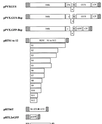
Plasmid construction.RDV gene segments S1 through S12 were individually cloned from the Fujian isolate and sequenced by using a previously described procedure (51). Restriction fragments containing open reading frames (ORFs) of each segment were ligated into aCauliflower mosaic virus(CaMV) 35S-based pRT transient-expression vector digested with BamHI and PstI (42). The recom-binant plasmids containing RDV gene segments were designated pRTS1 to pRTS12 (Fig. 1).
[image:2.603.118.477.65.501.2]The start codon for S6 ORF translation was altered to ATC by PCR with two primers: forward primer 5⬘-AATTCACCATCGACACAGAA-3⬘, including the FIG. 1. Schematic representation of plasmids used in this study. p, CaMV 35S promoter; o, nopaline synthase terminator; 166k, 166
kDa-protein; 25k, 25-kDa protein; a, 12-kDa protein; b, 8-kDa protein; c, 25-kDa mutant; CP, coat protein; ORFs are shown as rectangles, and the mutant S6 ORF lacking an ATG is shown as a line. pPVX.GUS-Bsp and pPVX.GFP-Bsp were provided by S. Y. Morozov, and pPVXGUS was provided by D. Baulcombe.
on November 8, 2019 by guest
http://jvi.asm.org/
5⬘portion of the ORF (the mutated start codon position is in boldface), and reverse primer 5⬘-GAGCTGCAGCTGCCCATTAGTCGACCCAG-3⬘, based on the S6 sequence of the RDV Fujian isolate (GenBank accession no. U36564). High-fidelityPfuDNA polymerase (Stratagene, La Jolla, Calif.) was used in the PCRs. The amplified PCR product was ligated into pGEM3Zf(⫹) digested with HincII. After sequencing, the mutated S6 sequence was digested with BamHI and PstI and ligated into the pRT vector digested with BamHI and PstI to yield pRTS6T (Fig. 1).
Movement-defective GUS- or GFP-tagged PVX, designated pPVX.GUS-Bsp or pPVX.GFP-Bsp (Fig. 1), respectively, and provided by S. Y. Morozov, were used for complementation experiments (28, 29). Both contain the same frame-shift mutation at nucleotide position 4959 within the 25-kDa protein ORF, resulting in the absence of 72 C-terminal amino acid residues in the translated protein. The cell-to-cell movement of infectious virus from these constructs was complemented by active 25-kDa protein and other movement-associated pro-teins (28). pPVXGUS, a derivative of pGC3 (5), was provided by David Baul-combe. All of the PVX constructs reside behind a CaMV 35S promoter (5, 28, 29).
A two-step recombinant PCR method (39) was used to generate the Pns6-eGFP (Clontech, Palo Alto, Calif.) fusion ORF, with the following modifications. Pns6 was amplified by using high-fidelityPfuDNA polymerase (Stratagene) in the presence of a forward primer (S6 forward primer, 5⬘-AAAATGGACACA GAAACTCTTTGC-3⬘) and a reverse primer containing complementary se-quences from the 3⬘end of the S6-coding sequence and the 5⬘ end of the eGFP-coding sequence (S6 reverse primer, 5⬘-CCTCGCCCTTGCTCACCATT TTGTACACGGTAATAGCA-3⬘). Similarly, the eGFP-coding region was am-plified by using a forward primer complementary to the S6 reverse primer (eGFP forward primer, 5⬘-TGCTATTACCGTGTACAAAATGGTGAGCAAGGGCG AGG-3⬘) and a reverse primer containing a KpnI site (eGFP reverse primer, 5⬘-GTCGGTACCTTTACTTGTACAGCTCGTCC-3⬘). The resulting PCR frag-ments containing the S6 and eGFP ORFs were purified by using a PCR product purification kit (Plasmid Midi; Qiagen, Valencia, Calif.). The purified PCR products and the S6 forward and eGFP reverse primers were then used in a second PCR to create the S6-eGFP fusion with a KpnI site at the 3⬘end. After purification and digestion with KpnI, the resulting chimeric cDNA was ligated into the plant transient-expression vector pRTL2 (4, 34), previously digested with KpnI and NcoI, to yield pRTL2S6:eGFP (Fig. 1).
All constructs were sequenced by automated dye-terminator sequencing (model 377; PE Applied Biosystems, Foster City, Calif.) according to the man-ufacturer’s protocol to confirm sequence authenticity.
Identification ofN. benthamianaleaf development stages.Carboxyfluorescein (CF) dye was used to identify source and sink leaves onN. benthamianaplants. CF dye was applied to the petioles of the most mature leaves, and fluorescence was monitored with a UV lamp. CF dye moves through the phloem in a source-to-sink direction, unloading in sink, but not source, leaves (32, 36).
Inoculation ofNicotiana and Oryzaleaves.Inoculation ofN. benthamiana,N. tabacum, andO. sativaleaves by particle bombardment was performed with a high-pressure helium-based apparatus and the flying disk method (model PDS-1000; Bio-Rad, Hercules, Calif.). For each series of bombardments, 5l of plasmid DNA (at 1.0g/l) was precipitated onto 3 mg of gold particles (1-m diameter; Bio-Rad) in the presence of a solution of calcium chloride (2.5 M) and spermidine (0.1 M). For the cobombardment, 2.5g of DNA of each plasmid was mixed and applied to gold particles in the same way as for the single bombardment. The plasmid DNA-coated gold particles were washed once with 70% ethanol and then maintained in suspension in absolute ethanol. After sonication, 20l of this mixture was placed on plastic flying disks and used for bombardment when the particles had dried. Source or sink leaves ofN. benthami-anaand source leaves ofN. tabacumorO. sativawere placed in the center of a plastic petri dish and bombarded on a solid support at a target distance of 8 cm. Bombardment was done with a pulse of 1,100 kPa of helium gas in a vacuum chamber. For each of the constructs used in this research, the bombardment was repeated at least three times.
Histochemical analysis of GUS activity.Leaves were sampled at 30 h post-bombardment forN. benthamianaand at 72 h postbombardment forN. tabacum. GUS activity was monitored by histochemical detection (20) with the following modification. Samples were infiltrated with 5-bromo-4-chloro-3-indolyl -D-gluc-uronide (X-Gluc), at 600g/ml, in a solution containing 0.115 M phosphate buffer [pH 7.0], 3 mM potassium ferricyanide, and 10 mM EDTA. The phos-phate-ferricyanide-EDTA mixture limits the diffusion of the intermediate prod-ucts of the reaction (8). After incubation overnight at 37°C, leaves were fixed in 70% ethanol and examined by light microscopy.
Confocal microscopy.The bombarded tobacco leaves expressing eGFP or fused Pns6-eGFP were harvested at 24 h postbombardment and examined with
a confocal imaging system (model 1024ES; Bio-Rad) attached to an upright microscope (Axioskop; Zeiss, Thornwood, N.Y.) equipped with 60⫻objective lens (Zeiss) as described previously (6). Serial optical sections were obtained at 0.5-m intervals, and the projections of optical sections were combined on a Power Edge 2200 computer (Dell, Austin, Tex.) by using Lasersharp software version 3.1 (Bio-Rad).
Immunocytochemical electron microscopy.Small pieces of tissue were excised from RDV-infected and noninfectedO. sativaleaves and fixed in a solution of 1% (vol/vol) glutaraldehyde and 4% (wt/vol) formaldehyde in 100 mM sodium cacodylate buffer (pH 7.2) for 3 h. After dehydration in a graded acetone series, the samples were embedded with Epon-araldite and polymerized at 60°C as described previously (21). For immunogold labeling, ultrathin tissue sections, attached to 200-mesh nick grids (Electron Microscopy Sciences, Fort Washing-ton, Pa.), were floated on a drop of blocking solution containing 1% (wt/vol) bovine serum albumin (Sigma-Aldrich, St. Louis, Mo.) in Tris-buffered saline (TBS) (10 mM Tris-HCl, 150 mM NaCl [pH 7.4]) for 20 min. The sections were then incubated for 2 h at room temperature with rabbit polyclonal antiserum against Pns6, which was expressed inEscherichia coliand purified by sodium dodecyl sulfate-polyacrylamide gel electrophoresis (Y. Li, unpublished results), diluted 1:200 (vol/vol) in the blocking solution. The sections were rinsed four times (10 min each time) in TBS and then incubated for 1 h with goat anti-rabbit immunoglobulin linked with 15-nm-diameter colloidal gold particles (GAR-gold, EM CARIgG15; Biocell Research Laboratories, Cardiff, United Kingdom) and diluted 1:30 in TBS. The sections were then washed in TBS and double-distilled water. After drying, the sections were contrasted with uranyl acetate and lead citrate and examined with an electron microscope (model EM10; Zeiss) at 80 kV. The specificity of immunogold labeling was determined by replacing primary antibody with rabbit preimmune serum or buffer.
RESULTS
RDV gene segments that support cell-to-cell movement of a movement-defective PVX inN. benthamiana.To identify genes involved in RDV cell-to-cell movement, RDV ORFs were individually inserted into the plant expression vector pRT (42), and, together with a movement-defective PVX expressing GUS (pPVX.GUS-Bsp [Fig. 1]), they were used for cobom-bardment onto detached source leaves fromN. benthamiana. In order to verify expression of the constructs which we used in cobombardment in the tissues, total protein extracts from bombardedN. benthamianaleaves were detected by Western blotting with RDV P3-, P8-, and P10-specific antisera. The results showed that RDV P3, P8, and P10 were expressed in the bombardedN. benthamianaleaves (results not shown). In addition, plasmids pPVXGUS (a plasmid yielding virus capa-ble of cell-to-cell movement [Fig. 1]) and pPVX.GUS-Bsp were used for bombardment ontoN. benthamianaleaves in the absence of RDV sequences.
Leaves bombarded with movement-competent pPVXGUS displayed large blue foci after histochemical staining (Fig. 2A). Cobombardment with pPVX.GUS-Bsp and plasmids contain-ing individual ORFs from S1 through S12 showed that only S6 allowed the production of multicellular blue foci on N.
benthamianaleaves (Fig. 2B and G and data not shown). The
blue foci produced in the presence of pRTS6 were smaller and displayed enhanced signal along minor veins compared with foci produced by the movement-competent virus (Fig. 2A and B and data not shown). In contrast with results from cobom-bardment with pPVX.GUS-Bsp and pRTS6, bomcobom-bardment with pPVX.GUS-Bsp alone yielded only small blue foci that were very difficult to see (Fig. 2C and H). These small blue foci represented initially bombarded individual cells. On rare oc-casions visible blue foci were observed after bombardment with pPVX.GUS-Bsp. These blue foci may have been formed
5384 LI ET AL. J. VIROL.
on November 8, 2019 by guest
http://jvi.asm.org/
by a small group of simultaneously infected epidermal or me-sophyll cells or by passive diffusion of the chromogenic dye between cells, despite our use of a method to limit diffusion (26, 28, 29). These results indicate that pPVX.GUS-Bsp was
unable to move from cell to cell. The same pattern of staining was produced after bombardment with a plasmid expressing only the GUS gene (data not shown) (28). The ability of the S6 segment to complement the movement of the
movement-de-FIG. 2. Cell-to-cell movement of a movement-defective PVX determined by transcomplementation with the S6 gene product of RDV in source leaves ofN. benthamianaandN. tabacum. Leaves ofN. benthamianaandN. tabacumwere bombarded with pPVX.GUS-Bsp (movement defective) alone or with pPVX.GUS-Bsp and pRTS6 or pPVXGUS (movement capable). Bombarded leaves ofN. benthamiana andN. tabacum were harvested and stained at 30 h or 3 days postbombardment, respectively. They were analyzed by histochemical staining and light microscopy. (A)N. benthamianaleaves bombarded with pPVXGUS, showing large blue infection foci (mean lesion size, 1,650.5m; standard deviation, 352m; standard error, 82m); (B and G)N. benthamianaleaves bombarded with pPVX.GUS-Bsp and pRTS6, showing intermediate-sized blue infection foci (mean, 683.3m; standard deviation, 168m; standard error, 27m [shown in panel B]); (C and H)N. benthamianaleaves bombarded with pPVX.GUS-Bsp alone, showing single-cell infections (mean, 38.5m; standard deviation, 9.3m; standard error, 1.8m [shown in panel C]); (D, E, and F)N. tabacumleaves bombarded with pPVXGUS, pPVX.GUS-Bsp and pRTS6, and pPVX.GUS-Bsp, respectively. Bars, 50m.
on November 8, 2019 by guest
http://jvi.asm.org/
fective PVX was similar in sink and source leaves of N.
benthamiana(data not shown).
To determine whether the S6 segment complemented the movement of the movement-defective PVX through its en-coded protein, the translational start codon of the S6 ORF was altered from ATG to ATC (Fig. 1). This mutant S6 ORF could not complement the cell-to-cell movement of the movement-defective PVX (Fig. 3A).
Analysis of the ability of the RDV S6 ORF to complement the movement of movement-defective PVX in the more restric-tive hostsN. tabacumandO. sativa.N. benthamianais a per-missive host to many plant viruses (7), and thus the results described above may reflect this host’s ability to allow comple-mentation that is not normally observed in more limiting hosts. To determine whether S6 could complement the cell-to-cell movement of movement-defective PVX in a host with a more limited susceptibility to viruses, pPVX.GUS-Bsp or pPVX. GFP-Bsp was used for cobombardment with pRTS6 onto source leaves ofN. tabacum. Leaves bombarded with pPVX-.GUS-Bsp and pRTS6 displayed blue foci with a size similar to that of foci produced by pPVXGUS (compare Fig. 2D and E). Leaves bombarded with pPVX.GUS-Bsp alone yielded mostly microscopic single-cell blue foci (Fig. 2F and data not shown). The results of multiple bombardments with the different com-binations of plasmids are summarized in Table 1. Multicellular expression was also observed when pPVX.GFP-Bsp was used for cobombardment with pRTS6 onto leaves of N. tabacum (Fig. 4B).
pPVX.GUS-Bsp was used for cobombardment with pRTS6 into cells of mature leaves ofO. sativato determine whether the S6-encoded protein complements the cell-to-cell move-ment of the movemove-ment-defective PVX in the natural host of RDV. The movement-competent PVX, pPVXGUS, was used as a control. No blue cells were observed in the rice tissues bombarded with either pPVXGUS alone or a combination of
pPVX.GUS-Bsp and pRTS6 by using histochemical staining and light microscopy. However, blue cells were observed when GUS alone was expressed (data not shown). Thus, PVX was not able to establish an infection in rice in the presence of S6, and we were unable to evaluate the ability of the S6-encoded protein to function as a movement protein in this host.
Subcellular location of Pns6-eGFP in leaves ofN. tabacum and of Pns6 in leaves ofO. sativa.To determine the subcellular location of RDV Pns6 in epidermal cells, the S6 ORF was fused with the 5⬘ end of the coding sequence of eGFP and expressed through the activity of an enhanced CaMV 35S promoter (pRTL2S6:eGFP [Fig. 1]). A free eGFP behind the same promoter (Fig. 1) was used as a control. The subcellular locations of the proteins expressed from these plasmids were determined by confocal laser scanning microscopy.
Expression of the Pns6-eGFP fusion yielded strongly
fluo-FIG. 3. Analysis of transcomplemented cell-to-cell movement by a movement-defective PVX in the presence of a translation-incompetent RDV S6 ORF or a Pns6-eGFP fusion in source leaves ofN. benthamiana. The bombarded leaves were harvested and analyzed at 30 h postbombardment by histochemical staining and light microscopy. (A) Leaves bombarded with pPVX.GUS-Bsp (movement defective) and pRTS6T (translation incompetent), showing infection in a single cell. (B) Leaves bombarded with pPVX.GUS-Bsp, showing infection in a single cell. (C and D) Leaves bombarded with pPVX.GUS-Bsp and pRTL2S6:eGFP (Pns6-eGFP fusion) and with pPVX25.GUS-Bsp and pRTS6, respectively, showing multicellular blue infection foci. Bar, 50m.
TABLE 1. Transcomplemented movement of movement-defective PVX (pPVX.GUS-Bsp) in leaves ofN. tabacumandN. benthamiana
in the presence of the S6 segment of RDV or the S6-eGFP fusiona
Plasmid(s) used for bombardment
No. (% of total foci) of foci with the following no. of infected cells:
1 2–5 6–10 ⱖ11
cpPVXGUSb 0 (0) 6 (20) 20 (67) 4 (13) pPVX.GUS-Bsp⫹pRTS6b 3 (10) 11 (37) 13 (43) 3 (10) pPVX.GUS-Bspb 29 (97) 1 (3) 0 (0) 0 (0) pPVX.GUS-Bsp⫹pRTS6c 2 (2) 42 (39) 37 (34) 26 (24) pPVX.GUS-Bsp⫹
pRTL2S6:eGFP fusionc 1 (1) 47 (43) 36 (33) 25 (23)
aLeaves were bombarded with pPVXGUS, pPVX.GUS-Bsp and pRTS6
(con-taining the S6 segment), pPVX.GUS-Bsp and the pRTL2S6:eGFP fusion, or pPVX.GUS-Bsp alone. The bombarded leaves ofN. tabacumandN. benthami-anawere harvested and analyzed by histochemical staining and light microscopy.
bData were recorded at 3 days postbombardment ofN. tabacumleaves. cData were recorded at 30 h postbombardment ofN. benthamianaleaves.
5386 LI ET AL. J. VIROL.
on November 8, 2019 by guest
http://jvi.asm.org/
rescing structures along the cell wall, possibly associated with PD (Fig. 5 and data not shown). Free eGFP did not target to the cell wall or form subcellular aggregates in epidermal cells (data not shown). To determine whether the Pns6-eGFP fu-sion functioned similarly to native Pns6 in complementing the
cell-to-cell movement of the movement-defective PVX, co-bombardment with pPVX.GUS-Bsp and pRTL2S6:eGFP was performed. The Pns6-eGFP fusion complemented the cell-to-cell movement of pPVX.GUS-Bsp onN. benthamianaleaves (Fig. 3C; Table 1). Thus, multicellular infection by the move-ment-incompetent PVX occurred even though the Pns6-eGFP fusion protein did not traffic from cell to cell when bombarded alone (Fig. 5).
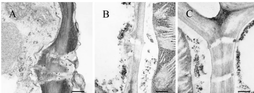
To confirm that RDV Pns6 associated with PD, tissue from RDV-infected leaves ofO. sativawas fixed and embedded, and sections were probed with gold-conjugated antibody against Pns6. Branched and simple PDs within RDV-infected rice leaves were labeled (Fig. 6A and B), and PDs from uninfected healthy rice leaf tissues were not labeled (Fig. 6C). These results are consistent with the results from transient expression of the Pns6-eGFP fusion in epidermal cells of tobacco leaves and further strengthen the conclusion that RDV Pns6 is lo-cated in PD within the cell wall.
DISCUSSION
Unlike the situation for single-stranded RNA viruses and some DNA viruses, proteins necessary for the cell-to-cell movement of phytoreoviruses were not previously identified. By cobombardment with plasmids containing reporter gene-tagged, movement-defective PVX with plasmids containing ORFs from RDV gene segments S1 to S12, we determined that S6 alone complemented the cell-to-cell movement of the movement-defective PVX mutant inN. benthamiana(Fig. 2B and G). Thus, a protein encoded by RDV that supports virus movement has been identified. The complemented cell-to-cell movement of the movement-defective PVX, however, was not as efficient as that observed for the movement-competent PVX
inN. benthamiana(Fig. 2A and B). This could be due to (i) a
lack of S6 product availability as the PVX mutant spread from the S6-expressing cell, (ii) the S6 product failing to comple-ment a unique function of the 25-kDa protein not associated with movement of PVX, or (iii) the S6 product being only partially active in complementing the movement function of the 25 kDa protein. The first possibility seems the most likely, since the lesion sizes of the complemented movement-defec-tive PVX increased only slowly as determined by visual obser-vation at later times postbombardment (Y. Li, unpublished observation). In addition, Pns6 fused with eGFP did not accu-mulate in multiple cells when used for bombardment alone (Fig. 5).
The complemented movement of the movement-defective PVX by Pns6 was more similar to that of the wild-type virus in
N. tabacumthan to that inN. benthamiana(compare Fig. 2A
and B with Fig. 2D and E). Since the constructs used for bombardment were identical, this result indicates that host factors important for spread of the viruses differ in these two species. N. benthamianais particularly susceptible to a wide range of viruses (7, 44). It is possible that the complementation exhibited inN. benthamianamay be due to an inability ofN.
benthamianato limit the accumulation and spread of the
[image:6.603.53.273.70.391.2]wild-type virus through a system not involving movement proteins. Further experiments quantifying virus accumulation and spread within the infected tissue will help to clarify this situa-tion.
FIG. 4. Transcomplemented cell-to-cell movement of movement-defective pPVX.GFP-Bsp in the presence of the S6 gene product of RDV in source leaves ofN. tabacum. Leaves were bombarded with pPVX.GFP-Bsp (movement defective) alone or with pPVX.GFP-Bsp and pRTS6. Images were taken with a confocal laser scanning micro-scope at 3 days postbombardment. (A) Leaves bombarded with pPVX.GFP-Bsp alone, showing infection in a single cell. (B) Leaves bombarded with pPVX.GFP-Bsp plus pRTS6, showing multicellular infection site. Each panel shows three independent cells expressing the product.
FIG. 5. Transient expression of Pns6-eGFP (pRTL2S6:eGFP) in epidermal cells of N. tabacum leaves. Leaves bombarded with pRTL2S6:eGFP were harvested at 30 h postbombardment, and images were taken with a confocal laser scanning microscope. (A) Fluorescent image of the expression pattern of Pns6-eGFP in a single cell. (B) Bright-field image under Nomarski illumination. (C) Merged im-age of panels A and B. Panels A and B are single optical sections from the same location within the cell.
on November 8, 2019 by guest
http://jvi.asm.org/
Although Pns6 fused with eGFP did not move from cell to cell, this fusion protein was as functional as native Pns6 in supporting the cell-to-cell movement of the movement-defec-tive PVX (Fig. 3C and Table 1). This result indicates that the presence of other viral components, such as RNA or other proteins, was necessary for the multicellular spread of the defective PVX. A similar conclusion was reached to explain how the MP ofTomato mosaic viruscomplemented the move-ment of a movemove-ment-defectiveTomato mosaic virus(40). For tobamoviruses, it is known that in addition to the MP, a viral protein(s) (i.e., the 126- and/or 183-kDa protein) involved in the accumulation of the virus within a cell also modulates virus cell-to-cell and systemic movement (10, 19, 24). Thus, it is possible that Pns6 functions in combination with a protein from PVX to allow movement of this virus. These studies indicate the power of transcomplementation experiments using movement-defective virus to identify proteins necessary for virus spread, in contrast to noncomplementation experiments studying the spread of the expressed putative movement pro-teins or transcomplementation experiments studying the spread of nonviral reporter proteins in the presence of only the putative movement protein.
The movement-defective PVX was altered to contain a frameshift leading to the loss of 72 C-terminal amino acid residues in the 25-kDa protein ORF (28). The ability of Pns6 to allow movement of the defective PVX indicates that this protein complements one of the functions of the 25-kDa pro-tein. The 25-kDa protein was localized in the cell wall as a small punctate structure within or adjacent to PDs (29). Pns6 fused with GFP was also observed localized to the cell wall of
N. tabacum cells, and unfused Pns6 was observed localized
specifically to the PD of rice cells (Fig. 5 and 6A and B). The 25-kDa protein has ATP/GTPase activity, binds RNA, targets to a specific cellular compartment(s), moves from cell to cell when fused with eGFP, and has a helicase-like domain (14, 29, 48). Since the mutation in the 25-kDa protein ORF would delete a portion of the helicase domain, it is logical that this region may be critical for PVX cell-to-cell movement and that Pns6 complements the helicase activity missing in the mutant protein. Pns6 has no significant sequence similarity to any known plant viral movement proteins but has sequence
simi-larity to yeast ATP-dependent RNA helicases (Li, unpublished results). Pns6 also is known to regulate the accumulation of and symptom severity induced by RDV (2). ForTobacco
mo-saic virus, it is known that the 126- or 183-kDa protein, which
contain identical helicase-like sequences, also regulates virus accumulation and symptom severity (10, 39). Thus, all of these proteins have significant structural and functional similarities, and the 25-, 126-, and 183-kDa proteins have been shown to aid the cell-to-cell movement of their encoding virus. Further work is needed to determine whether Pns6 has helicase activ-ity.
Recently, the 25-kDa protein has been shown to be a sup-pressor of RNA silencing (45). Although initial studies indicate that Pns6 is not a suppressor of silencing like the 2b, 19-kDa, 25-kDa, and HC-Pro viral proteins (15, 25, 45, 46; Li, unpub-lished results), further work is necessary to verify this finding. If Pns6 does not have suppressor activity, this would suggest that suppressor activity and virus movement are two different functions of the 25-kDa protein and that Pns6 complements only the movement function of this protein. Separating these activities would be of great interest to those studying virus movement and RNA silencing.
ACKNOWLEDGMENTS
We thank D.C. Baulcombe for providing pPVXGUS,N. benthami-ana16c plants, andAgrobacteriumcontaining the 35S-GFP construct; S. Y. Morozov for providing mutant PVX constructs; Shou-Wei Ding for advice on gene silencing; J. Carrington for providing the pRTL2 vector; E. Blancaflor for advice on confocal microscopy; and Stephen Chisholm and Xin Shun Ding for helpful comments on the manuscript. We thank Cuc Ly for preparation of the final figures.
This work was supported by a National Outstanding Youth Grant of China (30125004); Rockefeller Foundation grant award RF93022; Al-location no. 236, National High Tech (863), China (contract no. 2001AA212131); a Natural Science Foundation of China grant to Y.L.; and a National Key Basic Research Program (973), China grant (con-tract no. G62000016204) to C.H.W.
REFERENCES
1. Aaziz, R., S. Dinant, and B. L. Epel.2001. Plasmodesmata and plant cy-toskeleton. Trends Plant Sci.6:326–330.
[image:7.603.76.514.69.229.2]2. Ando, Y., I. Uyeda, K. Murao, and I. Kimura.1996. Naturally occurring phenotypic variants differing in symptom severity of rice dwarf phytoreovi-rus. Ann. Phytopathol. Soc. Jpn.62:466–471.
FIG. 6. Subcellular localization of Pns6 in RDV-infected leaves ofO. sativa(rice). Immunogold labeling of ultrathin sections from RDV-infected (A and B) and healthy (C) rice leaves with Pns6-specific antibody is shown. Bar, 250 nm.
5388 LI ET AL. J. VIROL.
on November 8, 2019 by guest
http://jvi.asm.org/
3. Boccardo, G., and R. G. Milne.1984. Plant reovirus group.InA. F. Morant and B. D. Harrison (ed.), CM/AAB descriptions of plant viruses, no. 294. Commonwealth Mycological Institute and Association of Applied Biologists, Unwin Brothers Ltd., The Gresham Press, Old Woking, England. 4. Carrington, J. C., and D. C. Freed.1990. Cap-independent enhancement of
translation by a plant potyvirus 5⬘nontranslated region. J. Virol.64:1590– 1597.
5. Chapman, S., T. Kavanagh, and D. C. Baulcombe.1992. Potato virus X as a vector for gene expression in plants. Plant J.2:549–557.
6. Cheng, N. H., C. L. Su, S. A. Carter, and R. S. Nelson.2000. Vascular invasion routes and systemic accumulation patterns of tobacco mosaic virus inNicotiana benthamiana. Plant J.23:349–362.
7. Christie, S. R., and W. E. Crawford.1978. Plant virus range ofNicotiana benthamiana. Plant Dis. Rep.62:20–22.
8. De Block, M., and D. Debrouwer.1992. In-situ enzyme histochemistry on plastic-embedded plant material. The development of an artifact-free glu-curonidase assay. Plant J.2:261–266.
9. De Jong, W., and P. Ahlquist.1992. A hybrid plant RNA virus made by transferring the noncapsid movement protein from a rod-shaped to an ico-sahedral virus is competent for systemic infection. Proc. Natl. Acad. Sci. USA89:6808–6812.
10. Derrick, P. M., S. A. Carter, and R. S. Nelson.1997. Mutation of the tobacco mosaic tobamovirus 126- and 183-kDa proteins: effects on phloem-depen-dent virus accumulation and synthesis of viral proteins. Mol. Plant-Microbe Interact.10:589–596.
11. Ding, B., A. Itaya, and Y. M. Woo.1999. Plasmodesmata and cell-to-cell communication in plants. Int. Rev. Cytol.190:251–316.
12. Fedorkin, O. N., A. Merits, J. Lucchesi, A. G. Solovyev, M. Saarma, S. Y. Morozov, and K. Makinen.2000. Complementation of the movement-defi-cient mutations in potato virus X: potyvirus coat protein mediates cell-to-cell trafficking of C-terminal truncation but not deletion mutant of potexvirus coat protein. Virology270:31–42.
13. Giesman-Cookmeyer, D., S. Silver, A. Vaewhongs, S. A. Lommel, and C. M. Deom.1995. Tobamovirus and dianthovirus movement proteins are func-tionally homologous. Virology213:38–45.
14. Gorbalenya, A. E., E. V. Koonin, A. P. Donchenko, and V. M. Blinov.1989. Two related superfamilies of putative helicases involved in replication, re-combination, repair and expression of DNA and RNA genomes. Nucleic Acids Res.17:4713–4730.
15. Guo, H. S., and S. W. Ding.2002. A viral protein inhibits the long range signaling activity of the gene silencing signal. EMBO J.21:398–407. 16. Hagiwara, K., J. Higashi, K. Namba, T. Uehara-Ichiki, and T. Omura.2003.
Assembly of single-shelled cores and double-shelled cores virus-like particles after baculovirus expression of major structural proteins P3, P7, and P8 of rice dwarf virus. J. Gen. Virol.84:981–984.
17. Haywood, V., F. Kragler, and W. J. Lucas.2002. Plasmodesmata: pathways for protein and ribonucleoprotein signaling. Plant Cell14:S303–S325. 18. Heinlein, M.2003. Plasmodesmata: dynamic regulation and role in
macro-molecular cell-to-cell signaling. Curr. Opin. Plant Biol.5:543–552. 19. Hirashima, K., and Y. Watanabe.2001. Tobamovirus replicase coding region
is involved in cell-to-cell movement. J. Virol.75:8831–8836.
20. Jefferson, R. A.1987. Assaying chimeric genes in plants: the GUS gene fusion system. Plant Mol. Rep.5:387–405.
21. Kang, Z.1996. Ultrastructure of plant pathogenic fungi. China Science and Technology Press, Beijing, China.
22. Krishnamurthy, K., R., M. Heppler, R. Mitra, E. Blancaflor, M. Payton, R. S. Nelson, and J. Verchot-Lubicz.2003. The potato virus X TGBp3 protein associates with the ER network for virus cell-to-cell movement. Virology309:135–151.
23. Lasarowitz, S. G., and R. N. Beachy.1999. Viral movement proteins as probes for intracellular and intercellular trafficking in plants. Plant Cell
11:535–548.
24. Lewandowski, D. J., and W. O. Dawson.2000. Functions of the 126- and 183-kDa proteins of tobacco mosaic virus. Virology271:90–98.
25. Llave, C., K. D. Kasschau, and J. C. Carrington.2000. Virus-encoded sup-pressor of posttranslational gene silencing targets a maintenance step in the silencing pathway. Proc. Natl. Acad. Sci. USA97:13401–13406.
26. Lough, T. J., N. E. Netzler, S. J. Emerson, P. Sutherland, F. Carr, D. L. Beck, W. J. Lucas, and L. S. Forster.2000. Cell-to-cell movement of potexviruses: evidence for a ribonucleoprotein complex involving the coat protein and first triple gene block protein. Mol. Plant-Microbe Interact.13:962–974. 27. Mao, Z., Y. Li, H. Xu, and Z. L. Chen.1998. The 42K protein of rice dwarf
virus is a post-translational cleavage product of the 46K outer capsid protein. Arch. Virol.143:1831–1838.
28. Morozov, S. Y., O. N. Fedorkin, G. Juttner, J. Schiemann, D. C. Baulcombe, and J. G. Atabekov.1997. Complementation of a potato virus X mutant
mediated by bombardment of plant tissues with cloned viral movement protein genes. J. Gen. Virol.78:2077–2083.
29. Morozov, S. Y., A. G. Solovyev, N. O. Kalinina, O. N. Fedorkin, O. V. Samuilova, J. Schiemann, and J. G. Atabekov.1999. Evidence for two non-overlaping functional domains in the potato virus X 25K movement protein. Virology260:55–63.
30. Nejidat, A., F. Cellier, C. A. Holt, and R. Gafny, A. L. Eggenberger, and R. N. Beachy.1991. Transfer of the movement protein gene between two tobamo-viruses: influence on local lesion development. Virology180:318–326. 31. Omura, T., K. Ishikawa, H. Hirano, M. Ugaki, Y. Minobe, T. Tsuchizaki, and
H. Kato.1989. The outer capsid protein of rice dwarf virus is encoded by genome S8. J. Gen. Virol.70:2759–2764.
32. Oparka, K. J., C. M. Duckett, D. A. M. Prior, and D. B. Fisher.1994. Real time imaging of phloem unloading in the root tip ofArabidopsis. Plant J.
6:759–766.
33. Reichel, C., P. Ma´s, and R. N. Beachy.1999. The role of the ER and cytoskeleton in plant viral trafficking. Trends Plant Sci.4:458–462. 34. Restrepo, M. A., D. D. Freed, and J. C. Carrington.1990. Nuclear transport
of plant potyviral proteins. Plant Cell2:987–988.
35. Roberts, A. G., and K. J. Oparka.2003. Plasmodesmata and the control of symplastic transport. Plant Cell Environ.26:103–124.
36. Roberts, A. G., S. Santa Cruz, I. M. Roberts, D. A. M. Prior, R. Turgeon.
1997. Phloem unloading in sink leaves ofNicotiana benthamiana: compari-son of a fluorescent solute with a fluorescent virus. Plant Cell9:1381–1396. 37. Santa Cruz, S., A. G. Roberts, D. A. M. Prior, S. Chapman, and K. J. Oparka.1998. Efficient cell-to-cell and phloem-mediated transport of potato virus X: the role of virions. Plant Cell10:495–510.
38. Sasaki, N., Y. Fujita, K. Mise, and I. Furusawa,.2001. Site-specific single amino acid changes to Lys or Arg in the central region of the movement protein of a hybrid bromovirus are required for adaptation to a nonhost. Virology279:47–57.
39. Shintaku, M. H., S. A. Carter, Y. Bao, and R. S. Nelson.1996. Mapping nucleotides in the 126kDa protein gene that control the differential symp-toms induced by two strains of tobacco mosaic virus. Virology221:218–225. 40. Tamai, A., and T. Meshi.2001. Tobamoviral movement protein transiently expressed in a single epidermal cell functions beyond multiple plasmodes-mata and spreads multicellularly in an infection-coupled manner. Mol. Plant-Microbe Interact.14:126–134.
41. Tamai, A., and T. Meshi.2001. Cell-to-cell movement of potato virus X: the role of p12 and p8 encoded by the second and third open reading frames of the triple gene block. Mol. Plant-Microbe Interact.14:1158–1167. 42. To¨pfer, R. V. Matzeit, B. Gronenborn, J. Schell, and H. H. Steinbiss.1987.
A set of plant expression vectors for transcriptional and translational fusions. Nucleic Acids Res.15:5890.
43. Uyeda, I., N. Suda, N. Yamada, H. Kudo, K. Murao, H. Suga, I. Kimura, E. Shikata, Y. Kitagaw, T. Kusno, M. Sugawara, and N. Suzuki.1994. Nucle-otide sequence of rice dwarf phytoreovirus genome segment 2: completion of sequence analysis of rice dwarf virus. Intervirology37:6–11.
44. Van Dijk, P., F. A. Van der Meer, and P. G. M. Prion.1987. Accessions of AustralianNicotianaspecies suitable as indicator hosts in the diagnosis of plant virus diseases. Neth. J. Plant Pathol.93:73–85.
45. Voinnet, O., C. Lederer, and D. C. Baulcombe.2000. A viral movement protein prevents spread of the gene silencing signal in Nicotiana benthami-ana. Cell103:157–167.
46. Voinnet, O., S. Rivas, P. Mestre, and D. Baulcombe.2003. An enhanced transient expression system in plants based on suppression of gene silencing by the p19 protein of tomato bushy stunt virus. Plant J.33:949–956. 47. Xu, H., Y. Li, Z. J. Mao, and Z. L. Chen.1998. Rice dwarf phytoreovirus
segment s11 encodes a nucleic acid binding protein. Virology240:267–272. 48. Yang, Y., B. Ding, D. C. Baulcombe, and J. Verchot.2000. Cell-to-cell movement of the 25K protein of potato virus X is regulated by three other viral proteins. Mol. Plant-Microbe Interact.13:599–605.
49. Zambryski, P., and K. Crawford.2000. Plasmodesmata: gatekeepers for cell-to-cell transport of developmental signals in plants. Annu. Rev. Cell. Dev. Biol.16:393–421.
50. Zambryski, P.1995. Plasmodesmata: plant channels for molecules on the move. Science270:1943–1944.
51. Zhang, F., Y. Li, Y. F. Liu, C. C. An, and Z. L. Chen.1997. Molecular cloning, sequencing, and functional analysis and expression inE. coliof major core protein gene (S3) of rice dwarf virus Chinese isolate. Acta Virol.141:161– 168.
52. Zheng, H. H., Y. Li, C. H. Wei, D. H. Wei, Y. P. Shen, Z. L. Chen, and Y. Li.
2000. Assembly of double-shelled, virus-like particles in transgenic rice ex-pressing two major structural proteins of rice dwarf virus. J. Virol.74:9808– 9810.