Copyright © 2003, American Society for Microbiology. All Rights Reserved.
Monocytes Treated with Human Immunodeficiency Virus Tat Kill
Uninfected CD4
⫹
Cells by a Tumor Necrosis Factor-Related
Apoptosis-Induced Ligand-Mediated Mechanism
Yida Yang, Ilia Tikhonov, Tracy J. Ruckwardt, Mahmoud Djavani, Juan Carlos Zapata,
C. David Pauza, and Maria S. Salvato*
Institute of Human Virology, University of Maryland Biotechnology Center, Baltimore, Maryland 21201
Received 25 October 2002/Accepted 26 March 2003
The human immunodeficiency virus (HIV) Tat protein has a critical role in viral transcription, but this study
focuses on its additional role as an extracellular effector of lymphocyte cell death. It is well known that Tat
induces tumor necrosis factor-related apoptosis-induced ligand (TRAIL) in peripheral blood mononuclear
cells (PBMC), and we show that the majority of TRAIL is produced by the monocyte subset of PBMC. Human
monocytes and U937 monoblastoid cells did not take up soluble HIV Tat-86, as T cells did, yet produced more
TRAIL than did T cells. TRAIL secretion was induced by Tat and by a cysteine-rich peptide of Tat but not by
sulfhydryl-modified Tat toxoid. Although there was only a slight increase in cell surface expression of TRAIL
on monocytes, sufficient TRAIL was secreted to be toxic for T cells. The cytotoxicity of Tat-stimulated monocyte
medium could be blocked by a TRAIL-neutralizing antibody. T cells treated with Tat did not secrete enough
TRAIL to mediate cell death in our assay. Remarkably, uninfected T cells are more susceptible to TRAIL than
are HIV-infected T cells. The production of TRAIL by Tat-stimulated monocytes provides a mechanism by
which HIV infection can destroy uninfected bystander cells.
The human immunodeficiency virus type 1 (HIV-1) Tat
pro-tein is essential for HIV-1 transcription and replication (11, 19,
55) and also modulates the expression of genes responsible for
cell survival and proliferation (15, 37, 48, 59). Tat is also a
secreted protein that can be detected in sera from
HIV-in-fected individuals (59) and binds to cell surfaces through
elec-trostatic interactions, chemokine receptors, or cell surface
in-tegrins (3, 6, 18). Cells treated with Tat showed increased
expression of chemokine receptors, overproduction of alpha
interferon (IFN-
␣
), and enhanced expression of Fas ligand
(FasL) on monocytes/macrophages that could cause cell death
in cultured peripheral blood mononuclear cells (PBMC) (7, 10,
27, 53, 62). Tat protein can also be taken up by infected and
uninfected PBMC, thus inducing biological effects through an
autocrine/paracrine mechanism (18, 63). Here we show that
Tat is internalized by T cells but not by monocytes. However,
Tat-treated monocytes secrete proapoptotic factors that cause
the death of uninfected bystander T cells.
Most circulating monocytes are in a nonactivated,
prediffer-entiated state on their way to becoming macrophages or
den-dritic cells (1). Activated monocytes mediate host defense
mechanisms by releasing inflammatory mediators, activating
major histocompatibility complex-restricted T lymphocytes
(21, 49), and killing virus-infected cells (12, 29). Activated
monocytes also express several soluble mediators of apoptosis,
including tumor necrosis factor alpha (TNF-
␣
), CD30L, FasL,
4-1BBL, and TNF-related apoptosis-inducing ligand (TRAIL)
(4, 14, 47, 50, 54). HIV infection activates monocyte
differen-tiation either indirectly by inducing IFN-
␣
(62) or directly
through the action of extracellular Tat protein (16, 27, 64).
TRAIL, a member of the TNF superfamily, plays a role in
HIV-mediated cell death (32, 64). TRAIL is expressed in
mouse and human T and B cells, natural killer cells, dendritic
cells, and monocytes (26, 31, 41) and is a potent inducer of
apoptosis in a variety of cell types (23, 28, 30, 42, 51). At least
five receptors for TRAIL have been identified. Two of these,
DR4 (TRAIL-R1) and DR5 (TRAIL-R2), contain a
cytoplas-mic death domain and are capable of transducing an apoptotic
signal. TRAIL-R3, TRAIL-R4, and osteoprotegerin lack
func-tional death domains and serve as decoy receptors to block
TRAIL-mediated apoptosis (24, 56). In addition to the
recep-tor distribution, intracellular regulation is important for
deter-mining the effects of binding soluble TRAIL to cells (23, 34).
This study demonstrates an intracellular and extracellular
increase in TRAIL once monocytes are exposed to HIV Tat or
to a cysteine-rich peptide of Tat. In contrast to monocytes, T
cells secreted relatively little TRAIL upon exposure to Tat.
Remarkably, HIV-infected Jurkat cells and primary CD4
⫹cells were relatively resistant to TRAIL-mediated killing
com-pared with uninfected cells. Cell death by secreted TRAIL is
one mechanism for the death of uninfected bystander cells that
is observed frequently in vivo after HIV infection (22).
MATERIALS AND METHODS
Reagents.HIV Tat protein, Tat-86, has a C-terminal truncation of 15 amino acids and was obtained from the National Institutes of Health AIDS Research and Reference Reagent Program (ARRRP). Antibodies included anti-Tat (ARRRP no. 705), mouse anti-human immunoglobulin G1 (IgG1; Sigma, St. Louis, Mo.), polyclonal rabbit anti-TRAIL (H-257; Santa Cruz Biotechnology, Inc., San Diego, Calif.), and a neutralizing monoclonal anti-TRAIL (N2B2; eBioscience Co., San Diego, Calif.). Tat was chemically inactivated by carboxym-ethylation to make Tat toxoid, a molecule with an average of two modified cysteines, as described previously (35). 7-Amino actinomycin D (7-AAD; Sigma),
* Corresponding author. Mailing address: Institute of Human
Vi-rology, University of Maryland Biotechnology Center, 725 W.
Lom-bard St., Baltimore, MD 21201. Phone: (410) 247-1368. Fax: (410)
247-5198. E-mail: salvato@umbi.umd.edu.
6700
on November 8, 2019 by guest
http://jvi.asm.org/
actinomycin D (Sigma), propidium iodide (Sigma), annexin V-fluorescein iso-thiocyanate (FITC), FITC–anti-human CD4⫹, and phycoerythrin
(PE)–anti-hu-man CD8⫹(Pharmingen, San Diego, Calif.) were used for flow cytometry. The
Tat peptides are as follows, with the included amino acid residues indicated in subscript: Tat1-20(peptide 1), MEPVDPRLEPWKHPGSQPKT; Tat16-35 (pep-tide 8), SQPKTACTNCYCKKCCFHCQ; Tat31-50(peptide 13), CFHCQVCFM TKALGISYGRK; Tat46-65 (peptide 19), SYGRKKRRQRRRAHQDSQTH; Tat61-80(peptide 25), DSQTHQASLSKQPTSQSRGD; and Tat76-95(peptide 31), QSRGDPTGPKESKKKVERET.
Cells and cell culture.The human leukemia Jurkat T-cell line (clone E6.1; American Type Culture Collection) and promonocytic U937 cell line (no. CRL2367; American Type Culture Collection) were cultured in RPMI 1640 medium supplemented with 10% fetal calf serum (FCS), 2 mML-glutamine, 100
U of penicillin/ml, and 100 g of streptomycin/ml. Human monocytes were isolated from the PBMC of healthy donors by adherence to plastic (i.e., they were incubated for 2 h in T125 flasks in RPMI 1640 medium containing 10% heat-inactivated AB serum). Fluorescence-activated cell sorter analysis demon-strated that more than 98% of the cells were CD14⫹, indicating monocytes of
high purity. CD4⫹lymphocytes were purified by using negative-selection
col-umns (R&D Systems, Minneapolis, Minn.). The purity of recovered cells was more than 90%, with no detectable cross-contamination by CD8⫹cells. CD4⫹
cells were incubated in RPMI 1640 medium supplemented with 10% heat-inactivated FCS, 2 mML-glutamine, 100 U of penicillin/ml, 100g of
strepto-mycin/ml, and 50 IU of interleukin-2/ml (RO 23-6019; Hoffmann-La Roche, Nutley, N.J.).
HIV infection of primary CD4ⴙcells and Jurkat cells.Before infection, cells were stimulated for 3 days with phytohemagglutinin (PHA; 5g/ml) and inter-leukin-2 (50 IU/ml) in RPMI 1640 medium containing 10% heat-inactivated FCS, 2 mM glutamine, 100 U of penicillin/ml, and 100g of streptomycin/ml. In preparation for infection, both CD4⫹cells and Jurkat cells were treated with 20 g of DEAE-dextran/ml for 30 min at 37°C. HIV IIIB was added to 107cells at 150 ng of p24gag (approximately 30 to 40 pg of p24 per 50% tissue culture infective dose). Cells were incubated at 37°C with occasional mixing for 2 h and then centrifuged and decanted. Cells were cultured for 5 days at 37°C in an atmosphere of 5% CO2to establish the infection and were then used for cell death studies by exposure to Tat-stimulated monocyte (TSM) medium.
Tat uptake into cells.CD4⫹cells, Jurkat cells, U937 cells, or fresh human
monocytes at 3⫻105cells/well were incubated with 100 ng of Tat/ml for 0, 30, 60, and 180 min and washed with phosphate-buffered saline (PBS) three times, and cell lysates were harvested according to the Ne-Per protocol (catalog no. 78833; Pierce Chemicals, Rockford, Ill.). This protocol includes detergent washes that strip proteins off the cell surface, resulting in some loss of cytoplasmic content, so most of the observed Tat is nuclear (57). For immunoblotting (see below), anti-Tat monoclonal antibody (ARRRP no. 705) was used.
Immunoblotting assays.U937 cells or monocytes were stimulated with HIV Tat (0 to 1,000 ng/ml) for 20 h, and then cell lysates and media were collected. The stimulation took place in 96-well plates at 37°C with 3⫻105cells/well in a volume of 100l of RPMI 1640 medium plus 10% FCS. At the end of these
incubations, media were collected and stored at⫺70°C for later use as TSM media. The cells were lysed at 4°C in a buffer containing 1% NP-40, 50 mM Tris-HCl (pH 7.5), 150 mM NaCl, 10 mM EDTA, 1 mM phenylmethylsulfonyl fluoride, 0.1% aprotinin, and 1 mM pepstatin A as described previously (13). The lysate was sonicated for 30 s on maximum setting (Ultrasonic Cleaner; Branson, Danbury, Conn.) and was centrifuged at 1,000⫻gfor 10 min at 4°C to pellet debris. TSM media, Tat-stimulated T-cell media, and cell lysates were analyzed by sodium dodecyl sulfate–10% polyacrylamide gel electrophoresis (SDS–10% PAGE) using 20l of undiluted medium per well or 6⫻104cell equivalents of lysate. The separated proteins were transferred to nitrocellulose membranes. Membranes were sequentially incubated with anti-TRAIL and then with a cor-responding secondary antibody conjugated with horseradish peroxidase and were detected with an electrochemiluminescent system (ECL; Amersham Pharmacia Biotech, Piscataway, N.J.).
Reverse transcription-PCR.Total RNA was extracted from 2⫻106cells by using Trizol reagent (Invitrogen, Carlsbad, Calif.), followed by treatment with RNase-free DNase (Promega, Madison, Wis.). cDNA was synthesized from 1g of RNA by using AMV reverse transcriptase (Promega) as described previously (60). PCRs were performed using the following primers: for TRAIL, 5⬘-GACC CCAATGACGAAGAGAGTATG-3⬘(forward) and 5⬘-GTTGCTCAGGAATG AATGCCC-3⬘ (reverse), and for-actin, 5⬘-GGGTCAGAAGGATTCCTAT G-3⬘ (forward) and 5⬘-GGTCTCAAACATGATCTGGG-3⬘ (reverse). The TRAIL PCR cycle conditions were 94°C for 1 min, 55°C for 1 min, and 72°C for 1 min for a total of 35 cycles. The-actin cycle conditions were 94°C for 45 s, 72°C for 1 min, and 55°C for 45 s for a total of 25 cycles. Samples were resolved on a 1% agarose gel and visualized with ethidium bromide.
Chromium release assay.Chromium release assays were performed as de-scribed previously (60, 61). Briefly, monocytes were treated with HIV Tat for 24 h and supernatants and cells were collected for use as effectors. The chromi-um-labeled target cells were either uninfected or HIV-infected Jurkat cells or primary human CD4⫹cells. Target cells were distributed to 96-well U-bottom
plates (Costar, Cambridge, Mass.) at 103cells/well, and effector monocytes were added at effector-to-target cell ratios of 6.25:1, 12.5:1, 25:1, and 50:1. Chromium release assay mixtures were incubated in triplicate for 20 h. The percentage of specific lysis was determined as follows: 100⫻[(experimental release⫺ spon-taneous release)/(maximum release⫺spontaneous release)]. Maximum release was determined by the lysis of targets in 1% Triton X-100, and spontaneous release was determined by lysis of targets in media without effectors and was found to be consistently less than 20%. Target cells were not lysed by cell-free Tat in this assay.
Cell viability and flow cytometry assay.To detect cell surface TRAIL, U937 cells, primary human monocytes, Jurkat cells, or primary human CD4⫹cells (2 ⫻106cells) were cultured with or without HIV Tat at 37°C for 24 h and then incubated with PE-labeled anti-TRAIL (RIK-2; Pharmingen) before a final wash and fixation in 1% paraformaldehyde–PBS for flow cytometry.
[image:2.603.140.452.69.224.2]To assay the cytotoxicity of Tat-stimulated monocytes, TSM media were col-lected from human monocyte cultures that had been exposed to 100 ng of Tat/ml for 20 h. Uninfected or HIV-infected target cells were incubated with 100l of
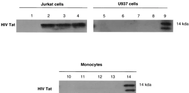
FIG. 1. HIV Tat enters Jurkat cells but not U937 cells or monocytes. Jurkat cells, U937 cells, and monocytes were exposed to HIV Tat (100
ng/ml) for 0 min (lanes 1, 5, and 10), 30 min (lanes 2, 6, and 11), 1 h (lanes 3, 7, and 12), or 3 h (lanes 4, 8, and 13). After the indicated times,
cells were collected, cell lysates were separated by SDS–10% PAGE, and cell proteins were transferred to nitrocellulose membranes and
immunoblotted with the TR1 murine monoclonal antibody to Tat (57). Lanes 9 and 14 contain recombinant Tat protein.
V
OL. 77, 2003
HIV Tat INDUCES TRAIL SECRETION BY MONOCYTES
6701
on November 8, 2019 by guest
http://jvi.asm.org/
TSM medium for 16 to 20 h at 37°C. Cell viability was routinely checked by trypan blue exclusion. For controls, TSM medium was preincubated with anti-human IgG1 at a concentration of 20g/ml or with anti-TRAIL (N2B2) at a concentration of 100 ng/ml for 1 h. For flow cytometry for detection of apoptosis, aliquots of 105cells were washed and incubated in the dark at 4°C for 20 min in PBS containing either 5l of annexin V-FITC (to detect early apoptosis) or 20
g of 7-AAD/ml (to detect late apoptosis) (58). Cells were processed according to the manufacturer’s protocol and fixed in a PBS-bovine serum albumin-NaN3 buffer containing 1% paraformaldehyde. Samples were analyzed 20 min later by FACScan flow cytometry (36).
RESULTS
HIV Tat is taken up by T cells but not by monocytes.
To
demonstrate how HIV Tat influences the activity of
mono-cytes, we exposed cells to HIV Tat protein and visualized its
uptake into monocytes and T cells. Immunoblotting results
indicated that HIV Tat was not internalized in U937 cells or
monocytes, while it appears in Jurkat cells as early as 30 min
after exposure (57) and can still be detected 3 h after the initial
exposure to Tat (Fig. 1).
HIV Tat increases the expression of TRAIL in U937 cells
and human monocytes.
Fresh human monocytes or the
promonocytic cell line U937 were stimulated with HIV Tat
protein (0 to 1,000 ng/ml) at 37°C, and lysates were collected
after 20 h. Immunoblotting results showed that HIV Tat
in-creases the expression of TRAIL in a dose-dependent manner,
with 100 ng/ml being the optimum concentration of HIV Tat
(Fig. 2A, part a). At a higher concentration of Tat (1,000
ng/ml), the expression of TRAIL was decreased. The same
results were obtained with U937 cells; however, no Tat
stim-ulation of TRAIL could be detected in fresh human T cells or
FIG. 2. HIV Tat increases TRAIL expression in U937 cells and human monocytes, but Tat toxoid does not. (A) TRAIL expression increases
with increasing doses of Tat protein. Monocytes (a) and CD4
⫹cells (b) were incubated with or without HIV Tat or Tat toxoid (10 to 1,000 ng/ml)
at 37°C for 20 h, and 10
l of cell lysate (6
⫻
10
4cell equivalents) was separated by SDS–10% PAGE. Cell proteins were transferred to
nitrocellulose membranes and immunoblotted with polyclonal anti-TRAIL (H-257; 1:1,000). Lanes (from left): 1, no HIV Tat protein; 2 to 5, cell
extracts exposed to 10, 50, 100, and 1,000 ng of Tat/ml, respectively. Actin (antibody A-2668; Sigma) was an internal control. Similar results were
obtained with fresh human monocytes and with the U937 cell line. TRAIL production in fresh human T cells or in Jurkat cells was not stimulated
by exposure to Tat. (B) HIV Tat induces the transcription of TRAIL mRNA in U937 cells and monocytes. Total RNA from U937 cells, monocytes,
or CD4
⫹cells was isolated and subjected to reverse transcription-PCR.

-Actin mRNA was amplified over 25 cycles and TRAIL mRNA was
amplified over 35 cycles of PCR. Transcription of control

-actin or TRAIL/APO2L mRNA in U937 cells is shown after 0, 2, 6, 12, or 24 h of
incubation in the presence of HIV Tat (100 ng/ml).
on November 8, 2019 by guest
http://jvi.asm.org/
in Jurkat cells (Fig. 2A, part b). Chemically modified Tat
tox-oid, with an average of two cysteines inactivated per molecule,
was incapable of inducing TRAIL in monocytic cells (Fig. 2A,
part a).
HIV Tat increases TRAIL mRNA transcription in U937 cells
and human monocytes.
The transcription of TRAIL mRNA
was rapidly induced within 2 h after the addition of Tat, and
expression was sustained throughout overnight culture (Fig.
2B) in both U937 cells and in freshly isolated human
mono-cytes. Since Tat is easily oxidized, mRNA expression dropped
off after 12 h. In parallel experiments with T cells, TRAIL
mRNA was not detectable.
HIV cysteine-rich peptide is responsible for TRAIL
induc-tion.
To identify the portion of Tat responsible for inducing
TRAIL, we synthesized six overlapping peptides covering
dif-ferent portions of Tat. Tat
16-35, which contains six of the seven
cysteines of Tat, induced TRAIL expression in monocytes,
while other peptides did not (Fig. 3).
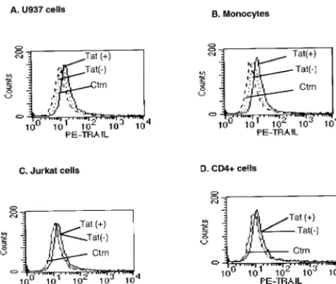
Tat induced little cell surface TRAIL on monocytes.
We
used flow cytometry to determine whether HIV Tat induced
the expression of TRAIL on the surface of human monocytes
or U937 cells. Tat treatment for 20 h only slightly increased the
membrane expression of TRAIL in U937 cells and monocytes
in comparison to what was seen with controls (Fig. 4A and B).
Tat did not affect surface expression of TRAIL on Jurkat cells
or on primary CD4
⫹cells (Fig. 4C and D).
Release of soluble TRAIL in the media from U937 cells and
human monocytes upon stimulation with HIV Tat.
In activated
human T cells, soluble TRAIL was released into the media in
the form of micelles (43). We tested whether HIV Tat
stimu-lation resulted in the majority of TRAIL being released from
monocytes or T cells. Using a Western blotting assay, we
clearly showed that Tat stimulation resulted in the appearance
of TRAIL in the media from U937 cell (Fig. 5A) or human
monocyte cultures (Fig. 5B). T cells did not release sufficient
TRAIL after 20 h as detected by Western blotting or the
cytotoxicity assay (Fig. 5C).
By comparing commercially available TRAIL protein with
Tat-induced TRAIL in extracts, we estimated that our Western
blots detected as little as 10 ng of TRAIL. Thus, in the weakest
bands of our Western blots, we saw approximately 10 ng of
TRAIL per 20
l of medium or per cell lysate (6
⫻
10
4cell
equivalents), and in the strongest bands, we saw approximately
200 ng of TRAIL per 6
⫻
10
4cell equivalents. At 20 h after Tat
exposure, a little more TRAIL was found intracellularly than
was found extracellularly (per cell) in the secreted form.
Uninfected T cells are more susceptible to TRAIL-mediated
cell death than are HIV-infected T cells.
Initially, we used a
20-h
51Cr release assay to check the cytotoxicity of
monocyte-associated TRAIL on Jurkat and primary CD4
⫹T cells (Fig.
6A and B). Cell-associated TRAIL has low cytotoxicity against
these T-cell targets. At the same time, both uninfected and
infected Jurkat and CD4
⫹cells were cultured with medium
from monocytes that had been stimulated with 100 ng of Tat
protein/ml (i.e., TSM medium). TSM medium caused
signifi-cant cytotoxicity, approximately two- to threefold over the
maximum cytotoxicity observed in the presence of Tat-treated
monocytes (Fig. 6C). The cytotoxicity of TSM medium could
be neutralized by anti-TRAIL monoclonal antibody but not by
anti-human IgG1. This indicates that soluble TRAIL is
re-leased from Tat-stimulated monocytes. Evidence that target T
cells underwent apoptosis was obtained by both annexin V
staining for early apoptotic surface changes and by 7-AAD
staining for later apoptotic DNA fragmentation. Figure 7
clearly shows that HIV TSM medium induced apoptosis in
Jurkat cells and primary CD4
⫹T cells. This apoptosis
de-creased when TSM medium was preincubated with
anti-TRAIL (N2B2). Jurkat cells and CD4
⫹cells infected with
HIV-1 IIIB were exposed to TSM medium at 5 days after
infection. Compared with uninfected cells, HIV-infected
Jur-kat cells and HIV-infected CD4
⫹cells were relatively resistant
to the apoptotic effects of TSM medium (Fig. 6C and 7).
FIG. 3. The cysteine-rich region of Tat is responsible for inducing
monocytes to express TRAIL. Peptides Tat
1-20(peptide 1), Tat
16-35(peptide 8), Tat
31-50(peptide 13), Tat
46-65(peptide 19), Tat
61-80(pep-tide 25), and Tat
76-95(peptide 31) at concentrations of 0 to 1,000 ng/ml
were incubated with monocytes at 37°C for 20 h, and then 10
l of cell
lysate was separated by SDS–10% PAGE. Cell proteins were
trans-ferred to nitrocellulose membranes and immunoblotted with
poly-clonal anti-TRAIL (H-257; 1:1,000) as described in Materials and
Methods.
V
OL. 77, 2003
HIV Tat INDUCES TRAIL SECRETION BY MONOCYTES
6703
on November 8, 2019 by guest
http://jvi.asm.org/
DISCUSSION
Extracellular HIV Tat has multiple effects on human
mono-cytes, including upregulation of cytokine expression,
upregu-lation of HIV coreceptor expression, promotion of chemotaxis,
and promotion of microvascular invasion (19). We
demon-strated that TRAIL expression by monocytes far exceeds
TRAIL expression by T cells after a brief exposure to Tat. Fifty
nanograms (or 5 nM) of Tat/ml initiates TRAIL production,
which is within range of the 1 ng/ml found in HIV-infected sera
(59), especially considering that Tat effects are more likely to
occur in solid tissues than in circulation. Exposure to Tat only
slightly increases membrane-bound TRAIL expression but
sig-nificantly increases de novo transcription and secretion of
TRAIL. The secreted TRAIL is highly toxic to uninfected T
cells.
The mechanism by which Tat induces monocyte TRAIL
expression is not known. HIV Tat functions as a transcriptional
activator when it arrives in the cell nucleus, so we first asked
whether HIV Tat is taken up by monocytes. In contrast to
previously reported results with T cells (57), Tat uptake could
not be demonstrated with U937 cells or monocytes. Either very
small amounts of intracellular Tat are required to induce
TRAIL or Tat can induce TRAIL expression via extracellular
signal transduction. Several previous studies have pointed to
the role of Tat in signal transduction. The basic domain of Tat
has homology with heparin-binding growth factors such as
vas-cular endothelial growth factor, and Tat can use the vasvas-cular
endothelial growth factor signaling pathways by its specific
binding to cognate receptors (44). In addition, a classical RGD
integrin-binding domain that can activate

integrins and bind
to extracellular matrix proteins is found in the N terminus of
Tat (6, 8, 66).
Recently, Martinez-Lorenzo et al. (42, 43) reported that
bioactive TRAIL is released in the form of microvesicles from
Jurkat cells. This group also observed that TRAIL-containing
vesicles were released into media after PHA stimulation of
PBMC (43, 46). More recently, Liabakk et al. (38)
demon-strated soluble TRAIL in both human serum and plasma
sam-ples as well as in media from PHA-stimulated PBMC. Here we
show that after stimulation with HIV Tat protein, human
monocytes upregulate TRAIL mRNA within 2 h and release
the soluble form of TRAIL into the medium. TRAIL can be
released from the cell surface by shedding, which involves
cleavage by cysteine proteases such as E64 and leupeptin (41),
but not by metalloproteases as is the case for TNF-
␣
and FasL
(33). TRAIL is more similar to TNF in that both
membrane-bound and soluble forms are biologically active, and it is
dis-similar to FasL in that membrane-bound FasL is much more
active than soluble FasL (sFasL) (52). We report here that
TRAIL released from Tat-stimulated monocytes is the major
cytotoxic agent responsible for killing T cells, since T cells
treated with Tat for the same amount of time did not
upregu-FIG. 4. HIV Tat only slightly increases membrane-associated
TRAIL expression in U937 cells and monocytes. U937 cells (A),
hu-man monocytes (B), Jurkat cells (C), and primary CD4
⫹cells (D) were
stimulated with HIV Tat (100 ng/ml) for 20 h, stained with
PE-conju-gated mouse anti-human TRAIL or mouse IgG1 control monoclonal
antibody as described in Materials and Methods, and then subjected to
flow cytometry. The histograms shown are representative of three
independent experiments.
FIG. 5. HIV Tat increases secretion of soluble TRAIL in media of
U937 cells and monocytes. For this experiment, 3
⫻
10
6U937 cells
(A), human monocytes (B), and CD4
⫹cells (C) in 100
l of medium
were incubated with or without HIV Tat (10 to 1,000 ng/ml) at 37°C for
20 h, and then 20
l of cell medium was separated by SDS–10%
PAGE. Proteins were transferred to nitrocellulose membrane and
incubated with polyclonal anti-TRAIL (H-257; 1:1,000). Lanes (from
left), 1, no HIV Tat protein; 2 to 5, extracellular media exposed to 10
to 1,000 ng of Tat/ml. TRAIL secretion could not be detected from
T-lymphocyte cultures after 20 h.
on November 8, 2019 by guest
http://jvi.asm.org/
[image:5.603.46.283.72.272.2]late any detectable TRAIL mRNA or produce enough TRAIL
to be detected by our immunoblotting or cytotoxicity assays.
Using flow cytometry, Zhang et al. (64) showed that HIV
Tat upregulated TRAIL on the surface of monocyte-derived
macrophages. In this study, we employed promonocytic U937
cells and human monocytes, but we failed to find a significant
increase in cell surface TRAIL in these cells after Tat
stimu-lation. Within the 24-h assay, we saw no cell surface increase in
T-cell TRAIL. The monocyte-derived macrophages are less
frequently found in circulating blood than the monocytes used
in our work, and hence, our findings with fresh monocytes are
more relevant to normal human PBMC.
To determine which portion of Tat was responsible for
in-ducing TRAIL, we used overlapping synthetic peptides from
all regions of Tat. Only one peptide, Tat
16-35, containing six of
the seven cysteines in Tat, was capable of inducing monocyte
TRAIL. TRAIL induction required 50 ng (about 20 nM) of
peptide/ml, so full-length Tat (inducing TRAIL at 5 nM) is at
least fourfold more potent than peptide. Others have reported
that a cysteine-rich peptide (Tat
21-40, containing all seven
cys-teines) activated NF-
B and mediated transcriptional
transac-tivation (9). Peptide Tat
16-35can induce TRAIL (this study)
and can activate NF-
B but cannot mediate transcriptional
transactivation (I. Tikhonov, unpublished results). Others have
shown that Tat
24-51, containing the same six cysteines, binds to
monocytes and enhances chemotaxis (3). Thus, the
cysteine-rich peptide we used is sufficient to induce TRAIL, and by
extrapolation from the data of others (5), it can also induce
NF-
B, a common transcription factor in monocyte activation.
Although monocyte activation results in a cascade of events,
we focused on the one factor, TRAIL, that has an overriding
toxic effect on bystander T cells.
The predominant view of the role of apoptosis in AIDS is
that uninfected bystander cells are depleted and that this
con-tributes to immunodeficiency (17, 22, 25, 56). Bystander
de-pletion is most apparent in lymphoid tissues, where in situ
assays for apoptosis have revealed a substantial level of cell
death that was not related directly to productive infection (17).
A recent study of SCID mice transplanted with PBMC from
HIV-infected humans also revealed a great deal more cell
death in uninfected than in HIV-infected cells (45). In the
murine study, splenocyte death could be inhibited by
adminis-tering anti-TRAIL, thereby indicating that TRAIL is an
im-portant effector of cell death in vivo. In contrast to our results,
that study demonstrated a prominent role for T-cell-associated
TRAIL by showing dying cells juxtaposed to cells that stained
positively for TRAIL as well as for CD3 or CD4. A possible
explanation for this difference is that we are describing
short-term TRAIL induction, whereas the murine results occurred 2
weeks after exposure to infected PBMC. It has long been
known that T cells make TRAIL (28, 41–43, 56) and, especially
in the environment of the spleen, may be important effectors of
TRAIL-mediated apoptosis.
We have shown that TRAIL is more toxic for uninfected
FIG. 6. (A and B) Tat induces cytotoxic activity in monocytes.
Human monocytes (effectors) were stimulated with 100 ng of Tat/ml
for 24 h and then incubated with chromium-labeled targets, Jurkat
(A) or CD4
⫹(B) cells, for a 20-h
51Cr-release assay. The percentage of
specific lysis was observed at several effector-to-target cell (E:T) ratios.
The data represent means
⫾
standard errors of the means (SEM) from
three independent experiments, each carried out in triplicate. (C) HIV
Tat-stimulated human monocytes release functional soluble TRAIL.
TSM medium was tested for cytotoxic activity against uninfected
Jur-kat cells, uninfected primary CD4
⫹cells, HIV-infected Jurkat cells, or
infected CD4
⫹cells. These target cells were
51Cr-labeled and then
incubated for 16 h with medium alone, 100
l of TSM medium, TSM
medium preincubated with anti-TRAIL (N2B2; 100 ng/ml), or TSM
medium preincubated with anti-mouse IgG1 (20
g/ml). Bars
repre-sent means
⫾
SEM from three independent experiments, each carried
out in triplicate.
V
OL. 77, 2003
HIV Tat INDUCES TRAIL SECRETION BY MONOCYTES
6705
on November 8, 2019 by guest
http://jvi.asm.org/
[image:6.603.48.282.69.613.2]cells than for infected cells, thereby providing a molecular basis
for observations of bystander cell death. We have not yet
identified a mechanism for the differential sensitivity to
TRAIL, although in another system, it has been shown that B
lymphocytes acquired resistance to TRAIL by downregulating
receptors TRAIL-R1 and TRAIL-R2 (40). Since infected
Ju-rkat cells have higher levels of TRAIL receptors than do
un-infected cells (39), TRAIL resistance is more likely due to
alteration of intracellular signaling. The resistance of
mono-cytes to HIV- and Tat-mediated apoptosis has been attributed
to upregulation of Bcl-2, a known intracellular inhibitor of
apoptosis (2, 65; Y.Yang, unpublished data). Others have
shown that Jurkat cells stably transfected with HIV-
tat
are less
susceptible to TRAIL-mediated cell death than are
untrans-fected cells (20). Unfortunately, the process of making stably
transfected cells often selects for cell death resistance that
might alter the outcome. Nevertheless, our comparisons of
HIV-infected and noninfected cell cultures are similar to the
results with transfected cells. Our results confirm that
HIV-infected cells are relatively resistant to TRAIL-mediated cell
death, a mechanism that might contribute to viral persistence
or establishment of the latent reservoir.
This study focused on CD4
⫹cell death because it represents
a large proportion of the cell death in AIDS. However, it will
be interesting in future studies to determine whether TRAIL
mediates any selective depletion of CD8
⫹T cells, since they
play a critical role in AIDS pathogenesis.
In summary, we described the rapid induction and secretion
of TRAIL in HIV Tat-stimulated monocytes, and its ability to
selectively eliminate uninfected T cells. This finding provides
important insight into the mechanism of lymphocyte cell death
during HIV infection.
ACKNOWLEDGMENTS
This study was supported by Public Health Service grants AI49805
(C.D.P.) and AI46244 (M.S.S.).
We thank Robert Gallo for critical comments on the manuscript and
Robert Powell and Christine Grassel of the Microquant Core Facility
for providing fresh human monocytes, lymphocytes, and HIV-infected
cultures.
REFERENCES
1. Adams, D. O., and T. A. Hamilton.1984. The cell biology of macrophage activation. Annu. Rev. Immunol.2:283–318.
2. Aillet, F., H. Masutani, C. Elbim, H. Raoul, L. Cene, M.-T. Nugeyre, C. Paya, F. Barre-Sinoussi, M.-A. Gougerot-Pocidalo, and N. Israel.1998. Human immunodeficiency virus induces a dual regulation of Bcl-2, resulting in per-sistent infection of CD4⫹T- or monocytic cell lines. J. Virol.72:9698–9705.
FIG. 7. Apoptosis was detected by annexin V and 7-AAD staining.
Jurkat cells and primary CD4
⫹cells were incubated with TSM media
for 20 h and then stained with either annexin V (a marker for early
stages of apoptosis) or 7-AAD (a marker for later stages of apoptosis)
as described in Materials and Methods. (A) 7-AAD staining of
in-fected and uninin-fected T cells. Bars represent the means
⫾
SEM from
three independent experiments, each carried out in triplicate. (B)
An-nexin V staining of uninfected Jurkat cells and CD4
⫹cells. (C)
An-nexin V staining of HIV-infected Jurkat and CD4
⫹cells. Data from
three independent experiments were similar.
on November 8, 2019 by guest
http://jvi.asm.org/
3. Albini, A., S. Ferrini, R. Benelli, S. Sforzini, D. Giunciuglio, and M. G. Aluigi.1998. HIV-1 Tat protein mimicry of chemokines. Proc. Natl. Acad. Sci. USA95:13153–13158.
4. Ashkenazi, A., R. C. Pai, S. Fong, S. Leung, D. A. Lawrence, S. A. Marsters, C. Blackie, L. Chang, A. E. McMurtrey, A. Hebert, L. DeForge, I. L. Kou-menis, D. Lewis, L. Harris, J. Bussiere, H. Koeppen, Z. Shahrokh, and R. H. Schwall.1999. Safety and antitumor activity of recombinant soluble Apo2 ligand. J. Clin. Investig.104:155–162.
5. Badou, A., Y. Bennasser, M. Moreau, C. Leclerc, M. Benkirane, and E. Bahraoui.2000. Tat protein of human immunodeficiency virus type 1 induces interleukin-10 in human peripheral blood monocytes: implication of protein kinase C-dependent pathway. J. Virol.74:10551–10562.
6. Barillari, G., R. Gendelman, R. C. Gallo, and B. Ensoli.1993. The Tat protein of human immunodeficiency virus type 1, a growth factor for AIDS Kaposi sarcoma and cytokine-activated vascular cells, induces adhesion of the same cell types by using integrin receptors recognizing the RGD amino acid sequence. Proc. Natl. Acad. Sci. USA90:7941–7945.
7. Bartz, S. R., and M. Emerman.1999. Human immunodeficiency virus type 1 Tat induces apoptosis and increases sensitivity to apoptotic signals by up-regulating FLICE/caspase-8. J. Virol.73:1956–1963.
8. Benelli, R., R. Mortarini, A. Anichini, D. Giunciuglio, D. M. Noonan, S. Montalti, and A. Albini.1998. Monocyte-derived dendritic cells and mono-cytes migrate to HIV-Tat RGD and basic peptides. AIDS12:261–265. 9. Boykins, R. A., R. Mahieux, U. T. Shankavaram, Y. S. Gho, S. F. Lee, I. K.
Hewlett, L. M. Wahl, H. K. Kleinman, J. N. Brady, K. M. Yamada, and S. Dhawan.1999. A short polypeptide domain of HIV-1-Tat protein mediates pathogenesis. J. Immunol.163:15–20.
10. Cohen, S. S., C. Li, L. Ding, A. B. Pardee, E. M. Shevach, and D. I. Cohen. 1999. Pronounced acute immunosuppression in vivo mediated by HIV Tat challenge. Proc. Natl. Acad. Sci. USA96:10842–10847.
11. Cullen, B. R.1986. Transcription of human immunodeficiency virus occurs via a bimodal mechanism. Cell46:937–982.
12. Ding, A. H., C. F. Nathan, and D. J. Stuehr.1988. Release of reactive nitrogen intermediates and reactive oxygen intermediates from mouse peri-toneal macrophages. Comparison of activating cytokines and evidence for independent production. J. Immunol.141:2407–2412.
13. Djavani, M., J. Rodas, I. S. Lukashevich, D. Horejsh, P. P. Pandolfi, K. L. Borden, and M. S. Salvato.2001. Role of the promyelocytic leukemia protein PML in the interferon sensitivity of lymphocytic choriomeningitis virus. J. Virol.75:6204–6208.
14. Dockrell, D. H., A. D. Badley, J. S. Villacian, C. J. Heppelmann, A. Algeciras, S. Ziesmer, H. Yagita, D. H. Lynch, P. C. Roche, P. J. Leibson, and C. V. Paya.1998. The expression of Fas ligand by macrophages and its upregula-tion by human immunodeficiency virus infecupregula-tion. J. Clin. Investig.101:2394– 2405.
15. Ensoli, B., G. Barillari, S. Salhuddin, R. C. Gallo, and F. Wong-Staal.1990. Tat protein of HIV-1 stimulates growth of cells derived from Kaposi’s sar-coma lesions of AIDS patients. Nature345:84–87.
16. Fanales-Belasio, E., S. Moretti, F. Nappi, G. Barillari, F. Michetti, A. Caf-aro, and B. Ensoli.2002. Native HIV-1 Tat protein targets monocyte-derived dendritic cells and enhances their maturation, function, and antigen-specific T cell responses. J. Immunol.168:197–206.
17. Finkel, T. H., G. Tudor-Williams, N. K. Banda, M. F. Cotton, T. Curiel, C. Monks, T. W. Baba, R. M. Ruprecht, and A. Kupfer.1995. Apoptosis occurs predominantly in bystander cells and not in productively infected cells of HIV- and SIV-infected lymph nodes. Nat. Med.1:129–132.
18. Frankel, A. D., and C. O. Pabo.1988. Cellular uptake of the Tat protein from human immunodeficiency virus. Cell55:1189–1193.
19. Gallo, R. C.1999. Tat as one key to HIV-induced immune pathogenesis and Tat toxoid as an important component of a vaccine. Proc. Natl. Acad. Sci. USA96:8324–8326.
20. Gibellini, D., M. C. Re, C. Ponti, C. Maldini, C. Celeghini, A. Cappellini, M. La Placa, and G. Zauli.2001. HIV-1 Tat protects CD4⫹Jurkat T
lympho-blastoid cells from apoptosis mediated by TNF-related apoptosis-inducing ligand. Cell. Immunol.207:89–99.
21. Gosselin, E. J., K. Wardwell, D. R. Gosselin, N. Alter, J. L. Fisher, and P. M. Guyre.1992. Enhanced antigen presentation using human Fc gamma recep-tor (monocyte/macrophage)-specific immunogens. J. Immunol.149:3477– 3481.
22. Gougeon, M. L., H. Lecour, A. Dulioust, M.-G. Enouf, M. Crouvoisier, C. Goujard, T. Dehord, and L. Montagnier.1996. Programmed cell death in peripheral lymphocytes from HIV-infected persons. J. Immunol.156:3509– 3515.
23. Griffith, T. S., W. A. Chin, G. C. Jackson, D. H. Lynch, and M. Z. Kubin. 1998. Intracellular regulation of TRAIL-induced apoptosis in human mela-noma cells. J. Immunol.6:2833–2840.
24. Griffith, T. S., and C. T. Lynch.1998. TRAIL: a molecule with multiple receptors and control mechanisms. Curr. Opin. Immunol.10:559–563. 25. Groux, H., G. Torpier, D. Monte, Y. Mouton, A. Capron, and J. C. Amieson.
1992. Activation-induced death by apoptosis in CD4⫹T cells from
HIV-infected asymptomatic individuals. J. Exp. Med.175:331–340.
26. Halaas, O., R. Vik, A. Asikenaza, and T. Espevik2000. Lipopolysaccharide
induces expression of Apo2L/TRAIL in human monocytes and macro-phages. Scand. J. Immunol.51:244–250.
27. Huang, L., I. Bosch, W. Hoffmann, J. Sodroski, and A. B. Pardee.1998. Tat protein induces human immunodeficiency virus type 1 (HIV-1) coreceptors and promotes infection with both macrophage-tropic and T-lymphotropic HIV-1 strains. J. Virol.72:8952–8960.
28. Jeremias, I., I. Herr, T. Boehler, and K. M. Debatin.1998. TRAIL/Apo-2-ligand-induced apoptosis in human T cells. Eur. J. Immunol.1:143–152. 29. Jewett, A., J. V. Giorgi, and B. Bonavida.1990. Antibody-dependent cellular
cytotoxicity against HIV-coated target cells by peripheral blood monocytes from HIV seropositive asymptomatic patients. J. Immunol.145:4065–4071. 30. Jo, M., T. H. Kim, D. W. Seol, J. E. Esplen, K. Dorko, T. R. Billiar, and S. Strom.2000. Apoptosis induced in normal human hepatocytes by tumor necrosis factor-related apoptosis-inducing ligand. Nat. Med.6:564–567. 31. Johnsen, A. C., J. Haux, B. Steinkjer, U. Nonstad, K. Egeberg, A. Sundan, A.
Ashkenazi, and T. Espevik.1999. Regulation of APO-2 ligand/trail expres-sion in NK cells—involvement in NK cell-mediated cytotoxicity. Cytokine 11:664–672.
32. Katsikis, P. D., M. E. Garcia-Ojeda, J. F. Torres-Roca, I. M. Tijoe, C. A. Smith, L. A. Herzenberg, and L. A. Herzenberg.1997. Interleukin-1 con-verting enzyme-like protease involvement in Fas-induced and activation-induced peripheral blood T cell apoptosis in HIV infection. TNF-related apoptosis-inducing ligand can mediate activation-induced T cell death in HIV infection. J. Exp. Med.186:1365–1372.
33. Kayagaki, N., A. Kawazaki, T. Ebata, H. Ohmato, S. Inoue, K. Yoshino, K. Okumura, and H. Yagita.1995. Metalloproteinase-mediate release of hu-man Fas ligand. J. Exp. Med.182:1777–1782.
34. Kothny-Wilkes, G., D. Kulms, B. Poppelmann, T. A. Luger, M. Kubin, and T. Schwarz.1998. Interleukin-1 protects transformed keratinocytes from tumor necrosis factor-related apoptosis-inducing ligand. J. Biol. Chem.44: 29247–29252.
35. Le Buanec, H., and B. Bizzini.2000. Procedures for preparing biologically inactive, but immunogenic HIV-1 Tat protein (Tat toxoid) for human use. Biomed. Pharmacother.54:41–44.
36. Lecoeur, H., E. Ledru, M. C. Prevost, and M. L. Gougeon.1997. Strategies for phenotyping apoptotic peripheral human lymphocytes comparing ISNT, annexin-V and 7-AAD cytofluorometric staining methods. J. Immunol. Methods209:111–123.
37. Li, C. J., D. J. Friedman, C. Wang, V. Metelev, and A. B. Pardee.1995. Induction of apoptosis in uninfected lymphocytes by HIV-1 Tat proteins. Science268:429–431.
38. Liabakk, N. B., A. Sundan, S. Torp, P. Aukrust, S. S. Froland, and T. Espevik.2002. Development, characterization and use of monoclonal anti-bodies against sTRAIL: measurement of sTRAIL by ELISA. J. Immunol. Methods259:119–128.
39. Lum, J. J., P. A. Andre, J. Sanchez-Dardon, B. N. Phenix, J. E. Kim, J. Mihowhich, K. Janison, N. Hawley-Foss, D. H. Lynch, and A. D. Badley. 2001. Induction of cell death in human immunodeficiency virus-infected macrophages and resting memory CD4 T cells by TRAIL/Apo2L. J. Virol. 75:11128–11136.
40. MacFarlane, M., N. Harper, R. T. Snowden, M. J. Dyer, G. A. Barnett, J. H. Pringle, and G. M. Cohen.2002. Mechanisms of resistance to TRAIL-induced apoptosis in primary B cell chronic lymphocytic leukaemia. Onco-gene21:6809–6818.
41. Mariani, S. M., and P. H. Krammer.1998. Surface expression of TRAIL/ Apo-2 ligand in activated mouse T and B cells. Eur. J. Immunol.5:1492– 1498.
42. Martinez-Lorenzo, M. J., M. A. Alava, S. Gamen, K. J. Kim, A. Chunt-harapai, A. Pineiro, J. Naval, and A. Anel.1998. Involvement of APO2 ligand/TRAIL in activation-induced death of Jurkat and human peripheral blood T cells. Eur. J. Immunol.28:2714–2725.
43. Martinez-Lorenzo, M. J., A. Anel, S. Gamen, I. Monle, P. Lasierra, L. Larrad, A. Pineiro, M. A. Alava, and J. Naval.1999. Activated human T cells release bioactive Fas ligand and APO2 ligand in microvesicles. J. Immunol. 163:1274–1281.
44. Mitola, S., S. Sozzani, W. Luini, L. Primo, A. Borsatti, H. Weich, and F. Bussolino.1997. Tat-human immunodeficiency virus-1 induces monocyte chemotaxis by activation of vascular endothelial growth factor receptor-1. Blood90:1365–1372.
45. Miura, Y., N. Misawa, N. Maeda, Y. Inagaki, Y. Tanaka, M. Ito, N. Kaya-gaki, N. Yamamoto, H. Yagita, H. Mizusawa, and Y. Koyanagi.2001. Critical contribution of tumor necrosis factor-related apoptosis-inducing ligand (TRAIL) to apoptosis of human CD4⫹T cells in HIV-1-infected
hu-PBL-NOD-SCID mice. J. Exp. Med.193:651–659.
46. Monleon, I., M. J. Martinez-Lorenzo, L. Monteagudo, P. Lasierra, M. Taules, M. Iturralde, A. Pineiro, L. Larrad, M. A. Alava, J. Naval, and A. Anel.2001. Differential secretion of Fas ligand- or APO2 ligand/TNF-related apoptosis-inducing ligand-carrying microvesicles during activation-induced death of human T cells. J. Immunol.167:6736–6742.
47. Old, T. J.1985. Tumor necrosis factor (TNF). Science230:630–635. 48. Pauza, C. D., P. Trivedi, M. Wallace, T. J. Ruckwardt, H. Le Buanec, W. Lu,
B. Bizzini, A. Burny, D. Zagury, and R. C. Gallo.2000. Vaccination with Tat
V
OL. 77, 2003
HIV Tat INDUCES TRAIL SECRETION BY MONOCYTES
6707
on November 8, 2019 by guest
http://jvi.asm.org/
toxoid attenuates disease in simian/HIV-challenged macaques. Proc. Natl. Acad. Sci. USA97:3515–3519.
49. Pfeifer, J. D., M. J. Wick, R. L. Roberts, K. Findlay, S. J. Normark, and C. V. Harding.1993. Phagocytic processing of bacterial antigens for class I MHC presentation to T cells. Nature361:359–362.
50. Pollok, K. E., Y. J. Kim, J. Hurtado, Z. Zhou, K. K. Kim, and B. S. Kwon. 1994. 4–1BB T-cell antigen binds to mature B cells and macrophages, and costimulates anti--primed splenic B cells. Eur. J. Immunol.24:367–370. 51. Rieger, J., U. Naumann, T. Glaser, A. Ashkenazi, and M. Weller.1998.
APO2 ligand: a novel lethal weapon against malignant glioma? FEBS Lett. 427:124–128.
52. Schneider, P., N. Holler, J. L. Bodmer, M. Hahne, K. Frei, A. Fontana, and J. Tschopp.1998. Conversion of membrane-bound Fas(CD95) ligand to its soluble form is associated with downregulation of its proapoptotic activity and loss of liver toxicity. J. Exp. Med.187:1205–1213.
53. Secchiero, P., D. Zella, S. Capitani, R. C. Gallo, and G. Zauli.1999. Extra-cellular HIV-1 Tat protein up-regulates the expression of surface CXC-chemokine receptor 4 in resting CD4⫹T cells. J. Immunol.162:2427–2431.
54. Smith, C. A., H. J. Gruss, T. Davis, D. Anderson, T. Farrah, E. Baker, G. R. Sutherland, C. I. Brannan, N. G. Copeland, and N. A. Jenkins.1993. CD30 antigen, a marker for Hodgkin’s lymphoma, is a receptor whose ligand defines an emerging family of cytokines with homology to TNF. Cell73: 1349–1360.
55. Sodroski, J., R. Patarca, C. Rosen, F. Wong-Staal, and W. Haseltine.1985. Location of the transactivating region on the genome of human T-cell lym-photropic virus type III. Science229:74–79.
56. Tan, K. B., J. Harrop, M. Reddy, P. Young, J. Terrett, J. Emery, G. Moore, and A. Truneh.1997. Characterization of a novel TNF-like ligand and re-cently described TNF ligand and TNF receptor superfamily genes and their constitutive and inducible expression in hematopoietic and non-hematopoi-etic cells. Gene204:35–39.
57. Tikhonov, I., T. J. Ruckwardt, G. S. Hatfield, and C. D. Pauza.2003. Tat-neutralizing antibodies in vaccinated macaques. J. Virol.77:3157–3166. 58. Wallace, M., P. M. Waterman, J. L. Mitchen, M. Djavani, C. Brown, P.
Trivedi, D. Horejsh, M. Dykhuizen, M. Kitabwalla, and C. D. Pauza.1999. Lymphocyte activation during acute simian/human immunodeficiency virus SHIV89.6 PD infection in macaques. J. Virol.73:10236–10244.
59. Westendorp, M. O., R. Frank, C. Ochsenbauer, K. Stricker, J. Dhein, H Walczak, K. M. Debatin, and P. H. Krammer.1995. Sensitization of T cells to CD95-mediated apoptosis by HIV-Tat and gp120. Nature375:497–500. 60. Yin, C., M. Djavani, A. R. Schenkel, D. S. Schmidt, C. D. Pauza, and M. S.
Salvato.1998. Dissemination of lymphocytic choriomeningitis virus from the gastric mucosa requires G protein-coupled signaling. J. Virol.72:8613–8619. 61. Yin, C., M. S. Wu, C. D. Pauza, and M. S. Salvato.1999. High major histocompatibility complex-unrestricted lysis of simian immunodeficiency virus envelope-expressing cells predisposes macaques to rapid AIDS pro-gression. J. Virol.73:3692–3701.
62. Zagury, D., A. Lachgar, V. Chams, L. S. Fall, J. Bernard, J. F. Zagury, B. Bizzini, A. Gringeri, E. Santagostino, and J. Rappaport.1998. Interferon alpha and Tat involvement in the immunosuppression of uninfected T cells and C-C chemokine decline in AIDS. Proc. Natl. Acad. Sci. USA95:3851– 3856.
63. Zauli, G., M. la Placa, M. Vignoli, M. C. Re, D. Gibellini, G. Furlini, D. Milani, M. Marchisio, M. Mazzoni, and S. Capitani.1995. An autocrine loop of HIV-1 Tat protein responsible for the improved survival/prolifera-tion capacity of permanently Tat-transfected cells and required for optimal human immunodeficiency virus type 1 long terminal repeat transactivating activity. J. Acquir. Immune Defic. Syndr.10:306–316.
64. Zhang, M., X. Li, X. Pang, L. Ding, O. Wood, K. Clouse, I. Hewlett, and A. I. Dayton.2001. Identification of a potential HIV-induced source of bystander-mediated apoptosis in T cells: upregulation of TRAIL in primary human macrophages by HIV-1 Tat. J. Biomed. Sci.8:290–296.
65. Zhang, M., X. Li, X. Pang, L. Ding, O. Wood, K. Clouse, I. Hewlett, and A. I. Dayton.2002. Bcl-2 upregulation by HIV-1 Tat during infection of primary human macrophages in culture. J. Biomed. Sci.9:133–139.
66. Zocchi, M. R., A. Poggi, and A. Rubartelli.1997. The RGD-containing domain of exogenous HIV Tat inhibits the engulfment of apoptotic bodies by dendritic cells. AIDS11:1227–1232.
on November 8, 2019 by guest
http://jvi.asm.org/