A Dissertation on
STUDY OF THYROID FUNCTION ABNORMALITIES
IN NEWLY DIAGNOSED HIV PATIENTS
Dissertation Submitted to
THE TAMILNADU Dr.M.G.R. MEDICAL UNIVERSITY
CHENNAI - 600 032
In partial fulfilment of the regulations For the award of the degree of
M.D. GENERAL MEDICINE
BRANCH-I
COIMBATORE MEDICAL COLLEGE
COIMBATORE
CERTIFICATE
This is to certify that the dissertation “STUDY OF THYROID FUNCTION ABNORMALITIES IN NEWLY DIAGNOSED HIV PATIENTS” is a bonafide research work done by Dr. DHANYA .S. KUMAR Post Graduate in M.D General Medicine under my direct guidance and supervision to my satisfaction, in partial fulfillment of the requirements for the degree of M.D General Medicine.
Date: Professor & Unit Chief M-4
Date: Professor & Head of Dept
Dept of General Medicine
Date: The Dean
DECLARATION
I hereby declare that this dissertation entitled “STUDY OF THYROID FUNCTION ABNORMALITIES IN NEWLY DIAGNOSED HIV PATIENTS” is a bonafide and genuine research work carried out by me under the guidance of Prof. M. RAVEENDRAN. MD., Department of medicine, Coimbatore medical college, Coimbatore.
Date:
ACKNOWLEDGEMENT
I would like to express my sincere thanks to The Dean, Prof. S. REVWATHY.M.D. D.G.O. D.N.B, for her able guidance and encouragement.
I would like to express my sincere thanks to Prof. K. MAHADEVAN.M.D.DV., Department of Dermatology and
Venereology, Coimbatore medical college, Coimbatore for his able guidance and encouragement.
I am grateful to the director, administrator and all the officials of Coimbatore medical college for their help and permission to carry out this study availing all the required facilities at this institution.
I am grateful to all my colleagues present and past in the department of medicine for being the good friends they are and for the all the cooperation and help I received while preparing this dissertation.
Most importantly I would like to express my sincere thanks to all my patients for their kind co-operation.
I am grateful to my parents my husband for being so supportive to me in this profession and the completion of this project.
LIST OF ABBREVIATIONS
AIDS = Acquired immunodeficiency syndrome
ART = Antiretroviral therapy
CD = Cluster differentiation
CDC = Centre for disease control
CMV = Cytomegalovirus
CI = Confidence interval
Cu.mm = Cubic millimetre
DNA = Deoxyribonucleic acid
ELISA = Enzyme linked immunosorbent assay
ESR = Erythrocyte sedimentation rate
T3- = triiodothyronine
FT3 = Free triiodothyronine
T4 = thyroxine
gp = glycoprotein
HAART = Highly active anti-retroviral therapy
Hb = Haemoglobin
HDL = High-density lipid
HIV = Human Immunodeficiency virus
I131 = Radiolabelled iodine
microL = Microlitre
MNG- = multinodular goitre
NACO = National AIDS control organisation
nm = nano-metres
OI = Oppurtunistic infection
PAS = Periodic acid Schiff
RNA = Ribo-nucleic acid
T3 = Triiodothyronine
T4 = Thyroxine
TG = Thyroglobulin
TPO = Thyroid peroxidase
TRH = Thyrotropin releasing hormone
TSH = Thyroid stimulating hormone
WBC = White blood cells
TABLE OF CONTENTS
SL NO: TITLE PAGE NO:
1. INTRODUCTION 1
2. AIMS & OBJECTIVES OF THE STUDY 3
3. REVIEW OF LITERATURE 4
4. MATERIALS AND METHODS 66
5. RESULTS 69
6. DISCUSSION 85
7. CONCLUSION 98
8. SUMMARY 100
9. BIBLIOGRAPHY 104
10. ANNEXURE 114
PROFORMA 114
CONSENT FORM 119
KEY TO MASTER CHART 121
LIST OF TABLES
SL NO: TITLE PAGE NO:
1 WHO Clinical Stages of HIV/AIDS 25
2 Thyroid dysfunction Classification 40
3 Primary Hypothyroidism 41
4 Secondary Hypothyroidism 42
5 Clinical features of Hypothyroidism 42
6 Primary and Secondary Hyperthyroidism 43
7 Clinical features of Hyperthyroidism 44
8. Madge S et al results 65
9 Age Distribution of the study group 69
10 Sex Wise Distribution of Patients in study group 70 11 Mean of TFT and CD4 of the study group 71 12 Thyroid status in the study group gross 71 13 Distribution of thyroid function in the study group 72 14 Distribution of CD4 count of patients in study group 73 15 Distribution of HIV patients based on WHO
Clinical stage
16 Haemoglobin levels in HIV patients 75 17 Distribution of pallor in HIV patients 76 18 Distribution of oral candidiasis in HIV patients 77 19 Association of Age with Thyroid dysfunction in HIV
patients
78
20 Association of Sex with Thyroid dysfunction in HIV patients
79
21 Association of CD4 count with Thyroid dysfunction 80 22 Association of WHO clinical Stage and Thyroid
dysfunction
81
23 Association of pallor with thyroid dysfunction in HIV patients
82
24 Association of oral candidiasis with thyroid dysfunction
83
25 Association between Haemoglobin and thyroid dysfunction
84
26 Comparison of percentage distribution of males and females in different studies
27 Comparison of thyroid dysfunction in male and female HIV patients
88
28 Comparison of thyroid dysfunction in HIV patients in different studies
89
29 Comparison of different types of thyroid function abnormalitites in HIV patients in different studies
90
LIST OF FIGURES
SL NO: TITLE PAGE NO:
1. HIV Morphology 6
2. HIV – 1 Genome 7
3. Modes of HIV transmission in India 8
4. HIV Replication cycle 10
5. HIV Pathogenesis 12
6. Immune Response To HIV 17
7. Course Of HIV Infection 19
8. Thyroid Hormone Synthesis 37
9. Hypothalamic pituitary thyroid axis 39
10 TFT results in Sick Euthyroid Syndrome 54 11. Age Distribution of patients in the study group 69 12 Sex Wise Distribution of Patients in study group 70 13 Thyroid status of the study group gross 71 14 Distribution of Thyroid function in HIV patients 72 15 Distribution of CD4 count of patients in study group 73 16 Distribution of HIV patients based on WHO
Clinical stage
74
18 Distribution of pallor in HIV patients 76 19 Distribution of oral candidiasis in HIV patients 77 20 Association of Age with Thyroid dysfunction in
HIV patients
78
21 Association of Sex with Thyroid dysfunction in HIV patients
79
22 Association of CD4 count with Thyroid dysfunction
80
23 Association of WHO clinical Stage and Thyroid dysfunction
81
24 Association of pallor with thyroid dysfunction in HIV patients
82
25 Association of oral candidiasis with thyroid dysfunction
83
26 Association between Hemoglobin and thyroid dysfunction
ABSTRACT
BACKGROUND
There has been a significant increase in number of HIV patients in India. Endocrinal involvement is common in these patients especially involvement of thyroid in the form of subclinical hypothyroidism in various studies. There is little data on abnormalities of thyroid function tests in newly diagnosed HIV patients and only few studies undertaken in India and hence this study is done
OBJECTIVE
(1) To study thyroid function abnormalities in newly diagnosed HIV patients.
(2) To find out relation between thyroid function abnormalities and severity of illness in HIV infected patients.
METHODS: Fifty patients who are diagnosed to have HIV infection according
to National AIDS Control Organisation (NACO) March 2007, who fulfill inclusion and exclusion criteria who attend Coimbatore medical college hospital from september 2013 to february 2014 were included with consent. Data was collected by using pre-tested proforma meeting the objectives of the study. All enrolled patients were investigated for total T3, total T4, TSH. It was compared with HIV status, CD4 count and WHO clinical stage of HIV disease
dysfunction; of them 20 % had subclinical hypothyroidism, 12% - hypothyroidism, 2% - hyperthyroidism and 4%-sick euthyroidism. 57.89% of thyroid dysfunction patients had CD4 count <200 / µL p value 0.023 ODDS RATIO-.253 ( CI-.075-.853). 46.67 % of WHO clinical stage 3 and 64.3 % of stage 4 patients had thyroid dysfunction and chi square value 10.025, p value 0.018. 57.89% of those with thyroid dysfunction had hemoglobin less than 9 g/dl p value- 0.003
CONCLUSION
There is significant association between age and thyroid dysfunction and gender had no statistical association with thyroid abnormalities in HIV patients. Subclinical hypothyroidism is the commonest thyroid dysfunction in newly diagnosed HIV. Thyroid dysfunction increases when CD4 count is <200/µL and WHO clinical stages 3 or 4. Hemoglobin <9 g/dl and pallor is associated with thyroid dysfunction. Hence thyroid function screening may be done in newly diagnosed HIV patients with – CD4 <200/ µL or WHO clinical stage 3 or 4 or if patient has hemoglobin <9 g/dl or pallor. This screening helps in early recognition of thyroid abnormalities before antiretroviral therapy initiation as these drugs themselves induce thyroid dysfunction.
1
INTRODUCTION
The AIDS- Acquired immunodeficiency syndrome was first reported in 1981.The etiologic virus Human immunodeficiency virus was discovered in 1983. For the discovery of the virus Luc Montagnier and Francoise Barre-Sinoussi were bestowed with nobel prize in 2008. (1)
HIV has become a pandemic with global statistics showing 35.3 million people living with HIV in 2012 and World wide around 2.3 million people have become newly infected with HIV and around 1.6 million people died from AIDS and its complications . From the beginning of the AIDS epidemic an estimated 36 million people have died of AIDS.
In India around 1.16 lakhs new HIV infections were reported in 2011 and estimated number of people living with HIV is 20.9 lakhs as per 2011 statistics. Wider access to Antiretroviral therapy has caused a decrease in estimated annual deaths due to AIDS by 29% in 2011 as compared to 2007 numbers.(3)
2
3
AIMS AND OBJECTIVES OF THE STUDY
(1) To study thyroid function abnormalities pattern in newly diagnosed HIV patients at varying severity of illness at diagnosis
4
REVIEW OF LITERATURE
Acquired Immunodeficiency syndrome is a secondary immunodeficiency condition which is caused due to infection by Human Immunodeficiency virus. In 1981 AIDS was first reported as an emerging fatal infectious disease. HIV is a retrovirus causing profound immunosuppression. This immunosuppression makes the infected person more susceptible to many unusual infectious organisms leading to opportunistic infections. This makes those infected vulnerable to many secondary neoplasms and autoimmune and endocrine and neurologic manifestations and thus is a multisystem disorder. It is the sudden increased prevalence of rare infections Pneumocystis carinii pneuomonia and kaposi’s sarcoma in young homosexual adults and injection drug abusers in 1981 that a suspicion towards the emergence of a new immunodeficiency disorder with new unrecognized pathogen arose. In 1983 the causative agent was identified from a patient with lymph node enlargement and by 1984 it was established beyond doubt that HIV was the etiologic agent responsible for AIDS. Enzyme Linked Immunosorbent assay a sensitive test for HIV was then developed by the year next.
5
heterosexual), sharing of drug injecting needles or Infected blood product transfusion, and vertically from mother to child.
INDIAN SCENARIO (1)
First case of HIV in India was reported in 1986. It was reported first in female sex workers in Chennai, Tamil Nadu. HIV epidemic in India is of Type IV pattern. In this kind of pattern new infections occur initially in high risk persons - female sex workers and injection drug abusers and after this it spreads to the so called Bridge Population that is the clients of FSW and sexual partners of drug users and then ultimately infects the general population.
Young adults (15-49 years) account for 89% of the burden of HIV infection. The male to female ratio is 3:2 . India has a ‘concentrated epidemic’, the prevalence being significantly higher among various high-risk groups (MSM, IDU, FSW and STD clinic attendees) than among antenatal subjects .
HIV -VIROLOGY
6
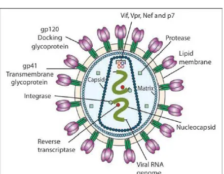
STRUCTURE OF HIV
HIV has an icosahedral structure .It is an enveloped virus .The diameter of HIV is 120 nm(6) . It also has a lipid bilayer with around 72 spikes of glycoprotein . The different types of glycoprotein are
• gp 120
• gp41 for HIV- 1
[image:25.595.109.564.348.701.2]• gp 36 for HIV – 2.
The gp 120 provide binding site for the cellular receptors. The virion has 2 RNA molecules within the protein capsid
viral RNA dependent DNA polymerase enzyme.The capsid (p24) is surrounded by enclosed by lipid bilayer.
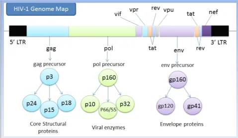
HIV genome
The genome of HIV contains 3 structural genes
7
The gp 120 provide binding site for the cellular receptors. The virion has RNA molecules within the protein capsid p24. Within the capsid are viral RNA dependent DNA polymerase Pol that is reverse transcriptase
The capsid (p24) is surrounded by a matrix layer (p1 enclosed by lipid bilayer.(5)
[image:26.595.85.565.377.655.2]The genome of HIV contains 3 structural genes - gag ,pol , env.
Figure 2 . HIV – 1 genome
The gp 120 provide binding site for the cellular receptors. The virion has Within the capsid are Pol that is reverse transcriptase
layer (p17) that is
8
In India the most common mode of transmission is heterosexual the statistics in India for mode of transmission is shown in the following figure
Figure3 . Modes of HIV transmission in India
The risk of transmission is
highest when blood or blood products are the source of infection that is more than 90% to 95 %(12)
15-40% for vertical transmission that is mother to child transmission
9
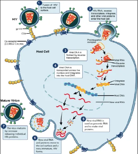
HIV REPLICATION
gp 120 protein binds to the CD4 molecule which is the gp 120 receptor(13)
This requires a co-receptor called CCR5 which helps the CD4 molecule for fusion and entry of the HIV 1 .
Fusion of the virion with the host membrane occurs via gp 41 HIV - RNA is uncoated and it is internalized to the target cell. The enzyme called reverse transcriptase of the virion causes the
reverse transcription of the virion RNA into ds DNA .
DNA translocates to nucleus and with the help of integrase , the DNA integrates with host chromosome
When cellular activation occurs this leads to transcription of the proviral DNA to mRNA and also to the virion genomic RNA Then transcribed mRNA is translated to proteins
Posttranscriptional modification occurs
The HIV proteins enzyme and also the genomic RNA assemble to form the core of the virion.
10
11
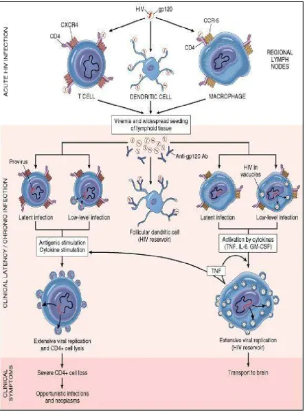
PATHOGENESIS
PRIMARY INFECTION :
• When there is infection by human immunodeficiency virus 1 initially in the acute stage there is viral replication in CD 4 + T lymphocytes. CD 4 + T cells in the GALT and MALT express HIV co-receptor CCR5 and hence are susceptible to HIV-1 infection. This infection leads to viral replication which in turn causes high HIV RNA titres . It reaches higher to about Ten lakh copies per millilitre within a fornight. Due to this high HIV load patients have a syndrome known as Acute Retroviral Syndrome.
• Following this fall in the peripheral CD4+ T lymphocyte cell counts occurs in the primary infection which causes opportunistic infections. In few weeks time the CD8 + T lymphocyte begin to form in response to HIV and causes control of multiplication of the virus.
• This leads to decrease in the load of HIV which leads to a state called as the SET POINT.
12
13
IMMUNITY THAT IS SPECIFIC TO HIV
• HIV activates dendritic cell through TLR
• This causes production of interferon IFN 1
• This interferon stimulates immunity against HIV 1
• Interferon 1 also activates CD4+ and CD 8+ cells
• Natural killer cells - activated when cell is infected with HIV
• NK cell activation leads to prevention of progression of illness in the long run may be by decreasing HIV multiplication.
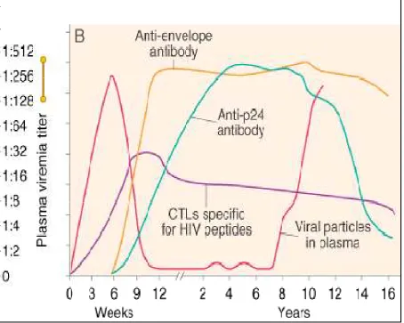
• Humoral immune activation has a latency , it is not reached till about maximum load of the virus is reached . this latency is called the window period
• Window period is the period in which there is viremia but the antibodies cannot be detected in blood.
14
• The depletion of CD 4 is very selective. This is responsible for the impaired adaptive immune response in human immunodeficiency virus.
• CD8+T lymphocytes control the human immunodeficiency virus infection by causing direct cytolysis of the cell which is infected by the virus.
• CD 8 + T cells also secrete soluble factors called macrophage inflammatory protein one beta which binds to chemokine receptor and thus prevents the entry of human immunodeficiency virus into the cell.
• Complete sterilizing immunity which is specific to HIV cannot be achieved by the CD8+ lymphocytes, because of the latent infection which remains within the CD4 + cells memory cells that are formed soon after the infection.
• These latent cell are not recognized by the CD 8 + T cells because the quiescent cells are not able to synthesise HIV proteins .
15
THE EFFECT OF HIV-1 REPLICATION ON THE
IMMUNE SYSTEM
There is immune system activation chronically due to continuous viral multiplication. This kind of chronically active immune mechanism can cause non - specific inflammatory activation and this leads to translocation of the microbes due to decrease in CD4+ T lymphocytes in GALT. It is the chronic activation of immune system which leads to the fall in the CD 4 + T lymphocytes.
The markers of immune system activation on the CD 4+ T lymphocytes and the CD 8 + T lymphocytes better correlates with rate at which the CD 4 falls than does the quanitity of the load of the virus in patients who are not treated. There are activation markers detected increasingly on the
NK cells B cells
CD 4 + lymphocytes
16
This activation of markers is associated with rise in the rate of turnover of the cells. NK cells don’t function effectively causing ineffective control over other viral infections
B cell function is also impaired causing hyper- gammaglobulinemia and accelerated generation of auto-antibodies. When vaccinated the response by production of antibody is impaired when CD4+ falls.
17
Figure 6 . Immune Response to HIV Infection
Autoimmune Phenomena
The autoimmune phenomena in HIV-infection is due to chronic immune system activation as well as molecular mimicry by viral components.
18
Autoimmune phenomena include antibodies to lymphocytes, to platelets or to neutrophils. Antiplatelet antibodies thus may cause thrombocytopenia of HIV disease.
Antibodies to nuclear and cytoplasmic components of cells and antibodies to cardiolipin; CD4 molecules; CD43 molecules; C1q-A; variable regions of the T cell receptor ; Fas; denatured collagen; and IL-2. In addition, autoantibodies to serum proteins, including albumin, immunoglobulin, and thyroglobulin, have been reported. There is antigenic cross-reactivity between HIV viral proteins (gp120
and gp41) and MHC class II determinants, and anti-MHC class II antibodies reported in HIV infection.
These antibodies could potentially lead to the elimination of MHC class II–bearing cells via antibody-dependent cellular cytotoxicity (ADCC), although this has not been clearly demonstrated to occur . In addition, regions of homology exist between HIV envelope glycoproteins and IL-2 as well as MHC class I molecules.
19
[image:38.595.136.543.338.659.2]• With the effective antiretroviral therapy, an immune reconstitution inflammatory syndrome (IRIS) has become common. IRIS is an autoimmune-like phenomenon characterized by a paradoxical deterioration of clinical condition, which is usually compartmentalized to a particular organ system, in individuals in whome cART has recently been initiated. It is associated with a decrease in viral load and at least partial recovery of immune competence, causing rise in CD4 count .
20
Clinical Features of AIDS
It can be of mild disease to severe. They range from a mild acute illness to severe disease. Symptoms range from fever with weight loss or diarrhea or generalized lymph node enlargement or opportunistic infections or neurological diseases and also secondary neoplasm. The typical infectious diseases and neoplasms in HIV and AIDS is detailed in the clinical stages of infections. Opportunistic infections are the most common cause of mortality in AIDS patients. The prevalence of infections can be reduced by the treatment with HAART.
NACO GUIDELINES (7)
In India the Case Definition for AIDS
Case definition for AIDS in India , was revised in October, 1999 by the national AIDS control organisation
Case Definition of AIDS in persons > 12 years
1. Two positive tests for HIV infection by ELISA/ RAPID / SIMPLE test AND
2. Any one of the following criteria:-
21
b) Tuberculosis: may be Extensive pulmonary or disseminated, or miliary, or extrapulmonary tuberculosis.
c) Neurological impairment is present that is preventing independent activities of daily living, not known to be caused by conditions that are unrelated to HIV
d) esophageal candidiasis
e) Clinically life -threatening / recurrent episodes of pneumonitis
f) Kaposi’s Sarcoma g) Other conditions:- Cryptococcal meninigitis Neurological Toxoplasmosis
Cytomegalovirus retinitis
22
DIAGNOSIS OF HIV
The diagnosis of HIV infection is by one of the following The demonstration of antibodies against HIV
The direct detection of HIV or one of its components. (10)
Antibodies to HIV generally appear in the circulation 2 to 12 weeks after infection. The standard screening test for HIV infection is the ELISA- Enzyme linked immunosorbent assay. This solid phase assay considered as a screening test with a sensitivity of >99.5%.(6)
The western blot test is a highly specific test; it detects specific antibody to viral core protein (p24) and envelop glycoprotein gp41.(8)
Quantitative nucleic acid testing. :
• Polymerase chain reaction (PCR) • the branched DNA (b-DNA)
• the nucleic acid sequence-based amplification (NASBA) • ligase chain reaction (LCR)
23
I. The screening tests
1) ELISA test for different viral antigens 2) Rapid tests
a) Dot blot assay
b) Particle agglutination c) HIV spot and coombs test
d) Flowmetric microparticulate technologies
The sensitivity of the ELISA is > 99.5%, the specificity of positive results by two different techniques approaches to 100% even in low risk population.(10) Factors associated with false positive tests are autoantibodies, hepatic disease, recent influenza vaccination and acute viral infection.
II. The confirmatory tests are:
1) Western blot 2) Immunoblot
3) Line immuno assay
4) ELISA with different antigen system or with different principle of test which makes the test more specific
24
7) Polymerase Chain reaction ( PCR) for HIV RNA
The confirmatory test commonly used is the western blot test. Its specificity when combined with ELISA is >99.99%. The indeterminate results occur with early HIV infection, HIV-2 infection, autoimmune diseases, pregnancy and recent tetanus toxoid injection.(10) But as the test is cumbersome and also costly the practice now is to perform two different types of ELISA or an ELISA with any of the rapid tests. A serum positive in both tests is considered positive. This is used in the diagnosis of HIV infection by NACO guidelines.
Revised World Health Organization clinical staging of
HIV/AIDS for adults and adolescents (2005)
(75)World Health Organization developed the clinical staging
and immunological classification of HIV and related illness to help those
countries out where inadequate resource makes lab measurements of CD
4 counts and HIV viral load in plasma not feasible. The WHO clinical
stage can be used to determine the eligibility of the patient for
antiretroviral treatment. Medical decision for HIV patients is made
entirely based on the clinical features. This staging system is a practical
and also accurate way to manage HIV patients with many international
studies showing agreement between the clinical features in staging
25
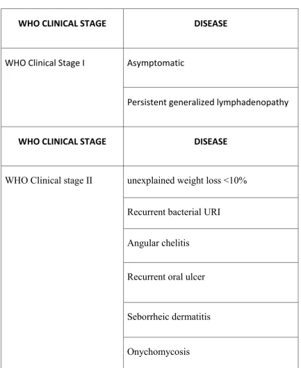
Table 1: WHO CLINICAL STAGES OF HIV/AIDS
WHO CLINICAL STAGE DISEASE
WHO Clinical Stage I Asymptomatic
Persistent generalized lymphadenopathy
WHO CLINICAL STAGE DISEASE
WHO Clinical stage II unexplained weight loss <10%
Recurrent bacterial URI
Angular chelitis
Recurrent oral ulcer
Seborrheic dermatitis
26
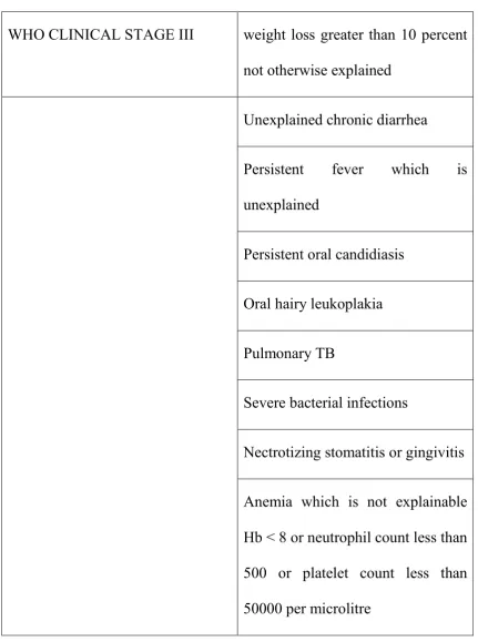
Table 1: WHO CLINICAL STAGE of HIV / AIDS
WHO CLINICAL STAGE III weight loss greater than 10 percent not otherwise explained
Unexplained chronic diarrhea
Persistent fever which is unexplained
Persistent oral candidiasis
Oral hairy leukoplakia
Pulmonary TB
Severe bacterial infections
Nectrotizing stomatitis or gingivitis
27
WHO CLINICAL STAGE IV Extrapulmonary TB PCP pneumonia
Recurrent severe bacterial pneumonia Central nervous system toxoplasmosis HIV encephalopathy
HIV wasting syndrome Cryptococcosis
Progressive multifocal leukoencephalopathy Esophageal candidiasis CMV infection
Lymphoma Kaposi sarcoma
HIV Associated Nephropathy HIV associated Cardiomyopathy
Invasive carcinoma cervix
28
HIV A MULTISYSTEM DISEASE
Respiratory System
Infectious pulmonary process(15) : Mycobacteria tuberculosis
pneumococcal pneumonia with sepsis
Haemophilus influenza pneumonia Pseudomonas aeruginosa
Pneumocystis jerovecii pneumonia
Non infectious pulmonary disease: Kaposi’s sarcoma Non Hodgkin’s lymphoma Interstitial pneumonitis Sinusitis : Bacterial sinusitis
Fungal sinusitis
29
Central Nervous System (15)
• Toxoplasmosis – most common space occupying lesion in patients with HIV
• CNS lymphoma – Non Hodgkin’s lymphoma
• AIDS dementia complex – most common cause of altered sensorium in HIV patients.
• Cryptococcal meningitis
• HIV myelopathy : spastic paraparesis and sensory ataxia
• Progressive multifocal leukoencephalopaty : viral infection – JC virus which destroys myelin by infecting the oligodendrocytes .It usually Presents with aphasia, limb apraxia , ataxia, hemiparesis, cortical blindness, focal signs related to occipital lobe.
Peripheral Nervous System Diseases
• Sensory neuropathy , Mononeuropathy
• Inflammatory demyelinating polyneuropathy
30
Myopathy
Proximal muscle myopathy
Zidovudine can also induce myopathy
Retinitis
CMV retinitis –fluffy opacities / exudates perivascular Hemorrheage
Oral Lesions
• Oral candidiasis
• Hairy leukoplakia
• Angular cheilitis
• Aphthous ulcers
• Gingival disease
Gastro intestinal involvebilaiment
• Candidial oesophagitis
31
Hepatitis B virus hepatitis
Cytomegalovirus infection of the liver
Mycobacterial disease of the liver Lymphoma involving the liver
• Biliary disease : these diseases are induced by Cytomegalovirus , cryptosporidia and microsporidia
Acalculous cholecystitis , Sclerosing cholangitis , Papillary stenosis
• Enterocolitis :
Bacterial : campylobacter , Salmonella , Shigella Virus : Cytomegalovirus , Adenovirus
Protozoans : entamoeba, Giardia etc
32
Skin Manifestations(16)
Herpes simplex infection is common and often more severe in AIDS patients .Herpes zoster is also common in HIV disease
Molluscum contagiosum : spread widely over the patients skin
Staphylococcus: most common bacterial skin disease. Presents as folliculitis , superficial abscess, bullous impetigo. It can progress to sepsis Bacillary angiomatosis : Bartonella mimic lesions of kaposi’s sarcoma Fungal infections : dermatophytes and candida
Seborrhoeic dermatitis caused by Malasezzia furfur Xerosis : severe pruritis in HIV patients with no rash
Psoriasis can be very severe in patients who are HIV seropositive
HIV Related Malignancies(15)
AIDS defining malignancies are
1.Kaposi’s sarcoma , 2.Non Hodgkins lymphoma, 3.primary lymphoma of the central nervous system and 4. invasive cervical carcinoma
Endocrine manifestations in HIV infection
33
autopsy there is found to be high incidence of adrenal involvement.(17,19 ) CMV infection of adrenal gland is found.(18) But clinically and biochemically, adrenal insufficiency is uncommon. The trio of hyperkalemia hyponatremia and hypotension in HIV patients is not always adrenal insufficiency, it can occur due to many causes in advanced HIV disease. Drugs like ketoconazole used to treat fungal infection in HIV patients causes inhibition of adrenal corticosteroid synthesis(20) and it also causes blunting of the cortisol response when ACTH is given(21). Rifampicin alters the metabolism of the glucocorticoids causing increased excretion of the steroids making increased glucocorticoid replacement to achieve therapeutic levels.(22)
HYPOGONADISM
34
1000 to 1100 ng/dl . Those found to have <450 to 500 ng/dl need to be treated. Serum estradiol level is used in women to assess for hypogonadism. The therapy is testosterone intramuscular injection every one to three weeks so that approximately 100mg/week is provided. Oxandrolone an oral androgen is now approved for use in AIDS-associated weight loss and the dose used is 20 mg.(27)
PITUITARY DYSFUNCTION
There are reports of Toxoplasma gondii infiltrating the pituitary(28) in advanced stages of HIV disease. Inadequate ACTH response to adrenal insufficiency and hypogonadotropic hypogonadism. Elevated levels of prolactin is also reported in advanced HIV disease. Posterior pituitary dysfunction can present as hyponatremia.(29) Hyponatremia is due to stress or drug induced SIADH (32). Another differential diagnosis is renal salt wasting syndrome.(30) Isolated hyponatremia is commonly seen in HIV disease in advanced stage of illness and it had a poor prognosis.(31)
PANCREATIC DYSFUNCTION
35
destruction of beta cell causes rapid release of large quantity of insulin leading to hypoglycemia.(34) The drug remains in the system for weeks due to long half life hence hypoglycemia may be persistent for days to weeks. Later on patient can go in for diabetes mellitus with or without ketoacidosis.(35)
THYROID GLAND FUNCTION
36
THYROID OVERVIEW
Normal thyroid gland is made up of two lobes joined by the isthmus. It is 0.5 cm thick, 2cm wide, and 2cm in height. It lies anterior to trachea that is it lies between the cricoid and the suprasternal notch.
PHYSIOLOGY OF THYROID HORMONE(42)
37
about 10% of T4 but little T3. In the periphery T4 is converted to T3 by the deiodinase enzymes. Type I deiodinase, which is located primarily in thyroid, liver, and kidneys, has a relatively low afinity for T4. Type II deiodinase that has a higher affinity for T4 and is found primarily in the pituitary gland, brain, brown fat, and thyroid gland. Expression of type I deiodinase allows it to regulate T3 concentrations , this is important in levothyroxine (T4) replacement.
38
MECHANISM OF ACTION:
Thyroid hormone enters the cell and the T3 gets bound to thyroid receptors located inside the nuclei. TR is of two types TR alpha and TR beta. TR alpha is abundant in brain, kidneys, muscle, gonads and heart and the TR beta is present in pituitary and liver. T4 can also get bound but the binding is not as avid. The hormone receptor complex is then bound to the DNA via the Zinc fingers thus regulate expression of different genes that code for enzymes and thus regulates cell function. T3 binds with receptor 10–15 times greater affinity than T4, which explains its increased hormonal potency. T4 is produced in excess of T3 but receptors are occupied mainly by T3.
Regulation of the Thyroid Axis
39
Figure 9 : Hypothalamic – Pituitary – Thyroid Axis
40
The TSH is extremely sensitive to the levels of thyroid hormones in circulation and can be used as a useful tool in detection of thyroid abnormalities rather than using T4 or T3 levels. The thyroid dysfunction is classified as hypothyroidism, hyperthyroidism, subclinical hyperthyroidism depending upon the TSH and thyroid hormone levels.
Table 2: Thyroid dysfunction classification
Thyroid status TSH Thyroid hormone
Normal Normal Normal
Hypothyroidism High Low
Hyperthyroidism Low High
Subclinical hypothyroidism High Normal
Subclinical hyperthyroidism Low Normal
HYPOTHYROIDISM
41
Hypothyroidism is of two types primary and secondary
Table 3 : PRIMARY HYPOTHYROIDISM
Autoimmune Hashimoto’s and atrophic thyroiditis
Iodine deficiency Environmental or nutritional
Iatrogenic I 131 use, thyroidectomy radiation therapy to neck
Medications Amiodarone, Lithium, antithyroid drugs
Congenital Absent or ectopic thyroid Dyshormonogenesis
TSH receptor mutation
Infiltrative disorder Amyloidosis, Sarcoidosis scleroderma Hemochromatosis
42
Table 4: SECONDARY HYPERTHYROIDISM
Hypopituitarism
Tumors, Pituitary surgery
Irradiation, Sheehans syndrome
Hypothalamic disease Tumors, Trauma, Infiltrative disorder
Table 5 :Clinical features of hypothyroidism
Symptoms
Signs
Weakness, Tiredness
Carpal tunnel syndrome
Dry skin, hair loss
Dry coarse skin, Diffuse alopecia
Cold intolerance
Cold peripheries
Poor concentration and
memory, Paraesthesia
Delayed DTR, Pseudomyotonic
reflex
Weight gain and poor appetite Puffy hands and feet
Constipation
Dyspnoea
Pericardial and pleural effusion
Hoarseness
43
HYPERTHYROIDISM
Hyperthyroidism(45) is due to excessive action of thyroid hormones in the body tissues. Thyrotoxicosis is a synonym. Grave’s disease is the commonest cause of hyperthyroidism. Approximately 0.5% to 1% of the population suffers from hyperthyroidism. The TSH levels are suppressed, <0.1mU/L and associated with high levels of thyroid hormones. It is of 2 types that is primary and secondary, the different causes of each is included in the table below.
Table 6: HYPERTHYROIDISM
Primary hyperthyroidism Secondary hyperthyroidism
Graves disease TSH secreting pituitary adenoma
Toxic MNG Thyroid hormone resistance
Toxic adenoma Gestational thyrotoxicosis
Thyroid malignancy
Thyrotoxicosis + hyperthyroidism
Subacute granulomatous thyroiditis
44
Table 7 : CLINICAL FEATURES OF HYPERTHYROIDISM
SYMPTOMS SIGNS
Hyperactive Tremors
Irritable Goitre
Dysphoria Warm moist skin
Palpitation Tachycardia and atrial fibrillation Fatigue Lid retraction lid lag
Weight loss and increased appetite Tachycardia
Diarrhoea Proximal muscle weakness Polyuria
Oligomenorrhoea ,loss of libido Gynaecomastia
SUBCLINICAL HYPOTHYROIDISM
45
SUBCLINICAL HYPERTHYROIDISM
Subclinical hyperthyroidism is defined as low serum TSH levels (0.1mU/L to 0.4mU/L) asociated with normal T4 and T3 levels.(40) Subclinical hyperthyroidism prevalence is about 2%. Most often subclinical disease is asymptomatic but some patients show symptoms of deficiency of thyroid hormone in subclinical hypothyroidism.
SICK EUTHYROID SYNDROME
Thyroid hormone alteration occurs in many diseases which affect mainly the T3 level in the absence of any intrinsic thyroid glandular disease. It is also called Low T3 syndrome, Non thyroidal illness syndrome or Thyroid hormone adaptation syndrome. Sick euthyroidism syndrome occurs in the following conditions
• Acute critical illness – life threatening infections or Myocardial infarction
• Burns or polytrauma • Post operative period • Diabetes mellitus • Renal diseases • Liver disease • Malignancy
46
Sick euthyroid syndrome is most often associated with fall in total and free T3 with normal T4 and TSH. The magnitude of fall in the T3 correlates with the severity of the disease. In this condition the peripheral conversion of T4 to T3 by deiodinase is abnormally low and instead is converted to Reverse T3. This is one mechanism although the main mechanism is decreased clearance than production for increased rT3. T4 by alternate metabolism is converted to inactive T3 sulfate. With increase in disease severity there is decrease in serum T4 also called the low T3 T4 syndrome. Though T level falls the free T4 remains within normal limits or slightly on the lower side. T3 T4 even if is lower, serum TSH levels are normal or reduced , this is the main difference from primary hypothyroidism where TSH level rises with fall in T3 T4 levels.
LABORATORY EVALUATION
Estimation of Thyroid Hormones
47
T4 and the T3 are greatly bound to proteins and this protein binding is affected by many factors like medications pregnancy or genetic make up. So it is useful to measure free T3. Moreover in pregnancy there is increase in thyroid binding globulin which leads to increase in total thyroid hormone levels similarly when there is decrease in thyroid binding globulin as in nephrotic syndrome, total thyroid hormone level is decreased.
Tests to determine Thyroid Dysfunction etiology
TPO antibodies: autoimmune thyroid disease is detected by measuring antibodies versus Thyroid peroxidase and thyroglobulin. It is found that around 10 % of euthyroid women and 2 % of euthyroid men have thyroid antibodies. In autoimmune hypothyroidism TPO antibodies is universally found whereas in graves disease upto 80 % have this.
THYROID STIMULATING ANTIBODIES : These stimulate the TSH-R in Graves disease.
THYROGLOBULIN LEVELS : It is elevated in thyrotoxicosis of all kinds except thyrotoxicosis factitia. Thyroglobulin levels are increased in thyroiditis. n of thyroid hormone.
48
Nuclear imaging of Graves disease is found to have increase in size of the gland and more tracer uptake .Thyroid scan has also role in monitoring patients with thyroid cancer.
d) Thyroid Ultrasound
USG is used in the diagnosis of nodular thyroid disease.
MANAGEMENT
Hypothyroidism:
• Levothyroxine (47) is the treatment of choice. Levothyroxin is converted to thyroxin in the body. It is partially, the more active thyroid hormone. If there is no residual thyroid function, the daily replacement dose of levothyroxine is usualy 1. 6 microgram/kg body weight (typicaly 10–150 microgram).
• Adult patients who are less than sixty years without evidence of heart disease may be started on 50-100 microgram levothyroxine (T4) daily.
49
Hyperthyroidism :
The hyperthyroidism of Graves' disease is treated by decreasing the synthesis of the thyroid hormones using the drugs called antithyroid medications and by decreasing the thyroid tissue by radio- iodine ablation or by thyroidectomy. Antithyroid drugs are propylthiouracil, carbimazole and its active metabolite methimazole. All of these block TPO enzyme , thus reducing oxidation of and organification of the iodide molecule.
• Propylthiouracil(47) blocks deiodination of T4 to Triiodothyronine
• The starting dose of carbimazole or methimazole is usually 10–20 mg every 8 or 12 h, and changed to daily once dosage after euthyroidism is attained.
• Propylthiouracil is given at a dose of 10–20 mg once in 6–8 hours
• TFT and clinically patient is reviewed 3–4 weeks after initiating therapy and then the dose is altered based on the free T4 levels.
• It takes about six to eight weeks after therapy initiation to achieve euthyroidism
50
• The usual daily maintenance doses of antithyroid drugs in the titration regimen are 2.5–10 mg of carbimazole or methimazole and 50–100 mg of propylthiouracil.
• In the block-replace regimen, the initial dose of antithyroid drug is held constant, and the dose of levothyroxine is adjusted to maintain normal unbound T4 levels. When TSH suppression is alleviated, TSH levels can also be used to monitor the effectiveness of treatment
• Maximum remission rates of up to 30– 50% are achieved by around 2 years for the titration regimen and by 6 months for the block-replace regimen.
• Propranolol (20–40 mg Q6H) or atenolol, are used for controlling the adrenergic symptoms mostly in the early stages of diseases before the antithyroid drugs action becomes effective.
• Radioiodine acts by destroying thyroid cells and may be used as a initial therapy or for relapse after a trial with the antithyroid drugs.
51
THYROID DYSFUNCTION IN HIV DISEASE
HIV disease is associated with many endocrine abnormalities. Many of these occur in early and many in late stage of the disease. Among those with HIV disease around 2 % develop frank thyroid illness. But what is more prevalent is subtle dysfunction in thyroid(48). Thyroid profile derangements are common in HIV patients. Thyroid binding globulin level is raised in them and there is inverse correlation with CD 4 count.(49 ) Abnormal TFT may be due to the stress of the disease in
patients with advanced disease. Subclinical hypothyroidism is common among the adult HIV infected patients. Abnormal TFT results may be caused by the stress of illness in patients especially those with low CD 4 counts. Thyroid dysfunction has been reported with the use of HAART.
PATTERNS OF THYROID DYSFUNCTION
Hypothyroidism
52
SUBCLINICAL HYPOTHYROIDISM
53
Isolated decreased FT4 levels
Decreased Free T4 levels along with normal TSH levels are found in HIV patients, with prevalence which is of 1.3%–6.8%. In adults didanosine stavudine and also the drug ritonavir are associated with this pattern of thyroid profile. Decreased free T4 may be due to the hypothalamus or the anterior pituitary dysfunction. In one study TSH releasing hormone when administered to those with decreased free T4 had no delayed and no absent TSH release, thus ruling out pituitary or hypothalamic dysfunction as a cause of decreased free T4. Those taking antiepileptic drugs had low free T4 possibly due to artifact due to the intereference of the drug in the estimation of the free T4. The patients with low free T4 do not have signs and symptoms of hypothyroidism hence the significance of free T4 clinically is not very clear.
TFT abnormalities due to nonthyroidal illness.
54
abnormality pattern. But this TFT abnormality is due to body’s response to severe illness rather than due to thyroid glandular function abnormality. Since AIDS due to the above described process can produce abnormal TFT this should be kept in the mind when interpreting TFT in patients with advanced HIV infection. Decreased triiodothyronine and elevated RT3 and variable free T4 and a low or normal thyroid stimulating hormone level , different patterns based on the grades of severe illness.
When the patient improves the thyroid stimulating hormone level rises and in some it rises above the normal level as both free T4 and triiodothyronine come back to baseline and this is similar to subclinical hypothyroidism pattern.
55
Sick euthyroidism was most prevalent approximating 16 % in those with advanced AIDS before highly active antiretroviral therapy became extensively used.
Treatment of sick euthyroidism requires treatment of the underlying severe disease and not hormone replacement. Repeat TFT after 1 to one and half months after the severe illness is controlled with HAART confirms the diagnosis.
CONDITIONS CAUSING THYROID FUNCTION
ABNORMALITIES IN AIDS
Opportunistic infection and malignancies cause thyroid dysfunction in AIDS
Thyroditis due to Pneumocystis jiroveci and Cryptococcus neoformans
Bacterial infection leading to suppuration of thyroid gland Infiltration of the gland with lymphoma or kaposi sarcoma .
Hyperthyroidism
56
antibodies to TSH receptor is the most common etiology of hyperthyroidism in general as well as HIV infected individuals. The IRIS which occurs after highly active retroviral therapy initiation is the state at which graves usually occurs in HIV infected patients. The IRIS causing exacerbation of Mycobacteria tuberculosis and and other infection usually occur in almost three months of antiretroviral therapy initiation. Where as the graves disease occurs approximately one to three years after starting highly active antiretroviral therapy. It is found that CD4 count increases in two phases after highly active antiretroviral therapy is started. One is by CD4 memory cells getting redistributed from the lymphoid organs and second phase occurs many months afterwards when there is increase in naïve CD 4 lymphocytes.
BIPHASIC INCREASE IN CD 4 CELLS DURING IRIS
Phase 1
• CD 4 cells redistributed from lymphoid organs
Phase 2
57
In patients with TFT patterns of overt hyperthyroidism radioiodine uptake study and the scanning of the thyroid gland can discriminate between the different etiologies of hyperthyroidism. In Graves there is increased uptake of radioiodine and shows homogenous pattern by scan whereas in MNG thyroid or thyroid adenoma shows increased radioiodine uptake but heterogenous .
Decreased radiiodine uptake is found in thyroiditis both subacute granulomatous and painless thyroiditis. Subacute granulomatous thyroiditis occurs after respiratory tract infection by some viruses causes increased thyroid hormone state initially, then around a month or a month and a half later causes decreased thyroid hormone status. Painless thyroiditis is an autoimmune thyroid inflammation which is quite often transient and has anti TPO antibodies.
Treatment of hyperthyroidism
58
Subacute thyroiditis is treated with NSAID s and if not cured with steroids for pain. Beta blockers relieve the adrenergic symptoms. Painless thyroiditis only treatment is beta blockers.
Subclinical hyperthyroidism
Subclinical hyperthyroidism predate hyperthyroidism and is characterized by decreased TSH, normal free T4 and T3 levels, and no symptoms of thyrotoxicosis. If symptoms are there then antithyroid medication or ablation of gland with radioiodine may be required. subclinical hyperthyroidism can lead to the following complications – atrial fibrillation and decreased mineral density of the bone . So it is told to treat those patients with the TSH level less than 0.1 mU/L or are elderly that is more than 60 years or those with osteopenia. Treatment is not needed as per guidelines in those with milder disease that is thyroid stimulating hormone level of 0.1 to 0.45 mU/L and guidelines ask for repeated TFT half yearly or yearly.
HAART and thyroid dysfunction (76)
59
ROLE OF SCREENING THYROID FUNCTION TEST IN
THYROID DISEASE
Older patients may be screened for thyroid dysfunction as there is increased incidence of subclinical hypothyroidism
TSH level measurement in those with symptoms of thyroid function abnormalities or decreased bone mineral density or altered lipid profile or depression or atrial fibrillation.
If TSH is found to be elevated FT4 is to be estimated
If TSH is low , Both free T4 and T3 need to estimated to rule out T3 toxicosis
Sick euthyroidism is to be considered in the interpretation of altered thyroid function test especially in advanced HIV disease
Correlation with the type of thyroid dysfunction observed:
Belttrans et al(52) studied 350 HIV+ patients showing that 16% suffered from hypothyroidism
Subclinical hypothyroidism 6.6 %
60
Jain G et al a prevalence study which included fifty HIV patients at different stages of illness. Patients were divided into two groups(53)
Group-1 had 25 patients those who had AIDS
Group-2 had 25 patients who were HIV+ but who were not having AIDS
18% had free T 3 levels below the normal range 20% patients had decreased FT-4 levels
24% patients had TSH levels above the normal range.
20% of those who had hypothyroidism were found to be among the AIDS patients
subclinical hypothyroidism predominated among the HIV infected who did not have AIDS
Sunder S et al studied 150 HIV patients. (59) They divided the study group into 3
61
In group A, fifteen of them had subclinical hypothyroidism and eleven had overt hypothyroidism.
In group B, 18 had subclinical hypothyroidism and 4 had overt hypothyroidism.
In group C, 12 patients had subclinical and 1 had clinical hypothyroidism
hypothyroid as well as subclinical hypothyroid states were seen in advanced HIV, and in non-AIDS groups, subclinical hypothyroid state was dominant (55,56)
Palanisamy et al(57)
Studied alteration in thyroid function in AIDS patients in relation to infection with HIV and healthy subjects.
No statistically significant difference in the thyroxin concentrations
in those with AIDS was found in comparison to those with HIV
infection and those who are healthy subjects.
FT4 concentration was statistically significantly decreased in HIV
and those with AIDS
Total triiodothyronine concentrations were normal in those
62
T3 level was decreased in AIDS patients.
The level of FT3 was slightly but significantly raised in HIV
infected and significantly decreased in those with AIDS
The study concluded that thyroid dysfunction is frequent in HIV
infection and with progression of disease there is a primary
hypothyroid like stage that occurs in patients with HIV infection.
Relationship with ART(59,60,61)
Beltran et al study 350 HIV patients for thyroid dysfunction. 32% had
thyroid dysfunction. In a substudy ,age and sex matched groups of
patients naïve of ART and those on ART were studied for changes. They
found no difference between the 2 groups which were statistically
significant.
Bongiovanni et al (58)observed statistically significant changes, in the
form of increased cases of subclinical hypothyroidism(15%) in the
patients receiving HAART.
63
of 49 subjects on HAART (12.2%), while no case of dysthyroidism was observed in the group of antiretroviral-naive patients (p<0.05). A long prior exposure to HAART was reported in all patients (ranging from 27 to 58 months), and ongoing antiretroviral therapy included stavudine in all the 6 cases, lamivudine in four, didanosine in two, saquinavir in three, indinavir, nelfinavir and lopinavir/ritonavir in one case each; duration of HAART ranged from 21 to 41 months.
Relationship with CD4 counts(63,64)
Most study series have found an inverse correlation of the CD4 counts with TSH values. There were no conclusions drawn in relation to their clinical manifestations in the different stages according to CD4 count.
Sunder et al, studied patients in 3 groups(CD4<200cells/mm3,200-
350cells/mm3 and >350cells/mm3). Each group had 50 subjects. Percentage of low TSH was maximum amongst subjects with CD4 <200cells/mm3(52%) and least amongst those with CD4 > 350 cells/mm3 (26%). All the studies owe this inverse correlation to the immune reconstitution occurring with advanced disease or the institution of ART.
Beltran et al, had 2 groups, A:CD4 >200 cells/mm3 and
64
in group B. Low CD4 cell count was a risk factor for hypothyroidism in the present study. (38)
Madge S et al (65,66)
It was a retrospective analysis of thyroid function test in HIV patients. The prevalence of and factors which are associated with both the clinical and the subclinical thyroid dysfunction were studied. Those patients who had normal thyroid function tests but had thyroid disease in the past were found from the medical records department and was made part of the overt category.
73% of the clinic population had at least one thyroid function test done since 2001.
1233 (79%) were male 1043 (66%) were white
365 (23%) were black African
969 (62%) - the main risk factor for HIV was homosexual mode of transmission
Median age of the study was 37 years.
65
Table 8 : Madge S et al results
Thyroid dysfunction Percentage affected
Overt hypothyroidism 2.5 %
Overt hyperthyroidism Less than 1 %
Subclinical hypothyroidism 4 %
Subclinical hyperthyroidism <1%
Sick euthyroidism 17%
Euthyroidism 75.5 %
By Multivariate analysis : no independent variable had statistical
66
MATERIALS AND METHODS
Research Design:
The present study is an observational and prospective study.
Source of data:
Patients admitted to and those attending Coimbatore medical college hospital as out patients who are newly diagnosed as HIV positive as per NACO March 2007 criteria.
Methodology
Sample size – fifty
Sampling method – simple random sampling
67
abnormalities were compared with HIV status, CD4 count and WHO clinical stage of HIV disease. Patient confidentiality was maintained.
INCLUSION CRITERIA:
Newly diagnosed HIV seropositive patients by ELISA testing by NACO March 2007 guidelines in the age group 20 to 60 years
EXCLUSION CRITERIA
1. Patients already known thyroid disease 2. Patients already on Thyroxin treatment.
3. Patient who had undergone thyroid surgery in the past 4. patients on drugs altering thyroid function – like
Amiodarone , Lithium, Phenobarbitone, Carbamazepine, Phenytoin , Para amino salicylic acid, Antiretroviral drugs especially stavudine.
5. Pregnant patients.
6. patients on oral contraceptive pills
Investigations done:
HIV serology by ELISA
CD4 count by Flow cytometry
Thyroid function test T3 T4 TSH by electrochemiluminescence method
68
Renal function test Liver function test Chest X ray
Statistical analysis:
Statistical methods applied(67,68,69,70)
The data that was collected was subjected to descriptive statistical analysis. Results on continuous measurement were expressed as Mean and standard deviation and results on categorical measurements are presented in numbers and percentage. Chi square test was used to assess the significance of the results. Odds ratio was used when applicable. The results were considered significant if p value was below 0.05.
SOFTWARE used :
• Microsoft word 2007 and Microsoft excel is used to generate graphs and tables.
69
20-30 years 31-40 years 41-50 years 51-60 years 0
5 10 15 20 25 30
No: of patients
No: of patients
RESULTS
Table 9 : Age Distribution of the study group
Age in years
Figure 11 : Age distribution of patients in the study group
Age in years No: of patients Percentage
20-30 8 16
31-40 24 48
41-50 11 22
51-60 7 14
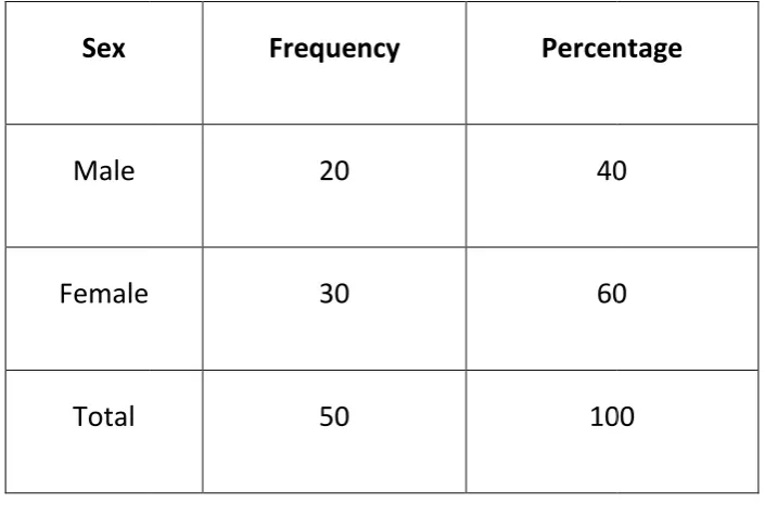
Table 10. : Sex wise distribution of Patients in study group
Sex
Male
Female
[image:89.595.146.499.148.392.2]Total
Figure 12. :
70
: Sex wise distribution of Patients in study group
Frequency Percentage
20 40
30 60
50 100
. : Sex wise distribution of patients in study group
number of patients
: Sex wise distribution of Patients in study group
Percentage
40
60
100
thyroid status of study group gross
[image:90.595.126.498.102.726.2]Table 11 : Mean of TFT and CD4 & Standard Deviation of the study group
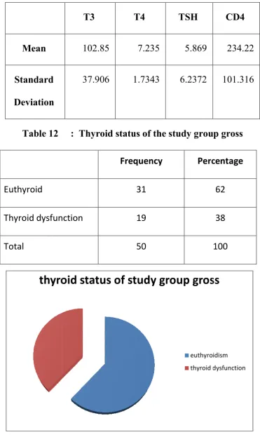
Table 12 Euthyroid Thyroid dysfunction Total Figure Mean Standard Deviation 71
thyroid status of study group gross
euthyroidism thyroid dysfunction : Mean of TFT and CD4 & Standard Deviation of the study group
: Thyroid status of the study group gross
Frequency Percentage
31
Thyroid dysfunction 19
50 100
Figure 13. Thyroid status of the study group gross
T3 T4 TSH
102.85 7.235 5.869
37.906 1.7343 6.2372
thyroid status of study group gross
euthyroidism thyroid dysfunction
: Mean of TFT and CD4 & Standard Deviation of the study group
: Thyroid status of the study group gross
Percentage
62
38
100
Thyroid status of the study group gross
CD4
234.22
72
0 10 20 30 40 50 60 70
thyroid status of the study group
[image:91.595.114.527.117.450.2]THYROID STATUS OF THE STUDY GROUP
Table 13 : Distribution of thyroid function in the study group
Thyroid status Number of patients Percentage
Euthyroidism 31 62
Subclinical hypothyroidism 10 20
Hypothyroidism 6 12
Hyperthyroidism 1 2
Low T3 2 4
[image:91.595.114.535.207.653.2]Total 50 100
0 5 10 15 20 25 30 35 <200 200
Table 14 : Distribution of CD4 count of patients in study group
CD4 count
<200
201-500
>500
Total
Figure 15: Distribution of CD4 count of patients in study group
73
200-500 >500
Distribution of CD4 count of patients in study group
CD4 count Number of patients Percentage
19 38
31 62
0
50 100
Distribution of CD4 count of patients in study group
number of patients
Distribution of CD4 count of patients in study group
Percentage
38
62
0
100
[image:92.595.96.529.400.660.2]0 2 4 6 8 10 12 14 16 18 1
Table 15 : Distribution of HIV patients based on WHO Clinical
WHO Clinical stage
1
2
3
4
Total
Figure 16 : Distribution of HIV patients based on WHO Clinical
74
2
3
4
: Distribution of HIV patients based on WHO Clinical
stage
WHO Clinical stage Number of patients Percentage
4
17
15
14
50
: Distribution of HIV patients based on WHO Clinical
stage
number of patients
: Distribution of HIV patients based on WHO Clinical
Percentage 8 34 30 28 100
[image:93.595.114.529.175.651.2]75
Table 16 : Haemoglobin levels in HIV patients
Haemoglobin level Number of patients Percentage
<6 1 2
6.1 – 9 13 26
9.1 – 12 23 46
>12.1 13 26
Total 50 100
Figure 17 : Hemoglobin levels in HIV patients
0 5 10 15 20 25
<6 6.1-9 9.1-12 >12
[image:94.595.110.544.400.658.2]76
23 23.5 24 24.5 25 25.5 26 26.5
present absent
[image:95.595.112.544.361.618.2]number of patients
Table 17 : Distribution of pallor in HIV patients
Pallor Number of patients Percentage
Present 24 48
Absent 26 52
Total 50 100
77
Table 18: Distribution of oral candidiasis in HIV patients
Oral candidiasis No : of patients Percentage
Present 30 60
Absent 20 40
Total 50 100
Figure 19: Distribution of oral candidiasis in HIV patients
0 5 10 15 20 25 30 35
present absent
78
0 2 4 6 8 10 12 14 16
20-30 31-40 41-50 51-60
[image:97.595.106.548.358.606.2]euthyroidism thyroid dysfunction
Table 19: Association of Age With Thyroid Dysfunction
Age Euthyroidism Thyroid dysfunction
21-30 8 0
31-40 15 9
41-50 4 7
51-60 4 3
Total 31 19
Figure 20 : Association of Age with Thyroid dysfunction
Pearson chi square test value : 8.044 P value 0.045
79
0 2 4 6 8 10 12 14 16 18
Male Female
euthyroidism thyroid dysfunction
Table 20 : Association of Sex with Thyroid dysfunction in HIV
patients
Sex Euthyroidism Thyroid dysfunction Total
Male 9 6 15
Female 22 13 35
[image:98.595.91.528.147.565.2]Total 31 19 50
Figure 21: Association of Sex with Thyroid dysfunction in HIV
patients
Pearson chi square value 2.391 P value 0.122
0 5 10 15 20 25 <200 200
Table 21 : Association
CD4 Count Number of Euthyroid
<200
200-500
>500
Total
Figure 22 : ASSOCIATION OF CD4 COUNT WITH THYROID
Pearson chi square value 5.148 ODDS RATIO-.253 ( CI
80
200-500 >500