R E S E A R C H
Open Access
Isolation and genotyping of
Acanthamoeba
spp. from
Acanthamoeba
meningitis/
meningoencephalitis (AME) patients in
India
Himanshu Sekhar Behera
1, Gita Satpathy
1*and Manjari Tripathi
2Abstract
Background:Acanthamoebaspp. are free-living ubiquitous protozoans capable of causingAcanthamoeba
meningitis/meningoencephalitis (AME) of the central nervous system in humans.Acanthamoebaspp. are divided into 20 different genotypes (T1–T20) on the basis of variation in nucleotide sequences of the 18S rRNA gene. The objective of this study was to identify the genotypes ofAcanthamoebaspp. in patients ofAcanthamoeba
meningitis/meningoencephalitis (AME) using 18S rRNA gene-based PCR assay. The present study provides information regarding the involvement of the most prevalent and predominant genotype ofAcanthamoebaspp. inAcanthamoebameningitis/meningoencephalitis infections in India.
Methods:Cerebrospinal fluid (CSF) was collected from 149 clinically suspectedAcanthamoebameningitis/ meningoencephalitis (AME) patients reporting to the outpatient department/causality services of the Neurosciences Centre, AIIMS, New Delhi, India during the past five years. Samples were inoculated onto 2 % non-nutrient agar plates overlaid withE. coliand incubated at 30 °C for 14 days. Among 149 suspected patients, ten were found culture-positive forAcanthamoebaspp. out of which six isolates were established in axenic culture for molecular analysis. DNA was isolated and a PCR assay was performed for amplification of the Diagnostic fragment 3 (DF3) (~280 bp) region of the 18S rRNA gene from axenic culture of sixAcanthamoebaspp. isolates. Rnsgenotyping was performed on the basis of the variation in nucleotide sequences of DF3 region of the 18S rRNA gene.
Results:In the phylogenetic analysis, all of the sixAcanthamoebaspp. isolates were found to belong to genotype T4. The sequence homology search for these six isolates in the NCBI databank showed homology with the available strains ofAcanthamoebaspp.The newly generated sequences are available in the GenBank database under accession numbers KT004416–KT004421.
Conclusions:In the present study, genotype T4 was found as the most prevalent and predominant genotype in
Acanthamoebameningitis/ meningoencephalitis infections. Hence further studies are needed to develop optimal therapeutic strategy againstAcanthamoebaspp. of genotype T4 to combat against the infections.
Keywords:Acanthamoebameningoencephalitis,Acanthamoebameningitis, Genotyping, Culture ofAcanthamoeba
* Correspondence:gita.satpathy@gmail.com
1Ocular Microbiology, Dr. R. P. Centre for Ophthalmic Sciences, All India Institute of Medical Sciences, New Delhi 110029, India
Full list of author information is available at the end of the article
Background
Acanthamoebaspp. are free-living ubiquitous protozoans capable of causing Acanthamoeba meningitis/meningo-encephalitis (AME) of the central nervous system and fatal granulomatous amoebic encephalitis (GAE) of the brain in humans [1]. AME is a slow progressive infection of the central nervous system that occurs mostly in immuno-compromised individuals with HIV/AIDS, tuberculosis, systemic lupus erythematosis (SLE), diabetes or undergo-ing cancer chemotherapy/radiotherapy treatment and can also occur in immunocompetent individuals [2–5]. Although a person may be infected with Acanthamoeba spp., the severity of infection depends on the size of the inoculum, the immunity of the patient and the virulence properties of the infective amoeba strain. AME results from the haematogenous spread of the amoebae from ini-tial portals of entry, i.e. either skin or respiratory system to brain parenchyma through olfactory nerve [6]. The clinical symptoms of AME resemble viral, bacterial or tubercular meningitis such as fever, headache, stiff neck, lethargy, vomiting, nausea, etc. [7]. With advancement of the disease, seizures, behavioural changes like diplopia, aphasia, ataxia, altered mental state and lethargy were seen as other major symptoms [7].
The first case of amoebic meningitis in a 9 year-old boy was reported by Fowler & Carter in 1965 [8]. The first case of amoebic meningoencephalitis in two Indian children was reported by Pan et al. in 1971 [9]. Thereafter, several cases of AME had been reported from various parts of India [10, 11].
Previously Acanthamoeba spp. was divided into three groups based on morphological characteristics i.e. cyst size, shape and growth temperature conditions which were confusing [12]. However with the advancement of molecular biology techniques, researchers could differ-entiate Acanthamoeba spp. into 20 different genotypes (T1–T20) based on the variation in nucleotide se-quences of the 18S rRNA gene [13]. These genotypes are discriminated from one another by a minimum of 5 % sequence divergence and 0–4.3 % sequence dissimilarity exists within genotype T4 [14, 15]. Although genotypes T1, T2, T4, T5, T10 and T12 are responsible for causing meningitis in humans, yet genotype T4 predominates among them [16–18]. PCR assay for amplification of a 464 bp region termed as“Acanthamoeba specific ampli-mer (ASA.S1)”, within the 18S rRNA gene was found suitable for genotyping of Acanthamoeba spp. [19]. ASA.S1 region includes two regions,“stem 29”is the con-served region and“stem 29-1”, a ~280 bp long highly vari-able region is designated as Diagnostic fragment 3 (DF3) [20].Rnsgenotyping ofAcanthamoebaspp. with variation in nucleotide sequences of small DF3 region was found to be as robust as based on long ASA.S1 region (19). The present study aimed to determine the prevailing
genotypes of Acanthamoeba spp. in amoebic meningi-tis/meningoencephalitis infections in humans.
Methods
Collection of clinical specimens
After informed consent and thorough clinical examination, cerebrospinal fluid was collected by the neurologists from 149 clinically suspectedAcanthamoeba meningitis/menin-goencephalitis patients with the following symptoms: head-ache, stiff neck, lethargy, vomiting, nausea, reporting to the Outpatient Department/ causality services of Neurosciences Centre, All India Institute of Medical Sciences, New Delhi, India in the past five years. Some patients were also having symptoms such as seizures or aphasia or ataxia or altered mental state and lethargy that were similar to symptoms in Acanthamoebainfections. A part of the specimen was inoc-ulated onto a 2 % non-nutrient agar plate overlaid with E. coli for culturing and a second part of the speci-men was placed in 500 μl PBS buffer (pH 7.4) for mo-lecular diagnosis by PCR assay. Inoculated non-nutrient agar plates were sealed incubated at 30 °C for 14 days. Plates were examined regularly for 14 days post-incubation under a light microscope (at a magnification of 100–400×) (Nikon) for the appearance of growth of Acanthamoeba spp. over the agar surface. Among 149 suspected patients ofAcanthamoeba meningitis/meningo-encephalitis 10 (6.71 %) cases were found culture posi-tive, 11 (7.38 %) cases were found PCR positive and 10 (6.71 %) cases were positive by both culture and PCR assay forAcanthamoebaspp. Six samples out of ten culture-positive cases were further sub-cultured for mo-lecular analysis by transferring a square shape agar surface withAcanthamoebaspp. on to new agar plates previously seeded withE. coli.
Establishment of axenic culture forAcanthamoebaspp. isolates
incubated at 30 °C for 5 days [21]. Flasks were exam-ined daily for 5–10 days under an inverted microscope (Nikon) until full growth of amoebas was seen in the medium. Initial culture flasks were subsequently sub cultured in 25 cm2 cell culture flasks containing PYG medium with antibiotics for at least 3 consecutive passages to achieve complete axenization.
PCR assay
Briefly, axenic culture of 6 Acanthamoeba spp. isolates were centrifuged at 500×gfor 10 min followed by washing the pellets with PBS buffer (pH 7.4) to make it free from remaining culture medium. DNA was extracted from the pellet using QIAmp DNA Mini Kit (QIAgen) following the manufacturer’s instructions and used for PCR assay. PCR amplification of the Diagnostic fragment 3 (DF3) (~280 bp) of the 18S rRNA gene of Acanthamoeba spp. was performed using the genus-specific forward primer 892C (5'-GTC AGA GGT GAA ATT CTT GG-3') and re-verse primer JDP2 (5'-TCT CAC AAG CTG CTA GGG G AG TCA-3') [20, 22]. PCR amplification was carried out in 25 μl of final reaction volume containing 1× reaction buffer (Fermentas), 0.2 mM dNTPs (Fermentas), 0.40μM of each primer and 1.25U Taq polymerase (Fermentas). The temperature profile of the PCR assay was as follows: initial denaturation for 10 min at 94 °C, followed by 35 cy-cles of denaturation for 1 min at 94 °C, primer annealing for 1 min at 57 °C, strand elongation for 1 min at 72 °C, with the final elongation for 10mins at 72 °C. DNA iso-lated from known isolates ofAcanthamoebaspp. was used as a positive control and reaction mixture with 5μl of dis-tilled water was used as a negative control in the PCR re-action. Amplified PCR products were electrophoresed on 1.5 % agarose gel, which was visualised under a Gel docu-mentation system (Syngene).
Sequence homology analysis and construction of phylogenetic tree
Amplified DNA bands for DF3 region (~280 bp in length) were cut from the agarose gel and DNA was extracted using QIAquick Gel Extraction Kit (QIAgen) as per the manufacturer’s instructions. Nucleotide sequences of the purified DNA were determined commercially (Biolink) using the PCR primers 892C and JDP2 (sequences
described above). Nucleotide sequences of the DF3 region of 6Acanthamoeba spp. from this study were searched for homology analysis with available sequences found in the GenBank database with NCBI BLAST computer programme (NCBI, USA) and DF3 nucleotide sequences of all existing genotypes of Acanthamoeba spp. were re-trieved from the NCBI databank (http://www.ncbi.nlm. nih.gov/pubmed). Nucleotide sequences from the present study and the homologous sequences from reference strains were analysed using MEGA6 computer programme [23]. Sequences were aligned (both pair wise and multiple sequence wise) using CLUSTAL W alignment programme implemented in MEGA6 [23]. The phylogenetic tree was reconstructed using Kimura two-parameter distance algo-rithm with 1000 bootstrap replicates. Acanthamoebaspp. strain V006 (T1 genotype) was used to root the tree. The tree was generated using the neighbour-joining method.
Results
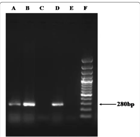
The partial nucleotide sequences of DF3 region of Acanthamoebaspp. from six Acanthamoebameningitis/ meningoencephalitis patients (RCSF1–RCSF6) aligned using ClustalW and showing highest variation are shown in Fig. 1. Sequence homology search for these six Acanth-amoeba spp. in the NCBI databank revealed homology with the available strains ofAcanthamoebaspp. presented in Table 1.The sequences generated in the present study are submitted to the GenBank database under accession numbers KT004416–KT004421 (Fig. 2). Nucleotide se-quences for the DF3 region produced a clear band of ~280 bp length when electrophoresed in 1.5 % agarose gel for all positive specimens (Fig. 3).
Phylogenetic tree reconstructions with neighbour-joining and UPGMA methods provided similar tree topologies, which placed the six Acanthamoeba spp. examined here within the genotype T4 clade (Fig. 2). We found that, RCSF1 showed 100 % similarity with the previously identi-fied strain AcaL7 of genotype T4; RCSF2 showed 100 % similarity with strain Ac_E4c of genotype T4; RCSF3 showed 97 % similarity with strain LC-2012 of genotype T4; RCSF4 showed 97 % similarity with AG-2012 of geno-type T4; RCSF5 showed 99 % similarity with strain LC-2012 of genotype T4 and RCSF6 showed 99 % similarity with strain Ac_E4c of genotype T4. Out of the six
Acanthamoebaspp. isolates, four were not 100 % identical to any available strain in the GeneBank revealing that, cer-tain polymorphisms exists within the nucleotide sequences.
Discussion
Free-living amoebae of the genus Acanthamoeba are widely distributed in nature, in the soil, water and air [7]. These are responsible for several central nervous system infections such as Acanthamoeba meningitis/ meningoencephalitis (AME) and fatal granulomatous amoebic encephalitis (GAE). Acanthamoebae were first re-ported as the causative agents of acute pyogenic meningitis in 1965, and thereafter several cases of Acanthamoeba infections have been recorded worldwide [8, 16, 24]. Martinez & Visvesvara [6] reported that, out of 166 cases of amoebic encephalitis taken for consideration, 103 were due to Acanthamoeba and 63 were due to
Balamuthia. Opportunistic infections due to Acanth-amoebaare increasing nowadays due to decrease in im-munity of the individuals mainly because of either HIV/ AIDS infections or organ transplantation or chemother-apeutic treatments or administration of steroids or other debilitating diseases.
Previously Acanthamoeba spp. comprised three mor-phological groups (groups I–III) based on mormor-phological characteristics (cyst size, shape and growth temperature requirements), which were difficult and confusing [12]. Later genotyping of Acanthamoeba spp. was introduced, using analysis of complete nucleotide sequences of the 18S rRNA gene, of 2,300–2,700 bp in length [19]. GTSA.B1, a large region of the 18S rRNA gene of ~1475 bp in length (approximately 65 % of the complete 18S rRNA gene), was amplified using the primers CRN5 and 1137, which was proven reliable as that of the complete 18S rRNA gene for genotyping [19]. Subsequently“ASA.S1”region, a 460 bp region within the 18S rRNA gene, was found use-ful as that of complete nucleotide sequences for genotyp-ing [19]. Schroder et al. also showed that, instead of sequencing whole regions of GTSA.B1, which include six variable regions, sequencing of only one small variable re-gion, i.e. Acanthamoeba Specific Amplimer (ASA.S1) of ~464 bp in length, was an useful substitute for genotyping [19]. Thereafter the DF3 region, the ~280 bp variable re-gion within ASA.S1 rere-gion, is being widely used for geno-typing studies, since it provides equivalent results as that of ASA.S1 [20].
[image:4.595.56.291.124.236.2]Among all of the known 20 genotypes (T1–T20) of Acanthamoeba spp. T4 is the most predominant and
Table 1Comparison ofAcanthamoebaspp. isolates obtained
from sixAcanthamoebameningitis/ meningoencephalitis patients with reference strains available in the GenBank database
Isolates Accession number
Genotype Name of strain with highest homology
Accession number
Region of origin
RCSF1 KT004416 T4 AcaL7 KJ094680 Italy
RCSF2 KT004417 T4 Ac_E4c GU808286 Thailand
RCSF3 KT004418 T4 LC-2012 KC164227 Switzerland
RCSF4 KT004419 T4 AG-2012 JQ678632 Spain
RCSF5 KT004420 T4 LC-2012 KC164227 Switzerland
RCSF6 KT004421 T4 Ac_E4c GU808286 Thailand
[image:4.595.57.540.474.713.2]prevalent genotype responsible for causingAcanthamoeba meningitis/meningoencephalitis (AME) followed by geno-types T1, T10, and T12(16). The genotype data obtained in this study from sixAcanthamoebaspp. isolates further confirmed that, “T4 genotype is the most common and predominant genotype causingAcanthamoebameningitis/ meningoencephalitis” as postulated from previous studies [16, 24]. In addition to T4, several genotypes of Acanth-amoeba spp. were also reported from different countries from patients with meningitis or granulomatous amoebic encephalitis. In a recent phylogenetic study, Acanth-amoeba spp. of genotypes with T2, T4 and T5 were iso-lated from patients with GAE [18]. Booton et al. reported that, Acanthamoeba spp. of genotypes T1, T10 and T12 were also responsible for causing GAE in humans [16]. Acanthamoebaof genotype T2 was identified as the causa-tive agent of GAE in an immunodeficiency virus-negacausa-tive patient with underlying tuberculosis [17]. Recently, Acanthamoebaspp. isolates of genotype T18 has been isolated from a patient with fatal GAE [25].
Conclusions
This study supports the conclusion that, among all of the genotypes of Acanthamoeba spp. reported to date, T4 appears as the most prevalent and predominant genotype associated with Acanthamoeba meningitis/ meningoencephalitis infections. Further research is needed in order to develop the optimal therapy against the T4
genotype of Acanthamoeba spp. to combat these fatal brain infections.
Abbreviations
AME,Acanthamoebameningoencephalitis; CSF, cerebro spinal fluid; GAE, granulomatous amoebic encephalitis
Acknowledgments
This study was supported by an Adhoc Research grant from Indian Council of Medical Research to Dr. Gita Satpathy.
Funding
There are no funding agencies to report for this submission.
Availability of data and materials
Essential data are presented in the main text of the paper. Nucleotide sequence data of six positive isolates ofAcanthamoebaspp. obtained from the patients ofAcanthamoebameningitis/ meningoencephalitis (AME) were submitted to the GenBank database under accession nos. KT004416– KT004421.
Authors’contributions
HB: Performed all laboratory experiments and drafted the manuscript; MT: Clinical team leader, did clinical examinations and provided clinical specimens. GS: Conceived the idea, arranged the funding and supervised the work, shaped the drafted manuscript. All authors read and approved the final version of the manuscript.
Competing interests
The authors declare that they have no competing interests.
Consent for publication Not applicable.
Ethical approval and consent to participate
Individual consent to participate in this study was obtained from each patient. All procedures performed in this study involving human participants were in accordance with the ethical standards of the institute (AIIMS, New Delhi) and ethical clearance was obtained from the institute.
Author details
1Ocular Microbiology, Dr. R. P. Centre for Ophthalmic Sciences, All India Institute of Medical Sciences, New Delhi 110029, India.2Neurosciences Centre, All India Institute of Medical Sciences, New Delhi 110029, India.
Received: 1 April 2016 Accepted: 26 July 2016
References
1. Marciano-Cabral F.Acanthamoebaspp. as agents of disease in humans. Clin Microbiol Rev. 2003;16:273–307.
2. Harwood CR, Rich GE, McAleer R, Cherian G. Isolation ofAcanthamoeba from a cerebral abscess. Med J Aust. 1988;148(1):47–9.
3. Shirwadkar CG, Samant R, Sankhe M, et al.Acanthamoebaencephalitis in patient with systemic lupus, India. Emerg Infect Dis. 2006;12(6):984–86. 4. Lalitha MK, Anandi V, Srivastava A, Thomas K, Cherian AM, Chandi SM. Isolation ofAcanthamoeba culbertsonifrom a patient with meningitis. J Clin Microbiol. 1985;21(4):666–67.
5. Ringsted J, Jager BV, Suk D, Visvesvara GS. ProbableAcanthamoeba meningoencephalitis in a Korean child. Am J Clin Pathol. 1976;66:723–30. 6. Martinez AJ, Visvesvara GS. Free-living, amphizoic and opportunistic amebas.
Brain Pathol. 1997;7:583–98.
7. Visvesvara GS, Moura H, Schuster L. Pathogenic and opportunistic free-living amoebae:Acanthamoebaspp.,Balamuthia mandrillaris,Naegleria fowleri, and Sappinia diploidea. FEMS Immunol Med Microbiol. 2007;50:1–26.
8. Fowler M, Carter RF. Acute pyogenic meningitis probably due to Acanthamoebasp.: a preliminary report. Br Med J. 1965;2(5464):740–42. 9. Pan NR, Ghosh TN. Primary amoebic meningoencephalitis in two Indian
children. J Indian Med Assoc. 1971;56(5):134–37.
[image:5.595.57.290.85.313.2]10. Sharma PP, Gupta P, Murali MV, Ramachandran VG. Primary amoebic meningoencephalitis caused byAcanthamoeba: successfully treated with cotrimoxazole. Indian Pediatr. 1993;30(10):1219–22.
11. Singhal T, Bajpai A, Kalra V, et al. Successful treatment ofAcanthamoeba meningitis with combination oral antimicrobials. Pediatr Infect Dis J. 2001;20:623–27.
12. Pussard M, Pons R. Morphologies de la paroi kystique et taxonomie du genreAcanthamoeba(Protozoa: Amoeba). Protistologica. 1977;13:557–610. 13. Fuerst PA, Booton GC, Crary M. Phylogenetic analysis and the evolution of the 18S rRNA gene typing system ofAcanthamoeba. J Eukaryot Microbiol. 2015;62:69–84.
14. Gast RJ, Ledee DR, Fuerst PA, Byers TJ. Subgenus systematics of
Acanthamoeba: four nuclear 18S rDNA sequence types. J Eukaryot Microbiol. 1996;43:498–504.
15. Stothard DR, Schroeder-Diedrich JM, Awwad MH, et al. The evolutionary history of the genusAcanthamoebaand the identification of eight new 18S rRNA gene sequence types. J Eukaryot Microbiol. 1998;45:45–54.
16. Booton GC, Visvesvara GS, Byers TJ. Identification and distribution of Acanthamoebaspecies Genotypes associated with non keratitis infections. J Clinical Microbiol. 2005;43(4):1689–93.
17. Walochnik J, Aichelburg A, Assadian O, et al. Granulomatous amoebic encephalitis caused byAcanthamoebaamoebae of genotype T2 in a human immunodeficiency virus-negative patient. J Clin Microbiol. 2008;46:338–40. 18. Walochnik J, Scheikl U, Haller-Schober EM. Twenty years ofAcanthamoeba
diagnostics in Austria. J Eukaryot Microbiol. 2014;62(1):3–11.
19. Schroeder JM, Booton GC, Hay J, et al. Use of subgenic 18S ribosomal DNA PCR and sequencing for genus and genotype identification ofAcanthamoebae from humans with keratitis and from sewage sludge. J Clin Microbiol. 2001;39:1903–11.
20. Booton GC, Kelly DJ, Chu YW, et al. 18S ribosomal DNA typing and tracking ofAcanthamoebaspecies isolates from corneal scrap specimens, contact lenses, lense cases and home water supplies ofAcanthamoebakeratitis patients in Hong Kong. J Clin Microbiol. 2002;40:1621–25.
21. Khan NA.Acanthamoeba: biology and pathogenesis. Norfolk: Caister Academic Press; 2009. p. 290. ISBN 978-1-904455-43-1.
22. Behera HS, Panda A, Satpathy G, Bandivadekar P, Vanathi M, Agarwal T, et al. Genotyping ofAcanthamoebaspp. and characterization of the prevalent T4 type along with T10 and unassigned genotypes from amoebic keratitis (AK) patients in India. J Med Microbiol. 2016; doi:10.1099/jmm.0.000234. 23. Tamura K, Stecher G, Peterson D, et al. MEGA6: molecular evolutionary
genetics analysis version 6.0. Mol Biol Evol. 2013;30:2725–29.
24. Liang SY, Ji DR, Hsia KT, Hung CC, et al. Isolation and identification of Acanthamoebaspecies related to amoebic encephalitis and non-pathogenic free-livingAmoebaspecies from the rice field. J Appl Microbiol. 2010;109:1422–29.
25. Qvarnstrom Y, Nerad TA, Visvesvara GS. Characterization of a new pathogenic Acanthamoebaspecies, A. byersi n. sp., isolated from a human with fatal amoebic encephalitis. J Eukaryot Microbiol. 2013;60(6):626–33.
• We accept pre-submission inquiries
• Our selector tool helps you to find the most relevant journal • We provide round the clock customer support
• Convenient online submission • Thorough peer review
• Inclusion in PubMed and all major indexing services • Maximum visibility for your research
Submit your manuscript at www.biomedcentral.com/submit