0022-538X/92/095399-10$02.00/0
Copyright
©1992, American Society for MicrobiologyCanine and Feline Host
Ranges of
Canine
Parvovirus
and
Feline Panleukopenia
Virus:
Distinct
Host Cell
Tropisms
of
Each Virus
In
Vitro
and
In
Vivo
UWETRUYENAND COLINR.PARRISH*
James A. Baker Institute for AnimalHealth, NewYork State College ofVeterinaryMedicine,
Cornell
University,
Ithaca,
NewYork 14853Received 13 February 1992/Accepted29 May 1992
Canine parvovirus (CPV)emergedas anapparentlynewvirus duringthemid-1970s.The originof CPV is
unknown, but a variation from feline panleukopenia virus (FPV) or another closely related parvovirus is
suspected. Here we examine the in vitro and in vivo canine and feline host ranges of CPV and FPV.
Examination of three canineand sixfeline cell lines and mitogen-stimulatedcanine andfeline peripheralblood lymphocytesrevealedthatCPVreplicates in both canine and felinecells, whereas FPV replicates efficientlyonly
infeline cells. The in vivo hostrangeswereunexpectedlycomplex and distinct fromthe invitro hostranges.
Inoculation of dogs with FPVrevealed efficient replication in the thymus and, to somedegree, in the bone marrow, asshown by virus isolation, viral DNArecovery,andSouthernblotting and by strand-specific insitu
hybridization. FPV replication couldnotbedemonstrated inmesenteric lymph nodesorin the small intestine,
whichareimportanttargettissues in CPV infection. Although CPVreplicated well in all the feline cells tested invitro, it didnotreplicate inanytissue ofcatsafter intramuscularorintravenous inoculation.These results
indicate that these viruses have complex and overlapping hostranges and thatdistincttissuetropisms exist in
the homologous and heterologous hosts.
Canine parvovirus (CPV) and feline panleukopenia virus (FPV) are naturally derived viruses which are classified
accordingtothe host from which theywere isolated. CPV
and FPVareconsideredtobehostrangevariantsamongthe
feline parvoviruses in thegenusParvovirus (51).
CPV and FPV isolates differ in less than 2% of their genomic DNAsequences(27, 35,44, 45), and theyarevery
similarantigenetically. However, theycanbe distinguished
by using specific monoclonal antibodies (MAbs) (38, 40, 53), and they do show differences in their pH dependence of hemagglutination. CPV hemagglutinates rhesusmacaqueor
pigerythrocytesover abroadpHrange,betweenpH 6.0 and
pH 8.0, whereas FPV isolateshemagglutinate these erythro-cytesonly below about pH 6.8 (11, 31, 36).
Afurther striking difference is in host range, since FPV strainshave been shownto replicatein feline cells andcats
but not in cultured canine cells. In contrast, CPV isolates have been shown to replicate in canine and feline cells in cultureaswellasindogs (3, 10, 36).As with other
autono-mously replicating parvoviruses, these viruses have a
tro-pism for tissues containing dividing cells, and in older animals both viruses replicate in cells in the lymphoid tissues, as well as in the rapidly dividing cells of the epitheliumof the small intestine. Theremaybe differencesin
the cell tropism of the twoviruses, becauseFPV causes a
profoundleukopeniaincatswhereas CPV infectionofdogs results inonlyarelative lymphopenia.
The invivo hostrangesof the viruses have notbeen well
defined.CPV-like isolates havebeen isolated from domestic
dogs, wolves,andcoyotes,aswellasfrom othermembersof
the family Canidae (16, 25, 34). FPV and related virus
isolates replicatewell in cats(manymembers of thefamily
Felidae), as well as in common raccoons, mink, and also
* Correspondingauthor.
probably in foxes (2, 5, 34). However,little is knownabout theheterologous hostrangesof theviruses.Previous studies suggestthat FPVisolatesprobably replicatetosomedegree
indogs but that the virus isnotshed in thefeces (43). Little is known about CPV infection ofcats, although one study
hasindicated that CPVreplicated incatsin apatternsimilar
toFPV (17).
Adetailedknowledge of the hostrangesofthese viruses is important for understanding the origin and evolution of CPV. CPVwasfirstrecognized around 1978, and thevirus
spread rapidly into mostpopulations of domestic and wild canids(4, 20, 33, 41, 42, 48, 58, 59). Retrospective serolog-ical studies indicate that CPV first infected dogs in the mid-1970s (20, 37, 48) and subsequently spread widely
amongdogs during 1978 (forareview,seereference34). The
origin of CPVis stillunknown,but it ispossiblethatCPVis
avariant of FPVorarelatedvirus which mutatedtogain the abilitytoefficiently replicate in andspreadamongdogs.
Hereweexamine the in vivo and in vitro canine andfeline
host ranges ofCPV and FPV. Virus replication in animals
wasdefinedby virusrecoveryandbydemonstration of viral DNA in tissues by Southern blot analysis or by strand-specific in situhybridization. Virusreplicationin anumber of canine and feline cultured cells was also examined by replicative-form (RF)DNA isolation andbyvirus titrationof tissueculturesupernatants. The data contribute toafurther understandingof therelationships between the twoviruses and of thechangeswhichwerenecessaryfor theemergence
ofCPVas a newcaninepathogen.
MATERIALS AND METHODS
Cell lines. The A72 canine fibroma-derived cell line
(ap-proximatelypassage 150)(6) and theNLFK feline cellline (derived from the Crandell feline kidney cell line [CRFK] [13])were grown as previously described (39). The CRFK
5399
on November 9, 2019 by guest
http://jvi.asm.org/
5400 TRUYEN AND PARRISH
TABLE 1. PFU titers ofsupernatants of various permanent cell lines inoculated with FPV or CPV
PFUtiterAin:
Virus Days Felinecells Canine cells Canine cells
p.i. (MOI,0.1PFU/cell) (MOI,0.1PFU/cell) (MOI,100PFU/cell)
NLFK CRFK 3201 Fc2Lu AK-D Fc3Tg A72 Cf2Th cT45-S A72 Cf2Th CT45-S
FPV ob 1.0 <1 <1 <1 <1 <1 1.3 <1 <1 2 <1 <1
3 6.0 5.0 4.3 4.0 5.8 2.6 2.3 <1 1.8 2 <1 <1
CPV 0 1.0 <1 <1 <1 <1 <1 <1 <1 <1 2.3 2.3 <1
3 5.6 5.9 4.0 3.9 4.6 2.8 5.3 4.3 1.6 6 4 2
a Determinedbytheimmune-staining plaqueassay in NLFKcells.Expressedaslog1oofPFU/0.3ml ofsupernatant. bDay 0indicates1hpostinoculation(p.i.).
cells (CCL94), feline tongue cells
Fc3Tg (CCL 176),
felinelung cells Fc2Lu (CCL 217), and feline lung cells AK-D (CCL 150)wereobtained fromthe American
Type
CultureCollection. The canine
thymus
epithelium
cell lineCf2Th,
thefelinelymphomacell line 3201(46),and the canineT-cell line CT 45-S (21) were kindly provided by,respectively,
F. W. Quimby, R.J.Avery,and M. J. G.Appel,NewYork StateCollege ofVeterinaryMedicine.Cellswere grown in RPMI 1640 medium with 20% fetal bovine serum (CT45-S), Dulbecco's minimal essential me-dium with 10% fetal bovine serum (AK-D, Fc2Lu, and Cf2Th),or a50% mixtureofMcCoy's 5AandLeibowitzL15 media with 5% fetal bovine serum (3201, NLFK, CRFK,
A72, and Fc3Tg).
Lymphocyte culture. Canine and feline peripheral blood lymphocytes (PBLs) were isolated from heparinized blood
samples ofparvovirus-seronegative animals by Ficoll-Paque densitycentrifugation. Freshly separatedPBLsat adensity
of 2 x
106/ml
were incubated with 5 ,ug ofconcanavalin A(ConA)per mlfor 48 h and then culturedin the samemedium with 100 U of human recombinant interleukin-2 (rIL-2; Boehringer-Mannheim) permlwithout ConA.
[3HJthymidine
incorporation test. PBLs (1 x 10⁵ cells) wereincubated for 16 h with 0.5,uCiof[3H]thymidine. Afterthe cells were harvested with a Skatron cell harvester, the filter-boundradioactivitywascounted.
Viruses.Viruses derived from the molecular clonesof the
A
:n f
9fi
feline
CPV strain CPV-d(CPVtype2
antigenic
type) andthe FPVstrain FPV-b (35, 40, 49)were usedthroughout the
experi-ments.The uncloned FPV-bwasalso used insomestudies.
Infection of cells.Cells of the various cell lineswereseeded
at adensity of2 x
104/cm2
andthen,after20hofincubation, inoculated with virusat amultiplicityofinfection (MOI)of 0.1 PFU per cell. Insome studiesthe canine celllineswerealsoinoculatedwith CPVorFPVat aMOIof 100 PFU per cell. The viruswas allowed toadsorb to the cells for 1 h at
37°C, and the cells were washed once with Dulbecco's minimal essential medium before andafter the virus adsorp-tion. Culture supernatants collected 1 h and 3 days after
inoculation were tested for virus by plaque titration in NLFKcells.
PBLs cultured at a cell density of 2 x 10⁶/ml were
inoculated withCPVorFPVatMOIs between
10-1
and10-5 PFU per cell after 48 h ofConA stimulation. The cells were then cultured for a further 3 days in rIL-2-supplemented RPMI1640medium with50 ,uM 2-mercaptoethanol and 20% fetal bovine serumwithout ConA. Unstimulated cells were treated the sameway exceptthe ConA was omitted.Virus titer determination. Virustiters were determined by
using an immune-staining plaque assay in NLFK cells (23,
39).Virusantigen was also assayed in the hemagglutination testbyusingrhesus macaque erythrocytes in barbital acetate buffer(pH 6.2) asdescribedpreviously (39, 40).
Hemagglutination inhibition assay and virus antigenic
typ-canine
NLFK CRFK 3201 Fc2Lu AK-D Fc3Tg A72 Cf2Th CT45-S
\ \ o
il11
14
C) c"ip'..
s
s1
B
dRF
mRF
ss
A72 C02Th CT45-S
- .*
I
I
FIG. 1. Southern blot analysis of low-molecular-weight DNA recovered from the various cell lines 3 days after inoculation. Samples representing the DNA from approximately 2 x10⁴cells were electrophoresed in a 1% agarosegel. The autoradiograph is exposed to make thesmalleramountsof RF DNA in certain cell lines visible. (A) Feline and canine cells inoculated with CPV or FPV at a MOI of 0.1 PFU percell.(B)Canine cell lines inoculated with CPV or FPV at a MOI of 100 PFU per cell. Abbreviations: dRF, dimer RFDNA;mRF, monomer RFDNA;ss, single-stranded virion DNA.
J. VIROL.
on November 9, 2019 by guest
http://jvi.asm.org/
[image:2.612.80.561.527.678.2]TABLE 2. PFU titers ofsupernatantsofConA-stimulated canine orfeline PBLs inoculated with FPV or CPV
PFU titer' in:
MOI Virus
Canine PBLs FelinePBLs
0.1 CPV 6.3 5.3
FPV 1.8 6.3
0.01 CPV 5.6 5.0
FPV 1.6 6.0
0.001 CPV 6.3 <1
FPV 1.4 5.8
a Expressed aslog10 PFU/0.3ml.Titers of cell culturesupernatants3 days after inoculation as determined by the immune-staining plaque assay in NLFK cells.
ing. The inhibition assay was performed with
heat-inacti-vated and erythrocyte-preincubated sera or with MAbs,
using rhesus macaqueerythrocytes and 8 hemagglutination
unitsof virus(11). TheMAbs used forantigenic typingof the
viruses (MAb7, MAb 14, MAbG, andMAb H) havebeen describedpreviously (38).
Isolation and
analysis
of viral DNA. Genomic andlow-molecular-weight DNAwas isolated frominfected-cell cul-turesoranimal tissue samples by standardprocedures (18, 28, 47). For total DNA preparation,
high-molecular-weight
DNAwas digested with BamHI prior to electrophoresis in 1% agarose gels in Tris-acetate-EDTA (TAE)buffer,with 1 ,gofethidium bromideperml.Low-molecular-weight
DNA was not digested but was electrophoresed under the sameconditions. DNAwas transferred to
nylon
membranes andthen hybridized with a
32P-labeled probe
representing
mapunits 59to98.5 (ca. 2,000bases)of the CPV genome. Animal infections and
analysis.
Twelve-week-old specific-pathogen-freebeagles(J.
A. BakerInstitute)
or12-week-oldA
feline canine PBLs PBLs
specific-pathogen-freekittens (Harlan-Sprague-Dawley Inc., Indianapolis,Ind.) were inoculated intranasally, intramuscu-larly, or intravenously with 0.5 x 10⁶to 1.0 x 10⁶PFU of either FPV or CPV. Animals were bled, and their tempera-turesweremonitored daily. They were killed between days 4 and 6 postinfection (see Table 3). Tissue samples were snap-frozen in liquid nitrogen or immediately fixed in ice-cold periodate-lysine-paraformaldehyde-glutaraldehyde (PLPG) so-lution(1). Aspirates of bone marrow cells collected from the femur were spun onto poly-L-lysine-coated microslides, fixed in ice-cold PLPG solution, and stored at -20°C.
Frozen tissues were stored at -70°C, and PLPG-fixed tis-sues were paraffin embedded.
Insitu hybridization. In situ hybridization of frozen tissue sections orPLPG-fixed cytospins was performed essentially as described elsewhere (1, 56) with strand-specific RNA probes. The probesweretranscribed, by using eitherT7 or SP6 polymerase, from plasmid pBI264, which represents
about2,000bases oftheCPV genome(map units 59 to 98.5) inthevectorpGEM3Z(Promega).The strandspecificities of the probes were confirmed by Southern blotting with viral RF DNA and virion single-stranded DNA preparations of CPV(data notshown). Since the viral genome of FPV and
CPVrepresents asingle-strandedDNAofnegative polarity,
the minus-sense probe hybridizes with both mRNA and
positive-sense DNA. Both are synthesized only in an
in-fectedcell, and ahybridizationsignal with the minus-sense probethereforeindicatesDNAreplication and/or transcrip-tion.
RESULTS
In vitrostudies.(i) Virus replication in permanent cell lines. FPVand CPVreplicated in all feline cell linestosimilar PFU titers(Table 1). NLFK, CRFK, and AK-D, 3201, and Fe2Lu cells replicated FPV and CPV efficiently, as measured by virus titers in thesupematant (Table 1), and FPV andCPV
B
C
feline PBLs FPV
0' OXy(zr Ye
dRF
mRF
ss
CPV
I.
.% Q
canine PBLs CPV
"I%
FIG. 2. Southern blotanalysisof
low-molecular-weight
DNAof FPV-orCPV-inoculated ConA-stimulated canineorfeline PBLs. Cellswereinoculated withtheMOIs(PFUper
cell)
indicated,and the DNAwasharvested3dayslater. All DNA recovered from PBLs showedacertaindegreeof DNAdegradation.Abbreviations:dRF,dimer RFDNA; mRF,monomerRF DNA; andss,single-stranded virion DNA. (A) Feline and caninePBLsinoculatedwith CPVorFPVataMOI of 0.1 PFU per cell. DNA from mock-inoculated control cultures and that
from canine PBLsinoculatedwith theunclonedFPV-bisolatearealso shown.(B)Titer determination of FPV and CPV in feline PBLs by
inoculationwithMOIsbetween 0.1 and 0.0001 PFU percellor
by
mock inoculation.(C)Titration of CPV in canine PBLs by inoculation withMOIsbetween 0.1and 0.00001 PFU per cellor
by
mockinoculation.on November 9, 2019 by guest
http://jvi.asm.org/
[image:3.612.139.487.479.668.2]5402 TRUYEN AND PARRISH
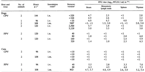
TABLE 3.
Summary
oftheinoculationsusedinthisstudyPFUtiter(logl0PFU/0.3ml)ind,e.
Hostand No.of Hours Inoculation
Serocon-virus animals pjia routeb version' Ileum Mesenteric Spleen Thymus
Dogs
CPV 2 144 i.n. <320 4.7 2.3 <1 2.0
<320 4.9 2.6 <1 2.2
1 96 i.v. <10 6.0 6.3 4.0 5.9
2 108 i.m. NDf <1, <1 1.5, 3.0 <1, <1 2.0,5.0
2 120 i.v. 2,560 4.7 1.0 <1 3.6
5,120 5.0 3.3 <1 3.8
FPV 2 120 i.n. 40 <1 <1 <1 <1
80 1.8 <1 2.0 6.0
2 120 i.v. 160 <1 <1 1.4 5.9
320 1.4 1.0 2.3 6.3
Cats
CPV 2 96 i.v. <10 <1 <1 <1 <1
<10 <1 <1 <1 <1
2 108 i.m. <10 <1 <1 <1 <1
<10 <1 <1 <1 <1
FPV 2 96 i.v. 40 5.5 2.8 3.3 7.0
80 6.3 4.5 4.0 7.3
2 108 i.m. ND 4.7,5.7 4.0,4.9 2.6,3.0 5.2, 5.4
aTheanimalswerekilledatthe times indicatedpostinoculation(p.i.).
b Abbreviations: i.v.,intravenous;i.m.,intramuscular;i.n. intranasal.
Virus-specificantibody titerasdeterminedbythehemagglutinationinhibitiontest.Allanimalswereseronegativebeforeinoculation. dTiterdeterminationof10%tissuehomogenatesin theimmune-staining plaqueassayinNLFKcells.
e ThePFUtiter of the tissue from each animal isgiven separately. fND,notdetermined.
RFDNAs couldbedemonstrated in extractsof feline cells
collected3 days afterinoculation
(Fig. 1A).
The amountof RF DNA inFc2Lu cellswascomparatively
small(Fig.
1A),but thiscell line clearly supportedreplication ofboth CPV and FPV.
CPVreplicatedefficientlyintwoofthreecaninecelllines
investigated, A72 and Cf2Th, but none of the canine cell lines supported efficient replication of FPV, even when
inoculatedwith FPV at aMOI of 100 PFU per cell (Fig. 1; Table 1).ThecanineT-cell-derived cell lineCT45-S was not
permissivefor either CPVorFPV(Fig. 1; Table1).Afaint RF DNA band was seen occasionally in extracts of FPV-inoculated A72 cells, indicating a very low level of DNA replication (Fig. 1A).
(ii) Virus replication in lymphoid cells of cats and dogs. PBL cultures showed a similar pattern of viral susceptibility to
that seen for permanent cell lines. Both CPV and FPV replicated efficiently in feline PBLs, whereas only CPV replicated in canine PBLs, as determined by PFU titration of tissueculture supernatants (Table 2) and by the presence of RF DNAfrom infected cells (Fig. 2). Viral RF DNAs could be recoveredfrom CPV-inoculated canine and feline PBLs and from inoculated feline PBLs but not from FPV-inoculated canine cells (Fig. 2A). The low FPV titers recov-eredfrom supernatants of canine PBLs after 3 days (Table 2) are most probably residual inoculum, since no RF DNA could be demonstrated in Hirt supernatants of those cells
(Fig. 2A). Both inocula gave efficient infections of the
homologoushost cells even at very lowMOIs,down to10-4
PFU percell(Fig. 2B and C). The [3H]thymidine incorpora-tionby the PBLs decreased during the course of infection,
thedegree of decrease being dependentonthe inoculum titer (datanotshown).
In vivo studies.A total ofsevendogswereinoculatedwith 5 x
105
to 1 x 10⁶ PFU of CPV-d as detailed in Table 3. Viruswasinoculatedintranasally, intramuscularly, or intra-venously. Another four dogs wereinoculated either intrana-sallyorintravenously with FPV-b. Groups oftwocatseach wereinoculatedintramuscularlyorintravenously with 0.5 x10⁶to 1 x 10⁶ PFU of FPV-d orCPV-d (Table3).
CPV dog CPVdog FPVdog CPV cat FPV cat day 4 day 6 day5 day 4 day 4
FIG. 3. Southern blot analysis of total DNA extracts from the
thymus,
spleen, mesenteric lymph nodes(ln.mes.),
and ileum of CPV- orFPV-inoculated dogs andcats. TheCPV-inoculated dogswereinoculatedintravenouslyorintranasallyandkilledonday4or
6postinoculation, respectively.TheFPV-inoculateddogwas inoc-ulatedintravenouslyandkilledonday5postinoculation. Bothcats
for which tissueanalysesareshownwereinoculatedintravenously
and killedonday4postinoculation.Theamountsof DNAloadedon
eachlaneareequivalenttoabout20 ergfromthethymus,mesenteric
lymphnode,orileum and about 50 ofgfrom the spleen. J. VIROL.
on November 9, 2019 by guest
http://jvi.asm.org/
[image:4.612.62.567.90.341.2] [image:4.612.330.559.538.642.2]FPV AND CPV HOST RANGES 5403
.-. . ~".
. .
;ss t t .. ;.g-gist;-4....B
INv *.
C.,",,? .
**''l
'
iSt' 'Af'j;--s l+ 'iWxv 1 2i<t
~'
iK
**
;
4
+ W 7 .9
I V
-I,:',I,,, b[
A I
Al
,6~~~~~~~i
''f*0'Bot wBb@
t.-E
4I,~~~~~~~~~~~~~~~~~~~
~~~~j~.r^
JV wi
I~~~~~~~~~~~~~~~~~
FIG. 4. Insituhybridizationofthymus(AandB),mesenteric
lymph
nodes(C
andD),
andileum(E
andF)
of CPV-(panels
A,C,andE)
or FPV (panels B, D, andF)-inoculated dogs. The animals shown were killed on
day
4(CPV)
orday
5(FPV)
after inoculation. The minus-senseprobe usedhybridizeswithviralRF DNAandmRNA,andpositive
signals
therefore indicate active viralDNAreplication
and/or transcription,allowingthediscrimination ofvirus-replicating
cells. Panelscontaining
thesametissueareatthesamemagnification.
Bar,100 ,um.(i) Canine inoculations. All dogs inoculated with CPV
acquired a systemic infection. Virus could be recovered
from severalregionsof the smallandlargeintestineand from
avarietyof tissues of the lymphatic system. The ileum and
thymus consistently had the highest virus titers (Table 3). ViralDNA could be demonstratedbySouthernblotanalysis
of total DNA recovered from these tissues (Fig. 3). Virus
replicationwasalso demonstratedinthethymus,mesenteric
lymph
node, and ileumby
in situhybridization
with the minus-sense RNAprobe
(Fig. 4A,
C,
andE).
At latertimes after inoculation,high
virus titers could not be recovered fromsomeCPV-inoculateddogs,
probably
asaconsequence of thedeveloping
neutralizing-antibody
response.However,
DNAcouldbedemonstratedin the tissuesof those animals bySouthern blotanalysis
(Fig.
3)
andby
insituhybridization
(datanot
shown),
indicating
an active infection.VOL. 66,1992
on November 9, 2019 by guest
http://jvi.asm.org/
[image:5.612.71.550.78.581.2]5404 TRUYEN AND PARRISH
,.'-;
+ s} M a- ,' sk X -xPn
AW
~ ~FIG. 5. Insituhybridizationofbonemarrowcellsofadog4days
after inoculation with FPV. The minus-sense probe was used,
allowingthedetectionofviralRFDNAormRNA.Bar, 100,um.
Hightiters ofFPVcouldberecoveredfromthethymusof inoculated dogs,whereasmuchlowertiterswererecovered
from the spleen (Table 3). The replication of FPV in the caninethymuswasconfirmedbybothSouthernblotanalysis (Fig.3)andinsituhybridizationwiththeminus-senseprobe (Fig. 4B). FPV-replicatingcells could also be readily
dem-onstrated in thebone marrowbyin situhybridization (Fig.
5). No virus orviral DNA could be detected in the small
intestine ormesentericlymphnodes(Table3;Fig. 3 and4D
andF).
(ii) Felineinoculations.FPVwasrecovered ondays4and
5 postinoculationfromseveralregionsofthesmallintestine
andfrom lymphatic tissues ofthe cats after either
intrave-nousor intramuscularinoculation.The identitiesofviruses
reisolated from each animal were confirmedwith CPV- or
FPV-specificMAbs.TheFPVreplicatedtohightitersinthe thymus and the small intestine, whereas lower titerswere recovered from the spleen and mesenteric lymph nodes
(Table 3). Viral DNA was recovered from the thymus,
mesenteric lymph nodes, and ileum (Fig. 3), and cells
containingFPVDNAormRNAwerealsodemonstrated in theseorgans by in situ hybridization withthe minus-sense probe(Fig. 6B, D, andF).
Intramuscularorintravenousinoculationof0.5 x106to 1 x 10⁶ PFU ofCPV-d didnot result ininfectionofthe four
animalsinoculated. Noviruscould berecovered from any
tissue(Table3),noviralDNAorvirus-replicatingcellswere
foundinthetissuesinvestigated bySouthernblotanalysisor
bystrand-specificinsituhybridization (Fig.3and6A, C,and
E), and noanimal hadseroconverted by day4 after
inocu-lation (Table3).
DISCUSSION
CPVemergedsuddenlyin1978as a newpathogenofdogs.
Currentevidence suggeststhat CPVis avariantofFPVor
somecloselyrelatedviruswhichmutated togain the canine
host range. We have shown previously that only a small
number of nucleotide differences between FPV and CPV
determinetheabilityofthelattervirustoreplicateincanine
cellsin culture(35,36). However, therelationship between
the invitro hostranges ofthevirusesand their abilitiesto replicateinvarioushostanimals isnotwellunderstood, and
fewstudies have
critically
compared
the in vitro and in vivo host ranges.The presentstudies of the in vitro host rangesextend and
confirm
previous
reports
(3,
10,
36).
We demonstrated that all feline cell lines testedreplicated
both FPV and CPV tocomparable
degrees,
whereastwocanine cell linesreplicated
CPV but not FPV. No FPVreplication
was detected inCf2Th
cells,
andonly
a very low level of FPV DNA wasdetected
occasionally
inA72 cellsby
Southern
blotanalysis
(Table
1; Fig.
1).
Lymphoid
cells are amajor
target for CPV and FPVreplication
indogs
and cats(8,
9,
12, 14,
29,
30).
CPVreplicated
efficiently
in both feline and caninePBLs,
whereas FPV
replicated
only
infeline PBLs(Table 2; Fig.
2).
The low virus titers in the supernatant of FPV-inoculated
canine PBLs were
probably
derived from theinoculum,
sincenoviral RF DNA couldbedemonstrated in those cells
(Fig.
2A;
datanotshown).
In contrastto the invitro
results,
the animal hostrangeswere
unexpectedly
complex. FPV,
which didnotreplicate
efficiently
in any caninecellculture,
replicated
toveryhigh
titers in thethymus
ofintravenously
(two
oftwo)
andintranasally
(one
oftwo)
inoculateddogs
(Table 3).
The replicationwasconfirmedbothby
Southernblotting
oftotal DNAextracts(Fig.
3)
andby
insituhybridization
(Fig.
4B).
Virus-replicating
cellswere alsofound in the bone marrow (Fig.5)
andoccasionally
in thespleen
(results
notshown).
No FPV
replication
was detectable in the canine small intestineormesentericlymph
nodes(Table
3;
Fig.
3and 4D andF).
The
histological
patternofFPVreplication
in thecanine thymus, withpositive
cellsmainly
in the cortex, suggestssome
differentiation-dependent
T-celltropism
of FPVrepli-cation. Stem cellsderived from thebonemarroware
initially
located as
subcapsular lymphoblasts
in the thymic cortex.These CD4- CD8- cells divide
rapidly;
most of themdifferentiate into CD4+CD8+progeny cells, and a propor-tionfinallymatureintoCD4+or
CD8+
lymphocytes.Asthe immatureprogenitor
cellsdifferentiate
in thethymiccortex(26,
57),
it ispossible
that one ofthose populations in thecanine
thymus
(CD4-
CD8-orCD4+
CD8+)
isparticularly
permissivefor FPV. The pathwayof entryof thevirusinto
the thymus is unknown. The blood-thymus barrier in the cortex
region
isrelatively impermeable
for high-molecular-mass molecules. Since FPV-replicatingcells were found in the bonemarrow(Fig. 5),
andsince bothfeline myeloid anderythroidprogenitorand precursorcells have been shown to besusceptibleto FPV infection in vitro (22),infected bone marrow-derived cells could be responsible for the viral
invasion of the thymus. Experiments to further define the
FPV-susceptible canine cells and the possible mechanisms
of entry arein progress.
Themolecularlycloned CPV-d strain replicated efficiently in dogs, with the typical tissue tropism described forCPV (24, 29, 30). The virus replicated in a variety of lymphoid tissues and in the small andlarge intestine, and anantibody
response was observed as early as 4 days postinfection (Table 3). The virus PFU titers recovered from inoculated animalsvaried greatly depending on the degree of serocon-version,indicating that virus-neutralizing antibodies affected the recovery ofinfectious virus from ground tissues. The demonstration of RF DNA in these tissues (Fig. 3) and of positive signals by the in situ hybridization was a more reliable criterion for viral replication at later times after infection.
CPVinoculation ofcatsalso revealed unexpected results.
J. VIROL.
on November 9, 2019 by guest
http://jvi.asm.org/
[image:6.612.69.302.77.253.2]4~~~
4;Z,
~~
~
~
Kek-
AV
vgw--.g
A~~~~~~~~
;:'
'
A--FG6siuInh
oraFP(nB, at
FIG.used aloing
stuhybrdetection
ofthymus (A
aDN andmentRNA.lyPhstv
sinodes(Cindicate
andtileumviandDNA ofepViato(anels/orC
trncrpind,Eallowingthediscrimination ofvirus-replicatingcells. Panelscontainingthesametissueareatthesamemagnification. Bar,100FLm.
4a~~i ,5i~vap;P
In twoseparate experiments, intramuscular or intravenous inoculation of CPV into cats resulted in no detectable
infection. Novirus could beisolatedfrom anytissue(Table 3),noRFDNAcould bedemonstrated by
Southern
blottingin DNA extracts (Fig. 3), and in situ hybridization was
negative for all tissues (Fig. 6A, C, andE). The reason for this lackofreplication is unknown. However,onepossibility is that the virus is clearedormaskedbefore itcanadsorbto asusceptible target cell. For example, since CPV, but not
FPV,hemagglutinates felineerythrocytesunder
physiologi-cal conditions (37°C and pH 7.4 [data not shown]), it is possible that binding byerythrocytesorsomeotherligand in thetissues removes CPV from the circulation after parent-eral application. Further experiments are necessary to ad-dress thisphenomenon.
Our findings on the feline replication of CPV differ from those of a study published by Goto et al. (17). They de-scribed a systemic infection ofspecific-pathogen-free cats
on November 9, 2019 by guest
http://jvi.asm.org/
[image:7.612.64.549.69.573.2]5406 TRUYEN AND PARRISH
with CPV after subcutaneous inoculation with virus doses similartothose used here. Viruswasrecovered from several organsbetween
days
3 and5postinoculation.
In thatstudy
a CPV isolate from 1982 wasused,
mostprobably
theanti-genic
type
CPV-2a(37,
41,
42).
Ourstudy
was done witha CPV-2 strain. Whether the different strainsmight
have resulted in the differentfindings
has not been determined.Also,
owing
to thehigh
environmental resistance ofparvo-viruses,
cross-contaminationby
a contaminatedenviron-mentisa
potential
threat. In thepresent
study
weconfirmedthe virus
types
used for the inoculation and also thosereisolatedfrominoculated
animals,
andwe canexclude anycontaminationwith FPV inour
experiments.
InfectionsofcatswithFPV-b derived from the infectious clone induced a
systemic
infection with a tissuetropism
typical
of FPV(8,
14).
Virus was recovered from varioustissues of the
lymphatic
system
and from the intestine(Table
3),
andDNAanalysis
revealed active virusreplication
in theileum,
mesentericlymph
node,
thymus,
bone marrow, andspleen
(Fig.
3 and6B, D,
andF;
data notshown).
The catsdeveloped
virus-specific
antibodies when tested 4days
afterinoculation
(Table
3).
The tissue
tropism
of CPV indogs
and FPVin catswasvery similar to that seen in mink infected with the
closely
related mink enteritis virus
(56).
Inthatstudy,
viralDNAorRNAwas observed
by
in situhybridization
up to 8days
postinoculation,
with thepeak
of DNAbeing
detectedatday
4.
These distinct tissue
tropisms
ofvirusreplication
indif-ferent hosts have notbeen
clearly
defined forotherparvo-viruses.
However,
mouseinfection studieswithbiotypes
ofthe minute virusofmice
MVM(p)
andMVM(i),
whichhave distinct host ranges in vitro(15,
52,
54),
showed thatdifferences in cell
tropisms
were seen after infection ofneonatal animals
(7,
19).
Theimportance
of thenonstruc-tural
protein
NS2 for efficient in vivoreplication
was alsodemonstrated
(7), although
it is notrequired
for efficientreplication
in certaincell lines invitro(32).
The results ofour studies
require
a reassessment oftheevents thatwere necessaryfor the emergence ofCPVas a newcanine
pathogen.
BecauseFPVwasabletoreplicate
inthe canine
thymus
and CPV did notreplicate
in cats, the emergence ofCPVas anefficient caninepathogen
mayhaverequired
achange
oftissuetropism
but notof animal hostrange. The ancestor of CPV
(probably
FPV or aclosely
related
virus)
hadtogain
theability
toinfectdogs efficiently
andalso the
ability
toreplicate
in cells of the smallintestinalepithelium
to allow efficientshedding
and transmission.Analysis
of recombinants between FPV and CPV hasshown that thecanineinvivoand invitro hostrangeofCPV
maps within the
capsid protein
gene, and themajor
determi-nantislocatedbetween59 and 73 map units. Therearefour
coding
amino acidchanges
between FPV-b and CPV-d in thatregion
of theVP1/VP2
gene(35, 36).
Recentgenetic
studies havefurther definedthenecessarychanges
toasfewasthree amino acids
(VP2
residues93,
103,
and323[11a]).
Some ofthese
coding changes
affect amino acids whichareexposed
on the surface of the viralcapsid (55),
andresidue93 alsoaffects the
CPV-specific
epitope.
Hypotheses
put
forward toexplain
the emergence and selection of CPV froman FPV-likeancestorhavesuggestedthat it was derived from an FPV variant strain which
contaminated canine vaccines
(50).
Our results do notex-cludethis
hypothesis
butsuggest
that suchmechanismswere notnecessarily required,
becauseFPVcanalready
replicate
insome caninetissuesevenafteroronasalinfection.
Only
asmallnumberof changes were apparentlyrequired to allow FPVto efficientlyreplicate in other canine tissues,including
those ofthe small intestine(36).
CPV has changed rapidly since its emergence,givingrise toat leasttwo distinctantigenic types(designatedCPVtype
2a and CPV type 2b), each of which largely replaced the previous antigenictype of virus(37,41,42).In the course of that evolution, the CPV DNA sequences continued to di-vergefromthose of FPV(37).The hostrangeof FPV defined here and the genetic and evolutionary studies of CPV and FPVsupportthepossibility that CPVoriginatedas avariant of an FPV-like virus and that the virus has
subsequently
evolvedto becomebetteradaptedtodogs.In future studies we will seekto definethe mechanisms which determine the altered tissuetropisms revealed here and thespecific differ-encesbetween the CPV varianttypes.ACKNOWLEDGMENTS
M. L. Strassheim provided experttechnical assistance. S.
Alex-andersen and M. E. Bloomgenerously providedthe in situ hybrid-izationprotocol. We thank M. J. G. Appeland L. E. Carmichaelfor
helpful discussions.
U.T. is the recipient of a postdoctoral fellowship from the
Deutsche Forschungsgemeinschaft (Tr 282/1-1). This work was
supported by grantAl28385 from the NationalInstitute ofAllergy and Infectious Diseases and in part byanaward from theMarilynM.
Simpson Trust.
REFERENCES
1. Alexandersen, S., M. E.Bloom, J.Wolfinbarger, and R. E. Race.
1987. In situ molecularhybridizationfordetection of Aleutian
disease minkparvovirus DNA by using strand-specificprobes:
identification oftarget cells for viral replication in cell cultures
and in mink kits with virus-induced interstitial pneumonia. J.
Virol. 61:2407-2419.
2. Appel, M. J. G., and C. R. Parrish. 1982. Raccoons are not
susceptible to canine parvovirus. J. Am. Vet. Med. Assoc.
181:489.
3. Appel, M. J. G., F. W. Scott, and L. E. Carmichael. 1979.
Isolation and immunisation studiesof a canineparvo-likevirus
fromdogs withhaemorrhagicenteritis. Vet. Rec.105:156-159.
4. Azetaka, M., T. Hirasawa, S. Konishi, and M. Ogata. 1981.
Studiesoncanineparvovirusisolation, experimental infection
andserologic survey. Jpn. J. Vet. Sci.43:243-255.
5. Barker, I. K., R. C. Povey,and D. R.Voigt. 1983. Responseof
mink, skunk, red fox andraccoonto inoculationwith mink virus
enteritis, felinepanleukopeniaand canineparvovirusand prev-alence ofantibodytoparvovirusin wild carnivoresinOntario. Can. J.Comp.Med. 47:188-197.
6. Binn, L. N., R. H. Marchwicki, and E. H. Stephenson. 1980. Establishment ofacanine cell line:derivation,characterization, and viralspectrum. Am. J. Vet. Res.41:855-860.
7. Brownstein,D.G.,A. I.Smith,E. A.Johnson,D. J. Pintel, L.K. Naeger, and P. Tattersall. 1992. Thepathogenesis ofinfection with minute virus of mice depends on the expression ofthe small nonstructural protein NS2 and on the genotype of the
allotropicdeterminant VP1 andVP2.J. Virol. 66:3118-3124. 8. Carlson, J. H., F. W. Scott, and J. R. Duncan. 1978. Feline
panleukopenia. III. Development of lesions in the lymphoid tissues. Vet. Pathol. 15:383-392.
9. Carman,P. S., and R. C.Povey.1985. Pathogenesisofcanine parvovirus-2indogs: histopathologyand antigenidentification in tissues. Res. Vet. Sci. 38:141-150.
10. Carmichael,L.E.,and L.N.Binn.1981. New enteric virusesin thedog.Adv. Vet. Sci. Comp.Med.25:1-37.
11. Carmichael,L. E.,J. C.Joubert, andR. V. H. Pollock. 1980.
Hemagglutination bycanine parvovirus: serologic studiesand
diagnostic
applications.
Am. J.Vet. Res. 40:784-791.lla.Chang, S. F., J.-Y. Sgro, and C. R. Parrish. Submitted for
publication.
J. VIROL.
on November 9, 2019 by guest
http://jvi.asm.org/
12. Cooper, B. J., L. E. Carmichael, M. J. G. Appel, and H. E. Greisen. 1979. Canine viral enteritis. II. Morphologic lesions in naturally occurring parvovirus infection. Cornell Vet. 69:134-144.
13. Crandell, R. A., C. G. Fabricant, and W. A. Nelson-Rees. 1973. Development, characterization and viral susceptibility of a feline (Felis catus) renal cell line (CRFK). In Vitro (Rockville) 9:176-185.
14. Csiza, C. K., F. W. Scott, A. DeLahunta, and J. H. Gillespie. 1971. Pathogenesis of feline panleukopenia virus in susceptible newborn kittens. I. Clinical signs, hematology, serology, and virology. Infect. Immun. 3:833-837.
15. Engers, H. D., J. A. Louis, R. H. Zubler, and B. Hirt. 1981. Inhibition of T cell-mediated functions by MVM(i), a parvovirus closely related to minute virus of mice. J. Immunol. 127:2280-2285.
16. Evermann, J. F., W. Foreyt, L. Maag-Miller, C. W. Leathers, A. J. McKeirnan, and B. LeaMaster. 1980. Acute hemorrhagic enteritis associated with canine coronavirus and parvovirus infections in a captive coyote population. J. Am. Vet. Med.
Assoc. 177:784-786.
17. Goto, H., E. Uchida, S.IchUo,K. Shimizu, Y. Morohoshi, and K. Nakano. 1984. Experimental infection of canine parvovirus in specific pathogen-freecats.Jpn.J.Vet. Sci. 46:729-731.
18. Hirt, B. 1967. Selective extraction of polyoma DNA from infectedmousecell cultures. J. Mol. Biol. 26:365-369. 19. Kimsey,P.B.,H. D.Engers,B.Hirt,andC.V.Jongeneel.1986.
Pathogenicityof fibroblast- andlymphocyte-specific variants of minute virus of mice. J. Virol. 59:8-13.
20. Koptopoulos,G.,0. Papadopoulos,M.Papanastasopoulou,and H. J. C. Cornwell. 1986. Presence of antibody cross-reacting with canineparvovirus in the sera ofdogs from Greece. Vet. Rec. 118:332-333.
21. Krakowka,S.,R.Olsen,andG. Cockerell. 1977. The effect of cell synchronization upon the detection ofT-and B-lymphoid cell receptorson twocontinuous lymphoidcell lines. InVitro (Rockville)13:119-124.
22. Kurtzman, G. J., L. Platanias, L. Lustig, N. Frickhofen, and N. S. Young. 1989. Feline parvovirus propagates in cat bone
marrowculturesandinhibitshematopoietic colony formation in vitro. Blood 74:71-81.
23. Lemon, S. M., L. N. Binn, and R. H. Marchwicki. 1983.
Radioimmunofocus assay forquantitation ofhepatitisAvirus in cellculture. J. Clin. Microbiol. 17:834-839.
24. Macartney, L.,I. A. P.McCandlish,H.Thompson,and H.J.C.
Cornwell. 1984. Canine parvovirus enteritis 1: clinical,
hema-tological and pathological features of experimental infection. Vet. Rec. 115:201-210.
25. Mann,P.C.,M.Bush,M.J. G.Appel,B. A.Beehler,and R.J. Montali. 1980. Canineparvovirus infectionin South American canids. J.Am.Vet. Med.Assoc.177:779-783.
26. Martinez-A.,C., J. C.Gutierrez-Ramos, J.L. De LaPompa,E.
Leonardo,M.J.Sanchez, J.M.Alonso,and M. L. Toribio.1990. Thethousand andonewaysofbeingaTcell.Thymus 16:173-185.
27. Martyn, J. C., B. E. Davidson, and M. J. Studdert. 1990.
Nucleotide sequence of felinepanleukopeniavirus:comparison with canine parvovirus identifies host-specific differences. J.
Gen.Virol. 71:2747-2753.
28. McMaster,G.K., P. Beard,H. D.Engers, and B. Hirt. 1981.
Characterization ofan immunosuppressive parvovirus related totheminutevirusof mice.J.Virol.38:317-326.
29. Meunier,P.C.,B.J.Cooper,M.J.G.Appel,M. E.Lanieu,and D.0.Slauson. 1985. Pathogenesisof canineparvovirus
enteri-tis:sequentialvirusdistribution andpassiveimmunization
stud-ies. Vet.Pathol. 22:617-624.
30. Meunier,P.C.,B.J.Cooper,M.J.G.Appel,and D.0. Slauson. 1985. Pathogenesis ofcanine parvovirus enteritis: the
impor-tanceofviremia.Vet. Pathol.22:60-71.
31. Moraillon,A.,and R.Moraillon.1982. Distinction des
parvovi-rus feline et canins par hemagglutination. Rec. Med. Vet.
158:799-804.
32. Naeger, L. K., J. Carter, and D. J. Pintel. 1990. The small
nonstructural protein(NS2)of the parvovirus minute virus of
mice is required for efficient DNA replication and infectious
virus production in a cell-type specific manner. J. Virol. 64: 6166-6175.
33. Osterhaus, A. D. M. E., G. A. Drost, R. M. S. Wirahadiredja, and T. S. G. A. M. van denIngh. 1980. Canine viral enteritis:
prevalence ofparvo-, corona, and rotavirus infections in dogs in
theNetherlands.Vet. Q. 2:181-236.
34. Parrish, C. R. 1990. Emergence, natural history and variation of
canine, mink,andfeline parvoviruses. Adv. Virus Res.
38:403-450.
35. Parrish, C. R. 1991. Mapping specific functions in the capsid structureof canine parvovirus and feline panleukopenia virus
using infectious plasmid clones. Virology 183:195-205.
36. Parrish, C. R., C. F. Aquadro, and L. E. Carmichael. 1988.
Canine hostrangeand a specific epitope map along with variant sequencesin the capsidprotein gene of canine parvovirus and
related feline, mink, and raccoon parvoviruses. Virology 166:
293-307.
37. Parrish, C. R., C. F. Aquadro, M. L. Strassheim, J. F. Ever-mann, J.-Y. Sgro, and H.0. Mohammed. 1991. Rapid antigenic-type replacementand DNA sequence evolution of canine
par-vovirus. J.Virol.65:6544-6552.
38. Parrish, C. R., and L. E. Carmichael. 1983. Antigenic structure
and variation ofcanineparvovirustype-2,feline panleukopenia
virus, and minkenteritisvirus.Virology 129:401-414. 39. Parrish, C. R., and L. E. Carmichael. 1986. Characterization
and recombination mapping of an antigenic and host range
mutationof canineparvovirus. Virology 148:121-132. 40. Parrish, C. R., L. E. Carmichael, and D. F. Antczak. 1982.
Antigenic relationships between canine parvovirustype2, feline panleukopenia virus, and mink enteritis virus using
conven-tionalseraandmonoclonal antibodies. Arch. Virol. 72:267-278. 41. Parrish,C.R., P. Have, W. J. Foreyt, J. F. Everman, M.Senda,
and L. E.Carmichael.1988.Theglobal spreadandreplacement of canineparvovirus strains. J. Gen. Virol.69:1111-1116. 42. Parrish, C. R., P. H. O'Connell, J. F. Evermann, and L. E.
Carmichael. 1985. Naturalvariation ofcanineparvovirus.
Sci-ence230:1046-1048.
43. Pollock, R. V. H., and L. E. Carmichael. 1983. Use ofmodified
live feline panleukopenia virus vaccine to immunize dogs against canineparvovirus.Am.J. Vet. Res.44:169-175.
44. Reed, A. P., E. V. Jones, and T. J. Miller. 1988. Nucleotide
sequence and genome organization of canine parvovirus. J.
Virol. 62:266-276.
45. Rhode, S. L., III. 1985.Nucleotidesequenceof thecoatprotein
of canineparvovirus. J.Virol. 54:630-633.
46. Rojko, J. L., G. J. Kociba, J. L. Abkovitz, K. L. Hamilton,
W. D. Hardy, Jr., J. N. Ihle, andS.J. O'Brien. 1988. Feline lymphomas:immunologicalandcytochemicalcharacterization. CancerRes.49:345-351.
47. Sambrook, J.,E. F.Fritsch, and T.Maniatis. 1989. Molecular
cloning: a laboratory manual, 2nd ed., Cold Spring Harbor
Laboratory, ColdSpring Harbor,N.Y.
48. Schwers, A., P.-P.Pastoret,G.Burtonboy,and E.Thiry.1979.
FrequenceenBelgiquedel'infectionaparvoviruschez lechien,
avant et apresl'observation des premierscas cliniques. Ann. Med. Vet. 123:561-566.
49. Scott, F. W., C. K. Csiza, and J. H. Gillespie. 1970. Feline viruses. IV. Isolation andcharacterization of feline panleuko-peniavirus intissueculture andcomparisonof
cytopathogenic-ity with felinepicornavirus, herpesvirusandreovirus. Cornell Vet.60:163-183.
50. Siegl,G. 1984. Canineparvovirus: originandsignificance ofa
"new"pathogen,p.363-388. In K. I. Berns(ed.),The parvo-viruses. PlenumPublishing Corp.,NewYork.
51. Siegl, G.,R.C.Bates,K.I.Berns,B.J.Carter,D.C.Kelly,E.
Kurstak,and P. Tattersall. 1985.Characteristicsandtaxonomy
ofParvoviridae.Intervirology23:61-73.
52. Spalholz, B.A., andP. Tattersall. 1983. Interaction of minute virus of mice withdifferentiated cells: strain-dependenttarget
cell specificity is mediated by intracellular factors. J. Virol.
46:937-947.
on November 9, 2019 by guest
http://jvi.asm.org/
5408 TRUYEN AND PARRISH
53. Surleraux, M., M. Bodeus, and G.Burtonboy. 1987. Studyof
canineparvovirus polypeptides byimmunoblotanalysis. Arch. Virol. 95:271-281.
54. Tattersall, P., and J. Bratton. 1983. Reciprocal productive and
restrictive virus-cell interactions of immunosuppressive and prototypestrains of minute virus of mice. J. Virol. 46:944-955. 55. Tsao, J., M.S.Chapman,M.Agbandje,W.Keller,K.Smith,H. Wu, M. Luo, T. J.Smith,M.G.Rossmann,R. W.Compans,and C. R. Parrish.1991. The three-dimensionalstructureofcanine
parvovirus and its functional implications. Science
251:1456-1464.
56. Uttenthal, A.,S. Larsen,E. Lund, M. E. Bloom, T. Storgard,
and S. Alexandersen. 1990. Analysis of experimental mink enteritis virusinfection in mink: insituhybridization,serology, andhistopathology. J. Virol. 64:2768-2779.
57. vonBoehmer,H.1988. Thedevelopmental biologyof T
lympho-cytes.Annu. Rev.Immunol.6:309-326.
58. Walker, S.T., C.P.Feilen, M. Sabine,D. N. Love, andR. F.
Jones.1980.Aserologicalsurveyof canine parvovirus infection in NewSouth Wales,Australia.Vet. Rec. 106:324-325.
59. Zarnke,R. L., and W. B. Ballard. 1987. Serologicsurveyfor
selectedmicrobial pathogensofwolves in Alaska, 1975-1982.J.
Wild. Dis.23:77-85.
J. VIROL.