Vol.50, No.2 JOURNALOFVIROLOGY, May1984, p.623-628
0022-538X/84/050623-06$02.00/0
Copyright C) 1984, American Society forMicrobiology
Localization of the Simian Virus
40 Small
t
Antigen
in
the
Nucleus
and
Cytoplasm of Monkey
and
Mouse
Cells
MATTHEW ELLMAN, ILAN BIKEL, JAMES FIGGE, THOMAS ROBERTS, ROBERT SCHLOSSMAN, ANDDAVID
M. LIVINGSTON*
The Dana-Farber CancerInstitute andDepartments ofMedicine andPathology, Harv ard MedicalSchool, Boston, Massachusetts 02115
Received4 November1983/Accepted 10January 1984
Monkey and mousecells producing simian virus40 small t antigen in the absence ofclearly detectable intact or truncated large T antigens were subjected to indirect immunofluorescence and biochemical cell compartment analyses. Results revealed specific immunofluorescence and small t polypeptide inboth the nucleus andcytoplasm of these cells.
The early region of simian virus 40 (SV40) encodes two known proteins, largeT antigen (T) and small tantigen (t). Both ofthese elements are active in the process of
virus-inducedneoplastictransformation,although the mechanisms
by which they operate in this regard are wholly unknown. Unlike T, it appears that t isdispensable during productive
infection(14), althoughunder someconditions bothproteins
play essential, albeitcomplementary, roles in the
transform-ingprocess (4, 7, 13, 16, 18). Tcan be readily identified in
infected cells by a variety of techniques, including indirect immunofluorescence, which shows ittobelargely anuclear
constituent. Bycontrast,ithas not beenpossibletodetect t
immunofluorescencein thepast. The cellularlocation ofthis
protein could,
therefore,
be assessedonly by
biochemicalextraction studies which suggested that it was largely a
constituent of the postnuclear soluble fraction and might, therefore, be largely, if notwholly, a cytoplasmic constitu-ent(10, 17).Comparable results wereobtained for polyoma-virus (15). However, in view ofthe indirect nature ofthe
analytical
approach employed in all of thesestudies,
adetailed understanding of the intracellular geography of t was not possible. In an effort to address this problem, we have raised an antibody against homogeneous SV40 t
syn-thesized in Escherichia coli (2, 12) and used it to decorate cellswhich synthesize substantial levels oft, but noreadily detectable intactortruncated Tby indirect
immunofluores-cence. Resultsofthese experiments revealedthat t
fluores-cence can be
readily
identified in both the nucleus andcytoplasm oftwo different cell types. As expected, in the sametypes ofexperiments, Tfluorescence was notedto be
largely, ifnotexclusively, nuclear.
We have recently described a plasmid (pHR402) and a
derivative SV40 mutant virus (SV402), which, upon intro-duction into rodentormonkey cells,lead to thesynthesis oft but notclearlyorreproduciblydetectablequantities of T or a
truncated derivative thereof(13). The 402genome contains twotandemly oriented viral replication originsupstream of an intact t-coding unit. The 402 early region has also
sustained a -1.5-kilobase deletion ofT-unique coding se-quences.
When pHR402 was cotransfected onto NIH-3T3 cells in the presence ofpSV2 Ecogpt (8), some of the clones which grew in medium containing mycophenolic acid also synthe-sized intact SV40 20-kilodalton t and failed to accumulate readily detectable quantities of intact or truncated T, as
*Correspondingauthor.
assayedbyspecific immunoprecipitation of
[35S]methionine-labeled cell extracts. One such example is shown in Fig. 1. The rabbit antiserum used in these experiments was raised against sodium dodecyl sulfate (SDS)-gel band-purified t synthesized in E. coli (2). This serum recognizes both t and T but failed to bind t after its adsorption to an extract of monkey cells infected by aT+/t- virus, dl884 (2, 14).Hence,
the vast majority if not all of the relevantantibody molecules in this serum recognize an antigen(s) contained within the commonT/t sequence. In an experiment analogous to that reported above, CV-1P cells were acutely infected with
SV402,and extracts ofthese cells were exposed toanti-T/t
immunoprecipitation (Fig. 1). No cytopathic effect or pro-duction of T was observed in such SV402-infectedcultures,
and in keeping with this observation, only t was clearly
identified by specific immunoprecipitation. In this regard, others have shown that mouse LTK cellstransformed by a recombinant genome, pVBtTK-1, which containsan analo-gously deleted SV40early region, also failed to accumulate
readily detectabletruncated T with the seraemployedin the relevant immunoprecipitation assays (11). As a test of the
possibilitythatexpressionof the 402genomemightlead to a truncated Twhichcomigratedwith t inSDS-polyacrylamide
gels, extractsofNIH-3T3clone 5-4 (oneof theabove-noted
t+/T- gpt+ cotransformants) and Cos-1 cells (a T+/t+
mon-keycellline)wereimmunoprecipitated withthe above-noted serum. Aliquots of the eluted products were then reduced
withdithiothreitoland alkylated withN-ethylmaleimide (3). As noted inFig. 1, comigratingtbands werenoted inthese twoextracts before and afterreductionand alkylation, and
in each case, the migration rate of t slowed to the same
degree after chemical modification, in keeping with the
original observation of Crawford and O'Farrell (3). By contrast, no major alteration in Cos-1 T migration was
detected;furthermore, no newimmunoreactive bands were seen after the above-noted treatment. Since the putative truncated T molecule encoded bythe 402 early region (13) would lack t-unique coding sequences and be expected to have a different cysteine content than t (4 versus 11), the failure to detect a previously "hidden" 20-kilodalton band
after reduction andalkylation is consistent with the notion that expression of the 402 genome does not lead to the intracellular accumulation of a truncated form of T which normally comigrates with t.Furthermore,aone-dimensional
isoelectric focusing gel analysis of
[35S]-methionine-labeled
pHR402 t from the NIH-3T3 gpt+cotransformant,clone5-4, revealed that all of the specifically immupoprecipitated
623
on November 10, 2019 by guest
http://jvi.asm.org/
C
D
[
;-
f
C
IJ
K
LA
B
C
D E
F
G
..".,.,....
am
*-f-4w:40
T~~~~~~~~~~~~~~~~~~~~~~~~~~.
-EM
-8 Kd
t
._ ..', .:. - d t
-8Kd
t
A
B C D EF G H
- _w ..
--T
-
tALK
-.. -t
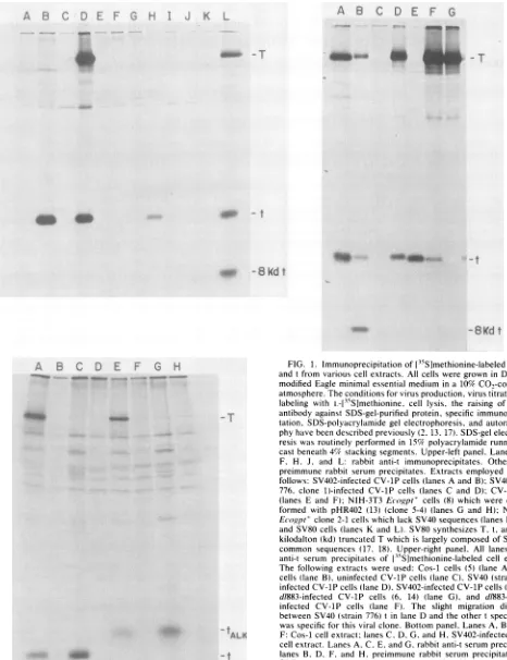
FIG. 1. Immunoprecipitationof[15S]methionine-labeled SV40T and tfrom various cellextracts. All cellsweregrownin Dulbecco modifiedEagle minimal essential medium in a 10% CO.-containing atmosphere.Theconditionsforvirusproduction, virustitration,cell labeling with L-[35S]methionine, cell lysis, the raising ofSV40 t
antibodyagainstSDS-gel-purified protein, specific immunoprecipi-tation, SDS-polyacrylamide gel electrophoresis, and autoradiogra-phyhave beendescribedpreviously (2,13,17). SDS-gel electropho-resis wasroutinely performed in 15% polyacrylamide running gels castbeneath 4% stackingsegments. Upper-left panel,Lanes B,D, F, H. J, and L: rabbit anti-t immunoprecipitates. Other lanes: preimmune rabbit serum precipitates. Extracts employed were as
follows: SV402-infectedCV-1P cells(lanesAand B);SV40(strain 776, clone 1)-infected CV-1Pcells (lanes C and D); CV-1P cells (lanes E and F); NIH-3T3 Ecogpt' cells (8) which were
cotrans-formed with pHR402 (13) (clone 5-4) (lanes G and H); NIH-3T3 Ecogpt'clone 2-1 cells which lackSV40sequences(lanesIandJ); andSV80 cells (lanes K and L). SV80 synthesizes T, t, andan 8-kilodalton (kd)truncated Twhich is largelycomposed of SV40T/t common sequences (17, 18). Upper-right panel, All lanes: rabbit anti-t serum precipitates of13-5S]methionine-labeled cell extracts.
The following extracts were used: Cos-1 cells (5) (lane A), SV80 cells (lane B), uninfected CV-1P cells (laneC), SV40(strain 776)-infected CV-1P cells(laneD).SV402-infected CV-1P cells(laneE). d/883-infected CV-1P cells (6, 14) (lane G), and d/883-SV402-infected CV-1P cells (lane F). The slight migration difference between SV40(strain 776)tin lane D and the othertspecies here was specificfor this viral clone. Bottompanel, LanesA, B, E,and F: Cos-1 cellextract; lanesC, D,G, andH, SV402-infectedCV-1P
cellextract. LanesA,C,E, andG,rabbit anti-tserumprecipitates;
lanes B, D, F, and H, preimmune rabbit serum precipitates. The SDS-elutedantigen(s) in lanes EthroughHhad beenexposedto20
mMdithiothreitol and40mMN-ethylmaleimidebefore
electropho-resis, as described previously (3). tALK, Migration position of
reduced andalkylated t.
A R
_*ww,
-T
me
4
on November 10, 2019 by guest
http://jvi.asm.org/
[image:2.612.69.535.82.688.2]NOTES 625
radioactivity which wasdetected appeared tocomigratewith
authentic
SV40
t synthesizedfrom an intact t-coding unit in E. coli (2, 12) (data notshown). Hence,by these criteria, all of the radioactivity in the anti-t-reactive 20-kilodalton band in cells bearing the 402 genome isSV40
t.With the availability of a monospecific antibody which recognized one or more segments of the common
T/t
se-quence and cells which, as characterized with this reagent, appear toaccumulate t and not T, itwas possible to search for this protein in fixed cells by indirectimmunofluores-cence. Thus,
t+/T- Ecogpt+-cotransformed
NIH-3T3 and CV-1P cells acutely infected withSV402
were sequentiallyfixed in 3.7% formaldehyde followed by absolute methanol and then incubated with this antibody (Fig. 2 and 3). Aftera reaction with fluoresceinated goat anti-rabbit immunoglob-ulin G, distinct, bright fluorescence was noted in the nucleus and cytoplasm of both cell types. The intranuclear fluores-cence wasfound to bediffuse and to spare evident nucleoli.
Cytoplasmic fluorescence in many cells was noted to be "lacy" and, in part, to decorate a tight, albeit irregular,
network of particulate material. Additional examples of
these effects are noted in Fig. 3. No fluorescence was noted in NIH-3T3 cellstransformed only with pSV2 Ecogpt (Fig. 2B), in uninfected CV-1P cells reacted with anti-t serum (Fig. 2E), or in
SV40-infected
CV-1P cells reacted with preimmune rabbit serum (data not shown). By contrast,CV-1P
cells infected by thet-/T+
viral mutantdl883
(6, 14) contained only T (cf. Fig. 1, upper right panel, lane G) and revealed bright nuclearfluorescence without detectable cy-toplasmic staining under theconditions employed (Fig.2C).
This result is in keeping with the widelyappreciated obser-vation that
-90%
of T in such cells is a nuclear constituent. Results qualitatively similar to those noted in the t-contain-ing cells described here were obtained with the above-notedt+lEcogpt+
NIH-3T3 clone 5-4 after fixation with methanol alone or with 3.7% formaldehyde followed by 1% Triton X-100 or acetone. In addition, identical results to those noted in Fig. 2 and 3 were obtained with the two othert+IEcogpt+
NIH-3T3 clones (5-1 and 5-10) available in our laboratory. Hence, the nuclear and cytoplasmic staining effect is a reproducible finding in more than one cell type which synthesizesSV40
t in the absence of T. Moreover, similar, although less intense staining of fixed, 402 genome-contain-ingt+/T-
cells was obtained after incubation with one lot of pooled hamster anti-SV40 tumor serum. In keeping with the contention that the observed fluorescence is due to an immune reaction with t and not the predicted z20-kilodalton truncated species of T (which can, in theory, be encoded by the 402 genome), only t was detected by specific immunopre-cipitation under the conditions employed with the same serum used in the indicated immunofluorescence experi-ments. Moreover, others have found that with hamsteranti-SV40
tumor serum, T extending less than 272 amino acids from the normal N terminus failed to score in immunofluo-rescence assays (9).In an independent test of the suggestion that t is present in both the nuclear and cytoplasmic cell compartments, clone 5-4 cells were fractionated into nuclei and a postnuclear soluble fraction after hypotonic swelling at 80 mM NaCl and Dounce homogenization performed by a modification of a previously described procedure (1). Nuclei were purified by differential centrifugation and washing. As determined by phase-contrast microscopy, the final pellet fraction was composed of
-95%
intact nuclei, and .-2%intact cells were identified after examination of 100 intact nuclei. Purified nuclei were extracted in RIPA buffer (0.05 MTris-hydro-chloride [pH 7.4], 1% Triton X-100, 1% sodium
deoxycho-late, 0.1% SDS), and the resulting fractions were subjected
to excess anti-SDS-gel band t immunoprecipitation. The results (Fig. 4) showed that t was present inboth the nuclear and postnuclear soluble fractions. Densitometric tracing of the t gel bands revealed that -78% of the total
immunoreac-tive t extracted from these two fractions was in the latter fraction, whereas '=22% was in the nuclear fraction. No t was detected in the nuclear wash, and thefinding ofnuclear
and soluble fraction-specific gel bands strongly suggested
that little or no significant cross-contamination ofthese three fractions had occurred (Fig. 4, right panel). It should also be noted that when the same cells or SV402-infected CV-1P cells were swollen at 10 mM
Na+
before homogenization,S5%
of the extracted t was present in the nuclear fraction (data not shown), suggesting that retention oft in thenucleus during and after cell disruption is, at least in part, a salt-sensitive phenomenon.Thus, by two criteria, indirect immunofluorescence and biochemical extraction analysis, t appears to be distributed
in two major cell compartments. Thequantitative
intracellu-lar distribution between these locations is difficult to estab-lish accurately, as the relative in situ immunoreactivities of
nuclear and cytoplasmic t may not prove to be the same as they are in vitro. Moreover, it seems likelythat some t leaks from the nucleus during hypotonic swelling or Dounce homogenization or both, and this may also have occurred
during the experiment described in Fig. 4. In a similar
experiment performed on SV40- or d1883-infected CV-1P cells, little or no T was detected in the postnuclear soluble fraction; nearly all of the antigen was present in the nuclear fraction (data not shown). Hence, the cell fractionation
technique per se does not lead to indiscriminate loss of
another known nuclear constituent.
In many cells, cytoplasmic t appeared to be distributed in a tightly packed, inhomogeneous pattern. This raises the possibility that it might, at least in part, be bound to one or more organized cell structures, e.g., a cytoskeletal compo-nent or a structure like the Golgi apparatus or endoplasmic
reticulum. t has been shown to be able to bind tothree cell proteins in vitro (19, C. Murphy, I. Bikel, and D. M. Livingston, submitted for publication). One of them is tubulin. Whether the others are associated with such struc-tures is unknown.
The data presented here represent the first in situ demon-stration ofSV40 t and lead to the surprisingconclusionthat in addition to existing in the cytoplasm, t is also a nuclear component. Recently, a similar conclusion has been drawn from immunofluorescence experiments in otherlaboratories
for polyomavirus (R. Kamen, personal communication) and BK virus (D. McCance and A. E. Smith,personal
communi-cation) t, two papovaviral proteins which are closelyrelated toSV40t. T is also present andfunctions in the nucleus, but since t was detected there in cells lacking T, its entry into that compartment is not a T-dependent process.
The significance of the dualdistribution of t isunclear, but certain relevant possibilities can be cited. (i) Perhaps t is biologically active only in the nucleus and is transported
there slowly or inefficiently, thereby creatingasizeable pool of nonfunctioning cytoplasmic t;(ii) tmayexistlargely in the nucleus and leak out into the cytoplasm during cell fixation or immunofluorescence manipulations orboth; (iii) perhapst functions in both the nucleus andthecytoplasm and, if so, in at least partially different ways inthe two cellcompartments;
(iv) it is theoretically possible that t moves from the cyto-plasm to the nucleus in the course ofperforming adiscrete VOL.50, 1984
on November 10, 2019 by guest
http://jvi.asm.org/
A
.,i 9 J!~~~7.
1'
...G.
B
C
D
E
H
I
;M.) a;S" *2g7>Rs *
A:~~~~~~~~~~~~~~~~A
FIG. 2. Immunofluorescenceof SV40 Tand t in various cell lines. Cells were grown on glass coverslips,as noted in the'egend to Fig. 1. They were then rinsed in phosphate-buffered saline (PBS) (0.01 M sodium phosphate [pH
7.4]1
0.14 M NaCl) and immersed in 3.7% formaldehyde in PBSfor 20 min at roomtemperature. Cover slips were againrinsed three times in PBS and then immersed in ice-cold absolutemethanolfor2min at -23°C. After three morerinses in roomtemperaturePBS,thecover slips were drained of obvious surface fluid and then incubatedwithrabbitanti-SDS-gel band-purified tserum (1:40 dilution) for 30minat 37°C in ahumidifiedatmosphere. Subsequently, they wererinsed three times in PBS and thenincubated withfluoresceinatedgoatanti-rabbit
F(ab')2
immunoglobulinG(1:100dilution) (Cappel Laboratories), rinsedagain three times in PBS, and then dried and mounted in 50% glycerol inPBS.Cells were viewed and photographed at x40 magnification with aZeissinverted UV microscope under oilimmersion. Leftpanels, Fluorescent images.Rightpanels, Bright-field, phase-contrastimages. All panels show cells after incubation with the same rabbitanti-SDS-gel band-purifiedtserumdescribedin the legend toFig. 1.(A and F)NIH-3T3 Ecogpt+lt+ clone5-4:(B and G)NIH-3T3 Ecogpt' clone2-1,acell line which was transfected only with pSV2V !:.i
I ol * k
.11 0
6
z-II
. .1 i..
.,
" i
7
on November 10, 2019 by guest
http://jvi.asm.org/
[image:4.612.122.515.30.665.2]NOTES 627
A
C
B
ID
FIG. 3. Anti-timmunofluorescence analysis of SV402-infectedCV-1P andNIH-3T3 clone 5-4 cells.Thesameadsorbed rabbit anti-tserum
asthatdescribedin the legend to Fig. 2 was employed here.Cellswerefixed andreactedwithantibody.asnotedpreviously(seelegendtoFig. 2). SV402-infected CV-1P cellswereviewedat a x63 magnification(top panels),and SV402-infected clone 5-4 cells wereviewedat a x40 magnification (bottompanels).
ABCDE
F
G
H
ao
0
-t
k
A B
C
AB
I_
f-FIG. 4. Anti-t immunoprecipitation of various 135S-labeled sub-cellular fractions of clone 5-4 cells. Twoconfluent,100-mm dishesof cells werelabeled for 3.5 h with [35S]methionine(60 pCi/ml). and. afterwashing, werelysedwith 1.2 ml of buffer A(50 mM Tris[pH 7.4],1%sodium deoxycholate, 0.1%SDS, 1%TritonX-100. 1mg of bovineserumalbuminperml).Aftercentrifugation,the supernatant was labeled"extract A" (whole-cell lysate). Cells on the other dish were scraped into 1.2 ml of buffer B (10 mM morpholineethanesul-fonicacid [pH 6.2], 80 mM NaCI, 1.5 mM MgCI., 1 mg of bovine serumalbumin perml), swollenonicefor40min,andhomogenized with 25 strokes in a Dounce homogenizer, leading tobreakage of .95% of the cells as defined by phase-contrast microscopy. After centrifugation at 1,600 rpm for 10 min at 4°C in a Beckman J6B centrifuge, the supernatant was labeled "extract B" (postnuclear solublefraction).Thepelletwassuspendedin 1.2 ml of bufferB,and afterextensive, gentlemixing, this fractionwasagaincentrifugedas
noted above. The supernatant was labeled "extract C" (nuclear wash). The pellet, composed of-95% intact nuclei, wasdissolvedin 1.2 ml of buffer A to yield "extract D' (nuclear extract). All fractions were centrifuged as noted above, and a 25-pLl portion of eachwasremovedfor directSDS-gelelectrophoretic analysis (right panel). The remainder was immunoprecipitated with either rabbit anti-SV40tserum orpreimmune rabbit serum, as notedpreviously (13). Siliconized plastic centrifuge tubes immersed in a 1-mg/mi solution of bovine serum albumin for 20 min before use were employed throughout. Left panel. Lanes B. D, F. and H: anti-t serumprecipitates; lanes A.C, E, andG:preimmune rabbitserum precipitates. Extractsemployedwere asfollows:extract A(lanesG and H): extract B (lanes Eand F): extract C(lanes C and D):and
extract D (lanes A and B). Right panel. Direct electrophoretic
analysisof thefollowing: A. extractB: B. extract C: andC.extract D.
Ecogpt: (Cand H)d/883-infected CV-1Pcells: (D and1) SV402-infected CV-1Pcells: (Eand J) uninfected CV-1Pcells. The rabbit serum employed was thesame one used in the experiments described in the legend to Fig. 1 and was seriallv adsorbed at a 1:1 dilution (in PBS) against CH3OH-fixedmonolayers of NIH-3T3 cells (five successive incubations of 1 h at
23WC
with confluent monolayers seeded on 60-mm plates).VOL. 50, 1984
on November 10, 2019 by guest
http://jvi.asm.org/
[image:5.612.112.498.71.314.2] [image:5.612.74.560.375.679.2]function(s)or at aspecial pointin thecellcycle or both; and (v) in addition, it remainspossible that the distribution of t in a cell which also contains functional T might be different
from what wasobserved in the cells described here. Once
t-specific antibody becomesavailable, it might bepossibleto testthis possibility directly. Clearly, withoutmore informa-tion,one cannotreadily conclude which,if any,ofthe above arecorrect. However, resolution of these possibilities might shed newlighton how t functions in the SV40-transforming
LITERATURE CITED
1. Abrams, H., L. Rohrschneider, and R. Eisenman. 1982.Nuclear location of the putativetransforming protein of avian myelocy-tomatosisvirus. Cell29:427-439.
2. Bikel, I., T. M. Roberts, M. T. Bladon, R. Green, E. Amann, and D. M.Livingston.1983.Purification of biologically active simian virus 40 small tumorantigen. Proc. Natl. Acad. Sci. U.S.A. 80:906-910.
3. Crawford, L. Y., and P. Z. O'Farrell.1979. Effect ofalkylation onthephysicalpropertiesof simianvirus40T-antigen species. J. Virol. 29:587-596.
4. Fluck, M. M., and T. L. Benjamin. 1979. Comparisonsoftwo early gene functions essential for transformation in polyoma virus and SV40. Virology96:205-228.
5. Gluzman, Y. 1981. SV40-transformed simian cells support the replication of early SV40mutants.Cell 23:175-182.
6. Khoury,G.,P.Gruss, R. Dhar, andC.-J.Lai.1979. Processing andexpression of early SV40mRNA: arole forRNA conforma-tion in splicing. Cell 18:85-92.
7. Martin, R.G.,V. P.Setlow,C. A. F.Edwards,and D. Vembu. 1979. The roles of the simian virus 40 tumor antigens in transformation of Chinese hamsterlung cells. Cell 17:635-643. 8. Mulligan, R.,and P. Berg. 1981.Selection for animal cellsthat express the Escherichia coligene coding forxanthine-guanine
phosphoribosyl-transferase. Proc. Natl. Acad. Sci. U.S.A. 78:2072-2076.
9. Pipas, J. M., K. W. C. Peden, and D. Nathans. 1983.Mutational analysis of simian virus40 Tantigen: isolation and characteriza-tionofmutantswith deletions intheT-antigengene. Mol. Cell. Biol. 3:203-213.
10. Prives, C., E. Gilboa, M. Revel, and E. Winocour. 1977. Cell-free translation of simian virus40earlymessenger RNAcoding for viralTantigen.Proc. Natl. Acad. Sci. U.S.A. 74:457-462. 11. Reddy, V. B., S.Tevethia,M.Tevethia, and S. Weissman.1982. Nonselectiveexpression of simian virus 40 largetumorantigen fragments in mouse cells. Proc. Natl. Acad. Sci. U.S.A. 79:2064-2067.
12. Roberts, T., I. Bikel, R. Yocum, D. M. Livingston, and M. Ptashne. 1979. Synthesis of simian virus40 tantigen in Esche-richia coli. Proc. Natl. Acad. Sci. U.S.A. 76:5596-5600. 13. Rubin, H., J. Figge, M. T. Bladon, L. B. Chen, M. ElIman,I.
Bikel, M. P. Farrell, and D. M.Livingston. 1982.Roleof smallt antigen in theacutetransforming activity of SV40. Cell 30:469-480.
14. Shenk, T. E., J. Carbon, and P. Berg. 1976.Construction and analysis of viable deletionmutantsof simian virus40. J. Virol. 18:664-671.
15. Silver, J., B. Schaffhausen, and T. L. Benjamin. 1978. Tumor antigens induced by nontransformingmutantsof polyomavirus. Cell 15:485-496.
16. Sleigh, M. J., W. C. Topp, R. Hanich, and J. F. Sambrook.1978. Mutantsof SV40 withanalteredsmall tproteinarereduced in their ability totransform cells. Cell 14:79-88.
17. Spangler, G. J., J. D. Griffin, H. Rubin, and D. M. Livingston. 1980. Identification and initial characterization ofa new low-molecular-weight virus-encoded T antigen in a line of simian virus40-transformedcells. J. Virol. 36:488-498.
18. Tooze, J. (ed.). 1980. DNA tumorviruses. ColdSpringHarbor Laboratory, ColdSpring Harbor, N.Y.
19. Yang,Y.-C.,P.Hearing, and K. Rundell. 1979.Cellularproteins associated with simianvirus 40 earlygene products in newly infected cells. J. Virol. 32:147-154.