0022-538X/85/050265-06$02.00/0
Copyright © 1985,American Society forMicrobiology
Isolation
by High-Performance Liquid Chromatography and Partial
Characterization of
a
57,000-Dalton Herpes Simplex Virus
Type
1Polypeptide
SYTSKE
WELLING-WESTER,'*
TINEKE POPKEN-BOER,' JAN B. WILTERDINK,' JOZEFVAN BEEUMEN,2 AND GJALTW. WELLING'Laboratorium voorMedischeMicrobiologie,
Rijksuniversiteit
Groningen, 9713 EZ Groningen, TheNetherlands,' andLaboratorium voor
Microbiologie
enMicrobieleGenetica,
Rijksuniversiteit,
B-9000Gent,Belgium2
Received13 August 1984/Accepted 12 December 1984A Nonidet P-40 extract of HSV-1-purified virions was fractionated by reversed-phase high-performance
liquidchromatography (RP-HPLC). The first peak fraction elutedat25% organicsolvent. Polyacrylamide gel electrophoresis showed that it contained a57,000-dalton polypeptide. The polypeptide was characterized by
determination of the amino acid composition and the N-terminal amino acid sequence. Adsorption of the
detergent extract before RP-HPLC showed that the polypeptide reacted with monoclonal antibodies LP1 directed against herpes simplexviruspolypeptide VP-16.
Muchattention has beenpaid totheisolationand
charac-terizationofthemajor glycoproteins ofherpes simplex virus types 1 and 2 (HSV-1 and HSV-2, respectively): gB, gC-1,
gD, gE,gG,and an HSV-2 130,000-dalton(130K)protein (2,
3,4,30,31,35,41,42, 52, 55). However, in additiontothese
glycoproteins, HSV containsalsonon-glycosylated proteins which may be ofimportance during the infection process. Immune precipitationand immunoblot studies have indeed
shown thatduring infection antibodies areproduced prima-rily directed againstthemajorglycoproteins,butin addition,
that antibodies were found directed against other compo-nentsofthevirion with lowermolecular massesofbetween 66K and 34K (1, 14, 18, 54).
Since HSVpolypeptides whichcanbe extracted byamild nonionic detergent generally are located at or close to the
surface ofvirions (44) itwasargued thatcharacterization of detergent extracts ofpurified virions could indicate which proteins other than the major glycoproteins are surface
located andmightplay a role in theinfectionprocess orhave someimmunologicalrelevance. Oneofthesepolypeptidesis themajortegumentpolypeptideVP16(8, 22, 43), or Vmw65,
M65,P65, and ICSP 31asdesignated by others (31, 39). The VP16polypeptide is phosphorylated (17, 26, 28) and can be extractedtogether with themajor glycoproteins.Theprotein
VP16 appears to play an important role in the induction of
the immediate early functions during the infection process
(7).
In this study we report the isolation of a detergent-ex-tracted 57KHSV-1 polypeptide by reversed-phase high-per-formance chromatography (RP-HPLC). Further characteri-zation was achieved by aminoacid analysis, determination of the N-terminal amino acid sequence in a gas-phase sequencer and by reaction with monoclonal antibodies di-rected againstVP16 before RP-HPLC.
MATERIALSANDMETHODS
Cells and viruses. HSV-1 strain McIntyre was used. The
propagation of thisstrain has been described elsewhere (50). Vero cells were cultured in roller bottles in medium 199
supplementedwith10% newborncalf serum, antibiotics (100
*Correspondingauthor.
U ofpenicillin G per ml, 100 ,ug ofstreptomycin per ml), 2.5 ,ugof amphotericin B perml, and 2 mM L-glutamine. Cells were infected for 18 to 20 hwith HSV-1 at amultiplicity of
10PFU percell. Infected cells were harvested by scraping off the wall of the roller bottles and collectedby low-speed
centrifugation. After two wash steps with Hanks solution, the cell pellet was resuspended in the same volume ofa
solution containing0.25 M sucrose, 0.05% (wt/vol) bovine
albumin, 0.02 mM MgSO4,
10'
mM N-a-p-tosyl-L-lysinechloromethyl ketone (TLCK) (Sigma Chemical Co., St. Louis, Mo.), and 1 mM N-2-hydroxyethylpiperazine-N-2-ethanesulfonic acid (HEPES), pH7.4. The cells were then
disrupted by nitrogencavitation accordingtothe method of
Spearand Roizman(43). Thenucleiwere removed fromthe cell homogenate and virions were purified by dextran T10
gradient centrifugation (43). After centrifugation for 1 h at
50,000 x g,the virus band was collected, and dextran was removedbydilution in 1 mMphosphate buffer (pH 7.4)and
subsequent centrifugation.
Extraction. Virion envelope extracts were prepared by suspending5to10mgofpurifiedHSV-1 virionsin 100 ,ulof
1 mMphosphatebuffer(pH 7.4)containing0.15 MNaCl and 0.5% Nonidet P-40 (NP-40). After 15 min of incubation at
0C, the extracted viral envelope
proteins
were separatedfrominsolubleresiduesby centrifugationat 140,000 x gfor
90 min.
RP-HPLC. The HSV-envelope extract was subjected to RP-HPLC. An HPLC system equipped with a Rheodyne
7125injector, aPye Unicam LC-UVdetector,andaWaters 6000A delivery system was used. The column (300
by
4.6 mm) was packed with Nucleosil 10 C-18. The elution wasperformed with a two-solvent gradient system. Solvent A
consisted of 87%water,3%n-butanol, 10%
2-methoxyetha-nol, and 0.1% trifluoroacetic acid (TFA). Solvent B con-tained70% ethanol, 20%n-butanol, 10%2-methoxyethanol, and0.025% TFA. Agradientwasgeneratedby low-pressure mixing, using ahomemade gradientcontrol system
consist-ing of an Acorn computer and a solenoid valve (47). The
following gradientwasusedfor elution: 7minwithisocratic solventAand 0to10%solvent Bover3min, followedby10 to 60% solvent B over 40 min and finally 60% solvent B
isocratically for another 6 min. Protein in the eluate was 265
on November 10, 2019 by guest
http://jvi.asm.org/
detected by monitoring the effluent at 215 nm. Pyridine was added to neutralize the collected fractions toafinal
concen-trationof0.1%. Theorganic solvent wasremoved by
evap-oration inaSpeed Vac model SVC-100Hcentrifuge (Savant Instruments, Hicksville, N.Y.). The remainingaqueous
so-lutionwas lyophilized.
SDS-PAGE. Sodium dodecyl sulfate-polyacrylamide gel
electrophoresis (SDS-PAGE) was carried out by using the
buffersystemof Laemmli(27).Samples of the collected peak fractions were analyzed on 12.5% SDS gels cross-linked
with N,N'-diallyltartardiamide (20). Proteins were visualized by the silver staining method of Wray et al. (51).
Amino acid analysis. Aminoacid analysis was performed with a Kontron Liquimat amino acid analyzer. Samples of 600pmol were hydrolyzed for 18 h in 0.4 ml of6 MHCl at 110°C in evacuated sealed glass tubes. The amounts of
cysteineand tryptophan were not determined.
Adsorption of NP-40 envelope extract with monoclonal antibodiesLP1.The monoclonal antibodiesLP1werekindly provided by A. C. Minson (Cambridge University). The
properties ofthemonoclonal antibodies, whicharedirected against VP16, have been described elsewhere (33). Mono-clonalantibodies(150
p.l)
wereadsorbedto40 mgof washed protein A-Sepharose CL-4B beads (Pharmacia, Uppsala,Sweden). Thebeads with the adsorbed LP1antibodieswere washed three timeswithphosphate-buffered saline, followed
by another four washes with the extraction buffer, 1 mM phosphate buffer (pH 7.4) containing 0.15 mNaCl and 0.5% NP-40. The HSV-1 envelope extract(400 p.l) wasadded to theimmobilized LP1 antibodies and incubated for 90 minat
0°C. After incubation, thebeads wereremovedby centrifu-gation, and the supernatant, designated as LP1-adsorbed HSV-1 envelope extract, wasapplied to RP-HPLC.
Amino acid sequencing. Microsequencingwascarried out in theApplied Biosystems gas-phasesequencer(FosterCity, Mich.), using the standard RUNTFA program with 25%
TFA asthe conversion reagent (21). The
phenylthiohydan-toin amino acid derivatives (PTHs) were identified and
quantified byHPLC on a5
p.m
IBMcyanopropyl column of (250 by 4.6 mm; Biotech, Hertford, U.K.), using gradient elution as described by Hunkapiller and Hood (23). Thegradient program described before (23) was adaptedto our
equipment configuration, which consisted of the Waters
Intellink System with two 6000A pumps, the 720 system
controller, the 730 data module, and the WISPautosampler
710B. The PTHs were detected both at 269and at 323 nm,
respectively, withaPerkin Elmer LC 75 andaPye Unicam
LC-UV detector.
RESULTS AND DISCUSSION
Isolation of the 57K HSV-1 polypeptide. Initial studies
showed that, dependingon theamountof the HSV-1
enve-lope extract subjected to RP-HPLC, the elution time of particular polypeptides varied. Most consistent elution pat-terns wereobserved upon chromatography ofatleast 200
p.l
ofextract originating from 10 to 20 mg ofpurified virions. The HPLC elution profile is shown in Fig. la. The peakswere collected,andsamples were analyzed bySDS-PAGE. The proteins were visualized by silver staining (Fig. lb),
using arelatively long development time of 20to25 min. InFig. lb theextract(E) and thepeakfractions(lanes1 to 10) are compared by SDS-PAGE. Aconsiderable degree of
purification wasobtained. Especiallythefractionat a reten-tion time of 33 min contained predominantly a ca. 57K
polypeptide. Occasionally, apossiblemultimerof this poly-peptide was observed in the gels. Of interest also is that
V-A215 "0.1
lA215
| l
ionj
1.' !
u0.1 i'/ ,.' !
:
*1,
.j
j
I
J i
! ;0
\\
j \_
LII
II;
\ I LLA_r- LL-3m.ii ...i
I..
30 min
b
E
R1
50
2
3
4
5 6 7 8 9 10
R
210
*0
94
.94
Is
*50
I*43
.0,w
"23.5
FIG. 1. (a) HPLC elutionprofileofaNP-40detergentextractof purifiedHSV-1virions.Theabsorbedproteinswereelutedatroom
temperaturefrom areversed-phase Nucleosil 10C-18 column (300 by 4.6 mm) at a flow rate of 1.0 ml/min. The absorbance was monitored at 215 nm (A215). A two-solvent gradient was used. Solvent A contained 87% water, 3% n-butanol, 10% 2-methoxy-ethanol, and 0.1% TFA. Solvent B contained 70% ethanol, 20% n-butanol, 10%2-methoxyethanol, and0.025% TFA. Thegradient usedwas asfollows:7minwith isocratic solventA,0to10%solvent Bover3min, 10to60% solventBover40min,and60%solvent B isocratically. The fractions were collected as indicated. *, NP-40 micelles. (b)SDS-PAGEanalysisofthecollectedpeakfractions of the RP-HPLC. The fractions were made 0.1% in pyridine. The organic solvent was removed by centrifugation in a Speed Vac centrifugeand theremaining solutionwas lyophilized. The freeze-dried fractions were dissolved in water and samples of15
p.g
of protein were prepared for electrophoresis on SDS gels. After electrophoresis gels were silver stained. The lane numberscorre-spond tothe fraction numbers in (a). R, Molecular massmarkers myosin (210K), phosphorylase b (94K), bovine serum albumin (68K), immunoglobulin heavy chain(50K), ovalbumin (43K), and chymotrypsinogen (23.5K);*,NP-40micelles;arrows,artifactbands duetothe silverstaining procedure (32, 45).
during
silverstaining
of thispolypeptide,
anegative
image
developed,
whichslowly
turned intoa red-brownish band. The 57Kprotein
could beeasily
identified in thegels
by
its red-brownishcolor;
the other HSVpolypeptides
showed adarkbrownish color upon silver
staining.
Besides the 57K
polypeptide,
other HSV-1proteins
eluted insubsequent
fractions. Fractions 4through
8(Fig.
la)
containedproteins
of110K,
80K, 75K, 56K, 53K, 42K,
and30K,
of which the 80to75K and56to53Kproteins
werethe mostabundant. We identified the 56to 53Kpolypeptide
as=t:
r
otj9M
W" .f i.. W. "3:.,-?2j,j
vk-w.
on November 10, 2019 by guest
http://jvi.asm.org/
[image:2.612.320.558.68.404.2]R
1
2
3
4
5
6
68
50.
43*
23.5
am
FIG. 2. SDS-PAGE analysis of RP-HPLCrunsof different HSV-1 envelopeextractpreparations.Only the 57K peakfractionsfromsix differentrunswereanalyzed (lanes 1 to6). Thelightgreyof the57K band reflects its red-brownish coloruponsilverstaining incontrast to the darkcolor of the other bands. R, Molecular mass markers bovineserumalbumin (68K), immunoglobulin G heavy chain(50K), ovalbumin (43K), andchymotrypsinogen (23.5K). Arrows indicate artifact bands due tothesilverstaining procedure (32, 45). gDby reaction with monoclonal antibodies against gD.Ithas been reported (36, 37, 53) that HSV polypeptides in Vero cells are subject toproteolysis. We added 10-3mMTLCK during the extraction procedure, but thiswas not sufficient since we found lower molecular masses for the proteins in the extractofpurified virionsthan thosereportedearlier(42) for HSVglycoproteins in HEp-2cells. Despite this
proteo-lysis,theinfectivity ofthepurified virionswasnotaffected.
Onthebasis of theirmolecular weights only itis difficultto
designate the proteins in the different HPLC fractions. Additional studies with monoclonal antibodies against the
individual glycoproteins are needed to indicate which glycoproteins are present in the different fractions.
Amino acid composition and N-terminal amino acid se-quence of the 57Kpolypeptide.Theelution
profile
of different HSV-1 envelope extract preparations showed that thepro-cedure for the isolation of the 57K polypeptide was very
reproducible. The analysis of the fractions containing this polypeptide from six different RP-HPLC runs of different envelopeextractsisshowninFig.2. Occasionally, fractions (Fig. 2, lane 5) were contaminated by other polypeptides. The fractions were pooled, and the 57K polypeptide was
further purified by RP-HPLC under the same conditions.
The peak was collected as two different fractions (Fig. 3). The leftpartcontained, in additiontothe 57K polypeptide,
contaminating
proteins, whereas the right part contained only the 57Kpolypeptide.Theelution position of the 57K polypeptide suggests that it is a relatively hydrophilic polypeptide (46). Generally, proteinsare denatured bythe lowpH and thehigh concen-tration of theorganicmodifier in the solvent. Theaminoacid
composition of the 57Kpolypeptide (Table 1) reflected the
hydrophilic/hydrophobic
properties of the protein. Inaddi-tion, posttranslational modificationmay affect these proper-ties. The 57K polypeptide contained 44 serine residues which may be phosphorylated.
The amino acid composition of the 57K polypeptide was
compared with the average composition of 314 proteins of different families (10)and withthe composition of HSV gD (without signal sequence) (15, 48), HSV gC (13, 16), and HSV gB (6), both without the proposed signal sequences.
Thepercentageofhydrophobicresidues(Leu, Ile,Val, Phe,
and Met) in the 57Kpolypeptidewas21.0%, while it is25.0 in the average composition. In HSV glycoproteins gB, gC,
andgD,it is25.0,21.4,and25.8, respectively. The percent-ages ofhydrophilic residues (Asx, Glx, Lys, Arg, andHis)
were 41.4, 34.8, 34.7, 26.7, and 29.8 for the 57Kpolypeptide,
the average composition, HSV gB, gC, and gD,
respec-tively. This shows thatthe 57K polypeptide wasrelatively hydrophilic. If only the percentage of basic amino acids is compared for these proteins, the 57K polypeptide wasstill
the most hydrophilic. It has been shown that HSV gD and gCcontainrelatively muchproline,12.1 and13.2%, respec-tively. The proline content of the 57K polypeptide was
almost similar to that of the average protein, but it was
characterized by its relatively high content (4.4%) of histi-dine, of which HSV gB, gC, gD, and the average protein contained 2.7, 2.6, 2.5, and2.1%, respectively.
Automatic Edmandegradation in theAppliedBiosystems
gas-phase sequencer wasperformedtodetermine the
N-ter-minal amino acid sequence, using 8 and 11 ,ug ofthe 57K
polypeptide. Thefirstrunyielded thesequencegiven in Fig.
4. The N-terminal residue was shown to be alanine. At position 2, anincrease of both proline and valine occurred by factors of 1.4 and 1.1, respectively. At the nextposition therewas afurther 1.8-fold increase ofPTH-proline butan 8.0-fold increase ofPTH-tyrosine. The datacanbeexplained by assuming that the sequence isPro-Tyr with an incomplete cleavage reactionatproline 2. In spite ofbackground PTHs,
residues3 to20 couldeasily beassigned (Fig.4). Atposition
MINUTES
FIG. 3. Purification of the 57K polypeptide from the NP-40 detergentextractofpurified HSV-1virions. The upperprofileshows the elution pattern for the NP-40 detergent extract applied to a Nucleosil 10 C-18 column. Lyophilized pools of the fractions containingthe 57Kpolypeptide (^) were injected forthe second time on the same column(lowerprofile). Elution conditions were
identical. The left part of the peak in the lowerprofile contained contaminatingproteins,andtherightpart (O)containedthepurified 57K polypeptide. This fractionwas used fordetermination of the aminoacidcomposition andforthesecondrun onthe sequencer.
on November 10, 2019 by guest
http://jvi.asm.org/
[image:3.612.82.267.75.281.2] [image:3.612.350.513.434.636.2]TABLE 1. Amino acidcomposition ofthe57KHSV-1 polypeptide in comparison with theaverageamino acid composition ofaprotein and of HSV-1glycoproteins gB,
gC,and gD'
57Kpolypep-
AvggC
Aminoacid tide protein HSVgB` HSV
gC"
HSVgD'Amino (% (% () (%
acid
Ala 39 7.2 9.0 10.1 8.3 10.2
Arg 22 4.0 5.1 7.7 6.8 6.2
Asn - - 4.5 4.7 2.8 3.6
Asp - 5.8 5.9 4.1 4.8
Asx 63 11.7 10.3 10.6 6.8 8.4
Cys nd nd ni ni ni ni
Gln 4.1 2.7 3.6 4.5
Glu - 6.1 6.4 4.9 4.8
Glx 78 14.5 10.4 9.1 8.6 9.3
Gly 45 8.3 10.5 7.0 7.7 5.6
His 24 4.4 2.1 2.7 2.6 2.5
Ile 20 3.7 4.7 3.2 3.6 6.2
Leu 39 7.2 7.8 6.8 5.1 9.5
Lys 38 7.0 6.9 4.6 1.9 3.4
Met 11 2.0 1.8 2.8 1.7 2.0
Phe 18 3.3 3.8 4.6 2.8 2.5
Pro 32 6.0 5.5 5.9 13.7 12.1
Ser 44 8.1 7.3 5.2 7.3 6.2
Thr 27 5.0 6.4 8.0 12.0 5.6
Trp nd nd ni ni ni ni
Tyr 13 2.4 3.6 4.4 3.0 4.8
Val 26 4.8 6.9 7.6 8.1 5.6
Hydrophobic' 21.0 25.0 25.0 21.4 25.8 Hydrophilica 41.6 34.8 34.7 26.7 29.8
Basic" 15.4 14.1 15.0 11.3 12.1
" Determinationon 600pmolof 57Kpolypeptide,20-h acidhydrolysis. nd,
Notdetermined;ni,notincluded.
bAverage amino acidcompositionof 314proteinsfromdifferentfamilies (8).
' Composition ofHSV-1gB(6) corrected for theproposedsignal peptide. d Composition of HSV-1 gC (13, 16) corrected for the proposed signal peptide.
eComposition ofHSV-1gD(48)correctedfor thesignalpeptide (15).
fLeu, Val, Ile, Phe, andMet.
g Asx,Glx,Arg, Lys,and His. Toallowcomparisonof the57Kpolypeptide withHSV-1glycoproteinsgB, gC, andgD and with the averagecomposition, thevaluesof asparagine andaspartic acidand ofglutamineandglutamic acid
werecombined.
hArg,Lys,and His.
18, noPTH amino acid could be identified, although there was evidence for two further Edman steps. Several expla-nations may bepossible. This position may be occupied by
cysteine or methionine, which may not be detectedby the method usedforthe PTH identification (23). Residue 18 may
alsobe a posttranslationally modifiedaminoacid. The sec-ond sequence run confirmed residues 1 to 13 but did not
yieldanyfurther sequence information beyondposition 14.
Effect of LP1 adsorption of envelope extract on the RP-HPLC elution profile. Since the 57K polypeptide was ex-tracted from thepurified virions under the same conditions
as the HSV glycoproteins, and also the molecular mass
indicated a possible relationship with HSV polypeptide
1 2
MINUTES
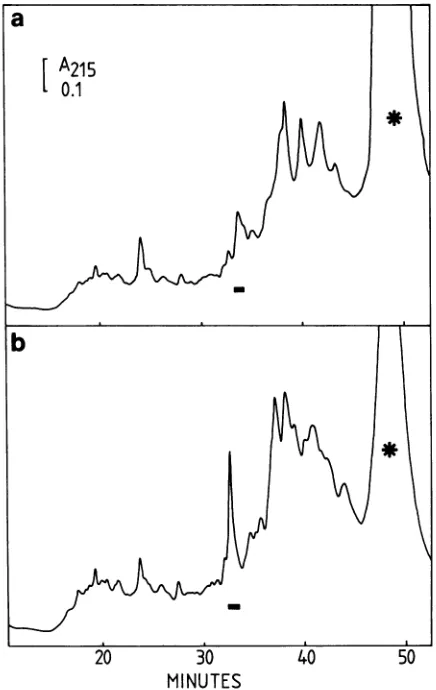
FIG. 5. The elution profiles of a NP-40 detergent extract of HSV-1 virions on RP-HPLC before and after adsorption with monoclonal antibodiesLP1. Elution conditionswereasindicated in the legend to Fig. la. (a) Detergent extract was adsorbed with immobilized monoclonal antibodiesLP1priortoRP-HPLC. Adsorp-tion was carried out with 400
p.l
of NP-40 extract and40 mg of protein A-SepharoseC1-4B,towhich 150p.l
of monoclonal antibod-ies was adsorbed. (b) Without adsorption with immrrobilized LP1. Symbols:-, elutionposition57Kpolypeptide; *, NP-40micelles.VP16,weinvestigatedthe reactionof monoclonal antibodies LP1, which are directed
against
VP16, with thedetergent
extractofpurifiedHSV-1 virions. SDS-PAGE ofthe deter-gent extracts before and after reaction with monoclonal antibodiesLP1
against
VP16showedthatonly
the red-brown-ish polypeptide band of the 57Kpolypeptide
could be eliminatedfrom the extract.RP-HPLC is performed in
high
concentrations oforganic
solvents andusually
results in conformationalchange
andloss of activity of proteins (29, 49). Rather than
doing
immunoprecipitation
studies with thepurified
57Kpolypep-tide, we
subjected
the extractto RP-HPLC before and after reaction with immobilized monoclonal antibodies LP1. The elution patternsare shown inFig.
5.Although
the57Kpeak
3 4 5 6 7 8 9 10 11 12 13 14 15 16 17 18 19 20
Ala-Pro/Val-jyr-Phe-Lys-Glu-Gln-Phe-Leu-Asp-Gly-Asp-
Tly-Jrp-Thr-Asn-Asp- X-Ile-Asp-FIG. 4. N-terminalaminoacid sequenceofthe 57KHSV-1polypeptide.The amino acid sequencegivenwasobtained aftertwoseparate
runsinagas-phase sequencer, using140and 200pmol of theprotein. Theunderlined residues wereidentifiedin both runs, whereas the evidenceforthe otherresidueswasobtainedfromthefirstrunonly.Xisanunidentifiedresidueatposition18.The PTHswereanalyzed by HPLConanIBMcyanocolumn, usingtheprotocol ofHunkapillerand Hood(23) with somemodifications.
on November 10, 2019 by guest
http://jvi.asm.org/
[image:4.612.329.547.69.414.2] [image:4.612.62.298.108.390.2]was not entirely eliminated from the elution pattern (Fig. 5a),asignificant decreaseinabsorbancewasobserved. This was confirmed by SDS-PAGE of the eluent fractions.
Thereaction between the LP1 monoclonal antibodiesand the 57Kpolypeptide strongly suggeststhat the 57K protein
was related to VP16. Others have estimated for VP16 a
molecularmass ofapproximately 65K (8, 22, 31, 33, 39, 43).
Recentstudies, however, reportlower molecularmassesof 63K and 60K(7, 25). Onthe otherhand,thepossibilitythat the 57Kpolypeptide isamajor fragment ofVP16 derivedby proteolytic cleavage may notbe excluded.
In conclusion, RP-HPLC was used for the isolation ofa
57K HSV-1 polypeptide. In this particular case, 50
mt
of purified virions were extracted, resulting after one RP-HPLCruninca.45 jig of the 57K polypeptide, whichis 774 pmol.It has been shown that HPLC isanexcellenttechniquefor the separation ofmany compounds (12). Now methods are
available for HPLC ofproteins (40). Reportson the
separa-tion of viralproteins by HPLC are scarce (5, 9, 11, 19, 34,
38, 49) and reflect the problems whichare encountered when proteins with aggregating properties have to be purified. New elution systems had to be developed which imply detergents in the solventsorhigh organicsolvent concentra-tions toavoid aggregation. Generally onlysmallamountsof virus proteins are available, therefore the combination of
HPLC and microsequencing supplementeach other. Partial amino acid sequences from virus proteins obtained in this way will be a useful addition to amino acid sequences
deduced from nucleotide sequencesdataof virus DNA (24). ACKNOWLEDGMENTS
We gratefully acknowledge the technical assistance of Anneke Samallo-Droppers.
This work was supported in part by grant GUKC 83-19 from
Stichting KoninginWilhelmina FondstoS.W.W. J.v.B. is indebted
tothe National Fund of ScientificResearch for thegrantproviding thegas-phasesequencer(FKFO 2.0007.82).
LITERATURE CITED
1. Ashley,R.L.,and L.Corey.1984. Effect ofacyclovirtreatment ofprimary genital herpes on the antibody response to herpes simplexvirus.J.Clin. Invest. 73:681-688.
2. Balachandran, N., D. Harnish, R. A. Killington, S. Bacchetti, andW. E.Rawls.1981. Monoclonal antibodiesto two glycopro-teins ofherpes simplex virus type 2. J. Virol. 39:438-446.
3. Balachandran, N., D. Harnish,W. E. Rawls, and S. Bacchetti.
1982.Glycoproteinsofherpes simplexvirustype2asdefinedby monoclonal antibodies. J. Virol. 44:344-355.
4. Baucke, R. B., and P. G. Spear. 1979. Membrane proteins specified by herpes simplex viruses. V. Identification of an
Fc-bindingglycoprotein.J. Virol. 32:779-789.
5. Brown, A. S., J. C.Bennett, J. E. Mole, and E. Hunter. 1981.
Purification and characterization of the gag gene products of
avian-typeC retroviruses by high-pressure liquid chromatogra-phy. Anal. Biochem. 112:128-134.
6. Bzik, D. J., B. A. Fox, N. A. DeLuca, and S. Person. 1984.
Nucleotide sequence specifyingthe glycoprotein gene, gB, of
herpes simplexvirus type 1.Virology 133:301-314.
7. Campbell, M. E. M., J. W. Palfreyman, and C. M. Preston.
1984. Identification of herpes simplex virus DNA sequences which encodeatrans-acting polypeptide responsiblefor
stimu-lation of immediate early transcription. J. Mol. Biol. 179:
699-703.
8. Cassai,E.N.,M.Sarmiento,and P. G.Spear.1975.Comparison
of the virionproteinsspecifiedby herpes simplexvirus types 1
and 2.J. Virol. 16:1327-1331.
9. Clark,R.B., J.A.Zaia,L.Balce-Directo,and Y.-P.Ting.1984.
Isolation and partial chemical characterization of a 64,000-dalton glycoprotein of human cytomegalovirus. J. Virol. 49:279-282.
10. Dayhoff, M. O., L. T. Hunt, and S. Hurst-Calderone. 1978. Composition ofproteins, p. 363-373. In M. 0. Dayhoff (ed.),
Atlasofproteinsequenceand structure, vol. 5, supplement 3. National Biomedical ResearchFoundation, Washington,D.C. 11. Dernick, R., J.Heukeshoven,and M.Hilbrig.1983.Induction of
neutralizing antibodies by all three poliovirus polypeptides.
Virology130:243-246.
12. Done,J.N., G.J.Kennedy,andJ.H. Knox.1972. Revolutionin
liquid
chromatography. Nature(London)237:77-81.13. Dowbenko, D.J.,and L. A. Lasky. 1984. Extensivehomology
between the herpessimplex virus type 2 glycoproteinF gene and the herpes simplex virus type 1 glycoprotein C gene. J. Virol.52:154-163.
14. Eberle, R.,andSiao-WenMou.1983.Relative titers of antibod-iesto individual polypeptideantigens ofherpes simplexvirus type 1inhumansera. J.Infect. Dis. 148:436444.
15. Eisenberg,R.J.+D.Long,R.Hogue-Angeletti,and G. H. Cohen. 1984. Amino-terminal sequence of glycoprotein D of herpes simplexvirus types 1and 2. J. Virol.49:265-268.
16. Frink,R.J.,R. Eisenberg,G.Cohen,and E. K.Wagner. 1983. Detailedanalysisof theportionof theherpessimplexvirus type 1genomeencodingglycoproteinC. J. Virol. 45:634 647. 17. Gibson, W.,andB.Roizman. 1974.Proteinsspecified by herpes
simplex virus. X. Staining and
radiolabelling
properties
of Bcapsid and virion proteins in polyacrylamide gels. J. Virol. 13:155-165.
18. Gilman,S.C.,J. J.Docherty,and W.E. Rawls.1981.
Antibody
responses in humans to individual proteins ofherpes simplex
viruses. Infect. Immun. 34:880-887.
19. Green, M., and K. H. Brackmann. 1982. The application of high-performance liquid chromatography for the resolution of proteinsencodedbythe humanadenovirus type 2 cell transfor-mationregion.Anal. Biochem. 124:209-216.
20. Heine, J. W., R. W.Honess,E.Cassai, and B.Roizman. 1974. Proteins specified by herpes simplex virus. XII. The virion
polypeptidesof type 1strains-. J. Virol. 14:640-651.
21. Hewick, R. M., M. W. Hunkapiller, L. E. Hood, and W. J. Dreyer. 1981. A gas-liquid solid phase
peptide
andprotein
sequenator. J. Biol. Chem. 256:7990-7997.
22. Honess, R. W., and B. Roizman. 1973. Proteins
specified by
herpessimplexvirus. XI.Identification and relativemolarrates ofsynthesisofstructural and non-structural herpesviruspoly-peptidesin the infected cell. J.Virol. 12:1347-1365.
23. Hunkapiller,M.,and L. E. Hood.1983.Analysisof
phenylthio-hydantoins by ultrasensitive
gradient
high-performance liquid
chromatography. Methods Enzymol.91:486-493.
24. Hunkapiller,M.,S.Kent,M.Caruthers,W.Dreyer,J.Firca,G. Giffin, S. Horvath, T. Hunkapillar, P. Tempst, and L.-Hood. 1984.A microchemicalfacilityforthe
analysis
andsynthesis
of genes andproteins.Nature (London)310:105-111.25. Johnson, D. C., M. Wittels, and P. G.Spear. 1984.
Binding
to cells of virosomescontaining herpes
simplex
virus type 1glycoproteins andevidence for fusion. J. Virol. 52:238-247. 26. Knopf, K. W., and H. C. Kaerner. 1980. Virus
specific
basicphosphoproteins associated with herpes
simplex
virus type 1(HSV-1) particlesand thechromatin ofHSV-1-infected cells.J. Gen. Virol. 46:405-414.
27. Laemmli,U. K. 1970.Cleavageof structural
proteins during
theassemblyoftheheadof thebacteriophageT4. Nature(London)
227:680-685.
28. Lemaster, S., and B. Roizman. 1980. Herpes
simplex
virus phosphoproteins. II. Characterization of the virion proteinkinaseand of the polypeptides phophorylated in the virion. J. Virol. 35:798-811.
29. Luiken, J.,R.vanderZee,andG. W.Welling. 1984.Structure and activityofproteinsafter reversed-phase
high-performance
liquid chromatography. J. Chromatogr.284:482-486.
30. Marsden,H.S., A.Buckmaster,J.W.Palfreyman,R.G.Hope,
andA. C. Minson. 1984. Characterizationof the 92,000-dalton
glycoprotein induced byherpes simplex virustype2. J. Virol.
on November 10, 2019 by guest
http://jvi.asm.org/
50:547-554.
31. Marsden, H. S.,N. D. Stow, V. G. Preston, M. C. Timbury,and N. M. Wilkie. 1978. Physical mapping of herpes simplex virus-inducedpolypeptides. J. Virol. 28:624-642.
32. Marshall, T., and K. M. Williams. 1984. Artifacts associated with 2-mercaptoethanol upon highresolution two-dimensional electrophoresis. Anal. Biochem. 139:502-505.
33. McLean, C., A.Buckmaster, D.Hancock,A. Buchan, A. Fuller, and A. C. Minson. 1982. Monoclonalantibodies to three non-glycosylated antigens of herpes simplex virus type 2. J. Gen. Virol.63:297-305.
34. Montelaro,R. C., M. West,andC. J.Issel.1981. High-perform-ancegelpermneation chromatography of proteins in denaturing solvents and its application to the analysis of enveloped virus polypeptides. Anal. Biochem. 114:398-406.
35. Para, M. F., K. M. Zezulak, A. J. Conley,M. Weinberger, K. Snitzer, andP. G. Spear. 1983. Useofmonoclonal antibodies against two75,000-molecular-weightglycoproteins specified by herpes simplex virus type 2 in glycoprotein identification and genemapping. J. Virol. 45:1223-1227.
36. Pereira, L., D. Dondero, B. Norrild, and B. Roizman. 1981. Differential immunologic reactivity andprocessing of glycopro-teins gAandgBof herpessimplex virus types 1 and 2 made in Vero and HEp-2 cells. Proc. Natl. Acad. Sci. U.S.A. 78: 5202-5206.
37. Pereira, L.,D.Dondero,and B. Roizman. 1982.Herpessimplex virus glycoprotein gA/B: evidence that the infected Verocell products comapand arise by proteolysis. J. Virol.44:88-97. 38. Phelan,M.A.,andK. A. Cohen. 1983.Gradientoptimalization
principles in reversed-phase high-performance liquid chroma-tography and the separation of influenza virus components. J. Chromatogr.266:55-66.
39. Powell, K.L., and R.J. Courtney. 1975. Polypeptides synthe-sized in herpes simplex virus type 2 infected HEp-2 cells. Virology 66:217-228.
40. Regnier, F. E. 1983. High-performance liquid chromatography ofbiopolymers. Science 222:245-252.
41. Roizman, B., B. Norrild, C. Chan, and L. Pereira. 1984. Identification and preliminary mapping with monoclonal anti-bodiesofaherpessimplex virus2glycoproteinlackingaknown type 1counterpart. Virology 133:242-247.
42. Spear, P. G. 1976. Membrane proteins specified by herpes simplex viruses. I.Identificationof fourglycoprotein precursors andtheir products in type 1-infected cells.J.Virol.17:991-1008. 43. Spear, P. G., and B. Roizman. 1972. Proteins specified by
herpessimplexvirus. V. Purification and structural proteins of theherpes-virion. J. Virol. 9:143-159.
44. Spear,P.G.,and B. Roizman.1980.Herpessimplex viruses. p. 630-635. In J. Tooze (ed.), DNA tumor viruses, ColdSpring Harbor Laboratory, ColdSpring Harbor, N.Y.
45. Tasheva, B., and G. Dessev. 1983. Artifacts in sodiumdodecyl sulfate-polyacrylamide gel electrophoresis due to 2-mercapto-ethanol. Anal. Biochem. 129:98-102.
46. van der Zee, R., and G. W. Welling. 1982. Molecularsieving during reversed-phase high-performance liquidchromatography of proteins. J. Chromatogr. 244:134-136.
47. vanderZee, R.,andG.W.Welling. 1984. Apersonal comput-er-basedgradient system forhigh-performance liquid chroma-tograpy with low-pressure mixing. J.Chromatogr. 292:412-417. 48. Watson,R.J., J. H.Weis, J. S. Salstrom,andL. W. Enquist. 1982. Herpessimplex virus type1glycoproteinDgene: nucleo-tide sequence and expression in Escherichia coli. Science 218:381-384.
49. Welling,G. W., J.R.J. Nijmeijer, 1R. vanderZee,G. Groen, J.B. Wilterdink, and S. Welling-Wester. 1984. Isolation of detergent-extractedSendai virus proteins by gel-filtration, ion exchange andreversed-phase high-performance liquid chroma-tography and the effectonimmunological activity. J. Chroma-togr. 297:101-109.
50. Welling-Wester, S., J. Vos,andJ. B.Wilterdink. 1984. Differ-ences inantigenic properties ofFc-bindingactivity during infec-tion withherpes simplex virus type1.Arch. Virol. 80:183-193. 51. Wray, W., T. Boulikas, V. P. Wray, and R. Hancock. 1981. Silverstaining of proteins in polyacrylamide gels. Anal. Bio-chem. 118:197-203.
52. Zezulak, K. M., andP. G. Spear. 1983. Characterization ofa herpes simplex virus type2 75,000-molecular-weight glycopro-teinaritigenically relatedtoherpes simplex virus type1 glycopro-tein C. J. Virol.47:553-562.
53. Zezulak,K. M.,and P. G.Spear. 1984. Limitedproteolysisof herpes simplex virus glycoproteins that occurs during their extractionfrom Vero cells. J. Virol. 50:258-262.
54. Zweerink,H.J.,and L. W. Stanton.1981. Immune responseto herpessimplex virus infections:virus-specificantibodies insera frompatients with recurrent facial infections. Infect. Immun. 31:624-630.
55. Zweig, M., S. D. Showalter, S. V.Bladen, C. J.Heilnan, Jr., and B.Hampar. 1983. Herpessimplex virus type2glycoprotein gFandtype 1 glycoproteingC have relatedantigenic determi-nants.J. Virol. 47:185-192.