Vol. 58, No. 3 JOURNALOFVIROLOGY,June 1986,p. 876-883
0022-538X/86/060876-08$02.00/0
Copyright C 1986, American Society for Microbiology
Intracellular Localization and Processing of pp6Ov-src Proteins
Expressed by Two Distinct
Temperature-Sensitive
Mutants
of
Rous
Sarcoma Virus
ANDREW W. STOKER,* STUART KELLIE, ANDJOHN A. WYKE
Imperial Cancer Research FundLaboratories, St. Bartholomew's Hospital, Dominion House, London ECIA 7BE, UnitedKingdom
Received 9December 1985/Accepted 17February 1986
Thetransforming protein of Roussarcomavirus,pp6Ov-src,isknowntobeatyrosineprotein kinase, but the
mechanismofcelltransformation remainsunclear.Infurther investigatingpp6ovsrc structureandfunction,we
have analyzed twotemperature-sensitive (ts) Rous sarcomavirussrcgene mutants, tsLA29 and tsLA32. The
mutations in tsLA29 and tsLA32mapinthecarboxy-terminal regionand the amino-terminal half ofpp6O-s'c,
respectively, and encode mutant proteins witheither temperature-labile (tsLA29)or -stable (tsLA32) kinase
activities. Here we examined the intracellular processing and localization of these pp6o-src mutants and extendedourcharacterization of transformationparametersexpressed by cellsinfected by the Roussarcoma
virus variants. Noobvious defects in functional integrity of the tsLA32 pp6o-src couldyetbe demonstrated,
whereas thetsLA29 pp6Ovsrcwasperturbednotonlyinkinaseactivity,butalsoinaspectsofprotein processing
andlocalization.Analysis of transformationparametersexpressed by infected cells demonstrated the complete
temperaturelability of bothmutants.
Thev-src gene ofRous sarcoma virus (RSV) encodes a
phosphoprotein ofmolecularweight 60,000,pp60v-src, which
caninduce in vitroneoplastic transformation ofavariety of
cells (24, 38). This transformation is a complex process,
involving considerable structural and metabolic
perturba-tionsto thecell, andyetpp6Ov-src is capable of initiating and
maintainingthepleiotropic changes whichareseen (18). The
mechanisms by which pp6Ov-src induces transformation
re-main unclear, althoughaconsiderableamountof knowledge
has been gained in recent years concerning its structure,
location, and functional activity within the cell.
Theonly well-defined functionascribedtopp6Ov-src is that
ofatyrosineprotein kinase (2, 6, 7, 16, 25, 30, 31,42), for
which a number of putative target proteins have been
identified in the cell (1, 8, 34). These include two
phosphoproteins, pp36(15, 39) andpp5O (23), vinculin(41),
and threeglycolyticenzymes(10).However, inno casehas
specific phosphorylationof these substrates been shownto
benecessaryfortransformation, suggestingthat the critical
cellular targets have yet to beidentified.
An additional property of the pp6Ov-src protein is its
association with the plasma membrane, wherethe majority
ofmaturepp60v-src is found(for review, see reference 27).
The importance of this localization is demonstrated by
mutantsshowing defects in membranebindingthat are also
invariably defective in some orall aspects ofcell
transfor-mation (11, 20, 28). The mechanism by which pp6vsrc
associates withthemembrane isunclear,but several factors
appear to beofimportanceingoverningthe transport to or
association with this site. First, the lipid myristic acid is
foundcovalently attachedtotheN-terminalglycineresidue
of pp60v-src (5, 43). Absence ofthis lipid prevents stable
associationwith themembrane,thuspreventing
transforma-tion, but notapparently affectingthekinaseactivity(13, 21,
26). The N-terminal 8 kilodaltons ofpp6Ov-src isbelieved to
be involved in close contact with the membrane (28) and
*Corresponding author.
together withmyristic acid maymediate the stable
associa-tion.Inadditiontothesefactors,pp60v-srcis known toform
a transient complex with two cellular proteins, ppSO and
pp9O, soonafter itssynthesisoncytosolic polyribosomes(2,
4,36). The precise role ofthecomplexis unclear, but some
involvement in transport ofpp60v-src to the membrane has
beensuggested(11).
Ithasbecome clearthat thefunctions ascribedtopp60v-src
canbecrudely mappedtodomains of the protein(17,29, 37,
44). The kinase function is believed to lie in the
carboxy-terminal (C-terminal) half of the protein in an area with
sequence homology to other known tyrosine kinases (40).
The membrane binding is ascribed to the amino-terminal
(N-terminal)region, and additionalareasoftheproteinmay
provide specific binding points for the pp50-pp9O
pro-teins (46). Todissect furtherthe structureand functions of
pp60v-src, we have been studyingtemperature-sensitive (ts)
mutantsofthe v-src gene inwhich different domains ofthe pp60v-srcproteinhavebeen perturbed. We have focused on
twomutants, tsLA32 andtsLA29, andourprevious workhas shown that these mutants havelesionswhicheither do affect
(LA29)ordonotdirectly affect (LA32)the kinaseactivity of
theprotein(44).TheLA32 mutant isofparticularinterestas
it maintains elevated cellular phosphotyrosine levels and
phosphorylates pp36 (44) and vinculin (S. Kellie, B. Patel,
N. M. Wigglesworth,D. R. Critchley,and J.A. Wyke,Exp.
Cell Res., in press) at restrictive temperature, suggesting
that this mutant has alesionin anoveldomain necessary for the maintenance of transformation.
Ourwork didnotindicatewhether the lesion in LA32was
affectingaspects ofpp60v-src localization or maturation, or
whether the LA29 pp6Ov-src exhibited defects other than in thekinase function.Toinvestigatethesequestions,wehave
studiedboth the intracellularprocessingand thelocalization
of the mutant pp6Ov-src proteins. We show that the LA32
pp6Ov-src mutant is not significantly defective in the above parameters and contrast these findings with those of the LA29 mutant. In addition, having previously demonstrated
876
on November 10, 2019 by guest
http://jvi.asm.org/
the elevation of phosphotyrosine levels and the residual
soft-agar growth capacity of LA32-infected chicken embryo
fibroblasts (CEF) at41°C (44), we were interested in
exam-ining whether additional parameters oftransformationwere
expressed under restrictive conditions. We describe a
de-tailed study of the dissolution of actin- and
a-actinin-containing cables and the fibronectin matrix in the
mutant-infected cells and demonstratethefulltemperaturelability of
these transformation parameters.
MATERIALS AND METHODS
Cells and viruses. CEFwerepreparedfrom fertile eggsof
a white leghorn and brown leghorn cross (Wickham
Laboratories, Wickham, Hampshire, U.K.). Methods of cell
culture and infection were as described previously (47).
Isolation of viruses and initial characterization have been
described before (17,44, 49).
Antibodies and fluorescent reagents. Normal rabbitserum
was collected as described previously (44). Anti-pp6Ovsrc
antibodies were kindly provided by the following people:
JB327 monoclonal (J. Brugge [32]); anti-v-src-C (S. J.
Courtneidge); anti-src rabbit serum raised against pp60-src
expressedandpurifiedfrom Escherichia coli(R. L. Erikson
[22]). Rabbitanti-mouseserumMH1 waskindly supplied by
M.Hayman. Antiseraagainst chickengizzardoa-actininwere
raisedinrabbits andaffinity purifiedasdescribedpreviously
(26a). Fluorescein isothiocyanate-conjugated
affinity-purified goat anti-rabbit and goat anti-mouse
immunoglob-ulinswereobtained fromSigma Ltd. Nitrobenzo-oxadiazole
(NBD)-phallicidinwasobtained from MolecularProbesInc.,
Junction City, Ore., and was used as described by the
supplier.
Immunoprecipitation and gel electrophoresis. Cell lysis,
immunoprecipitations, and sodium dodecyl
sulfate-polyacrylamide slabgel electrophoresiswere performed as
described previously (44). The lysis buffers used were as
follows: modifiedRIPA buffer (10 mM Tris hydrochloride,
pH 7.2, 150 mMNaCl, 1% deoxycholate, 1% Triton X-100,
0.1% sodium dodecyl sulfate, 0.1 trypsin inhibitor unit per
ml ofaprotinin [Sigma], 20 mM NaPP1; buffer A (150 mM
NaCl, 10 mMTrishydrochloride, pH 7.0, 1% Nonidet P-40,
20 mM
NaPPi).
Immunofluorescence. Cells were grown on
13-mm-diameter cover slips for at least 18 h and fixed in 3.8%
formaldehydeinphosphate-bufferedsaline(PBS)for30min
followed by extensive washing in PBS. After
permeabiliza-tionby immersion in PBScontaining 1% NonidetP-40, the
cover slipswerewashed and overlaidwith 50,Iof the first
antibody in PBS (anti-a-actinin, 50 ,ug/ml; antifibronectin, 1:100dilution;JB327anti-pp60vsrc, 1:20dilution)for30min. Afterthorough washing, the coverslips were overlaidwith 50
RI
of a 1:20 dilution of fluoresceinisothiocyanate-conjugatedgoatanti-rabbitorgoatanti-mouse
immunoglob-ulin for 30 min. Cover slips were washed and mounted in
PBS-glycerol (1:9) containing 1 mg of p-phenylamine
diamineper ml toreducephotobleachingandviewed witha
Leitz Dialux microscope equipped with epifluorescence.
Photomicrographs weretaken withIlford HP5film.
Cell fractionation. Plates (35 mm) of RSV-infected CEF were incubated for 30 min in methionine-free medium and then labeledfor1hwith 200,uCiof
[35S]methionine
per mlat35 and 41°C. The label was removed and chased in cold,
complete medium for 2 h at the respective temperatures.
Uninfected CEF carrier cells
(107)
were suspended andwashed twice in PBS. The labeled cells were rinsed twice
with PBS, scraped into the carrier cell suspension with a
rubber policeman, and washed once with PBS. The cells
were resuspended in 3 ml ofhypotonicbuffer (10 mM Tris
hydrochloride, pH 7, 1 mM MgCl2)and lefttoswell on ice
for 4 min. NaPP1 to 10 mM and 2 trypsininhibitor units of
Aprotinin (Sigma)wereadded, and the cellswerelysedwith
30strokesofatight-fittingDouncehomogenizer.Thelysates
werecleared of unbrokencells andnuclei with alow-speed
spin(600 x g) for 10min. Half of the resultant supernatant
wasnotfurther treated(fraction MC),andtheother halfwas
further spun at 80,000 x g for 30 min to obtain a crude
membrane pellet(fraction M) and a supernatant of soluble
cytoplasmic constituents (fraction C). All fractions were
adjusted to RIPA concentration and immunoprecipitated with normalrabbitserumand theanti-v-src-C antisera.This
procedureprovided90to100%recoveryofpp6Ov-srcin theM
and Cfractionswhencomparedwiththe totalunfractionated
MC sample.
Immunoprecipitates
weresubjected
topoly-acrylamide gel electrophoresis, and the gels were impreg-nated with Amplify (Amersham International) and
fluorographed with Kodak XAR-5 film. pp6Ov-src in each
fraction was quantified by scanning densitometry of the
resulting fluorographs, using aJoyce-Loebldensitometer.
pp6&Vsrc complex analysis. (i) Complex immunoprecipita-tion.CEF infected with RSVwerelabeledfor18 hwith 100 ,uCi of[35S]methionine per ml in complete medium. Cells were washed twice with PBS and lysed in buffer A. The lysateswerenormalized forproteincontentandhalveswere
immunoprecipitatedwithnormalrabbitserumand
monoclo-nal JB237, followed by rabbit anti-mouse serum MH1.
Immunoprecipitatesweresubjectedtogelelectrophoresison 10%acrylamide gels.
(ii) Complex stability analysis. Cellswerelabeledasabove
but using 200 ,uCi of [35S]methionine per ml. Cells were
washedtwice in PBS andlysedin bufferA.
Immunoprecip-itation ofpp6Ov-src was performed initially as in (i) above.
Immune complexes were washed once with bufferA, and
then each sample wassplit intotwo aliquots. Eachaliquot was washed three times in either bufferA or RIPA buffer.
The immune complexes were subjected to polyacrylamide
gelelectrophoresis,and afterfluorography gelslices
contain-ingpp60v-src and pp9Owere cut out and the 35S counts per minute were quantitated in anLKB RackBeta scintillation counter. Countswerecorrectedfor background countsper
minute fromcontrol areasofthegel.
Myristic acid labeling.
9,10-[3H]myristic
acid (2mCi/ml;
AmershamInternational) supplied in toluenewasdried and
suspendedin calfserum. CEF eitherinfected with RSVor
uninfectedwerelabeledfor18 hwith40 ,uCiof
[3H]myristic
acidperml incompletemedium at35and41°C. Cells were
lysedin RIPAbuffer,andpp60V-srcwasimmuneprecipitated
withanti-src serum.Afterpolyacrylamide gel
electrophore-sis, gels wereimpregnated withAmplify (Amersham Inter-national) and fluorographed with KodakXAR-5 film for 28 days. pp60vsrc bands were scanned with a Joyce-Loebl densitometer,and valueswerenormalized for therespective lysate proteinconcentration.
RESULTS
pp6O-src
localization:cellfractionationand immunofluores-cence. After synthesis on cytosolic polyribosomes, and ashortlagperiod of10to 15 min, wild-type
pp60v-src
canbefound localizedin theplasmamembrane compartment of the
cell (29). It is believed that the membrane-bound form of
pp6Ov-srcis the functionalformof theprotein,and thus itwas
on November 10, 2019 by guest
http://jvi.asm.org/
878 STOKER ET AL.
ofimportance to determine the localization of the mutant
proteins. For this, apulse-chase labelingprotocol was used,
followed by cellular fractionation and immunoprecipitation
ofpp6Ov-sc. Given the kinetics of wild-type pp6Ov-src associ-ation with the plasma membrane, the majority of the labeled pp64?v-src proteins will be chased to their final intracellular location within the 2-h chase period.
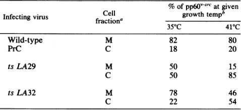
Cellular fractionation ofwild-type pp6Ovsrc reveals that 80% of the labeled protein is found in the crude membrane fraction at both 35 and 41°C (Table 1). The mutant protein encoded by LA32 behavesvery similarly to the wild type at permissive temperature, with comparable levelsof pp6Ov-src in the membrane fraction. At nonpermissive temperature nearly 50% of the labeled pp60-src is still found in the membrane fraction. A differentpatternisfound with mutant LA29. At permissive temperature only 50% of the labeled pp6Ov-src is found in the membrane, but this represents a level which is clearly sufficient for cell transformation. At
nonpermissive temperature only 15% of the pp6Ov-src
re-mains in themembrane, with85%as acytosolicform. We have not ruled out the possibility that the mutant proteins, particularly that of LA29, exhibit some instability
in membrane binding during the fractionation procedure.
Thus, to confirm the membrane association pattern, and in particular todemonstrate theassociation with plasma mem-branes, we analyzed pp6ov-src localization, using indirect
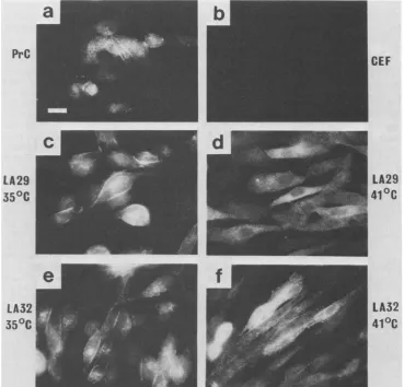
immunofluorescence. Both LA29- and LA32-infected CEF
displayprominent plasma membrane staining ofpp6Ov-srcat
permissive temperature, similar in pattern to
wild-type-transformedcells (Fig. 1). Atnonpermissive temperaturethe
pattern of fluorescence is quite different with LA29, the
pp60v-src distribution now being of a diffuse, cytoplasmic
nature, correlating well with our cell fractionation data. In contrast to LA29, and againin agreementwiththe
fraction-ationdata, CEFinfectedwith LA32 maintainstaining inthe plasma membrane at 41°C, with prominentsignalsatcell-cell
boundaries. Although notquantitative, themembrane
stain-ing of LA32-infected CEF is reduced to some degree at
restrictive versus permissive temperature, and this may in
part be due to the flatter nature of the cells and thus the greater spreading of the plasma membrane.Membrane
stain-ing in wild-type RSV-transformed CEF is unaffected by
growth temperature.
Association of mutantpp60vsrcproteins with thepp9O-ppSO
complex. Soon aftersynthesis pp6Ov-srcenters acytoplasmic
complex with cell proteins pp90 and pp50. Only a small
percentage of the total cellular pp9O is incorporated in the
complex, and newly synthesized pp9O enters alarge
cyto-TABLE 1. Localization ofpp6O-vsrCby cell fractionation
% ofpp6VS-Catgiven
Cell growthtempb
Infectingvirus fraction'
35"C 410C
Wild-type M 82 80
PrC C 18 20
tsLA29 M 50 15
C 50 85
tsLA32 M 78 46
C 22 54
aM, Crude membranefraction; C,cytoplasmicfraction.
b Each number represents the proportion of labeled
pp60.-sr
in each fractionas apercentageof the totalMplus C densitometricvalue andistheaverageoftwoindependent experiments.
solic poolrather than thecomplex (4). Becauseofthis,along
[35S]methionine labeling periodwasperformed to obtain the
complex with high levels of labeled pp90 associated with
pp60v-src forquantitation purposes. RSV-infected CEFwere
labeled for 18 h with [35S]methionine, and pp6Ov-src was
immunoprecipitated with the monoclonal JB327 which
effi-cientlyprecipitatesthecomplexedformofpp6Ov-src (32).By
this procedure, the efficiency of complex formation was
judged from the ratio of pp9O/pp60. The pp5O was not
quantitated because of the difficulty of distinguishing this
proteinfrompp6Ov-src breakdown products andbackground
proteins of a similar molecular weight.
The data in Fig. 2 demonstrate that the levels of pp9O
coprecipitated with mutant pp6Ov-src proteins were not
greatly affected bygrowth temperature, and the levels seen
were similar to those found with our wild-type pp6Ov-src (quantitation of35S counts per minute from pp60 and pp90
gelareas was confirmatory; data not shown). We did note a smalldecrease in the amount of pp9O coprecipitated at 35°C in all cases, the largest decrease was seen with thewild type, but the significance of this is unclear. The sensitivity of
pp9O-pp60 binding to variations in detergent conditions is shown by the following ratios ofpp90 remaining bound to
pp60v-srcinbufferA/RIPA: with wild-type PrC as the infect-ing virus, 2.1; with tsLA29, 9.7; and with tsLA32, 3.3. The pp6Ov-src proteins encoded by both mutants and wild type bind pp90 to similar degrees in Nonidet P-40-containing
buffer, buffer A. However, in the more denaturing RIPA
buffer, containing the ionic detergent sodium dodecyl sul-fate, a three to fourfold greater instability in pp60-pp9O
bindingis found with LA29 than with LA32 or wild-type PrC.
This in vitro instability is not dependent upon the growth temperature of the infected CEF from with the pp6Ov-src proteins are isolated (data not shown). Under physiological
conditions, however, such aninstability may well be
differ-entially expressed at 35 or41°C and could have a significant effect upon pp6ov-src processing and function. We are pres-ently investigating the in vivo kinetics of complex associa-tion with these mutants in an attempt to answer this ques-tion.
Myristylation ofpp6O-v'. The lipid myristic acid is cova-lently attached to the N-terminal glycine residue of the pp60v-srcprotein, and this modificationto pp6Ov-src has been shown to be essential for transforming activity. Given the importance of myristic acid attachment during pp6Ov-src maturation, it was of interest to determine whether temper-ature shift significantly affected the myristylation states of the ts mutant pp6Ov-src proteins.
CEFinfected with the mutant viruses were labeled for 18 h with [3H]myristic acid (providing steady-state levels of
incorporation; data not shown), and pp6Ovsrc was
im-munoprecipitated from whole-cell lysates. Thepeakareas of labeled pp6Ov-src were quantitated as described inMaterials andMethods. The incorporationoflabeledmyristic acidinto wild-type pp6Ov-src shows little change between the two growth temperatures (35°C/41°C ratio = 1.5). The mutant pp6Ov-src proteins were found to behave in a manner similar to thewild-type protein withrespect tomyristic acid incor-poration(35°C/41°Cratios:LA32, 1.5;LA29, 1.7). Bussetal. (5)recently reported similarfindings with the LA29 mutant, but with ashorter 2-hlabeling period andusing [3H]palmitic
acid.
It is clear from these data that the ts lesions in both mutants do not greatly affect the ability to incorporate myristic acid into the mutant pp60v-src proteins and that a
defect in myristylation, which is an early step in pp6O-src
J. VIROL.
on November 10, 2019 by guest
http://jvi.asm.org/
[image:3.612.63.302.579.689.2]PrC
LA29
350C
LA32
350C
CEF
LA29
41
°C
LA32
41°C
FIG. 1. Immunofluorescencelocalization of pp60v-src in transformed CEF. Cellswerestained with monoclonalanti-pp60srcasdescribed inMaterials and Methods. (a) PrC transformed; (b) untransformed CEF; (c) LA29-transformed CEFat35°C;(d)LA29-transformed CEFat 41°C; (f) LA32-transformed CEFat35°C;(g)LA32-transformed CEFat41°C. Bar= 10,um.
maturation (5, 19), is notresponsible for their temperature
lability.
Fibronectinmatrixproduction. The productionofa fibro-nectin matrix is dramatically reduced in CEF transformed by
RSV (35). When CEF infected with either LA29 orLA32
were grown at nonpermissive temperature, the cells
pro-duced a fibronectin matrix indistinguishable from that
syn-thesized by uninfected CEF (Fig. 3i and k). At permissive
temperature, the mutantvirus-infected cells showedalarge
reduction in matrix production as expected. However,
al-though the LA29-transformed CEF matrix was reduced to
the residual level seen in wild-type-transformed cells, the
LA32-transformed cells consistently produced a residual
fibronectinmatrixto agreaterdegree than either
wild-type-orLA29-transformed cells (Fig. 3jand1).This residual level
of fibronectin has notbeen analyzed quantitatively.
F-actin and a-actinin distribution. The two cytoskeletal
proteins actin and a-actinin undergo gross intracellular
re-distributionsupon cell transformation byRSV(14, 33). The
temperature-dependent effects of the mutant RSV upon
theseproteinswereexamined. The distribution of F-actin in RSV-infected CEF was examined by fluorescence micros-copy,usingNBD-phallicidin. At41°Cboth LA29- and
LA32-infectedcellsgrew asmorphologically normal, flat cellswith
manyprominent microfilamentbundles running through the
cytoplasmsimilartothose seen inuninfectedCEF(Fig. 3a andc).Atpermissivetemperaturebothmutantsinducedthe
expecteddissolution of actin cables in the transformed cells.
However, in the LA32-infected cells a sparse pattern of
cableswas consistently seencompared with LA29-or
wild-type-transformed CEF(Fig. 3b and d). Although this
obser-vation is not quantitative, it may be related to the more
fusiformmorphology oftheLA32-transformed cellsat35°C
(44). The mutant-andwild-type-transformed cells all
exhib-itedcharacteristicaggregatesofF-actin withinthecytoplasm
atpermissivetemperature.
The distribution of a-actinin inthe infected CEFclosely
paralleled that of F-actin. Upontemperatureshift from 35to
41°C, the diffuse, cytoplasmic pattern ofa-actinin staining changedtothe characteristicperiodic staining along
micro-filament bundles as seenin uninfected CEF (Fig. 3e toh).
The mutant-infected CEF thus exhibit full temperature
sen-sitivity in the redistributions of F-actin and a-actinin,
whereas the distributions ofthese proteinsinuninfectedor
wild-type-transformed CEFwereunaffected bygrowth
tem-perature.
DISCUSSION
Inthispaperweextendourcharacterization ofthemutant
pp60v-src proteins encoded by tsLA32 and tsLA29and
on November 10, 2019 by guest
http://jvi.asm.org/
[image:4.612.128.497.70.424.2]880 STOKER ET AL.
wtPrC tsLA29
35 41 35 41
-w
a b a b a b a b
FIG. 2. Immunoprecipitation of pp6C plex proteins.RSV-infected CEFwerela
for 18 h atpermissive (35°C) and nonp
tures. pp60v-src was immunoprecipitated Lanesa,JB327immunoprecipitates; lant
serumimmunoprecipitates.
lyze further parameters of transfc infected by these mutant RSV. TE current knowledge of thetwo mutar The mutations in LA32 and LA"
tsLA32 matelylocated bymarker rescuemapping: LA32 contains a
mutation(s) in the N-terminal 60% of pp6Ov-src, whereas
35 41 LA29 is mutated very near to the carboxy terminus ofthe
_
w protein (17).tsLA29.Althoughwe candetect no temperature sensitivity
in eithersteady-statecomplexlevels ormyristylationof this mutantprotein, it isclear that its intracellular localization is -_
~PP90
abnormal, particularly at restrictive temperature. Areduc-tion in membrane binding is seen at both temperatures,
-pp60src although at35°C there isno apparent
impairment
oftrans--I;;_m
formation.Although
at41°C themajority
ofthepp6OV-src
iscytosolic, we notethat the amount ofpp9O bound does not
increaseproportionately, in contrast toother ts mutants of
pp60v-src, suchasNY68 (11). Inaddition, wecandetect an
increased sensitivity of pp9O-pp6O complexing to ionic
de-a b a b tergents in vitro. We suggest, given that the LA29 mutation
liesoutside themembrane-bindingregion of pp6Ovsrc, thata
-src and associated com- defect in complex binding in the cell may be one aspect of
tbeled
with[35S]methionine
thisprotein'sdysfunction. This wouldappear to occurafter iermissive (41°C)tempera- initialcomplex
binding,andpossiblyaftermyristylation,andwith monoclonal JB327. p
s
b,
control normal rabbit may lead to premature dissolution of thecomplex.
Such amalfunctioncould result intheobserved failure ofpp6Ov-src
toassociate well with the membrane at41°Cand, bearingin
mind evidence that the C-terminal region of pp6Ov-src is
)rmation seen in CEF involved with complexbinding (46), it would not be
unex-able 2 summarizes our pected. In addition, Wilkerson et al. recently described
its in this study. site-directed variants of pp6Ov-src with alterations in the
29 have been approxi- C-terminaldomain oftheprotein (48). These proteins behave
LA
32
1. 350 350
[image:5.612.65.304.69.233.2]At
FIG. 3. Stainingofactin(upper panel),a-actinin(middle panel),and fibronectin(lower panel)intransformed CEF.Cellswerestainedwith NBD-phallicidin, rabbitanti-a-actinin, orrabbitanti-fibronectinasdescribedinMaterials andMethods.(a,e,i) LA29-transformedCEFat 41°C; (b, f, j) LA29-transformed CEFat35°C; (c,g,k) LA32-transformed CEFat41°C;(d, h, 1)LA32-transformed CEFat35°C. For(a)to (d)and(e)to(h), bar= 10,um. For (i) to(1),bar = 100,um.
93-68- ..-,
43- .11-1
II
~~~~~~~..._
ret4w-J. VIROL.
f A e)
Li A-\ L )
on November 10, 2019 by guest
http://jvi.asm.org/
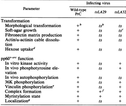
[image:5.612.70.557.360.685.2]TABLE 2. Parameters oftransformation and
pp60v-src
function in tsmutant-infected CEF'Infecting virus Parameter Wild-type tsLA29 tsLA32
Transformation
Morphological transformation +b tSb ts
Soft-agargrowth + ts tSC
Fibronectinmatrixproduction + ts ts
Actin/a-actinin cable dissolu- + ts ts
tion
Hexoseuptaked + ts ts
pp6fv-src
functionInvitro kinase activity + ts +
Invivophosphotyrosine ele- + ts +
vation
Invivoautophosphorylation + ts ts
36Kphosphorylation + ts +
Vinculinphosphorylatione + ts +
Complexformation + +f +
Myristylation state + + +
Localizationg + ts +
aDataare presented in this paper and Stoker et al. (44) unless otherwise
stated.
b+ and ts indicate absence and presence of temperature sensitivity,
respectively.
CSomeresidualgrowth is seen at41°C(44).
dStoker,unpublished data. 'Kellie et al., inpress.
fAlthough total levels of complex are unchanged, the protein may exhibit otherdefects in complex stability (see text).
8Localizationdatafrom both cell fractionation andimmunofluorescence of
pp6l0J-vrC(see text).
(nonconditionally) in a manner similar to LA29 at 41°C.
Thesemutants arekinase defective andpredominantly
cyto-solic,and there isalow level ofassociation with thecomplex
proteins. The mutation in LA29 lies in the region affected in
thesesite-directed variants, suggesting thatboth are
defec-tive inasimilar,possibly regulatory domain-of the protein.
We cannot yet say whether the temperature-dependent
kinase defect of LA29 results from, is causative in, or is
unrelatedtothetransportdefect. Given thatgeneticanalysis
indicates a single mutation towards the 3' end of this v-src
gene, itisnoteworthythatthismutantsuffers fromanumber
of processing and functional defects, suggesting that the
lesion has pleiotropic effects on a structural or temporal
basis.
tsLA32. The LA32 mutant pp60-src shows no obvious
defect in complex formation or myristylation. The protein
also remains membrane localized to a significant degree at
nonpermissive temperature; inaddition, we know the
pro-tein remains localized within adhesion plaques at 41°C
(Kellieetal., in press). Wehavetherefore ruledoutseveral causesofthetransformation defect inLA32 andsuggest that
thelesion intheproteindisruptsitsfunctionat alatestage in
maturationsubsequenttomembranebinding. Itislikelythat
theprotein isimpairedin some specific interactionrequired at or near the plasma membrane, disrupting either kinase
substrate specificity or a critical association with other
cellular structures necessary for the maintenance of the
transformedstate.Of interesthere aretwo recentreportsby
Garber etal. (19) and Cross et al. (12) describingpp60v-src
proteinswithdeletions between aminoacids 15 and 169. This
region is withinthe area towhichthe LA32lesion has been
mapped, and indeed the deletion mutants affect pp6v-src
behavior in a similar way. The kinase functions are not
apparently affected directly, and full morphological
transfor-mation of CEF expressing the proteins does not occur(cf.
LA32 at 35°C). These mutants and our LA32 datatogether
strongly suggestacritical functionalrole fordomains in the
N-terminal halfofpp6O-src.
It has recentlybeen reportedthat pp60vsrcmaybe ableto
causephosphorylationofphosphoinositidederivatives in the
cell, either directly or indirectly, thus influencing a cell
biochemical pathway ofimportance in cell growth control
(45). Given that we have eliminated several sources of the
lesion inLA32, we are at present analyzing the possibility
that themutant isdefective inthisfunction.
AlthoughCEFinfectedwith LA32 at41°C showincreased
phosphotyrosine levels and retain some
anchorage-independent growth (44), there are no residual structural
features of transformation inthese cells, nor is the hexose
uptake rateelevated (A. Stoker, unpublished data). Indeed,
even atpermissive temperature, cellstransformed byLA32
showa less dramaticloss offibronectin matrix and
redistri-bution of actin and a-actinin than similar cells transformed
by wild-type orLA29 virus. This correlates with the
some-whatfusiform morphologyofLA32transformants(44),andit
is of interest that we have mapped the lesion in a fully
fusiform variant of RSV (td SF/L0104)tothe amino-terminal
half ofpp60v-src (17). Sequencing of thev-srcgenesof LA32
(work in progress) and the fusiform virus should reveal whether the ts and fusiform phenotypes represent distinct
mutations ormanifestations of the samelesion.
In conclusion, we have analyzed two ts mutants of
pp6Ov-src which have lesions in different domains of the
protein and which present defects in pp6Ov-src function
varying in both pattern and degree. TheLA29 mutant is of
interest considering the pleiotropic nature of the defects expressed in pp60v-src. The mutation in LA32, in anas yet
undefined domain ofpp60v-src, abolishes almost all
transfor-mation parameters at41°C but has littleeffectondetectable
facets ofpp60v-src maturation andfunction, with the
excep-tion ofthepreviously demonstrated reduction in
phosphor-ylation of pp60v-src itself (44). This latter finding is under
continuing investigation at present.
Further studies with suchtsmutantsandelucidation of the
defectspresentshould be ofconsiderable valueinadvancing
our understanding of the mechanism of pp60v-srcmediated
cell transformation.
ACKNOWLEDGMENTS
We thank J. A.Schmidtforcritical comments on thismanuscript, P.J. Enrietto for additionalcommentsandadviceduring this work, and A. Sterlini for herpatienceduringpreparationof the manuscript.
LITERATURE CITED
1. Beemon, K., T. Ryden,and E.A.McNelly. 1982. Transformation by avian sarcomaviruses leadstophosphorylation of multiple cellular proteins ontyrosine residues. J. Virol. 42:742-747. 2. Brugge, J. S.,E.Erikson, andR. L.Erikson. 1981.Thespecific
interaction of the Rous sarcoma virus transforming protein, pp60Src. Cell 25:363-372.
3. Brugge, J. S., P. J. Steinbaugh, and R. L. Erikson. 1978. Characterisation of the avian sarcoma virus protein pp60s'c. Virology 91:130-140.
4. Brugge, J. S.,W. Yonemoto, and D.Darrow. 1983. Interaction between the Roussarcoma virus transforming protein andtwo cellularphosphoproteins: analysis of the turnoverand distribu-tionof this complex. Mol. Cell. Biol. 3:9-19.
5. Buss, J. E.,N. P.Kamps, and B.M. Sefton. 1984. Myristic acid
on November 10, 2019 by guest
http://jvi.asm.org/
882 STOKER ET AL.
is attached tothe transforming protein of Rous sarcoma virus during or immediately after synthesis and is present in both soluble and membrane-boundforms of the protein. Mol. Cell. Biol. 4:2697-2704.
6. Collett, M. S., and R. L. Erikson. 1978. Protein kinase activity associated with the aviansarcomavirussrcgeneproduct.Proc. Natl. Acad. Sci. USA75:2021-2024.
7. Collett, M. S., A. F. Purchio, andR. L. Erikson. 1980. Avian
sarcoma virus-transforming protein, pp60orc shows protein kinase activity specific for tyrosine. Nature (London) 285:167-169.
8. Cooper, J. A., and T. Hunter. 1981. Changes in protein phos-phorylationin Roussarcomavirus-transformed chicken embryo cells. Mol. Cell. Biol. 1:165-178.
9. Cooper, J. A., and T. Hunter. 1983. Identification of cellular
targets for tyrosine protein kinases. J. Biol. Chem. 258: 1108-1115.
10. Cooper, J. A., N. A. Reiss, R.J.Schwartz,and T.Hunter. 1983. Three glycolytic enzymes are phosphorylated on tyrosine in
cells transformed by Rous sarcoma virus. Nature (London) 302:218-222.
11. Courtneidge, S. A., and J. M. Bishop. 1982. Transit of pp6Ov-src
to the plasma membrane. Proc. Natl. Acad. Sci. USA 79:7117-7121.
12. Cross, F. R., E. A. Garber,andH.Hanafusa. 1985. N-terminal deletions in Rous sarcoma virus p6Osr: effects on tyrosine
kinaseand biological activitiesandonrecombination in tissue
culture with thecellularsrcgene.Mol. Cell. Biol.5:2789-2795. 13. Cross, F. R., E. A. Garber,D. Pellman, and H.Hanafusa. 1984.
Ashortsequence in the p6osrc N terminus is required forp60src
myristylation and membrane association and for cell transfor-mation. Mol. Cell. Biol. 4:1834-1842.
14. David-Pfeuty,T., and S. J. Singer. 1980.Altereddistributionsof cytoskeletal proteins vinculin and alpha-actinin in cultured fibroblasts transformed by Rous sarcoma virus. Proc. Natl. Acad.Sci.USA77:6687-6691.
15. Erikson, E., and R. L. Erikson.1980. Identification ofacellular protein substrate phosphorylated bypp6osrc. Cell21:829-836. 16. Erikson,R. L., M. S. Collett, E. Erikson, and A. F. Purchio.
1979. Evidence that theaviansarcomavirustransforminggene product is a cyclic-AMP-independent protein kinase. Proc.
Natl. Acad. Sci. USA76:6260-6264.
17. Fincham, V. J., D. J. Chiswell, and J. A. Wyke. 1982. Mapping ofnonconditional and conditional mutants in the src geneof
Prague strain Roussarcomavirus.Virology 116:72-83. 18. Friis, R. J., R. T. Schwartz, and M. F. G. Schmidt. 1977.
Phenotype of Rous sarcoma virus-transformed fibroblasts: an
argumentforamultifunctional src gene product. Med. Micro-biol. Immunol. 64:155-165.
19. Garber,E.A., F. R. Cross, and H. Hanafusa. 1985. Processing of pp6ov-src to its myristylated membrane-bound form. Mol. Cell.Biol. 5:2781-2788.
20. Garber, E. A., and H. Hanafusa. 1983. Temperature sensitive membrane association of pp6Osrc inNY68-infected cells
corre-lates with increased tyrosine phosphorylation of membrane-associatedproteins. Virology 126:73-86.
21. Garber, E. A., J. B. Krueger, H. Hanafusa, and A. R. Goldberg. 1983.Onlymembrane-associated RSVsrcproteins have
amino-terminallybound lipid. Nature(London)302:161-163. 22. Gilmer, T. M., and R. L. Erikson. 1983. Development of
anti-pp60srcserumwithantigenproducedinEscherichiacoli. J. Virol. 45:462-465.
23. Gilmore, T. D., K. Radke, and G. S. Martin. 1982. Tyrosine phosphorylation ofa50K cellularpolypeptide associated with
the Roussarcomavirustransforming proteinpp6Osrc.Mol. Cell.
Biol. 2:199-206.
24. Hanafusa H. 1977.Celltransformation by RNAtumorviruses,
p. 401-483. In H. Fraenkel-Conrat and R. R. Wagner (ed.), Comprehensive virology, vol. 10.PlenumPress, NewYork. 25. Hunter, T., and B.M.Sefton. 1980.Transforminggeneproduct
of Rous sarcoma virus phosphorylates tyrosine. Proc. Natl.
Acad. Sci. USA77:1311-1315.
26. Kamps, M. P., J. E. Buss, and B. M. Sefton. 1985. Mutation of
NH2-terminal glycine ofpp6Osrc prevents both myristylation and morphological transformation. Proc. Natl. Acad. Sci. USA 82:4625-4628.
26a.Kellie, S., B. Patel, E. Pierce, and D. R. Critchley. 1983. Capping ofcholeratoxin-ganglioside
GM,
complexes on mouse lympho-cytes is accompanied by co-capping of a-actinin. J. Cell. Biol. 97:447-454.27. Krueger, J. G., E. A. Garber, and A. R. Goldberg. 1984. Subcellular localisation of pp6Osrc in RSV-transformed cells. Curr. Top. Microbiol. Immunol. 107:51-112.
28. Krueger, J. G., E. R. Garber, A. R. Goldberg, and H. Hanafusa. 1982. Changes in amino-terminal sequences ofpp60src lead to decreased membrane association and decreased in vivo tumorigenicity. Cell 28:889-896.
29. Levinson, A. D., S. A. Courtneidge, and J. M. Bishop. 1981. Structural and functional domains of Rous sarcoma virus trans-forming protein pp6Osrc. Proc. Nati. Acad. Sci. USA 78:1624-1628.
30. Levinson, A. D., H. Opperman, L. Levintow, H. Varmus, and J. M. Bishop. 1978. Evidence that the transforming gene of avian sarcoma virus encodes a protein kinase associated with a phosphoprotein. Cell 15:561-572.
31. Levinson, A. D., H. Opperman, H. E. Varmus, and J. M. Bishop. 1980. The purified product of the transforming gene of avian sarcoma virus phosphorylates tyrosine. J. Biol. Chem. 255:11973-11980.
32. Lipsich, L. A., A. J. Lewis, and J. S.Brugge. 1983. Isolationof monoclonalantibodies that recognize the transforming proteins ofavian sarcoma virus. J. Virol. 48:352-360.
33. Maness, P. F., and B. T. Levy. 1983. Highly purified pp60rc induces the actin transformation in microinjected cells and phosphorylates selected cytoskeletal proteins in vitro. Mol. Cell. Biol.3:102-112.
34. Martinez, R., K. D. Nakamura, and M. J. Weber. 1982. Identi-fication of phosphotyrosine-containing proteins in untrans-formed and Rous sarcoma virus-transformed chicken embryo fibroblasts. Mol. Cell. Biol. 2:653-665.
35. Olden, K., and K. M.Yamada. 1977.Mechanism of the decrease in the major cell surface protein of chick embryo fibroblasts aftertransformation. Cell 11:957-969.
36. Opperman, H., A. D. Levinson, L. Levintow, H. E. Varmus, J. M. Bishop, and S. Kawai. 1981. Two cellular proteins that immune precipitate with the transforming protein of Rous sarcomavirus. Virology 113:736-751.
37. Parsons, J. T., D. Bryant, V. Wilkerson, G.Gilmartin, and S. J. Parsons. 1984. Site-directedmutagenesis of Rous sarcoma virus
pp6Osr: identification offunctional domains required for trans-formation, p. 37-42. In G. F. Van de Woude, A. J. Levine, W. C. Topp, and W. C. Watson (ed.), Cancer cells, vol. 2. Oncogenes and viral genes. Cold Spring Harbor Laboratory, ColdSpring Harbor, N.Y.
38. Purchio, A. F., E. Erikson, J. S. Brugge, and R. L. Erikson. 1978. Identification of a polypeptide encoded by the avian sarcoma virus src gene. Proc. Natl. Acad. Sci. USA 75: 1567-1571.
39. Radke, K., T.Gilmore, and G. S. Martin. 1980.Transformation by Rous sarcoma virus: acellular substrate for transformation-specificprotein phosphorylation contains phosphotyrosine. Cell 21:821-828.
40. Reddy,E.P.,M.J. Smith, and A. Srinivasan. 1983.Nucleotide sequence of Abelson murine leukemia virus genome: structural similarity of its transforminggene product to other onc gene products with tyrosine-specific kinase activity. Proc. Natl. Acad. Sci. USA80:3623-3627.
41. Sefton, B. M., T. Hunter, E. H. Ball, and S. J. Singer. 1981. Vinculin: a cytoskeletal target of the transforming protein of Roussarcoma virus. Cell 24:165-174.
42. Sefton, B.M., T. Hunter, K. Beemon, and W. Eckhart. 1980. Evidence that the phosphorylation of tyrosine is essential for cellulartransformation by Rous sarcoma virus. Cell 20:807-816. 43. Sefton, B. M., I. S. Trowbridge, J. A. Cooper, and E. M. Scolnick. 1982. The transforming proteins of Rous sarcoma virus, Harveysarcoma virus and Abelson sarcoma virus contain J.VIROL.
on November 10, 2019 by guest
http://jvi.asm.org/
tightlybound lipid. Cell 31:465-474.
44. Stoker,A.W., P. J.Enrietto, and J. A. Wyke.1984. Functional domains of the pp6v-src protein as revealed by analysis of temperature-sensitive Rous sarcomavirusmutants. Mol. Cell. Biol. 4:1508-1514.
45. Sugimoto, Y.,M. Whitman, L. C. Cantley, and R. L. Erikson. 1984. Evidence that theRoussarcomavirus transforminggene product phosphorylates phosphatidylinositol and diaglycerol. Proc.Natl. Acad. Sci. USA 81:2117-2121.
46. Tamura, T., H. Bauer, C.Birr,and R.Pipkorn. 1983. Antibod-ies against synthetic peptidesas atool for functional analysisof
the transforming protein pp6OYrc. Cell 34:587-5%.
47. Tato, F., J. A. Beamand, and J. A. Wyke. 1978. Amutant of Rous sarcoma virus with a thermolabile defect in the virus envelope. Virology88:71-81.
48. Wilkerson, V. W., D. L. Bryant, and J. T. Parsons. 1985. Rous
sarcomavirus variants that encodesrcproteins withanaltered
carboxy terminus are defective for cellular transformation. J. Virol. 55:314-321.
49. Wyke, J. A., and M. Linial.1973. Temperature-sensitive avian
sarcomaviruses:aphysiological comparisonoftwenty mutants.
Virology5:152-161.
on November 10, 2019 by guest
http://jvi.asm.org/
![FIG.iermissive (41°C) tempera-with monoclonal JB327.s-src b, and control associated latbeled with [35S]methionine normal com-fortures.plexLanesserum 2](https://thumb-us.123doks.com/thumbv2/123dok_us/1380728.91272/5.612.70.557.360.685/iermissive-monoclonal-control-associated-latbeled-methionine-fortures-plexlanesserum.webp)