Dissertation on
A STUDY OF SCIATIC NERVE AND ITS VARIATIONS
WITH ITS CLINICAL SIGNIFICANCE
SUBMITTED IN PARTIAL FULFILLMENT FOR M.D. DEGREE EXAMINATION
BRANCH - XXIII, ANATOMY
Upgraded Institute of Anatomy
Madras Medical College and Rajiv Gandhi Government General Hospital, Chennai – 600 003
THE TAMILNADU Dr.M.G.R. MEDICAL UNIVERSITY
CHENNAI – 600 032
TAMILNADU
CERTIFICATE
This is to certify that this dissertation entitled “A STUDY OF SCIATIC NERVE AND ITS VARIATIONS WITH CLINICAL SIGNIFICANCE” is a bonafide record of the research work done by
Dr.H.GEETHA SANGEETHA, Post Graduate in the Institute of Anatomy, Madras Medical College and Rajiv Gandhi Government General
Hospital, Chennai – 03, in partial fulfilment of the regulations laid down by
The Tamil Nadu Dr.M.G.R.Medical University for the award of
M.D.Degree Branch XXIII-Anatomy , under my guidance and supervision
during the academic year from 2016 – 2019.
Dr. R. Jayanthi, MD., FRCP (Glasg)
The Dean
Madras Medical College &
Rajiv Gandhi Govt. General Hospital, Chennai – 600003.
Dr. Sudha Seshayyan, M.B.B.S.,M.S.,
Director & Professor, Institute of Anatomy, Madras Medical College &
ACKNOWLEDGEMENT
I wish to express exquisite thankfulness and gratitude to my most
respected teacher and guide Dr. Sudha Seshayyan, M.S., Director and Professor, Institute of Anatomy, Madras Medical college, Chennai – 3, for her valuable guidance, persistent support and quest for perfection which has
made this dissertation take its present shape.
I am thankful to Dr. R. Jayanthi M.D., FRCP (Glasg), Dean,
Madras Medical College, Chennai – 3 for permitting me to avail the
facilities in this college for performing this study.
My heartfelt thanks to Dr.B .Chezhian, Professor Dr.V.Lokanayaki, Dr.B.Santhi Associate Professor, Dr. V. Lakshmi, Dr. T. Anitha, Dr.P. Kanagavalli, Dr.J.Sreevidya, Dr.ElamathiBose, Dr.S.Arrchana, Dr.B.J.Bhuvaneswari, Dr.MohanaPriya, Dr.S.Keerthi, Dr.P.R.Prefulla, Dr.M.K.Punitha Rani, Dr. N. Bama, Dr.K.Lavanya Devi, Assistant Professors and Dr. N. Sridharan, Mrs. S. Nirmala Devi, Tutors, Institute of Anatomy, Madras Medical College, Chennai for their valuable
I extend my heartfelt thanks to my colleagues Dr.S. Elavar kuzhali, Dr.P. Mythili and Dr.P.Soundarya for their constant encouragement and unstinted co – operation.
I am especially thankful to Mr.R.A.C. Mathews and Mr.E. Senthil Kumar technicians, who extended great support for this study all other staff members including Mr. Jagadesan, Mr. Maneesh, Mr. Narasimhalu and
Mr. Devaraj for helping me to carry out the study.
I am grateful to my Husband Dr. S. Giridharan, my parents, my
Daughter Ms. G. Rakshitha who have helped making this study a reality.
PLAGIARISM CERTIFICATE
This is to certify that this dissertation work titled “A STUDY OF SCIATIC NERVE AND ITS VARIATIONS WITH CLINICAL SIGNIFICANCE” of the candidate Dr.H. GEETHA SANGEETHA with registration Number 201633002 for the award of M.D in the branch of
ANATOMY. I personally verified the urkund.com website for the purpose of plagiarism Check. I found that the uploaded thesis file contains from
introduction to conclusion pages and result shows 14 percentage of plagiarism in the dissertation.
CONTENTS
SI.NO TITLE PAGE
NO
1. INTRODUCTION 1
2. AIM OF THE STUDY 17
3. REVIEW OF LITREATURE 20
4. EMBRYOLOGY 35
5. MATERIALS AND METHODS 38
6. OBSERVATION 42
7. DISCUSSION 53
8. CONCLUSION 73
LEGEND
BF - BICEPS FEMORIS
CPN - COMMON PERONEAL NERVE
GMX - GLUTEUS MAXIMUS
IG - INFERIOR GEMELLI
LCN - LATERAL CUTANEOUS NERVE
PF - PIRIFORMIS
QF - QUADRATUS FEMORIS
SG - SUPERIOR GEMELLI
SM - SEMIMEMBRANOSUS
SN - SCIATIC NERVE
ST - SEMITENDINOSUS
STL - SACROTUBEROUS LIGAMENT
TN - TIBIAL NERVE
et al - and others
1
INTRODUCTION
The Sciatic nerve is the largest and thickest nerve of the human body, also
known as the ischiadic nerve or ischial nerve. It is the continuation of the
lumbosacral plexus. It is 2 cms wide at its origin and courses through the gluteal
region and posterior aspect of thigh.
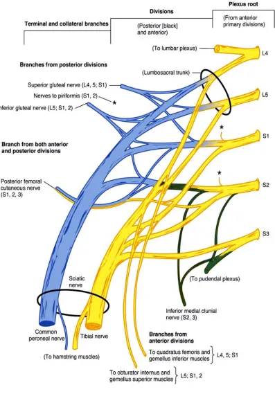
The nerve is formed by the convergence of the anterior primary rami of
spinal nerves L4, L5, S1, S2 and S3 on the anterior surface of piriformis muscle.
Sciatic nerve has two nerve components, Tibial nerve derived from anterior
(preaxial) division of the anterior primary rami, and Common peroneal nerve
derived from the posterior division (postaxial) division of anterior primary rami.
These two components are loosely bound together in the same connective tissue
2
3 COURSE OF SCIATIC NERVE
The Sciatic nerve leaves the pelvis through the greater sciatic foramen
below the piriformis and descends between greater trochanter and ischial
tuberosity along the back of thigh.
The Sciatic nerve divides into Tibial nerve and Common Peroneal Nerve at
a varying level proximal to the knee.
The Sciatic nerve enters the back of thigh at the lower border of Gluteus
maximus.
The Sciatic nerve runs vertically downwards and at the junction of the
middle and lower thirds of thigh, near the apex of the popliteal fossa, it terminates
4
5 RELATIONS
The Sciatic nerve lies deep to gluteus maximus, resting on the posterior ischial
surface with the nerve to quadratus femoris lying between them. The nerve then
crosses posterior to the obturator internus, the gemelli and the quadratus femoris,
separated by the latter from obturator externus and the hip joint. The nerve is
accompanied medially by the posterior femoral cutaneous nerve and the inferior
gluteal artery. More distally the Sciatic nerve lies behind adductor magnus and is
crossed posteriorly by the long head of biceps femoris58. The course of Sciatic nerve corresponds to a line drawn just medial to the midpoint between ischial
tuberosity and greater trochanter to the apex of the popliteal fossa.
BRANCHES OF SCIATIC NERVE
Articular branches arise proximally to supply the hip joint through its
posterior capsule.They may be sometimes derived directly from the sacral plexus.
Muscular branches are distributed to biceps femoris, semitendinosus,
semimembranosus and the ischial part of adductor magnus58.
The terminal branches of the Sciatic nerve are the Tibial nerve and
Common Peroneal Nerve. The point of division of Sciatic nerve into Tibial nerve
and the Common Peroneal Nerve is very variable. The common site is at the
junction of the middle and lower third of the thigh, near the apex of the popliteal
fossa. The division may occur at any level above this, though rarely below the
6
Sciatic nerve to leave the sacral plexus separately, in which case the common
peroneal component usually passes through the piriformis while the tibial
component passes below the piriformis muscle58. The Sciatic nerve supplies the flexors of the knee and all the muscles below the knee, through one of its terminal
branches, because of which, injury of Sciatic nerve results in a flail foot deformity
and severe difficulty in walking. A number of variations in the course and
distribution of Sciatic nerve have been reported.
The Sciatic nerve is vulnerable to injury in posterior dislocation of the hip. As the
Sciatic nerve leaves the pelvis it passes either behind the piriformis muscle or
sometimes through the piriformis muscle and at that point it may become
entrapped.This is called the piriformis syndrome, which is a common anatomical
variant but an extremely rare entrapment neuropathy.
External compression over the buttock which is common in patients who lie
immobile on hard surface for a considerable period of time can injure the Sciatic
nerve. Nevertheless, the most common cause of serious Sciatic nerve injury,and
consequent major medicolegal claims is iatrogenic. The Sciatic nerve may be
damaged in misplaced therapeutic injections into the gluteus maximus. The safe
zone for deep intramuscular injections here is the superolateral quadrant of the
buttock. Perhaps safer is to inject into the quadriceps femoris, though this can
produce problems of its own, like haemorrhage leading to contracture of the
7
Sciatic nerve palsy occurs following total hip replacement or similar
surgery in 1% of cases. It can be due to sharp injury from bone cement, traction
from instruments, manipulation of the hip, inadvertent lengthening of the femur,
or haematoma surrounding the nerve or within its soft tissue coverings. Complete
Sciatic nerve palsy is very rare phenomenon. For some reasons possibly
anatomical, the Common peroneal part of Sciatic nerve is more affected usually
alone. The patient will develop foot drop and a high stepping gait.
Several authors have reported variations in its terminal division into Tibial
nerve and Common Peroneal Nerves ranging from the sacral plexus to the
terminal division at the lower part of the popliteal fossa. These anatomical
variations of Sciatic nerve may contribute to the piriformis syndrome, sciatica,
coccygodynia and muscle atrophy. This should be considered by the clinicians
8
9 TIBIAL NERVE
The Tibial nerve, the larger terminal division of Sciatic nerve,is derived
from the anterior divisions L4,L5,S1,S2,S3 ventral rami.
The Tibial nerve ends under the flexor retinaculum by dividing into the medial
and lateral plantar nerves.
The branches of Tibial nerve are articular, muscular, sural, medial
calcaneal and medial and lateral plantar nerves.
Articular branches accompany the arteries to supply the knee joint. They
form a plexus with a branch from the obturator nerve and supply the oblique
posterior ligament. The branches accompanying the superior and inferior
genicular arteries also supply the oblique posterior ligament. The branches
accompanying the superior and inferior genicular arteries supply the medial part
of the capsule. Just before bifurcation the Tibial nerve supplies the ankle joint.
Proximal muscular branches arise between the heads of gastrocnemius and
supply the gastrocnemius, plantaris, soleus and popliteus. The nerve to popliteus
also supplies tibialis posterior, proximal tibiofibular joint and tibia. It gives off an
interosseous branch that descends near the fibula to reach the distal tibiofibular
10
Muscular branches in the leg, independently or by a common trunk, supply
soleus (on its deep surface), tibialis posterior, flexor digitorum longus and flexor
hallucis longus. The branch to flexor hallucis longus accompanies the peroneal
vessels.
THE MEDIAL AND LATERAL PLANTAR NERVES
These are derived from the Tibial nerve, under cover of the flexor
retinaculum.
The medial plantar nerve is larger than the lateral. It supplies fewer
muscles (though their bulk is considerable) than the lateral plantar nerve. The
nerve supplies abductor hallucis,flexor digitorium brevis,flexor hallucis brevis and
first lumbricals . In addition, it gives off digital cutaneous branches that supply the
medial three and half toes on their plantar surfaces and on their dorsal surfaces
proximal to the nail beds. Its most lateral cutaneous branch communicates with
the neighbouring lateral plantar digital branch across the plantar surface of the
fourth metatarsophalangeal joint, where pressure on the nerve may give rise to the
painful condition known as metatarsalgia.
The lateral plantar nerve gives off cutaneous branches to the sole that
perforate the plantar aponeurosis. It supplies flexor accessorius and abductor digiti
minimi and sends perforating branches through the plantar aponeurosis to supply
11
into superficial and deep branches. The superficial branch supplies the skin of
fourth cleft of foot and communicates with the medial plantar nerve and by a
lateral branch supplies the skin of the lateral side and distal dorsum of little toe. It
supplies three muscles, namely flexor digiti minimi brevis and the two interossei
of the fourth space (third plantar and fourth dorsal). The deep branch lies within
the concavity of the plantar arch and ends by sinking into the deep surface of the
oblique head of adductor hallucis. It gives off branches to the remaining
interossei, to the transverse head of adductor hallucis and to the three lateral
(bicipital) lumbricals. The branch to the second lumbrical passes dorsal to the
transverse head of adductor hallucis and recurves ventrally to enter the
lumbrical58.
COMMON PERONEAL NERVE
The Common Peroneal Nerve also known as common fibular nerve is
derived from the posterior divisions of L4,L5,S1,S2 ventral rami.
It curves lateral to the fibular neck, and divides into superficial peroneal
and deep peroneal nerves.
The Common Peroneal Nerve has articular and cutaneous branches. There
are three articular branches. Two of which accompany the superior, inferior and
lateral geniculate arteries and may arise in common. The third, the recurrent,
12
supplies the anterolateral part of the knee joint capsule and the proximal
tibiofibular joint58.
The two cutaneous branches, often from a common trunk are the lateral
sural and sural communicating nerves. The lateral sural nerve supplies the skin on
the anterior, posterior and lateral surfaces of the proximal leg.
The sural communicating nerve arises near the head of the fibula and
crosses the lateral head of gastrocnemius to join the sural nerve.
The deep peroneal nerve is the nerve of the anterior compartment of leg. It
is one of the two terminal branches of the common peroneal nerve. It also supplies
the intrinsic muscles and a small area of the skin of the dorsum of foot. A lesion
of this nerve results in an inability to dorsiflex the ankle.
The superficial peroneal nerve, is the nerve of lateral compartment of leg. After
supplying the Peroneus longus and Peroneus brevis, the superficial peroneal
nerve continues as a cutaneous nerve, supplying the skin on the distal part of the
13 SURAL NERVE
The sural nerve also called short saphenous nerve, is a sensory nerve
formed by the union of the medial sural cutaneous nerve and the peroneal
anastomotic branch of the lateral sural cutaneous nerve. It passes downward near
the lateral margin of the tendocalcaneus, lying close to the small saphenous vein.
It continues as the lateral dorsal cutaneous nerve along the lateral side of the foot
and little toe (via a dorsal digital nerve). On the dorsum of the foot the
intermediate dorsal cutaneous branch of superficial peroneal nerve communicates
with it. In the leg its branches communicate with those of the collateral branches
of the Tibial and Common Peroneal Nerves58.
HISTOLOGY
The Sciatic nerve has a whitish, homogenous, glistening appearance
because of the myelin and collagen content of the nerve sheath. Nerve fibres are
grouped in bundles.The nerve has an external fibrous coat of dense connective
tissue called epineurium, which also fills the spaces between the bundles of nerve
fibres. Each bundle is surrounded by perineurium, a sleeve formed by layers of
flattened epithelium like cells. The cells of each layer of the perineurium sleeve
are joined at the edges by tight junctions, an arrangement that makes the
perineurium a barrier to the passage of most macromolecules and has the
14
Within the perineurium sheath run the axons which are also sheathed by
Schwann cells and the developing connective tissue, endoneurium. The
endoneurium consists of a thin layer of reticular fibres, produced by Schwann
cells.
BLOOD SUPPLY OF SCIATIC NERVE
The Sciatic nerve is supplied by a branch from the inferior gluteal artery.
This artery represents the remnant of the original axis artery of lower limb.
15
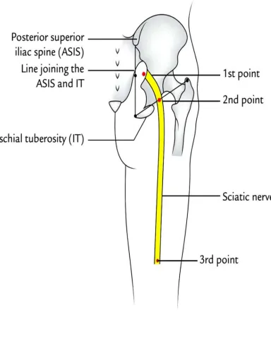
SURFACE MARKING OF SCIATIC NERVE
Draw a line joining the posterior superior iliac spine and ischial tuberosity.
Take a point about 2.5 centimetres lateral to the mid-point of that line, which
represents the entry of Sciatic nerve in the gluteal region14.
Take another point just medial to the mid-point of a line joining ischial tuberosity
to the apex of greater trochanter.
Put a point at the upper angle of popliteal fossa. Join these points by a broad line
which passes downwards and laterally. This broad line represents the Sciatic
16
17
AIM OF THE STUDY
The Sciatic nerve leaves the pelvis through the greater sciatic foramen
below piriformis and enters the gluteal region.The Sciatic nerve descends between
the greater trochanter and ischial tuberosity, along the back of thigh.The nerve
ends by dividing into the Tibial and Common Peroneal Nerves at a varying level
proximal to the knee58.
The gluteal region is a common site for intramuscular injection of drugs,
due to availability of large area for venous absorption of drugs. Intramuscular
injections, Wounds or surgery on the medial side may injure the Sciatic nerve and
its branches to the hamstrings. Paralysis of these muscles result in impairment of
thigh extension and leg flexion. Complications of improper technique include
nerve injury, hematoma, and abscess formation.So,intramuscular injections in the
gluteal region are safe in the supero lateral quadrant.
Sciatic nerve palsy is a known complication of total hip arthroplasty with
an incidence of 0.2% to 2.8% in the first time and 1.7% to 7.6% following
revision arthroplasty.
Sciatic nerve palsy can also result in severe spinal stenosis following the
18
Sciatic nerve exploration can be done by endoscopy as a minimal invasive
procedure to assess lesions of the Sciatic nerve. Endoscopic treatment for Sciatic
nerve entrapment has been implemented in deep gluteal syndrome.
Sciatica is a painful condition that is most commonly indicated by pain that
radiates through the leg. This can lead to muscle weakness, numbness or tingling
and significant leg pain. It is a common condition frequently caused by
degenerative spine lesion.
Piriformis syndrome characterised by entrapment of Sciatic nerve produces
leg pain without low back pain. The pain is worse during sitting and is
exacerbated by abduction and external rotation of the hip. It is characterised by
local tenderness in the buttock, no neurological signs, and normal imaging studies.
Sciatic nerve block or decompression is advocated for symptomatic management
of this condition.
The Sciatic nerve is accompanied by a companion artery (derived from the
inferior gluteal artery) which bleeds when the nerve is divided during an above
knee amputation.The artery must be neatly isolated and tied without any nerve
fibres being incorporated in the ligature, since this would be followed by severe
pain in the stump.Hence knowledge of Sciatic nerve length,width,course and
variations in terminal division is of great importance to surgeons, orthopaedicians,
anaesthetists and medical professionals in planning surgical interventions in the
19 PARAMETERS
1. The length of Sciatic nerve.
2. The width of Sciatic nerve at the level of inferior border of piriformis.
3. The width of Sciatic nerve between greater trochanter and ischial
tuberosity.
4. The distance between the mid-point of sacrotuberous ligament and Sciatic
nerve.
5. The course of Sciatic nerve in relation to piriformis.
6. Incidence of high division of Sciatic nerve.
20
REVIEW OF LITERATURE
1. LENGTH OF SCIATIC NERVE
Vicente et al63 (2007) observed that the mean length of Sciatic nerve was 7.63 cm
on the right side and 7.95 cm on the left side.
Prameela et al45 (2015) in their study on 15 cadaveric left sided lower limb
specimens stated that maximum length was 39 cm and minimum length was 2.5
cm.
Rajalakshmi et al47 (2015): study showed that the maximum length in their study
was 36.5 cm and the minimum length was 10.1 cm.
2. WIDTH OF SCIATIC NERVE AT THE LEVEL OF INFERIOR
BORDER OF PIRIFORMIS
Moore KL & Arthur F Dally32 (1982) The Sciatic nerve is the continuation of
the main part of the sacral plexus, arising from ventral rami of L4 to S3. The rami
converge at the inferior border of piriformis to form the Sciatic nerve, a thick,
21
Susan Standring Grays Anatomy 37th edition58 (1989) The Sciatic nerve is 2
cm broad at its origin and the broadest nerve in the body, and is the continuation
of the upper band of the sacral plexus. It leaves the pelvis via the greater sciatic
foramen below the piriformis.
Van de et al61 (2001) Sciatic nerve arises from L3 to S3. It leaves the pelvis
through the greater sciatic foramen distal to the piriformis and is broad (20mms),
descends lateral to the ischial tuberosity, then travels deep to the gluteus maximus
and long head of biceps femoris.
Vicente et al63 (2007) observed in their study that the Sciatic nerve thickness at
the level of inferior border of piriformis muscle was 18.85mm on the right side
and 22.32mm on the left side.
Marieb et al 29(2012) The Sciatic nerve leaves the pelvis via the greater sciatic
foramen, below the piriformis and is 20.10 mm wide and descends lateral to the
ischial tuberosity, and then travels deep to the gluteus maximus and long head of
22
3. WIDTH OF SCIATIC NERVE BETWEEN ISCHIAL TUBEROSITY
AND GREATER TROCHANTER.
Gabrielli et al19 (1994): stated that the average width of Sciatic nerve was 21.82
mm on the right side and 20.95 mm on the left side, at the level of ischial
tuberosity and greater trochanter.
Williams et al65(2005): observed that the width of Sciatic nerve at the level of
ischial tuberosity and greater trochanter was 20.0 mm. The average thickness was
found to be 26.46 mm on the right side and 29.68 mm on the left side.
Vicente et al63 (2007) in their study stated that the width of Sciatic nerve between
ischial tuberosity and greater trochanter was 18.85 mm on the right side and 22.32
mm on the left side.
Prameela et al45 (2015) stated that the mean width of Sciatic nerve between
ischial tuberosity and greater trochanter in their study was 10.6 mm on the right
side and 9.7 mm on the left side.
Rajalakshmi et al47 (2015): reported that the width of Sciatic nerve between
ischial tuberosity and greater trochanter was a maximum of 13.5 mm and
23
maximum width of 13 mm and minimum of 7.84 mm and the average of 9.7 mm
on the left side.
4. DISTANCE BETWEEN THE MIDPOINT OF SACROTUBEROUS
LIGAMENT AND SCIATIC NERVE.
Vicente et al63 (2007) in their study quoted that the mean distance observed
between the medial margin of the Sciatic nerve and the midpoint of the lateral
border of the Sacro tuberous ligament was 17.27 mm on the right and 17.83 mm
on the left side.
Joseph Bruno bidin brooks et al25 (2011) in their study quoted that the mean
distance between the medial margin of Sciatic nerve and the midpoint of the
lateral margin of Sacro tuberous ligament on the right side was 17.97mm and on
the left side was 18.42mm.
5. COURSE OF SCIATIC NERVE IN RELATION TO PIRIFORMIS
Beaton and Anson et al7 (1937) studied Sciatic nerve in 120 specimens and classified the same on the basis of the relation of Sciatic nerve to the piriformis
24
Classification of the relationship between piriformis and the Sciatic nerve (Beaton
and Anson)
Type A : Undivided nerve below undivided muscle
Type B : Division of nerve between and below undivided muscle
Type C : Divisions above and below undivided muscle
Type D : Undivided nerve between heads
Type E : Divisions between and above head
Type F : Undivided nerve above undivided muscle
Beaton and Anson et al7 (1937): studied 120 cadavers and reported that 84.2%
were Type A, 11.7% were Type B, 3.3% were Type C, and 0.8% Type D.
Pan et al38 (1941) studied 140 specimens of Chinese population and reported 92
(65.7%) was type A and 46 (32.9%) was type B and 2 (1.4%) was type D.
Misra et al31 (1954) in Indian population studied on 300 specimens and reported
262 (87.4%) were type A ,18 (6%) were type B, 5(2.1%) were type C and 2
(0.8%) were type D and 13(3.7%) were type E.
Kubota et al26 (1960) studied 38 specimens in Japanese population and observed
25
Nizankowski et al36 (1972) observed in 200 specimens in Poland population of
which 110 were male and 90 were female of which 157 (70.2%) was type A and
33 (19.6%) was type B, 7 (4.2%) was Type C and 3 (1.8%) Type D.
Pecina et al42 (1979) reported that of the 130 cadavers in about 22% Sciatic nerve
passed through the piriformis, and piriformis was pierced by common peroneal
nerve in 17% of the specimens.
Chiba et al12 (1994) study observed that common peroneal nerve passing through
the piriformis was seen in 34% of cases in 514 extremities.
Chen et al11 (1994) found an extremely rare variation which was type 4 in seven
cases in a total of 57 cases (12.2%).
Sayson et al52 (1994) reported 1 case of type 6 variation in routine cadaveric
dissection. Sciatic nerve lying posterior to the piriformis in a woman suspected
with piriformis syndrome.
Gabrielli et al19 (1997) in brazil conducted a study on 80 cadavers of which 69
(86.3%) type A, 9 (11.2%) type B, 2 (2.5%) was type C in his study.
Arifoglu et al3 (1997) reported one case of type B variation (2.32%) in his study
26
Moore et al32 (1999) study has reported a variation that he found common
peroneal nerve passing through the piriformis and tibial nerve passing below
piriformis in 12.2% of the specimens.
Vloka et al64 (2001) conducted a study in New York and studied the level of
division of the Sciatic nerve in the popliteal fossa and its relationship to the
common epineural sheath of the Sciatic nerve. The level of division of the Sciatic
nerve into the tibial and common peroneal nerve above the knee was measured in
28 cadavers. The Sciatic nerve was invariably formed of independent trunks
(Tibial Nerve and Common peroneal Nerve) encompassed in one common
epineural sheath. The Sciatic nerve divided at a mean distance of 60.5 +/-27.0 mm
above the popliteal fossa crease. The conclusion was that the Tibial Nerve and the
Common Peroneal Nerve leave the common epineural sheath at variable distances
from the popliteal crease. This finding and relationship of the Tibial Nerve and
Common Peroneal Nerve sheaths may have significant implications in popliteal
block. When performing popliteal block, insertion of needle at 100 mm above the
popliteal crease is more likely to result in placement of the needle proximal to the
division of the Sciatic nerve rather than placement at 50 or 70 mm according to
27
Babinski4 and Mas30 et al (2003) reported the passage of Common Peroneal
Nerve through the piriformis, and the passage of Tibial Nerve under the superior
gemellus is also a rare variation.
Machado et al27 (2003) observed gluteal regions in 100 cadaveric lower limbs
and reported three types of variations,
Type A – The common peroneal nerve piercing the piriformis and tibial nerve
passing below piriformis.
Type B – common peroneal nerve passing above piriformis and tibial nerve
below piriformis.
Type C – Sciatic nerve piercing the piriformis muscle.
Ugrenovic et al59 (2005) studied gluteal regions in 200 cadaveric lower limbs and
observed that 96% of Sciatic nerve was leaving the pelvis below piriformis. The
Common Peroneal Nerve has been seen passing below the piriformis in 2.5% of
specimens and the Common Peroneal Nerve passing above piriformis and Tibial
nerve below piriformis in 1.5% cadavers.
Pokorny et al43(2006) studied 91 cadavers and found individual variations in 19
cadavers. According to them Sciatic nerve exits below the piriformis muscle in
79.1% cases. Sciatic nerve separates into divisions 1 branch passing through the
piriformis muscle and the other below it in (14.3%). An unsplit nerve passes
28
Guvencer et al20 (2009) conducted a study in 50 cadaveric gluteal regions of
Turkish population. The results were in 52% of the cases, the Sciatic nerve exited
the pelvis as a whole nerve without any division, whereas in 48% high division
was observed. Branches of the Sciatic nerve left the pelvis below the piriformis
muscle as two separate nerves.
Prakash et al44 (2010) conducted a study in India and observed that the Sciatic
nerve division was observed in the lower part of the posterior compartment of
thigh in 40.7%. In 43.0% percent of the specimens, the Sciatic nerve divided into
tibial and common peroneal nerves in the popliteal fossa and 16.3 percent of
extremities showed Sciatic nerve division proximal to its entrance in the gluteal
region.
Shailesh M Patel et al53 (2011) study was conducted in 86 gluteal regions, and
observed 94.2% showed normal variation and 5.8% showed higher division of
Sciatic nerve bilaterally. The differences in exit routes of these two nerves are
important for surgeons, as this is the area of frequent surgical manipulation, nerve
injury during deep intramuscular injection of the gluteal region, failed Sciatic
nerve block in anaesthesia and injury during posterior hip operations.
Manisha.B. Sinha et al28 (2013) studied morphological variations of Sciatic
29
in 40 foetuses, that is 35 (80%) of the limbs. Sciatic nerve emerged below
piriformis muscle and in 5 foetuses, that is (12.5%) of the limbs
Sciatic nerve divided into common peroneal nerve and tibial nerve . Common
peroneal component passed through the piriformis resulting in splitting of the
piriformis, where as the tibial nerve passed below the piriformis in the other 3
foetuses (7.5%). The common peroneal component passed above the muscle and
the tibial component passed below the muscle.
A.D.Shewale et al56 (2013) study observed that the Sciatic nerve exited the pelvis
through the greater sciatic foramen below piriformis as a single nerve without
division in 66 of the 90 gluteal regions(73.3%) and 34 specimens (26.7%)showed
variations.
Natsis et al34 (2014) studied in 147 Caucasian cadavers (294) limbs the result
was that, the Sciatic nerve and piriformis muscle relationship followed the typical
anatomical pattern in 275 limbs (93.6%) and variations was observed in 19 limbs
(6.4%).
Hadbaa ghazi Hussain et al21 (2014) conducted a study in 50 dissected gluteal
and thigh regions and found that 92% of Sciatic nerve pass below the piriformis
muscle, and 8% of Sciatic nerve divide inside the pelvis. The site of division of
30
upper third of the thigh,1% in the middle third of thigh, 1.3% in the superior angle
of popliteal fossa and 1.2% at the centre of popliteal fossa.
6.INCIDENCE OF HIGH DIVISION OF SCIATIC NERVE
Ewa Okraszewska et al16 (2002) stated in a study conducted in Polish population
that three levels of Sciatic nerve division into terminal branches were observed:
1. High (pelvic) level 5 cases (13.89%) – Sciatic nerve divides inside lesser
pelvis or just below piriformis muscle.
2. Intermediate Level 5 cases (13.89%) – Sciatic nerve divides at lower 2/3 of
femur.
3. Low (popliteal) level 26 cases (72.22%) – Sciatic nerve divides in popliteal
fossa.
Ugrenovic et al59 (2005) have found high division of Sciatic nerve in 27.5% of
the specimens in a study performed in 100 foetuses.
Jigna Parmar et al 24(2005) studied 12 lower limbs and found that normal
division of Sciatic nerve at the apex of popliteal fossa was observed in five lower
limbs (41.67%) and variations was observed in 7 lower limbs 58.33% .
Guvencer et al20 (2009) studied 50 gluteal regions in 25 cadavers and observed
31
Prakash et al44 (2010) observed high division of Sciatic nerve in 16.3%
specimens.
A D Shewale et al56 (2013) conducted a cadaveric study in 105 specimens study
stated that Division of Sciatic nerve can occur in gluteal region in 24.44% and
75.56 % near the superior angle of popliteal fossa.
Rashmi C Koshi et al48(2015) in a case report observed that Sciatic nerve divided
even before entering the pelvic fossa into the Tibial Nerve and common peroneal
nerve and pierces the piriformis muscle dividing the muscle into upper and lower
slips of fibres. Tibial nerve reached the gluteal region by passing between the
lower slip of piriformis muscle and the gemellus superior muscle. These two
nerves remained separately in their entire course. The high division results in
Sciatic nerve injury during deep intramuscular injection in gluteal regions and
piriformis syndrome.
Birhane alem berihu et al10 (2015) conducted a study in 28 formalin fixed
cadavers comprising of 56 lower limbs, among them 42 lower limbs(75%)
showed a normal anatomy of the Sciatic nerve and 14 lower limbs (25%) showed
variations of Sciatic nerve ,of the 25% present 11% showed variations in relation
to piriformis, in 9% the Sciatic nerve emerged as separate components below the
32
piriformis and the tibial component emerges below the piriformis and descends
separately along their course.
Saravanan et al50 (2016) studied 18 embalmed cadavers and dissected 36 gluteal
regions and observed undivided Sciatic nerve emerging below the piriformis in 34
specimens (94.5%) and high division in 2 cadavers (5.5%). In both the specimens,
the division of Sciatic nerve was within the pelvis, the common peroneal nerve
was found passing through the fibres of piriformis and the tibial nerve passed
below the piriformis.
7. INCIDENCE OF TRIFURCATION OF SCIATIC NERVE
Satheesha nayak et al35 (2010) in their study observed a type of trifurcation of
Sciatic nerve. The Sciatic nerve gave an abnormal trunk in addition to the tibial
and common peroneal nerve. The abnormal trunk divided into lateral cutaneous
nerve of the calf and the peroneal communicating nerve. Since the point of
trifurcation was in the middle of popliteal fossa, it is of clinical and surgical
importance.
Sharad Kumar Pralhad Sawant et al54 (2013) during routine dissection for
MBBS students observed an unusual trifurcation of the Sciatic nerve on the back
of both the thighs in the middle of the popliteal fossa of a 70-year-old. The Sciatic
33
three branches given were tibial, superficial and deep peroneal nerves. The
superficial peroneal nerve was as thick as the deep peroneal nerve. There is high
division of superficial peroneal nerve into medial and lateral branches.
Malliga Arjun Adibatti et al1 (2014) in their study of 25 cadavers observed for
variation in the course and division of Sciatic nerve, while in 4 specimens (8%)
there was high division of Sciatic nerve within the pelvis. While in 2 specimens
(4%) trifurcation of Sciatic nerve was observed with an additional muscular
branch to soleus. In one limb the Sciatic nerve emerged above the piriformis into
the tibial and common peroneal later reunited at the back of thigh and re split
normally at the superior angle of the popliteal fossa thus having a varied course.
Birhane alem berihu et al10 (2015) conducted a study on 28 formalin fixed
cadavers comprising of 56 lower limbs. Among them 75% showed normal
anatomy and 25% showed variations in the Sciatic nerve, and the variation was
trifurcation of Sciatic nerve into three major divisions tibial nerve, common
peroneal nerve, and an unusual trunk) in the middle of the popliteal fossa in 11%.
The unusual trunk divided into the lateral cutaneous nerve of the calf and peroneal
communicating nerve. Additionally, another lower limb showed trifurcation of
Sciatic nerve into tibial, superficial and deep peroneal 9% and the other limb
34
Anbumani . T. L et al2 (2015) conducted a study on 50 lower limbs of which 41
specimens (82%) showed normal anatomy of Sciatic nerve, 9 specimens (18%)
showed variations in the Sciatic nerve and stated that trifurcation of Sciatic nerve
is a rare entity and in his study one specimen (2%) showed trifurcation of Sciatic
35
EMBRYOLOGY
Lateral plate mesoderm migrates and condenses along the central axis of
the lower limb buds to eventually form the vasculature and skeletal components of
lower limbs. Mesoderm from somites migrates into the lower limb buds and
condenses to eventually form the musculature of the lower limbs15.
Apical ectodermal ridge is a ridge of thickened ectoderm at the apex of
each limb bud. The ridge produces fibroblast growth factor, which interacts with
the underlying mesoderm to promote the growth of the limb by stimulating
mitosis and preventing terminal differentiation of the underlying mesoderm. The
Apical ectodermal ridge expresses the WNT7 gene, which directs the organisation
of the limb bud along the dorsoventral axis15.
Zone of polarising activity consists of mesodermal cells located at the base
of the limb bud. It contains Sonic Hedgehog which directs the organization of
limb bud along the antero-posterior axis and patterning of digits.
Sonic Hedgehog activates the gene for Bone Morphogenetic Protein and
the Hoxd - 9, Hoxd - 10, Hoxd – 11, Hoxd – 12 and Hoxd - 13 genes. Retinoic
36
Digit formation occurs as a result of selected apoptosis within the Apical
Ectodermal Ridge such that five separate regions remain at the tip of the future
digits.
NERVES: THE LUMBOSACRAL PLEXUS
Local cell biological messages produced at the base of the limb bud guide
the early nerve fibres into the limb bud. The muscles do not provide any specific
target messages to the in growing nerve fibres.
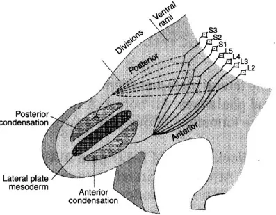
Ventral primary rami of L2 -L5 and S1-S3 arrive at the base of the limb
bud and divide into anterior and posterior divisions. Posterior divisions grow into
the posterior condensation of mesoderm. Anterior divisions grow into the anterior
condensation of mesoderm.
With further development of limb musculature, the posterior divisions
branch into Superior gluteal nerve (L4, L5, S1), Inferior gluteal nerve (L5, S1,
S2), Femoral nerve (L2, L3, L4) and the Common peroneal component of Sciatic
nerve (L4, L5, S1, S2), there by innervating all the muscles that develop from the
37
With the further development of limb musculature, the anterior divisions
branch into Tibial component of Sciatic nerve (L4, L5, S1, S2, S3) and Obturator
nerve (L2, L3, L4), there by innervating all the muscles that form from the
[image:50.595.111.508.314.623.2]anterior condensation of mesoderm15.
SCIATIC NERVE
GMX
SN
PF
TN
CPN
GMX
- Gluteus Maximus
PF
- Piriformis
SN
- Sciatic Nerve
TN
- Tibial Nerve
38
MATERIALS AND METHODS
STUDY MATERIALS
50 Embalmed human adult lower limb specimens
METHODS OF STUDY
Conventional dissection method.
SPECIMEN COLLECTION
Adult lower limb specimens were obtained from the embalmed cadavers
allotted for routine dissection to the first year MBBS and BDS students at the
Institute of Anatomy, Madras Medical College, Chennai.
CONVENTIONAL DISSECTION METHOD
Sciatic nerve was dissected as per the dissection steps given in
Cunningham’s textbook of anatomy. The iliac crest was traced forwards upto the
anterior superior iliac spine and backwards to the posterior superior iliac spine.
The latter lies at the level of second sacral spine and the middle of the Sacro-iliac
joint. A skin incision was made and the flap of skin and superficial fascia was
reflected laterally, cutaneous nerves which enter the gluteal region were looked
for. Two fingers were passed deep to the lower edge of the gluteus maximus
muscle 2 -3 cm medial to its femoral insertion and the muscle was cut between the
fingers to the upper border superior to the greater trochanter. The lateral part of
the muscle was reflected upto its insertion, the bursae was identified which
39
to the gluteal tuberosity. The medial part of the muscle was reflected. The deep
surface of the muscle was watched carefully to avoid injury to the posterior
cutaneous nerve of the thigh. The inferior gluteal vessels and the nerve was found
entering the lower part of the muscle and the superficial parts of the superior
gluteal vessels entering its upper part, as the ischial tuberosity was uncovered the
bursae was superficial to the origin of the hamstring muscles from the tuberosity.
The muscle was detached from the surface of the rigid Sacro tuberous ligament,
and the perforating cutaneous nerves were found.The relation between the Sciatic
nerve and piriformis muscle was noted13.
The posterior cutaneous nerve of the thigh was found posterior to the
Sciatic nerve. The fascia surrounding the Sciatic nerve was carefully dissected .
The nerve was traced upwards to the lower border of piriformis and downwards to
the point where its branches to the hamstrings muscle arises, near the level of
ischial tuberosity. The fascia was removed from the muscles anterior to the Sciatic
nerve, from above downwards these are the tendon of obturator internus
overlapped by the gemelli. The quadratus femoris was passing from the ischial
tuberosity to the back of the femur. Inferior to the quadratus femoris the posterior
surface of adductor magnus, branches of the medial circumflex femoral artery
appeared both above and below the quadratus femoris. The Sciatic nerve was
traced till the point of termination into Tibial nerve and Common Peroneal Nerve
40
The following parameters were noted
1. THE LENGTH OF SCIATIC NERVE: Point A was defined, where the
Sciatic nerve appeared in the gluteal region. Point B was defined at the
terminal division of Sciatic nerve. The distance between point A and point
B was measured using a measuring tape. This is the length of Sciatic nerve.
2. THE WIDTH OF SCIATIC NERVE AT THE LEVEL OF INFERIOR
BORDER OF PIRIFORMIS : The Sciatic nerve was measured at the
inferior border of piriformis muscle using Vernier calipers and measuring
tape.
3. THE WIDTH OF SCIATIC NERVE BETWEEN GREATER
TROCHANTER AND ISCHIAL TUBEROSITY : A line was drawn
connecting the ischial tuberosity and greater trochanter. At the point where
the Sciatic nerve intersects the line, the width of Sciatic nerve was
measured using Vernier calipers.
4. THE DISTANCE BETWEEN THE MID-POINT OF SACROTUBEROUS
LIGAMENT AND SCIATIC NERVE:The mid point of sacrotuberous
ligament was taken as the reference point. The distance between the lateral
border of the sacro tuberous ligament and the medial margin of the Sciatic
41
5. THE COURSE OF SCIATIC NERVE IN RELATION TO PIRIFORMIS:
The relation of Sciatic nerve or its Tibial and Common peroneal
components to the piriformis muscle was noted.
6. INCIDENCE OF HIGH DIVISION OF SCIATIC NERVE:
Division of Sciatic nerve above the level of apex of popliteal fossa was
considered as high division.
7. INCIDENCE OF TRIFURCATION OF SCIATIC NERVE:
LENGTH OF SCIATIC NERVE
GMX
- Gluteus Maximus
PF
- Piriformis
SN
- Sciatic Nerve
ST
- Semitendinosus
BF
- Biceps Femoris
PF
SN
ST
42
OBSERVATION
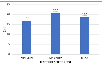
1.LENGTH OF SCIATIC NERVE
The minimum length of Sciatic nerve was 16.8 cm, the maximum length of
[image:59.595.95.524.420.695.2]Sciatic nerve was 20.6 cm and the mean length was 18.6 cm.
TABLE NO 1: LENGTH OF SCIATIC NERVE
LENGTH VALUES (cm)
MINIMUM 16.8
MAXIMUM 20.6
MEAN 18.6
CHART NO 1: LENGTH OF SCIATIC NERVE
16.8 20.6 18.6 0 5 10 15 20 25
MINIMUM MAXIMUM MEAN
(c
m
)
WIDTH OF SCIATIC NERVE AT THE LEVEL OF
INFERIOR BORDER OF PIRIFORMIS
GT
SN
IT
GMX
- Gluteus Maximus
SN
- Sciatic Nerve
IT
- Ischial Tuberosity
GT
- Greater Trochanter
43
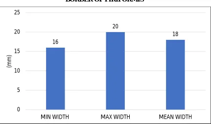
2. WIDTH OF SCIATIC NERVE AT THE LEVEL OF INFERIOR BORDER OF PIRIFORMIS
The width of Sciatic nerve was measured using standard Vernier calipers,
the results are given in millimetres.
In the present study the maximum width just below the piriformis was 20
[image:61.595.95.526.487.742.2]mm and the minimum width was 16 mm with an average of 18 mm.
TABLE 2: WIDTH OF SCIATIC NERVE AT THE LEVEL OF INFERIOR BORDER OF PIRIFORMIS
WIDTH AT INFERIOR BORDER OF PIRIFORMIS VALUE (mm)
MINIMUM 16
MAXIMUM 20
MEAN 18
CHART 2: WIDTH OF SCIATIC NERVE AT THE LEVEL OF INFERIOR BORDER OF PIRIFORMIS
16 20 18 0 5 10 15 20 25
MIN WIDTH MAX WIDTH MEAN WIDTH
WIDTH OF SCIATIC NERVE BETWEEN
GREATER TROCHANTER AND ISCHIAL TUBEROSITY
IT
SN
GT
STL
PF
STL
- Sacro Tuberous Ligament
PF
- Piriformis
44
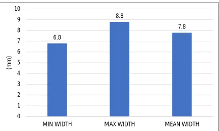
3. WIDTH OF SCIATIC NERVE BETWEEN GREATER TROCHANTER AND THE ISCHIAL TUBEROSITY
In the present study maximum width of Sciatic nerve between the greater
trochanter and ischial tuberosity was 8.8 mm and the minimum width was
[image:63.595.96.526.481.738.2]observed to be 6.8 mm, and the average width was 7.8 mm.
TABLE 3: WIDTH OF SCIATIC NERVE BETWEEN GREATER TROCHANTER AND THE ISCHIAL TUBEROSITY.
WIDTH BETWEEN GREATER TROCHANTER AND
ISCHIAL TUBEROSITY VALUE (mm)
MINIMUM 6.8
MAXIMUM 8.8
MEAN 7.8
CHART 3: WIDTH BETWEEN GREATER TROCHANTER AND THE ISCHIAL TUBEROSITY 6.8 8.8 7.8 0 1 2 3 4 5 6 7 8 9 10
MIN WIDTH MAX WIDTH MEAN WIDTH
DISTANCE BETWEEN THE MID POINT OF
SACROTUBEROUS LIGAMENT AND SCIATIC NERVE
PF
SN
STL
STL
- Sacro Tuberous Ligament
PF
- Piriformis
45
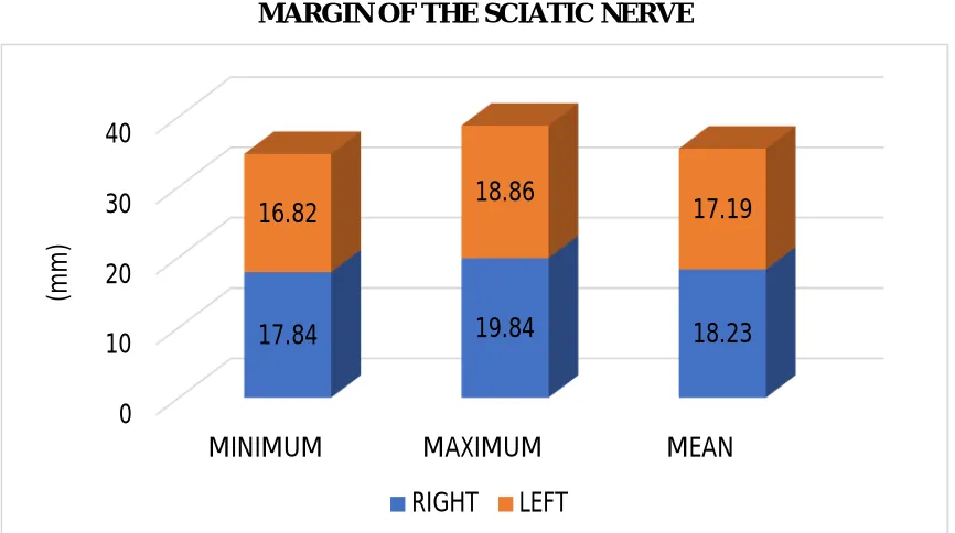
4. DISTANCE BETWEEN THE LATERAL MARGIN OF THE SACROTUBEROUS LIGAMENT AND THE MEDIAL MARGIN OF THE SCIATIC NERVE.
In the present study the mean distance between the lateral border of Sacro
tuberous ligament and the medial margin of Sciatic nerve was measured and
[image:65.595.93.526.507.749.2]found to be 18.23mm on the right side and 17.19mm on the left side.
TABLE 4: DISTANCE BETWEEN THE LATERAL MARGIN OF THE SACROTUBEROUS LIGAMENT AND THE MEDIAL MARGIN OF THE
SCIATIC NERVE
DISTANCE RIGHT SIDE(MM) LEFTSIDE(MM)
MINIMUM 17.84 16.82
MAXIMUM 19.84 18.86
MEAN 18.23 17.19
CHART 4: DISTANCE BETWEEN THE LATERAL MARGIN OF THE SACROTUBEROUS LIGAMENT AND THE MEDIAL
MARGIN OF THE SCIATIC NERVE
0 10 20 30 40
MINIMUM MAXIMUM MEAN
17.84 19.84 18.23
16.82 18.86 17.19
(mm
)
COURSE OF SCIATIC NERVE IN RELATION
TO PIRIFORMIS
PF
SG
IG
QF
GMX
SN
GMX
- Gluteus Maximus
PF
- Piriformis
46
5.COURSE OF SCIATIC NERVE IN RELATION TO PIRIFORMIS
In the present study, out of fifty specimens, forty-nine specimens showed
normal course of Sciatic nerve and in one specimen the Sciatic nerve showed
high division of Sciatic nerve that is nerve emerged as tibial component and
common peroneal component below the piriformis and descended separately
through their entire course as Tibial nerve and Common peroneal nerve.There was
no other variation observed in the course of Sciatic nerve.
CHART 5: COURSE OF SCIATIC NERVE IN RELATION TO PIRIFORMIS
In one specimen the Sciatic nerve showed high division of Sciatic nerve
that is nerve emerged as tibial component and common peroneal component
below the piriformis descended separately through their entire course as Tibial
nerve and Common peroneal nerve. to supply their regions.
HIGH DIVISION OF SCIATIC NERVE
GMX
- Gluteus Maximus
PF
- Piriformis
CPN
- Common Peroneal Nerve
TN
- Tibial Nerve
47
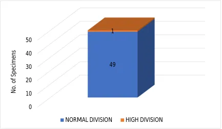
TABLE6: INCIDENCE OF HIGH DIVISION OF SCIATIC NERVE
In the present study of the fifty specimens, forty-nine specimens showed
normal pattern of terminal division and one specimen showed high division of
Sciatic nerve the tibial nerve and common peroneal nerve emerged separately
below the piriformis.
CHART 6: INCIDENCE OF HIGH DIVISION OF SCIATIC NERVE
0 10 20 30 40 50 49 1 N o . o f Sp e ci m e n s
NORMAL DIVISION HIGH DIVISION INCIDENCE OF
HIGH DIVISION
TOTAL NO. OF
SPECIMENS PERCENTAGE (%)
48
CHART 6: INCIDENCE OF HIGH DIVISION OF SCIATIC NERVE IN
PERCENTAGE
98% 2%
TRIFUCATION OF SCIATIC NERVE
SN
TN
LCN
CPN
SN
- Sciatic Nerve
TN
- Tibial Nerve
49
7. INCIDENCE OF TRIFURCATION OF SCIATIC NERVE
Sciatic nerve trifurcation was observed in 4 lower limbs. In the present
study the Sciatic nerve terminated bilaterally in the superior angle of popliteal
fossa in 2 lower limbs by giving three branches, which were (Tibial nerve,
common peroneal nerve and unusual trunk). The unusual trunk was traced and
found to supply the skin of the calf and hence named as lateral cutaneous nerve of
the calf. In the other 2 lower limbs bilateral trifurcation of Sciatic nerve in the
middle of popliteal fossa into the Tibial nerve, Common peroneal nerve and sural
[image:72.595.90.529.468.757.2]nerve was observed.
TABLE 7: INCIDENCE OF TRIFURCATION OF SCIATIC NERVE
TOTAL NO OF SPECIMENS
INCIDENCE OF
TRIFURCATION PERCENTAGE
50 2 4
CHART 7: INCIDENCE OF TRIFURCATION OF SCIATIC NERVE
0 10 20 30 40 50 NORMAL TERMINATION 48 2 N o . o f Sp e ci me n s
50
CHART 7: INCIDENCE OF TRIFURCATION OF SCIATIC NERVE IN
PERCENTAGE
In the present study Trifurcation of Sciatic nerve in one specimen (2%)
terminated as Tibial nerve, Common peroneal nerve and lateral cutaneous nerve
of calf, and in another specimen (2%) the Sciatic nerve terminated as Tibial nerve,
common peroneal nerve and sural nerve.
4%
96%
51
In the present study, out of fifty specimens, forty-seven specimens showed
normal pattern of termination.
In one specimen, Incidence of High division of Sciatic nerve was observed
In two specimen, Incidence of Trifurcation of Sciatic nerve was observed
THE VARIATIONS OF SCIATIC NERVE OBSERVED IN THE PRESENT STUDY IN PERCENTAGE
Termination of Sciatic nerve Variations of Sciatic nerve Percentage (%)
Normal Termination of Sciatic nerve 47 94
High Division of Sciatic nerve 1 2
Trifurcation of Sciatic nerve 2 4
52
VARIATIONS IN THE PRESENT STUDY
VARIATIONS IN THE PRESENT STUDY IN PERCENTAGE 47
1 2
94%
2% 4%
0 20 40 60 80 100
NO VARIATIONS HIGH DIVISION TRIFURCATION
P
e
rc
e
n
tag
e
NO VARIATIONS PERCENTAGE
94%
2% 4%
53
DISCUSSION
1. LENGTH OF SCIATIC NERVE
Prameela et al45 (2015) in her study on 15 cadaveric lower limb specimens
stated that maximum length was 39 cm , minimum length was 2.5 cm with an
average length of 23.5 cm .
Rajalakshmi et al47 (2015): study showed that the maximum length of Sciatic
nerve was 36.5 cm and the minimum length 10.1 cm with an average of 25.2 cm.
In the Present study the maximum length was 20.6 cm and the minimum length
[image:77.595.86.530.513.673.2]was observed to be 16.8 cm and the mean length was 18.6 cm.
TABLE NO 1: LENGTH OF SCIATIC NERVE
S.NO STUDY
MAX LENGTH (cm) MIN LENGTH (cm) MEAN LENGTH (cm)
1 Prameela et al 39.0 2.5 23.5
2 Rajalakshmi et al 36.5 10.1 25.2
3 Present study 20.6 16.8 18.6
54
CHART NO 1: LENGTH OF SCIATIC NERVE
The knowledge about the length of Sciatic nerve from its origin to
termination will be useful for surgeons, anaesthetists and orthopaedicians. The
understanding of normal anatomy of gluteal region and Sciatic nerve is essential
for professionals to be cautious and plan various surgical interventions in this
region.eg: Sciatic nerve block,total hip arthroplasty,posterior hip surgeries,Sciatic
nerve decompression,and during deep intramuscular gluteal injections.
23.5 25.2 18.6 0 5 10 15 20 25 30 35 40 45 50
Prameela et al Rajalakshmi et al Present study
(c
m
)
55
2. WIDTH OF SCIATIC NERVE AT THE LEVEL OF INFERIOR
BORDER OF PIRIFORMIS
Van de et al61 (2001) stated that Sciatic nerve leaves the pelvis through the
greater sciatic foramen distal to the piriformis and is broad (20mm), descends
lateral to the ischial tuberosity, then travels deep to the gluteus maximus and long
head of biceps femoris.
Vicente et al63 (2007) observed in their study that the Sciatic nerve thickness at
the level of inferior border of piriformis muscle was 18.85mm on the right side
and 22.32mm on the left side.
Marieb et al29 (2012) stated that the Sciatic nerve leaves the pelvis via the greater
sciatic foramen, below the piriformis and is 20.10 mm wide and descends lateral
to the ischial tuberosity, and then travels deep to the gluteus maximus and long
head of biceps femoris.
In the Present study the mean width of Sciatic nerve at the level of inferior
56
TABLE2 :WIDTH OF SCIATIC NERVE AT THE LEVEL OF INFERIOR BORDER OF PIRIFORMIS
NAME OF STUDY MEAN WIDTH(mm)
Van de et al (2001) 20.00
Marieb et al (2012) 20.10
Present study 18.00
CHART2:WIDTH OF SCIATIC NERVE AT THE LEVEL OF INFERIOR BORDER OF PIRIFORMIS
Sciatic nerve block is commonly used for lower limb surgeries.There are
several methods and approaches and hence knowledge about the dimensions and
course of Sciatic nerve in the gluteal region is very important. Previous studies
have suggested that the local anaesthetic injected into the subgluteal space under
ultrasound guidance is effective in producing Sciatic nerve block,therefore the
surface marking for the topographic relation of Sciatic nerve becomes important.
20 20.1 18 0 5 10 15 20 25
Van de et al Marieb et al Present study
(m
m)
57
3. WIDTH OF SCIATIC NERVE BETWEEN
GREATER TROCHANTER AND ISCHIAL TUBEROSITY
Gabrielli et al19 (1994): stated that the average width of Sciatic nerve was 21.82
mm on the right side and 20.95 mm on the left side, between ischial tuberosity
and greater trochanter.
Williams et al65 (2005): observed that the width of Sciatic nerve at the level of
ischial tuberosity and greater trochanter was 20.0 mm of Sciatic nerve. The
average thickness was found to be 26.46 mm on the right side and 29.68 mm on
the left side.
Vicente et al63 (2007) in his study stated that the width of Sciatic nerve was 18.85
mm on the right side and 22.32 mm on the left side.
Prameela et al45 (2015) stated that the mean width of Sciatic nerve in their study
was 10.6 mm on the right side and 9.7 mm on the left side.
In the present study the maximum width was 9.8mm and minimum width was 7.8
mm with an average width of 8.8mm on right side and the maximum width on
the left side was 8.8mm and minimum width was 6.8mm with an the average
58
TABLE 3: WIDTH OF SCIATIC NERVE BETWEEN GREATER TROCHANTER AND ISCHIAL TUBEROSITY.
STUDY MEANWIDTH (mm) RIGHT
MEANWIDTH (mm) LEFT
Prameela et al(2015) 10.6 9.7
Present study 8.8 7.8
CHART 3: WIDTH OF SCIATIC NERVE BETWEEN ISCHIAL TUBEROSITY AND GREATER TROCHANTER.
10.6 8.8 9.7 7.8 0 2 4 6 8 10 12
Prameela et al Present study
(m
m
)
59
The width of Sciatic nerve is important as knowledge about it will help in
preventing iatrogenic, Sciatic nerve injury during deep intramuscular injections
and Sciatic nerve blocks,The posterior approach to the Sciatic nerve combined
with a lumbar plexus block provides a complete anaesthesia of the Lower
extremity.The different approaches to Sciatic nerve are the Raj technique in which
the hip and knee are both flexed at 90 degree.Flexion of the hip in this
way,flattens the gluteal muscles and the Sciatic nerve becomes superficial.The
greater trochanter and ischial tuberosity are palpated and a line is drawn
connecting them.A 10 cm needle is inserted at the midpoint of this line,at
60
4. DISTANCE BETWEEN THE MIDPOINT OF SACROTUBEROUS
LIGAMENT AND SCIATIC NERVE.
Vicente et al63 (2007) in their study quoted that the mean distance observed
between the midpoint of the Sacro tuberous ligament and the medial margin of
Sciatic nerve was 17.27 mm on the right side and 17.83 mm on the left side.
Joseph Bruno Bidin Brooks et al25 (2011) stated that the mean distance between
the midpoint of sacrotuberous ligament and the medial margin of Sciatic nerve on
the right side was 17.97 mm and on the left side was 18.42 mm.
In the present study the mean distance between the medial margin of Sciatic
nerve and midpoint of sacrotuberous ligament on the right side was 18.23 mm and
[image:84.595.88.529.565.688.2]left side was 17.19 mm.
TABLE 4:DISTANCE BETWEEN THE MIDPOINT OF SACROTUBEROUS LIGAMENT AND SCIATIC NERVE
S.NO STUDY RIGHT SIDE
(mm)
LEFT SIDE (mm)
1 Vicente et al (2007) 17.27 17.83
2 Joseph bruno et al (2011) 17.97 18.42
61
CHART 4:DISTANCE BETWEEN THE MIDPOINT OF SACROTUBEROUS LIGAMENT AND SCIATIC NERVE
The knowledge of the distance of the Sciatic nerve from sacrotuberous
ligament is very helpful to avoid complications during intramuscular injections
and Sciatic nerve block. The gluteal region is a common site for intramuscular
injection of drugs, due to availability of large area for venous absorption of drugs.
Intramuscular injections, Wounds or surgery on the medial side may injure the
Sciatic nerve and its branches to the hamstrings. Paralysis of these muscles result
in impairment of thigh extension and leg flexion. Complications of improper
technique include nerve injury, hematoma, and abscess formation. So,
intramuscular injections in the gluteal region are safe in the supero lateral
quadrant.
17.27 17.83 17.97 18.42 18.23 17.19
0 2 4 6 8 10 12 14 16 18 20
Vicente et al (2007) Joseph et al (2011) Present study
(m
m
)
62
5. COURSE OF SCIATIC NERVE IN RELATION TO PIRIFORMIS
Parson and Keith et al40 (1897) observed 138 specimens of which 76 were male
and 36 were female specimens of which 118 (85.5%) was type A ,17
(12.3%) type B , 3 (3.2%) type D.
Bardeen et al5 (1901) in America conducted a study on 246 specimens of which
172 were Males and 74 were females. Of this, 220 (89.4%) type A, 25 (10.2%)
type B , and 1(0.4%) type C.
Fukumotto et al18 (1935): observed in 306 limbs of Japanese population and
200 (65.5%) in type A and 106 (34.6%) was in type B.
Beaton and Anson et al7 (1937): studied 120 cadavers and reported 84.2 % were
Type A, 11.7% Type B,3.3% Type C, and 0.8% Type D.
Panet et al38 (1941) studied in 140 specimens of Chinese population and reported
92 (65.7%) was type A and 46 (32.9%) was type B and 2 (1.4%) was type D.
Misra et al31(1954) studied in Indian population in 300 specimens of which 262
(87.4%) type A ,18 (6%) type B, 5(2.1%) type C and 2 (0.8%) type D.
Kubota et al26 (1960) studied in 38 specimens in Japanese population and
63
Nizankowski et al36 (1972) observed in 200 specimens in Poland population o