complexes are alleviated by RecG DNA repair helicase..
White Rose Research Online URL for this paper: http://eprints.whiterose.ac.uk/132969/
Version: Accepted Version
Article:
Killelea, Tom, Hawkins, Michelle Sarah orcid.org/0000-0001-9393-8206, Howard,
Jamieson Anthony Leyland orcid.org/0000-0002-4694-5427 et al. (2 more authors) (2018) DNA replication roadblocks caused by Cascade Interference complexes are alleviated by RecG DNA repair helicase. RNA Biology. pp. 1-6.
https://doi.org/10.1080/15476286.2018.1496773
[email protected] https://eprints.whiterose.ac.uk/
Reuse
This article is distributed under the terms of the Creative Commons Attribution (CC BY) licence. This licence allows you to distribute, remix, tweak, and build upon the work, even commercially, as long as you credit the authors for the original work. More information and the full terms of the licence here:
https://creativecommons.org/licenses/
Takedown
If you consider content in White Rose Research Online to be in breach of UK law, please notify us by
Killelea et al Cascade roadblock and RecG
DNA replication roadblocks caused by Cascade Interference complexes are
alleviated by RecG DNA repair helicase.
Tom Killelea, Michelle Hawkins*, Jamieson L. Howard*, Peter McGlynn*, Edward L. Bolt. University of Nottingham, School of Life Sciences, Queen’s Medical Centre, σG72UH. *University of York, Department of Biology.
Correspondence to [email protected]
ABSTRACT
Cascade complexes underpin E. coli CRISPR-Cas immunity systems by stimulating "adaptation" reactions that update immunity and by initiating "interference" reactions that destroy invader DNA. Recognition of invader DNA in Cascade catalysed R-loops provokes DNA capture and its subsequent integration into CRISPR loci by Cas1 and Cas2. DNA capture processes are unclear but may involve RecG helicase, which stimulates adaptation during its role responding to genome instability. We show that Cascade is a potential source of genome instability because it blocks DNA replication and that RecG helicase alleviates this by dissociating Cascade. This highlights how integrating in vitro CRISPR-Cas interference and adaptation reactions with DNA replication and repair reactions will help to determine precise mechanisms underpinning prokaryotic adaptive immunity.
INTRODUCTION
CRISPR-Cas prokaryotic adaptive immunity protects cells from predation by phage and limits movement of mobile genetic elements (MGEs, e.g. plasmids) between cells (reviewed most recently in (Hille et al. 2018). Immunity derives from host cell CRISPR loci that store DNA from previously encountered invader MGEs as fragments called
”spacers” that are precisely interspersed between repeat DNA sequences. Transcription of CRISPR and subsequent processing of RNA transcripts generates CRISPR RNA (crRNA) that matches originating MGE DNA sequence. Immunity is delivered when crRNA incorporated into CRISPR-Cas “Interference” complexes is targeted to MGE DNA leading to its destruction by nucleases.
dimers held together by a Cas2 dimer (Nunez et al. 2014; Nunez et al. 2015a). This forms a pre-integration complex with a flayed duplex DNA molecule that positions DNA 3« OH groups appropriately for integration into CRISPR as a new spacer (Nunez et al. 2015b; Wang et al. 2015). Integration occurs via transesterification reactions that have been elucidated in detail (Nunez et al. 2016; Pougach et al. 2010; Rollie et al. 2015; Wright et al. 2017). Adaptation events prior to DNA integration require DNA pre-processing into molecules suitable for capture by Cas1-Cas2. It is not clear how this occurs but genetic analysis has implicated various host cell nucleases and helicases including involvement of enzymes from host DNA replication and DNA repair pathways (Ivancic-Bace et al. 2015; Kunne et al. 2016; Levy et al. 2015). The genetic requirements for adaptation also vary according to whether Cas1-Cas2 is establishing new immunity when there is no interference because an MGE has not been previously encountered (“naïve” adaptation), or if Cas1-Cas2 is updating immunity after interference has recognized an MGE (“targeted/primed” adaptation).
“Primed” adaptation (Brouns et al. 2008; Datsenko et al. 2012) and “targeted” adaptation (Semenova et al. 2016; Staals et al. 2016) are triggered by interference reactions catalysed in E. coli by “Cascade” ribonucleoprotein complexes. Cascade recognizes MGE DNA by forming an R-loop of crRNA base-paired to one MGE DNA strand and the other is displaced as single-stranded DNA (Ivancic-Bace et al. 2012; Jore et al. 2012). This culminates in recruitment of Cas3 nuclease that destroys MGE DNA (Brouns et al. 2008; Sinkunas et al. 2011). E. coli Cascade is hetero-oligomer of five proteins assembled as Cse1-(Cse2)2-(Cas7)6-Cas5-Cas6e around a single crRNA payload that
comprises a 32 nucleotide spacer sequence flanked by a few nucleotides of repeat sequence (Jackson et al. 2014; Jore et al. 2012; Wiedenheft et al. 2011; Zhao et al. 2014). Major events of Cascade interference that lead to R-loop formation on MGE DNA begin with Cascade sampling dsDNA through electrostatic contacts between a lysine
RESULTS AND DISCUSSION
Genetic analysis has implicated RecG helicase in promoting primed and targeted adaptation by CRISPR-Cas systems in E. coli and P. aeruginosa (Heussler et al. 2016; Ivancic-Bace et al. 2015). We proposed that Cascade-catalysed R-loop interference reactions that target MGE DNA are a barrier to MGE DNA replication. This leads to recruitment of RecG as part of host cell responses to genome instability, and promotes adaptation. We began investigating this in vitro using defined DNA substrates and purified proteins.
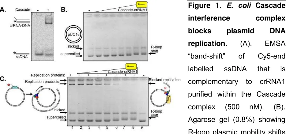
A Cascade complex was purified containing crRNA (crRNA1) to target 32 base pairs within lacZ of pUC18 plasmid to determine if this had any effect on plasmid DNA replication by purified E. coli replisome proteins (Figure 1). Targeting of Cascade-crRNA1 to lacZ was confirmed by hybridisation of crRNA to cognate DNA in EMSAs (Figure 1A), and by binding to pUC18 identifiable as pronounced altered mobility of supercoiled pUC and subtly shifted mobility of nicked pUC indicating R-loop formation (Westra et al. 2012) (Figure 1B). Replication assays were performed by loading the E. coli replisome onto pUC18 and observed as high molecular mass DNA products within
agarose gels (Figure 1C, lane 1). Plasmid replication is initiated in these assays from DnaC810 loading DnaB onto SSB-coated ssDNA generated at nicks or when DNA is supercoiled (Gilbert and Allan 2014; Xu and Marians 2000). Replication products disappeared on addition of increasing concentrations of Cascade-crRNA1 (lanes 2-8) corresponding to altered pUC18 mobility caused by Cascade R-loop formation (Figure 1C lanes 2-8).
Figure 1. E. coli Cascade
interference complex
blocks plasmid DNA
replication. (A). EMSA
[image:5.595.67.534.524.743.2]DNA as indicated (50 ng). Cascade was used at 4, 8, 16, 32, 64, 125 and 250 nM. (C). Titration of Cascade-crRNA1 into pUC18 plasmid DNA replication reactions cause loss of replication product. Full DNA replication product is shown in (lane 1), and Cascade-crRNA1 was used at the same concentrations as in part (B).
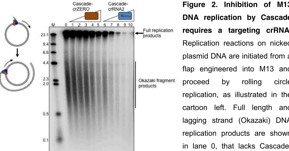
Replication blockage was also observed by using an alternative interference complex (Cascade-crRNA2) that binds to a target sequence within a nicked M13 DNA substrate (Figure 2, lanes 6 - 10). A purified Cascade complex lacking cRNA (Cascade-crZERO) and therefore unable to target M13 for binding had no significant effect on DNA replication (Figure 2 lanes 1 - 5). Cascade-crZERO was stable during purification (Supplementary Figure S1A) and gave an elution peak at the same position as for
Cascade-crRNA1 during gel filtration (Supplementary Figure S1B). These results indicate that a Cascade interference complex that is bound to a single target plasmid recognition sequence prevents DNA replication. Cascade R-loop formation most likely blocks DNA replication elongation at sites specific for crRNA base pairing, since Cascade-crZERO that would bind only non-specifically to DNA, if at all, did not block replication. We are currently developing assays to identify “pause site” Dσχ replication
products that would be expected to arise at or close to sites of blockage by Cascade.
Such pause DNA products would be further evidence that the elongation phase of
replication is being inhibited.
Figure 2. Inhibition of M13
DNA replication by Cascade
requires a targeting crRNA.
[image:6.595.65.529.465.708.2]DNA (see methods) caused a substantial decrease in observable product. Cascade protein complexes were each used at 4, 8, 16, 32 and 64 nM as indicated.
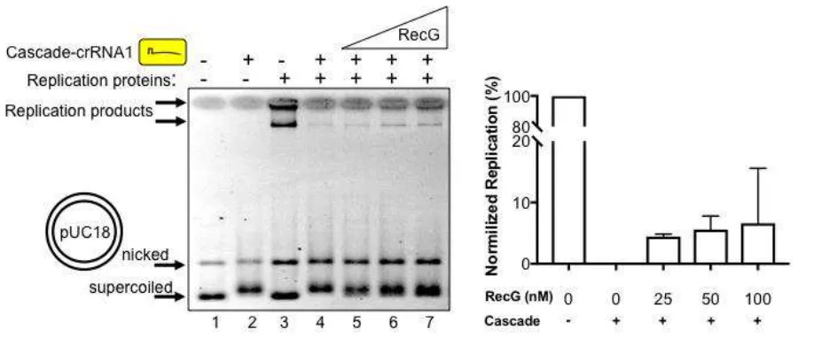
Nucleoprotein complexes are a major cause of genome instability because they provoke genetic rearrangements in cells when DNA replication is blocked (Gupta et al. 2013; Marians 2018; Syeda et al. 2014). Multiple pathways have evolved to overcome replication barriers, including involvement of bacterial RecG (Rudolph et al. 2010; Rudolph et al. 2013). The exact function of RecG in bacterial cells is not clear (Lloyd and Rudolph 2016) but it can dissociate R-loop structures and also helps cell cycle progression by re-modelling replication termination barriers (Rudolph et al. 2013; Vincent et al. 1996). We observed that DNA replication of pUC18 (Figure 3 lane 3) that had been blocked by 125 nM Cascade-crRNA1 (lane 4) resumed with the addition of E. coli RecG protein. This corresponds with re-appearance of replication products (lanes 5
- 7) to about 5-10% of the total product formed when Cascade was absent from reactions, summarised in Figure 3. Resumption of replication also corresponded to mobility of pUC18 changing from slower migrating plasmid bound by Cascade (Figure 3, lanes 2, 4 and 5) to faster migrating supercoiled plasmid (lanes 6 and 7). This suggested that replication was at least partially restored by RecG displacing Cascade from pUC18.
Figure 3. RecG alleviates replication blockage caused by a Cascade R-loop
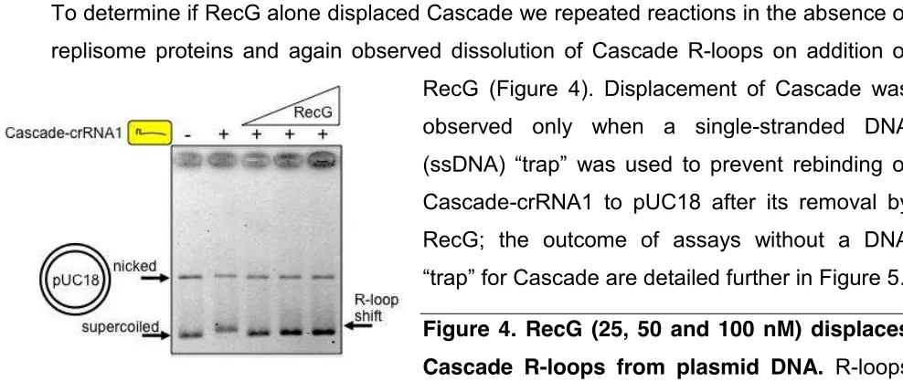
[image:7.595.65.524.428.623.2]To determine if RecG alone displaced Cascade we repeated reactions in the absence of replisome proteins and again observed dissolution of Cascade R-loops on addition of RecG (Figure 4). Displacement of Cascade was observed only when a single-stranded DNA
(ssDσχ) “trap” was used to prevent rebinding of Cascade-crRNA1 to pUC18 after its removal by RecG; the outcome of assays without a DNA
“trap” for Cascade are detailed further in Figure 5.
Figure 4. RecG (25, 50 and 100 nM) displaces
Cascade R-loops from plasmid DNA. R-loops were formed between Cascade-crRNA1 and pUC18 independently of DNA replication. A
ssDσχ “trap” in reactions prevents re-binding of Cascade-crRNA to plasmid if removed by RecG, detailed in Figure 5 and in the Methods section.
[image:8.595.35.529.63.272.2]Direct interaction between E. coli RecG and single strand DNA binding protein SSB promotes genome stability (Bianco and Lyubchenko 2017; Buss et al. 2008). This may also be relevant for recruitment of RecG to Cascade interference reactions through binding of SSB to ssDNA that is generated within R-loops. We repeated Cascade-crRNA1 reactions without the ssDNA “trap” that would also be bound strongly by SSB (Figure 5). Cascade R-loops formed on pUC18 (lane 2) but their displacement by RecG was not apparent, as expected in the absence of ssDσχ “trap” (lanes 3 – 6). Addition of SSB alone to Cascade-pUC18 R-loop resulted in a further plasmid mobility shift (lane 7), and titration of RecG into SSB pre-bound to Cascade-pUC18 resulted in further progressively increased shifts of pUC18 into more slowly migrating plasmid DNA (lanes 8 – 11). Neither SSB nor RecG alone had any observable effect on pUC18 mobility in these gels when Cascade-crRNA1 was absent (lanes 12 and 13).
Figure 5. Cascade
R-loops targeting pUC18
are also bound by RecG
and SSB proteins.
respectively 125 nM and 1 uM. RecG was used at 25, 50, 100 and 250 nM.
These results indicate a possible model that centres on Cascade-crRNA binding to target duplex DNA and generating a nucleoprotein R-loop. This forms a sequence-specific barrier to DNA replication that provokes genome instability and DNA repair responses. The displaced ssDσχ “loop” can be bound by SSB, and RecG is recruited either through its known physical interaction with SSB (Buss et al. 2008) or through RecG recognizing DNA structures within the R-loops. ATP-dependent dissociation of the cascade R-loop by RecG then generates DNA structures that are suitable for DNA capture during CRISPR-Cas adaptation, either directly by Cas1-Cas2 or mediated through another enzyme such as Cas3. This work also begins to demonstrate the feasibility of integrating E. coli DNA replication, CRISPR-Cas interference and DNA repair reactions by reconstitution in vitro to precisely determine the mechanisms involved. Further work is now required to couple these reactions to Cas1-Cas2 catalysed adaptation to determine how RecG might assist E. coli Cas1-Cas2 at Cascade barriers to replication in vitro. Similar in vitro analyses of na•ve adaptation should be able to determine how Cas1-Cas2 can establish immunity with the aid of other DNA repair enzymes.
MATERIALS & METHODS
Proteins
E. coli DNA replication proteins used were DNA polymerase III core (cgs), Clamp loader
( 3 ' ), clamp, SSB, DnaG and DnaB were purified as described in (Gupta et al. 2013), and DnaC810 was purified according to (Xu and Marians 2000). RecG helicase protein was purified as described in (Singleton et al. 2001). Purification of E. coli Cascade utilized over-expression from pET-Duet (Novagen) of Cascade Cse1 subunit from multiple cloning site 1 (MCS1 cloned NcoI – EcoRI) and of Cascade subunits Cse2-Cas7-Cas5-Cas6e as an operon from multiple cloning site 2 (MCS2 cloned NdeI – XhoI). crRNA1 for assembly into Cascade was generated by synthesis of DNA based on the E. coli CRISPR-1 Leader-Repeat1-Spacer-Repeat2 DNA sequence (GeneArt, Life Technologies) and its cloning into pACYC-Duet. DNA was synthesised with spacer
sequence 5«- AGGCCCGCACCGATCGCCCTTCCCAACAGTTG or
pETDuet construct. Plasmids co-transformed into BL21 AI cells were grown at 37 ˚C to OD600 of 0.6 for inducing expression of Cascade and crRNA1 by addition to growth media of 0.2% L-arabinose and 0.1 mM IPTG. Growth was continued for 18 hours at 18
˚C before harvesting cell pellets that were resuspended in 50 mM Tris pH 8.0 and 100 mM NaCl containing CompleteTM protease inhibitor cocktail (Roche). Soluble protein
obtained after cell lysis by sonication and clarification by centrifugation (60 min at 17000 rpm) was passed through a 5 ml StrepTrap column (GE Healthcare) followed by isocratic elution with 1 x buffer E (100 mM Tris pH8.0, 150 mM NaCl, 1 mM EDTA and 2.5 mM Desthiobiotin). Cascade containing fractions were further purified by HiPrep Sephacryl S-300 HR column (GE Helathcare) and eluted in storage buffer (50 mM Tris pH8.0, 150 mM NaCl, 2 mM DTT, 20 % glycerol) for flash freezing and storage at -80˚C.
In vitro reactions: Cascade EMSAs, R-loop formation and DNA replication
EMSA reactions binding Cascade-crRNA1 (0.5 M) to ssDNA (20 nM) (Figure 1A) were incubated for 15 min at 25 oC in 20 mM Tris pH 7.5, 75 mM NaCl, 1 mM DTT, 0.2 mM
Nicked M13 substrate utilized in rolling circle replication assays was generated by annealing single stranded M13 with the primer oJχ162 (5’ -TTTTTTTTTTTTTTTTTTTTTTTTTTTTTTTTTTTTGTCCACTATTAAAGAACGTGGACT CCAACG -3’) and extending with exo- T4 DNA polymerase exo- (NEB) using manufacturers instructions. As this polymerase is unable to engage in strand
displacement, polymerization arrested upon encountering the 5’ end of the primer. To
ensure substrate purity excess primer, nucleotides and polymerase were separated from the DNA substrate using a Micro BioSpin-6 column (Bio-Rad). Rolling circle replication reactions were assembled on ice, 4 nM nicked M13 substrate was added to reaction buffer supplemented with 0.2 mM G/C/UTP and 0.04mM dGTP/dCTP. Replication proteins were added at concentrations described above, followed by either Cascade-crZero or Cascade-crRNA2. Reactions were pre-incubated for 2 minutes at 37oC and
initiated by addition of 0.04 mM dχTP/dTTP, followed 2 minutes later by 5 Ci of
[ 32P]dCTP (Perkin Elmer). Reactions were quenched at T=14 minutes by addition of
ammonium acetate to a final concentration of 2.5 mM with samples immediately ethanol precipitated to remove unincorporated [ 32P]dCTP. Pellets were resuspended in 50 mM
NaOH and 30 mM EDTA prior to loading and overnight migration for 420 volt hours at 25 volts on a 0.7 % denaturing agarose gel (2mM EDTA, 30 mM NaOH). The gel was fixed by washing in a solution of 5 % TCA for 20 minutes followed by 10 minutes in H2O
before drying. After overnight exposure the gel was imaged using a Personal Molecular Imager system (Bio-Rad).
ACKNOWLEDGEMENTS
The work was funded by BBSRC grant BB/M020541/1 to ELB and BBSRC grants BB/N014863/1 and BB/P000746/1 to PM.
REFERENCES
Bianco, P. R. and Lyubchenko, Y. L. (2017), 'SSB and the RecG DNA helicase: an intimate association to rescue a stalled replication fork', Protein Sci, 26 (4), 638-49.
Brouns, S. J., et al. (2008), 'Small CRISPR RNAs guide antiviral defense in prokaryotes', Science, 321 (5891), 960-4.
Cooper, L. A., Stringer, A. M., and Wade, J. T. (2018), 'Determining the Specificity of Cascade Binding, Interference, and Primed Adaptation In Vivo in the Escherichia coli Type I-E CRISPR-Cas System', MBio, 9 (2).
Datsenko, K. A., et al. (2012), 'Molecular memory of prior infections activates the CRISPR/Cas adaptive bacterial immunity system', Nat Commun, 3, 945.
Gilbert, N. and Allan, J. (2014), 'Supercoiling in DNA and chromatin', Curr Opin Genet Dev, 25, 15-21.
Gupta, M. K., et al. (2013), 'Protein-DNA complexes are the primary sources of replication fork pausing in Escherichia coli', Proc Natl Acad Sci U S A, 110 (18), 7252-7.
Hayes, R. P., et al. (2016), 'Structural basis for promiscuous PAM recognition in type I-E Cascade from E. coli', Nature, 530 (7591), 499-503.
Heussler, G. E., et al. (2016), 'Requirements for Pseudomonas aeruginosa Type I-F CRISPR-Cas Adaptation Determined Using a Biofilm Enrichment Assay', J Bacteriol, 198 (22), 3080-90.
Hille, F., et al. (2018), 'The Biology of CRISPR-Cas: Backward and Forward', Cell, 172 (6), 1239-59.
Ivancic-Bace, I., Al Howard, J., and Bolt, E. L. (2012), 'Tuning in to Interference: R-Loops and Cascade Complexes in CRISPR Immunity', J Mol Biol, 422 (5), 607-16.
Ivancic-Bace, I., et al. (2015), 'Different genome stability proteins underpin primed and naive adaptation in E. coli CRISPR-Cas immunity', Nucleic Acids Res, 43 (22), 10821-30.
Jackson, R. N., et al. (2014), 'Structural biology. Crystal structure of the CRISPR RNA-guided surveillance complex from Escherichia coli', Science, 345 (6203), 1473-9. Jore, M. M., et al. (2012), 'Structural basis for CRISPR RNA-guided DNA recognition by
Cascade', Nat Struct Mol Biol, 18 (5), 529-36.
Kunne, T., et al. (2016), 'Cas3-Derived Target DNA Degradation Fragments Fuel Primed CRISPR Adaptation', Mol Cell, 63 (5), 852-64.
Levy, A., et al. (2015), 'CRISPR adaptation biases explain preference for acquisition of foreign DNA', Nature, 520 (7548), 505-10.
Lloyd, R. G. and Rudolph, C. J. (2016), '25 years on and no end in sight: a perspective on the role of RecG protein', Curr Genet.
Marians, K. J. (2018), 'Lesion Bypass and the Reactivation of Stalled Replication Forks', Annu Rev Biochem.
Nunez, J. K., et al. (2015a), 'Integrase-mediated spacer acquisition during CRISPR-Cas adaptive immunity', Nature, 519 (7542), 193-8.
Nunez, J. K., et al. (2015b), 'Foreign DNA capture during CRISPR-Cas adaptive immunity', Nature.
Nunez, J. K., et al. (2016), 'CRISPR Immunological Memory Requires a Host Factor for Specificity', Mol Cell, 62 (6), 824-33.
Nunez, J. K., et al. (2014), 'Cas1-Cas2 complex formation mediates spacer acquisition during CRISPR-Cas adaptive immunity', Nat Struct Mol Biol, 21 (6), 528-34. Pougach, K., et al. (2010), 'Transcription, processing and function of CRISPR cassettes
in Escherichia coli', Mol Microbiol, 77 (6), 1367-79.
Redding, S., et al. (2015), 'Surveillance and Processing of Foreign DNA by the Escherichia coli CRISPR-Cas System', Cell, 163 (4), 854-65.
Rollie, C., et al. (2015), 'Intrinsic sequence specificity of the Cas1 integrase directs new spacer acquisition', Elife, 4.
Rudolph, C. J., et al. (2013), 'Avoiding chromosome pathology when replication forks collide', Nature, 500 (7464), 608-11.
Rutkauskas, M., et al. (2015), 'Directional R-Loop Formation by the CRISPR-Cas Surveillance Complex Cascade Provides Efficient Off-Target Site Rejection', Cell Rep, 10, 1534-43.
Sashital, D. G., Wiedenheft, B., and Doudna, J. A. (2012), 'Mechanism of foreign DNA selection in a bacterial adaptive immune system', Mol Cell, 46 (5), 606-15.
Semenova, E., et al. (2016), 'Highly efficient primed spacer acquisition from targets destroyed by the Escherichia coli type I-E CRISPR-Cas interfering complex', Proc Natl Acad Sci U S A, 113 (27), 7626-31.
Singleton, M. R., Scaife, S., and Wigley, D. B. (2001), 'Structural analysis of DNA replication fork reversal by RecG.', Cell, 107 (1), 79-89.
Sinkunas, T., et al. (2011), 'Cas3 is a single-stranded DNA nuclease and ATP-dependent helicase in the CRISPR/Cas immune system', Embo J, 30 (7), 1335-42.
Staals, R. H., et al. (2016), 'Interference-driven spacer acquisition is dominant over naive and primed adaptation in a native CRISPR-Cas system', Nat Commun, 7, 12853.
Syeda, A. H., Hawkins, M., and McGlynn, P. (2014), 'Recombination and replication', Cold Spring Harb Perspect Biol, 6 (11), a016550.
Szczelkun, M. D., et al. (2014), 'Direct observation of R-loop formation by single RNA-guided Cas9 and Cascade effector complexes', Proc Natl Acad Sci U S A, 111 (27), 9798-803.
Vincent, S.D., Mahdi, A.A., and Lloyd, R.G. (1996), 'The RecG branch migration protein of Escherichia coli dissociates R-loops', J. Mol. Biol., 264, 713-21.
Wang, J., et al. (2015), 'Structural and Mechanistic Basis of PAM-Dependent Spacer Acquisition in CRISPR-Cas Systems', Cell.
Westra, E. R., et al. (2012), 'CRISPR Immunity Relies on the Consecutive Binding and Degradation of Negatively Supercoiled Invader DNA by Cascade and Cas3', Mol Cell, 46 (5), 595-605.
Wiedenheft, B., et al. (2011), 'Structures of the RNA-guided surveillance complex from a bacterial immune system', Nature, 477 (7365), 486-89.
Wright, A. V., et al. (2017), 'Structures of the CRISPR genome integration complex', Science, 357 (6356), 1113-18.
Xiao, Y., et al. (2017), 'Structure Basis for Directional R-loop Formation and Substrate Handover Mechanisms in Type I CRISPR-Cas System', Cell, 170 (1), 48-60 e11. Xu, L. and Marians, K. J. (2000), 'Purification and characterization of DnaC810, a
primosomal protein capable of bypassing PriA function', J Biol Chem, 275 (11), 8196-205.
Xue, C., et al. (2017), 'Real-Time Observation of Target Search by the CRISPR Surveillance Complex Cascade', Cell Rep, 21 (13), 3717-27.
Suppl. Figure 1. (A). SDS-PAGE analysis of purified Cascade complexes utilized in this work as labeled. (B). A gel filtration step utilized during purification of Cascade complexes showed that Cascade-crRNAZERO and Cascade–crRNA1 eluted in the same
5 10 15 20 25
-20 0 20 40 60 80 ml m A u
5 10 15 20 25