White Rose Research Online URL for this paper:
http://eprints.whiterose.ac.uk/128133/
Version: Published Version
Article:
Jonker, C.T.H. orcid.org/0000-0003-0906-9808, Galmes, R.
orcid.org/0000-0001-5616-5937, Veenendaal, T. et al. (8 more authors) (2018) Vps3 and
Vps8 control integrin trafficking from early to recycling endosomes and regulate
integrin-dependent functions. Nature Communications, 9. 792.
https://doi.org/10.1038/s41467-018-03226-8
[email protected]
https://eprints.whiterose.ac.uk/
Reuse
This article is distributed under the terms of the Creative Commons Attribution (CC BY) licence. This licence
allows you to distribute, remix, tweak, and build upon the work, even commercially, as long as you credit the
authors for the original work. More information and the full terms of the licence here:
https://creativecommons.org/licenses/
Takedown
If you consider content in White Rose Research Online to be in breach of UK law, please notify us by
Vps3 and Vps8 control integrin traf
fi
cking from
early to recycling endosomes and regulate
integrin-dependent functions
C.T.H. Jonker
1,5
, R. Galmes
1,6
, T. Veenendaal
1
, C. ten Brink
1
, R.E.N. van der Welle
1
, N. Liv
1
, J. de Rooij
2
,
A.A. Peden
3
, P. van der Sluijs
1,7
, C. Margadant
4
& J. Klumperman
1
Recycling endosomes maintain plasma membrane homeostasis and are important for cell
polarity, migration, and cytokinesis. Yet, the molecular machineries that drive endocytic
recycling remain largely unclear. The CORVET complex is a multi-subunit tether required for
fusion between early endosomes. Here we show that the CORVET-speci
fi
c subunits Vps3
and Vps8 also regulate vesicular transport from early to recycling endosomes. Vps3 and
Vps8 localise to Rab4-positive recycling vesicles and co-localise with the CHEVI complex on
Rab11-positive recycling endosomes. Depletion of Vps3 or Vps8 does not affect transferrin
recycling, but delays the delivery of internalised integrins to recycling endosomes and their
subsequent return to the plasma membrane. Consequently, Vps3/8 depletion results in
defects in integrin-dependent cell adhesion and spreading, focal adhesion formation, and cell
migration. These data reveal a role for Vps3 and Vps8 in a specialised recycling pathway
important for integrin traf
fi
cking.
DOI: 10.1038/s41467-018-03226-8
OPEN
1Section Cell Biology, Center for Molecular Medicine, University Medical Center Utrecht, Utrecht University, Heidelberglaan 100, 3584 CX Utrecht, The
Netherlands.2Section Molecular Cancer Research, Center for Molecular Medicine, University Medical Center Utrecht, Utrecht Universty, Heidelberglaan 100, 3584 CX Utrecht, The Netherlands.3Department of Biomedical Science, The University of Sheffield, Sheffield, S10 2TN, UK.4Department of Molecular Cell
Biology, Sanquin Research, Plesmanlaan 125, 1066 CX Amsterdam, The Netherlands.5Present address: Department of Ophthalmology, Weill Cornell Medicine, 1300 York Ave, New York, NY 10065, USA.6Present address: UCL Cancer Institute, University College London, 72 Huntley Street, London, WC1E 6DD, UK.7Present address: Cellular Protein Chemistry, Bijvoet Center for Biomolecular Research, Utrecht University, 3584 CH Utrecht, The Netherlands. C.T. H. Jonker and R. Galmes contributed equally to this work. C. Margadant and J. Klumperman jointly supervised this work. Correspondence and requests for materials should be addressed to J.K. (email:[email protected])
123456789
T
he endolysosomal system is important for a variety of
cellular processes, such as protein homeostasis, antigen
presentation, signal transduction and cell migration.
Hence, disruption of endolysosome function is found in a wide
range of diseases, from genetic lysosomal storage disorders to
cancer and neurodegenerative disorders
1–3. The progression of
cargo through the endolysosomal system, from early endosomes
(EEs) to late endosomes and lysosomes or from EEs to the plasma
membrane or recycling endosomes (REs), is tightly controlled by
dedicated protein machinery. Membrane fusion is coordinated by
the concerted action of Rab GTPases, tethers and soluble NSF
attachment protein receptors (SNAREs)
4,5. Rab GTPases drive
the process by recruiting effector machinery proteins to speci
fi
c
membrane domains
6,7. Contact between opposing membranes is
then initiated by tethering proteins, followed by
SNARE-mediated fusion.
EE-EE fusion is initiated by activation of Rab5, which recruits
multiple effector proteins, including the class C core vacuole/
endosome tethering (CORVET) complex
8–11. The hexameric
CORVET complex consists of a core (Vps11, Vps16, Vps18 and
Vps33A), which is shared with the late endosomal homotypic
fusion and protein sorting (HOPS) tethering complex, and
additionally contains the two CORVET-speci
fi
c subunits Vps8
and Vps3 (also named TGFBRAP1 or TRAP1)
9,12. Recycling of
endocytosed proteins and membranes from EEs is crucial to
maintain plasma membrane homeostasis and is essential for cell
polarity, cell migration and cytokinesis. Recycling occurs either
directly from EEs to the plasma membrane (fast recycling) or
indirectly via Rab11-positive REs (slow recycling)
13–15. Both
pathways involve Rab4, which resides on EEs as well as on
recycling vesicles that emerge from EEs
15–17. The class C
homologues in endosome-vesicle interaction (CHEVI) complex
consisting of Vps33B and VIPAS39, homologues of Vps33A and
Vps16, respectively, binds to Rab11 and localises to REs
18–20.
Mutations in Vps33B or VIPAS39 underlie arthrogryposis, renal
dysfunction and cholestasis (ARC) syndrome, a rare autosomal
recessive multisystem disorder that affects the transport of apical
and junctional proteins in polarised cells
18,19,21.
A poorly understood step in endosomal recycling is the
transport from EEs to REs. Since EEs are the major source of
membranes for REs, we here study a possible role for the
COR-VET complex in endosomal recycling. To our surprise we found
that the CORVET-speci
fi
c Vps3 and Vps8 subunits interact
directly with each other and localise to Rab4-positive recycling
vesicles and CHEVI-positive REs. Moreover, we show that Vps3
and Vps8 function in a specialised pathway required for integrin
recycling, and thereby regulates integrin-dependent cell adhesion
and migration.
Results
Vps3 and Vps8 localise to recycling vesicles
. The mammalian
CORVET complex functions as a tether between EEs and is
recruited to membranes via the interaction of Vps8 with Rab5
8.
To determine a possible role of the CORVET complex in
endo-somal recycling, we analysed the localisation of the
CORVET-speci
fi
c subunits Vps3 and Vps8 in ultrastructural detail. We
expressed GFP-Vps3 and HA-Vps8 in HeLa cells and performed
immuno-electron microscopy (IEM) by immunogold labelling of
ultrathin cryosections. Our IEM data con
fi
rmed the localisation
of Vps3 and Vps8 on EEs (Fig.
1
a, top panel), but in addition
revealed a substantial labelling on EE-associated tubules and
vesicles (Fig.
1
a, bottom panel). The Vps3- and Vps8-labelled
vesicles had a characteristic dense content, were often found in
clusters (Fig.
1
a, lower panel, and
1
b) and were consistently
negative for endocytosed BSA-Au
5, a marker of endocytic but not
recycling vesicles. These morphological characteristics putatively
de
fi
ne the Vps3- and Vps8-positive vesicles as recycling vesicles.
To determine whether the Vps3- and Vps8-positive vesicles
also contain CORVET core subunits, we expressed GFP-Vps3
together with FLAG-Vps18 for IEM analysis. Immunogold
double labelling showed that Vps18 co-localises with Vps3 on
EEs, but was absent from Vps3-positive vesicles (Fig.
1
b,
asterisks), indicating that Vps3 and Vps8 localise to recycling
vesicles, which lack CORVET core components. We then
co-expressed GFP-Vps3, HA-Vps8 or the CORVET core subunit
GFP-Vps11, together with mCherry-tagged Rab4, Rab5 or Rab11,
and assessed their co-localisation by quantitative immuno
fl
uor-escence (IF) microscopy. We found that Vps3 (Fig.
1
d and
Supplementary Fig.
1
a) and Vps8 (Fig.
1
c, d) signi
fi
cantly
co-localised with Rab5 and Rab4, which label EEs and EE-derived
recycling vesicles, respectively
17,22, and partially with the RE
marker Rab11 (Fig.
1
c, d and Supplementary Fig.
1
a), indicative
for the presence of Vps3 and Vps8 on EEs as well as on recycling
vesicles. By contrast, GFP-Vps11 only notably overlapped with
Rab5 (Fig.
1
d and Supplementary Fig.
1
b), reinforcing the notion
that CORVET core components are present on EEs but not on
recycling vesicles. To facilitate the distinction of Rab5 and
Rab4 subdomains within EEs by IF microscopy, we expressed the
constitutively active Rab5Q79L mutant, which increases the size
of EEs
22,23. Strikingly, co-expression of GFP-Rab5Q79L with
HA-Vps8 and FLAG-Rab4 revealed that HA-HA-Vps8 speci
fi
cally
concentrated in FLAG-Rab4-positive patches (Fig.
1
e, arrows)
and protrusions (Supplementary Fig.
1
c, arrows) of
GFP-Rab5Q79L-positive EEs. To further con
fi
rm that Vps3 and
Vps8 reside on the same recycling vesicles as Rab4 we performed
IEM of HeLa cells expressing HA-Vps8 or HA-Vps3 with
GFP-Rab4. This unequivocally demonstrated that Vps3 and Vps8
indeed co-localise with Rab4 on recycling tubules and vesicles
(Fig.
1
f, arrows).
In conclusion, these data reveal that Vps3 and Vps8 are present
on EE-derived Rab4-positive recycling vesicles and
Rab11-positive REs, which lack CORVET core components.
Vps3 and Vps8 interact directly with each other
. Previous
studies showed that Vps3 and Vps8 bind the CORVET core
complex
9,12. In addition, direct interactions between Vps3 and
Vps8 have been reported in yeast
24. Since we
fi
nd that Vps3 and
Vps8 co-localise on recycling vesicles that lack CORVET core
components, we performed co-immunoprecipitation (co-IP) and
glutathione
S
-transferase
(GST)-binding
experiments
to
investigate whether mammalian Vps3 and Vps8 bind directly to
each other. In agreement with this scenario, co-IP experiments in
HeLa cells revealed that endogenous Vps8 readily
co-immunoprecipitated with overexpressed GFP-Vps3, while less
Vps8 was recovered with GFP-tagged Vps11, Vps16 or Vps41, the
HOPS-speci
fi
c subunit that is most homologous to Vps8 (Fig.
2
a,
full blots in Supplementary Fig.
2
). To investigate whether Vps3
can directly bind Vps8 we probed the binding of full-length
GST-Vps3 puri
fi
ed from bacterial lysates with in vitro-translated
HA-Vps8. Importantly, GST-Vps3 interacted directly with HA-Vps8
(Fig.
2
b, full blots in Supplementary Fig.
3
). By expressing
trun-cation mutants of GST-Vps3, we could map the Vps8 interacting
domain to the C terminus of Vps3 (Fig.
2
b), which also con
fi
rms
the speci
fi
city of the binding assays. Finally, to quantify the
interaction of Vps3 with Vps8, Vps11 or Vps16 we performed a
proximity ligation assay (PLA) in HeLa cells. This showed a
signi
fi
cantly higher PLA signal between Vps3 and Vps8, than
between Vps3 and Vps11 or Vps16 (Fig.
2
c, d).
BSA-Au5, FLAG-Vps1810, GFP-Vps315
EE
*
*
*
*
*
a
c
e
f
b
BSA-Au5
HA-Vps810
GFP-Vps315
EE
EE
Rab4-GFP10
Vps8-V515
EE
E
N
Rab4-GFP10
HA-Vps315
EE
0.95 0.80 0.42
HA-Vps8 mCherry-Rab4
HA-Vps8 mCherry-Rab5
HA-Vps8 mCherry-Rab11
Vps8 Rab4 Vps8 Rab5 Vps8 Rab11
Rab5Q79L-GFP Rab4a-FLAG Vps8-V5 Merge
d
0.0 0.2 0.4 0.6 0.8 1.0
mCh-Rab4 mCh-Rab5 mCh-Rab11
Vps8 Vps3 Vps11
***
*
*
Fold change
BSA-Au5
HA-Vps810
GFP-Vps315
Fig. 1Vps3 and Vps8 localise to Rab4- and Rab11-positive recycling endosomes.aIEM of HeLa cells expressing HA-Vps8 and GFP-Vps3, and loaded with
BSA-Au5. HA-Vps8 (10 nm gold, arrows) and GFP-Vps3 (15 nm gold) co-localise on EEs (top panel) and on vesicles negative for BSA-Au5 (bottom panel). Bar, 100 nm.bIEM of HeLa cells expressing FLAG-Vps18, Vps3 and HA-Vps8, and loaded with BSA-Au5. FLAG-Vps18 (10 nm gold, arrows) and GFP-Vps3 (15 nm gold) co-localise on EEs but not on vesicles negative for BSA-Au5 (asterisk). Bar, 100 nm.cHeLa cells expressing HA-Vps8 together with mCherry-Rab4, mCherry-Rab5 or mCherry-Rab11. HA-Vps8 co-localises strongly with Rab4 and Rab5, and less with Rab11. Bar, 10µm.dQuantification of
triplicate experiments shown incbased onn> 30 cells per condition. Error bars represent the standard error of the correlation coefficient (SEr). *P≤0.05, ***P≤0.001, calculated using Eqs.4and 5.eIF of HeLa cells expressing Rab5Q79L-GFP, Rab4-FLAG and Vps8-V5. Vps8 localises to Rab4-positive patches on Rab5Q79L-positive enlarged endosomes (arrows). Bar, 10µM.fIEM of HeLa cells expressing Rab4-GFP together with Vps8-V5 or HA-Vps3,
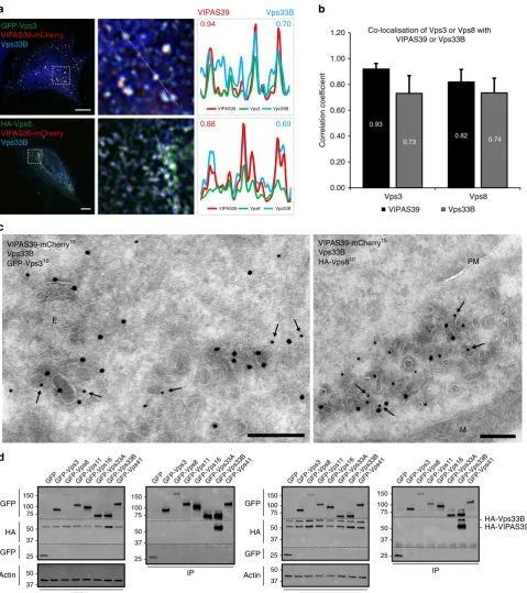
[image:4.595.118.480.48.618.2]Vps3 and Vps8 co-localise with CHEVI on recycling
mem-branes
. The CHEVI complex interacts with Rab11 and localises
to Rab11-positive REs
8,9,18,19,25,26. Since Vps3 and Vps8 partially
co-localise with Rab11 (Fig.
1
c, d) we investigated co-localisation
between Vps3 and Vps8 with the CHEVI complex. To allow
formation of the CHEVI complex we co-expressed
VIPAS39-mCherry and untagged Vps33B. Expression of the CHEVI
complex with either HA-Vps8 or GFP-Vps3 resulted in clear
co-localisation as determined by quantitative IF microscopy (Fig.
3
a,
b). By IEM, these foci of co-localisation were identi
fi
ed as Vps3-,
Vps8- and CHEVI-positive clusters of vesicles and tubules, which
is the typical morphology for REs
27(Fig.
3
c).
To examine possible interactions between the CHEVI complex
and Vps3 or Vps8, or between CHEVI and the CORVET core
subunits, we performed co-IP experiments after co-expression of
HA-VIPAS39 with GFP-tagged constructs of Vps3, Vps8, Vps11,
Vps16, Vps33A, Vps33B or Vps41. Because co-expression of
Vps33B together with VIPAS39 is needed for membrane
recruitment of the CHEVI complex
18,19, we included an internal
control by probing the interactions with HA-VIPAS39 in the
absence (Fig.
3
d, left panel) or presence (Fig.
3
d, right panel) of
overexpressed HA-Vps33B. As expected, we detected a strong
interaction between HA-VIPAS39 and GFP-Vps33B (Fig.
3
d, lane
7 both panels, full blots in Supplementary Fig.
4
). Strikingly,
however, no signi
fi
cant binding between CHEVI with any of the
other subunits was found (Fig.
3
d). Only after prolonged
exposure of the western blot a weak interaction between
GFP-Vps3 and HA-VIPAS39 was detected (Supplementary Fig.
5
and
6
). Previous studies using endogenous proteins failed to detect
speci
fi
c interactions between VIPAS39 with any of the HOPS or
CORVET subunits, including Vps3 and Vps8
8,9. Our studies are
in line with these
fi
ndings, however, the weak binding between
CHEVI and Vps3 could be indicative for transient interactions.
Taken together, our data show that in addition to their
localisation on Rab4-positive recycling vesicles (Fig.
1
f), Vps3 and
Vps8 partially co-localise with the CHEVI complex on REs.
Furthermore, these data suggest that a transient interaction of
Vps3 and Vps8 with the CHEVI complex may occur on REs.
Vps3 and Vps8 are required for recycling of
β1 integrins
. To
assess which cargo is transported by the Vps3/8-positive recycling
vesicles, we
fi
rst determined the effect of Vps3 and Vps8
deple-tion on transferrin (Tf) recycling. We depleted both subunits to
fully prevent any function related to the interaction between
residual Vps3 and Vps8. Endocytosed Tf recycles either directly
from EEs to the plasma membrane or indirectly via REs, while
dextran is transported to the lysosome for degradation
28. A block
in recycling increases inclusion of Tf in the degradative pathway
to the lysosome, and therefore increases the co-localisation
between Tf and dextran
29,30. Vps3/8 depletion had no effect on Tf
localisation, indicating that the recycling of Tf was not disturbed
1.00
0.24 0.44
0.0 0.2 0.4 0.6 0.8 1.0 1.2
Vps8+Vps3Vps16+Vps3Vps11+Vps3 Corrected PLA signal
Fold change
a
b
c
d
HeLa HA-Vps8 GFP-Vps3 PLA HeLa HA-Vps3 GFP-Vps11 PLA HeLa HA-Vps3 GFP-Vps16 PLA
GST
-Vps3 full length GST
-Vps3 1–550 GST
-Vps3 551–860
GST HA-Vps8 TCL GST
-Vps3 full length GST
-Vps3 1–550 GST
-Vps3 551–860 GST
GST Biotin
25 37 50 75 100
100
75
50 150 250
-GFP GFP-Vps3 GFP-Vps1 1
GFP-Vps16GFP-Vps41
IP: GFP 25
150
-GFP GFP-Vps3 GFP-Vps1 1
GFP-Vps16GFP-Vps41
TCL
α-Vps8
α-GFP
- 100 - 250
- 150
25
-150 - α-Vps8
α-GFP
- 100 - 250
- 150
Fig. 2Vps3 and Vps8 interact with each other.aIPs of GFP-tagged Vps3, Vps11, Vps16 or Vps41 probed for interaction with endogenous Vps8 (GFP
control lane shows area around 25 kDa from the same membrane).bIPs of GST-tagged Vps3 and truncation mutants of GST-Vps3 purified from bacterial lysates (left panel) probed for interaction with in vitro-translated HA-Vps8. In vitro translation was performed with biotinylated lysines using the Transcend Non-Radioactive Translation Detection System (Promega) according to the manufacturer’s protocol and detected using streptavidin-HRP. HA-Vps8 binds to the C terminus of Vps3 and full-length Vps3.cPLA on HeLa cells. The highest PLA signal is between Vps3 and Vps8. Bar, 10µm.dQuantification of
[image:5.595.88.505.49.367.2]c
d
a
b
0.00 0.20 0.40 0.60 0.80 1.00 1.20
Vps3 Vps8
VIPAS39 Vps33B
Co-localisation of Vps3 or Vps8 with VIPAS39 or Vps33B
0.82 0.74
0.73 0.93
Correlation coef
ficient
VIPAS39 Vps3 Vps33B
0.94 0.70
VIPAS39 Vps8 Vps33B
0.88 0.69
Vps33B VIPAS39
HA-Vps8
VIPAS39-mCherry Vps33B GFP-Vps3
VIPAS39-mCherry Vps33B
VIPAS39-mCherry15 Vps33B
GFP-Vps310
E
VIPAS39-mCherry15 Vps33B
HA-Vps810
M PM
TCL
IP
HA-VIPAS39 HA-Vps33B GFPGFP-Vps3GFP-Vps8GFP-Vps1
1
GFP-Vps16GFP-Vps33AGFP-Vps33BGFP-Vps41 GFPGFP-Vps3GFP-Vps8GFP-Vps1 1
GFP-Vps16GFP-Vps33AGFP-Vps33BGFP-Vps41
GFP
GFP HA
Actin 50
37 25 37 50 75 100 150
25 37 50 75 100 150
-GFPGFP-Vps3GFP-Vps8GFP-Vps1 1
GFP-Vps16GFP-Vps33AGFP-Vps33BGFP-Vps41 GFP GFP-Vps3GFP-Vps8GFP-Vps1 1
GFP-Vps16GFP-Vps33AGFP-Vps33BGFP-Vps41
TCL
IP GFP
GFP HA
Actin
25
50 37 37 50 75 100 150
25 37 50 75 100 150
-Fig. 3Vps3 and Vps8 co-localise with the CHEVI complex.aIF of HeLa cells expressing VIPAS39-mCherry, untagged Vps33B and either GFP-Vps3 or
HA-Vps8. Vps3 and Vps8 co-localise with both VIPAS39 and Vps33B (line profiles in right panel indicate co-localisation correlation, correlation coefficients indicated: VIPAS39=red; Vps33B=blue). Bar, 10µm.bQuantification of triplicate experiments shown inabased onn> 30 cells per condition by line
[image:6.595.58.538.48.588.2](Fig.
4
a, b). These data are in line with a previous study showing
that Vps3 is not required for Tf recycling
8.
In addition to the common recycling pathway taken by Tf,
speci
fi
c recycling pathways are described for cell-surface proteins
such as integrins
31–33. To study a potential role of Vps3 and Vps8
in the recycling of
β
1 integrins we used an established
antibody-based integrin recycling assay
34. HeLa cells were depleted of Vps3
and Vps8, and then serum-starved overnight, labelled with
anti-β
1 integrin, washed and chased for 2 h. In the absence of serum,
β
1 integrins accumulate in REs (Fig.
4
c, arrows). Vps3/8
knockdown prevented the accumulation of
β
1 integrins, which
instead retained a dispersed distribution throughout the cell
PM
E
β1 integrin15
GFP-Vps310
BSA-Au5
α5 integrin15
GFP-Vps310
BSA-Au5
E
Dextran-Alexa-488
Transferrin-Alexa-568 Scrambled
Vps3/8KD Endocytosed anti-β1 integrin Vps3/8KD Scrambled
Scrambled
β1 integrin uptake10
N
M
3/8 KD
β1 integrin uptake10
EE
% Cells containing accumulated β1 integrin in RE
0 10 20 30 40 50 60 70
No recycling 5′ recycling 15′ recycling Scrambled Vps3/8KD Scrambled Vps3/8KD
Scrambled Vps3/8KD
α5β1 integrin recycling assay
Minutes recycling
Scrambled Vps3/8 KD
% Recycled
α
5 integrin
0
0 2 4 6 8 10 12 14 16
10 20 30 40 50 60 70 80
a
c
b
d
e
g
h
f
0.000.20 0.40 0.60
Correlation coefficient
0.80
Scr Vps3/8KD
Dextran/transferrin co-localisation
0.0 0.5 1.0 1.5 2.0
(Fig.
4
c and Supplementary Fig.
7
). Using an adapted IEM
protocol to visualise the antibody uptake as described above, we
could identify the accumulations in control cells as large clusters
of recycling tubules and vesicles (Fig.
4
d, left panel). Importantly,
IEM of the Vps3/8 knockdown cells showed that internalised
β
1
integrins still entered EEs and EE-associated recycling vesicles
(Fig.
4
d, right panel). These data show that the block in
β
1
integrin recycling induced by Vps3/8 knockdown occurs after exit
from EEs, and interferes with the transport of recycling vesicles to
REs. Re-addition of serum induced integrin recycling in control
cells resulting in the disappearance of intracellular accumulations,
while the localisation of internalised
β
1 integrins in knockdown
cells remained unchanged (Fig.
4
e and Supplementary Fig.
7
).
Together these results imply that Vps3/8 knockdown prevents or
delays internalised
β
1 integrins to reach REs and thereby prevents
clearance of the intracellular pool.
We next investigated the impact of Vps3/8 knockdown on the
kinetics of integrin recycling by studying the
fi
bronectin
(FN)-binding integrin
α
5
β
1
35. In short, cells were surface-labelled with
cleavable biotin and endocytosis was allowed for 30 min in the
absence of serum. Surface biotin was removed and recycling was
induced by re-addition of serum. Surface biotin was removed
again and the reduction of intracellular biotinylated
α
5 integrin
before and after recycling was quanti
fi
ed using capture ELISA.
This assay showed that knockdown of Vps3/8 resulted in a
signi
fi
cant decrease of
α
5
β
1 recycling, especially after short
recycling times (Fig.
4
f), while the internalisation of
α
5
β
1 as not
affected (Fig.
4
g). Finally, to verify that
α
5
β
1 is indeed transported
by Vps3/8-positive recycling vesicles, we labelled the
α
5 and
β
1
integrin subunits for IEM. This unequivocally demonstrated that
integrin
α
5
β
1 is present in the Vps3/8-positive recycling vesicles
(Fig.
4
h).
Taken together, these data show that
β
1 integrins are
incorporated into Vps3/8-positive recycling vesicles, and that
Vps3 and Vps8 are required for ef
fi
cient
β
1 integrin recycling.
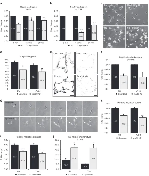
Vps3 and Vps8 regulate cell adhesion and migration
. Integrins
are the primary receptors for extracellular matrix (ECM)
pro-teins
36and integrin recycling is important for cell-ECM
inter-actions and cell migration
37–39. To investigate whether Vps3 and
Vps8 are required for integrin-dependent cell adhesion, we
see-ded HeLa Vps3/8 knockdown cells on FN or collagen-I (Col-I) in
the absence of serum. Non-adherent cells were washed away after
5, 15 or 30 min, and the number of attached cells was quanti
fi
ed.
This revealed that adhesion to both FN (Fig.
5
a) and Col-I
(Fig.
5
b) was signi
fi
cantly compromised in Vps3/8 knockdown
cells. Individual knockdown of Vps3 or Vps8 also reduced cell
adhesion, but the effect was less stringent, suggesting that residual
expression in single-knockdown cells still allows a functional
interaction between Vps3 and Vps8, while in the
double-knockdown cells this is more completely abrogated
(Supple-mentary Fig.
8
a). Importantly, different siRNA oligos induced
essentially the same results, rendering it unlikely that the
observed adhesion defect is due to off-target effects
(Supple-mentary Fig.
8
b).
Because integrin-dependent cell adhesion is followed by a
reorganisation of the cytoskeleton and cell spreading, we next
determined cell spreading on FN and Col-I 2 h after cell adhesion
(Fig.
5
c). Quanti
fi
cation of the number of spread cells showed
that Vps3/8 depletion reduces cell spreading on both FN and
Col-I (Fig.
5
d). Integrin-mediated cell spreading is linked to the
assembly of focal adhesions (FAs), which anchor the actin
cytoskeleton to the ECM
40. To determine if Vps3/8 knockdown
impacts on FA formation, we quanti
fi
ed the number of FAs per
cell using confocal microscopy on cell populations stained for
vinculin, a well-established marker of FAs (Fig.
5
e). Consistent
with reduced cell adhesion and spreading, Vps3/8 knockdown
cells assembled less FAs, both on FN and Col-I (Fig.
5
f).
Finally, we analysed cell motility by time-lapse microscopy
(Fig.
5
g, arrows indicate migration direction). Knockdown of
Vps3 and Vps8 resulted in reduced migration speed (Fig.
5
h) and
migration distance (Fig.
5
i) on both Col-I and FN. Moreover,
analysis of cell morphology revealed that the knockdown induced
a tail retraction defect (Fig.
5
g, j), which is a known effect of
impaired integrin traf
fi
cking
41,42.
Together these results show that interfering with
Vps3/Vps8-dependent integrin recycling leads to a variety of defects in
integrin-dependent processes, including cell adhesion and
migration.
Discussion
Previous studies on mammalian Vps3 and Vps8 established their
importance in EE-EE fusion as part of the CORVET complex
8.
Here we show that Vps3 and Vps8 independent of CORVET are
required for recycling of selected cargo proteins. By IEM and IF,
we show that Vps3 and Vps8 co-localise on EEs as well as on
Rab4-positive, EE-associated recycling vesicles and tubules that
lack CORVET core components. In addition, we
fi
nd Vps3/8 on
Rab11-positive REs. By co-IP and assays using puri
fi
ed proteins
we show strong interactions between Vps3 and Vps8,
demon-strating that they can interact directly with each other. In
addi-tion, we show by IEM that Vps3 and Vps8 co-localise with the
CHEVI complex on REs. Based on our data we propose that Vps3
and Vps8 are recruited to EEs together with CORVET core
Fig. 4Vps3 and Vps8 are required for recycling ofβ1 integrins but not Tf.aHeLa cells knocked down for Vps3, Vps8 or Vps3/8 were incubated with green
Col-I - Scr Col-I - 3/8 KD
FN - Scr FN - 3/8 KD
Scrambled
Vps3/8 KD
a
b
c
e
i
j
d
g
*
**
FN Col-I
92.5
81.6
65.6 59.2
0 10 20 30 40 50 60 70 80 90 100
% Spreading cells
Scrambled Vps3/8 KD
***
***
1.00 1.00 1.00
0.33
0.63
5 min 15 min 30 min
Relative adhesion to Col-I
Scr Vps3/8 KD
*
***
**
1.00 1.00 1.00
0.68 0.81 0.83
0.00 0.20 0.40 0.60 0.80 1.00 1.20
0.00 0.20 0.40 0.60 0.80 1.00 1.20
5 min 15 min 30 min
Relative adhesion to FN
Scr Vps3/8 KD
**
**
FN Col-I
18.1
10.8
44.2 43.6
0.0 10.0 20.0 30.0 40.0 50.0 60.0
Tail retraction phenotype % cells
Scrambled Vps3/8 KD
Fold change Fold change
h
f
FN Col-I
1.00 1.00
0.54 0.50
0.00 0.20 0.40 0.60 0.80 1.00 1.20
Relative focal adhesions per cell
Scrambled Vps3/8 KD
***
***
FN Col-I
1.00 1.00
0.72
0.63
0.00 0.20 0.40 0.60 0.80 1.00
1.20 Relative migration speed
Scrambled Vps3/8 KD
Fold change
Fold change
***
***
FN Col-I
1.00 1.00
0.73 0.64
0.00 0.20 0.40 0.60 0.80 1.00 1.20
Relative migration distance
Scrambled Vps3/8 KD
Fold change
Col-I - 3/8 KD Col-I - Scr
FN - Scr FN - 3/8 KD
Fig. 5Vps3 and Vps8 regulateβ1 integrin-dependent cell adhesion and migration.a,bCells knocked down for Vps3/8 show a decreased capability to
attach to FN (a) or Col-I (b). Error bars indicate SD.cHeLa cells knocked down for Vps3/8 show decreased spreading compared to the control cells 2 h after seeding on Col-I- or FN-coated surfaces. Bar, 100µm.dQuantification of triplicate experiments ofc. Error bars represent the SD.e
Background-subtracted and thresholded confocal images of HeLa cells knocked down for Vps3/8 and stained with rabbit anti-vinculin. Knockdown cells show a reduction in the number of FAs per cell both on Col-I and FN. Bar, 10µm.fQuantification of triplicate experiments ofe.gTime-lapse microscopy of HeLa
cells knocked down for Vps3/8 (FN condition, arrows indicate direction of migration) show defects in mean migration speed. Bar, 100µm. Relative
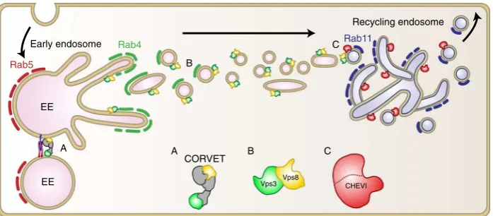
[image:9.595.50.547.45.634.2]components, after which they localise to Rab4-positive recycling
vesicles, which set out to fuse with Rab11- and CHEVI-positive
REs (Fig.
6
). The Vps3/Vps8-dependent recycling pathway is
required for ef
fi
cient recycling of internalised
β
1 integrins, and
perturbation of Vps3 or Vps8 impairs integrin-dependent cell
adhesion and spreading, FA assembly and cell migration.
Rab4 has been implicated in the short-loop recycling from EEs
to the plasma membrane, as well as in the long-loop recycling
from EEs to Rab11-positive REs
13,15–17,22,43,44. Since Vps3/8
knockdown prevents delivery of
β
1 integrins to REs (Fig.
4
c, f),
we conclude that Vps3 and Vps8 are involved in transport from
EEs to REs (Fig.
6
). Our
fi
nding that Vps3 and Vps8 interact with
each other (Fig.
2
) and localise to recycling vesicles that lack the
CORVET core subunit Vps18 (Fig.
1
) could indicate the
forma-tion of a Vps3/8 complex independently of the CORVET core
subunits. A direct interaction between Vps3 and Vps8 has been
found before
8, but seemed counterintuitive since in the current
CORVET model Vps3 and Vps8 are located at opposite ends of
the complex
45. The formation of a Vps3/8 complex without other
CORVET core components would provide a functional and
structural explanation for these
fi
ndings. Interestingly, Vps39 and
Vps41, the counterparts of Vps3 and Vps8 residing on the
opposite ends of the HOPS complex, can also directly interact
with each other
24, possibly by obtaining a closed post-fusion
conformation of HOPS following late endosome-lysosome
fusion
45,46. In analogy herewith, a closed post EE-EE fusion
conformation of the CORVET complex could precede the direct
interaction of Vps3 and Vps8. Future studies on endogenous
proteins are however required to determine if there is indeed a
functional Vps3/8 complex formed and whether this requires the
presence of additional proteins.
As a crucial mechanism to control integrin function, endocytic
recycling of integrins is a topic of extensive research
39,47–49.
Integrins use multiple recycling pathways depending on
hetero-dimer composition, conformation (active or inactive) and ligand
binding. A key question is how sorting to these different pathways
is achieved. Interestingly, we found that Vps3/8 knockdown had
no effect on Tf recycling (Fig.
4
a, b). Tf recycling depends on
Rab4 and Rab11, which are also involved in integrin
recy-cling
13,30,34,38,50. However, in contrast to Tf, recycling of
β
1
integrins depends on a NPxY motif that in EEs binds SNX17 or
SNX31 to sort integrins into recycling vesicles
51–53. This suggests
that within the Rab4/Rab11 pathway multiple sub-pathways exist.
Recently, a new protein complex, retriever, has been identi
fi
ed
that associates with SNX17 on EEs and sorts
α
5
β
1 integrin from
the early endosome to the recycling pathway
54. Retriever is
therefore a likely candidate to mediate sorting of
α
5
β
1 integrin
into the Vps3/8 recycling pathway. Together these data suggest
that the interplay between CORVET, retriever, CHEVI and a
possible Vps3/8 complex is important for integrin recycling. In
analogy with the concept of EE-associated tubular sorting
endo-somes or networks, machinery proteins involved in cargo sorting
might de
fi
ne subdomains on REs
27,55,56. At the level of REs, the
CHEVI protein Vps33B is a putative candidate to de
fi
ne exits
specialised for integrin recycling, since it interacts directly with
integrin
β
-subunits
57. Future studies are required to de
fi
ne
additional cargo for the retriever-Vps3/8-CHEVI-dependent
recycling pathway.
Mutations in the genes that encode for the CHEVI complex
cause ARC syndrome, by impairing polarised transport from REs
to the apical plasma membrane
18,19,21. Interestingly, a recently
identi
fi
ed patient with a mutation in VPS8 suffered from
arthrogryposis
58, one of the hallmarks of ARC patients. Our
model that CHEVI and Vps3/8 function in the same pathway
provide a possible explanation for this partially overlapping
phenotype.
Our data contribute to the emerging view that multi-subunit
tethering complexes are dynamic in composition in order to
facilitate multiple transport pathways
11,59. The increasing
num-ber of disease-causing mutations found in individual subunits
illustrates the importance to understand the role of each
indivi-dual subunit at a fundamental cellular level.
Methods
Cell culture and transfection. HeLa cells (American Type Culture Collection
clone ccl-2) were grown at 37 °C and 5% CO2in Dulbecco’s modified Eagle’s
medium (DMEM) supplemented with 10% heat-inactivated fetal bovine serum, 2mML-glutamine, 100 U/ml penicillin and 100 µg/ml streptomycin. Cells were transfected with cDNA using Effectene transfection reagent (Qiagen) according to the manufacturer’s protocol. siRNA transfection was performed using HiPerfect transfection Reagent (Qiagen) according to manufacturer’s protocol.
Antibodies. For IF and IEM labelling: mouse anti-HA (1:400, 901502, Covance);
mouse anti-β1 integrin (1:200, JB1A, Millipore); mouse anti-α5 integrin (1:200, VC5, BD Biosciences); goat anti-HA (1:400, A00168, Genscript); rabbit anti-GFP (TP401, Acris); rabbit anti-mCherry (1:400) was made on site as described60, and rabbit anti-vinculin was from Abcam (1:500, ab73412). For detection of GST fusion proteins, we used mouse anti-GST (1:5000, sc138, Santa Cruz). Rabbit anti-goat IgG (1:500, 7S, NORDIC) or rabbit anti-mouse IgG (1:250, Z0412, Dako) was used to bridge between goat or mouse antibodies and protein-A gold61. Fluorescent
secondary antibodies were obtained from Invitrogen (1:250). For immunopreci-pitation and western blotting we used mouse anti-GFP (1:2000, 11814460001, Roche), mouse anti-HA (1:2000, 901502, Covance), rabbit anti-mCherry (1:2000) and rabbit anti-GFP (1:2000, ab290, Abcam). Fluorescent secondary antibodies were obtained from Li-Cor (1:20,000).
Recycling endosome
A
B
C
CORVET A
Vps8 Vps3
B C
Rab11
Rab5
Rab4
EE
Early endosome
EE CHEVI
Fig. 6Model of the proposed function of Vps3 and Vps8 in endosomal recycling. The CORVET complex mediates the fusion between Rab5-positive EEs
[image:10.595.125.474.49.202.2]Reagents. Gold particles of 5 nm coupled to bovine serum albumin (BSA-Au5), and 10 nm or 15 nm gold particles conjugated to protein A were made on site (Cell Microscopy Core, UMC Utrecht, The Netherlands). HA-Vps8, V5 and Vps8-GFP, GFP-Rab4, mCherry-Rab4, FLAG-Rab4, mCherry-Rab5, mCherry-Rab7, mCherry-Rab11 and untagged Vps33B were cloned from cDNA purchased from Origen. HA-Vps3, GFP-Vps3, GFP-Vps11 and Vps41-GFP were a generous gift from Dr. J. Neefjes (NKI, Amsterdam, The Netherlands), VIPAS39-mCherry was a generous gift from Dr. P. Gissen (LMCB, UCL, London), and Vps33B-HA-V5-His was a generous gift from Dr. V. Faundez (Cell Biology, Emory University, Atlanta, GA, USA). Vps8, Vps3, Vps33B and VIPAS39 SMARTPool siRNAs were pur-chased from Dharmacon. FN and type-I collagen were purpur-chased from Sigma. MG132 was purchased from Enzo Life Sciences. EZ-link Sulfo-NHS-SS-biotin was from Thermo Fisher. Complete protease inhibitors were purchased from Roche. For detection of in vitro-translated proteins, peroxidase-conjugated streptavidin (0.1 µg/ml) was used (Jackson ImmunoResearch).
GST pulldown. Full-length Vps3 and Vps3 truncation mutants were cloned in
pGEX5 vectors. The proteins were produced inEscherichia coliBL21 (DE3) grown at 30 °C and lysed using sonification. Proteins were then immobilised on glu-tathione sepharose 4B beads (GE). HA-Vps8 was produced using a T7 in vitro translation kit (Promega) in combination with the Transcend Non-Radioactive Translation Detection System (Promega) that incorporates biotin-conjugated methionine. In vitro-translated proteins were incubated o/n at 4 °C with the loaded sepharose 4B Beads. After that beads were washed and proteins were eluted using 1× sample buffer for 30 min at 37 °C. Eluted proteins were separated using SDS-polyacrylamide gel electrophoresis (SDS-PAGE) and analysed using western blotting.
Co-immunoprecipitation. Cells were washed in ice-cold phosphate-buffered saline
(PBS) and scraped in lysis buffer (40 mM TRIS (pH 7.4), 100 mM NaCl, 0.1% TX-100 and 5 mM EDTA supplemented with complete protease inhibitors (Roche)). Cells were lysed for 30–60 min at 4 °C followed by 20 min centrifugation at 13,000 r.p.m. at 4 °C. Protein A beads were incubated with the desired antibody for 2 h at 4 °C, washed and added to the collected supernatant. Beads and supernatant were incubated in a rotator at 4 °C for 1 h. Beads were washed six times with lysis buffer minus protease inhibitors after which proteins were eluted using 1× sample buffer for 30 min at 37 °C. Eluted proteins were separated using SDS-PAGE and analysed by western blotting.
Western blotting. Cells were washed in ice-cold PBS and scraped in E1A lysis
buffer (40 mM TRIS (pH 7.4), 100 mM NaCl, 0.1%TX-100 and 5 mM EDTA supplemented with complete protease inhibitors (Roche)). Cells were lysed for 30 min at 4 °C followed by 20 min centrifugation at max speed at 4 °C to remove cell debris. Proteins were eluted using 1.5× sample buffer for 30 min at 37 °C, separated on a 7.5 or 10% SDS-PAGE and transferred to Immobilon-FL PVDF membrane (Millipore). Membranes were blocked at room temperature (RT) using Odyssey blocking buffer in PBS (1:1) for 1 h and incubated with selected primary antibodies diluted in Odyssey blocking buffer in PBS-T 0.2% (1:1). Membranes were washed in PBS-T 0.1% and incubated with secondary antibodies diluted in Odyssey blocking buffer in PBS-T 0.2% (1:1) for 1 h at RT. Finally, membranes were washed and imaged on an Odyssey imaging system (Li-Cor) for co-IP experiments. For GST-pulldown experiments, biotin-labelled proteins were detected using ECL (GE Healthcare), which were consequently exposed to Kodak XBfilms (Rochester). Complete, uncropped blots are presented in the Supplementary Information.
IF microscopy. For Tf recycling experiments, HeLa cells were incubated with
dextran conjugated to Alexa-488 (Thermo Fisher) for 120 min and with Tf con-jugated to Alexa-568 (Thermo Fisher) for 30 min and imaged in pre-warmed image medium (Sigma) on a climate-controlled DeltaVision widefield microscope using a ×100/1.4 A immersion objective. Forfixed-cell IF experiments, HeLa cells grown on sterile glass coverslips were washed with ice-cold PBS andfixed with 4% par-aformaldehyde (PFA) in PBS for 20 min at RT. Then, cells were permeabilised using 0.1% TX-100 in PBS for 5 min and blocked for 15 min using PBS supple-mented with 1% BSA. Cells were labelled with primary antibodies diluted in blocking buffer at RT for 1 h, washed and labelled withfluorescent secondary antibodies for 30 min in the dark. After labelling, the cells were washed and mounted using Prolong Gold antifade reagent with 4′,6-diamidino-2-phenylindole (DAPI; Thermo Fisher) and imaged on a DeltaVision widefield microscope using a ×100/1.4 A immersion objective. Pictures were deconvolved using Softworx soft-ware and analysed using Fiji ImageJ v1.48q.
Immuno-electron microscopy. Sample preparation, ultrathin cryosectioning and
immunolabelling were performed as described61. In brief, HeLa cells were grown on 60 mm dishes andfixed by the addition of freshly prepared 4% PFA in 0.1 M phosphate buffer (pH 7.4) to an equal volume of culture medium for 10 min, followed by postfixation with fresh 4% PFA overnight at 4 °C. Fixed cells were washed in PBS containing 0.05 M glycine and gently scraped in PBS containing 1% gelatine. Cells were pelleted in 12% gelatin in PBS, which was then left to solidify on ice and cut into small blocks. The blocks were infiltrated overnight in 2.3 M
sucrose at 4 °C, mounted on aluminium pins and frozen in liquid nitrogen. The 70 nm ultrathin cryosections were cut on a Leica ultracut UCT cryomicrotome and picked up in a freshly prepared 1:1 mixture of 2.3 M sucrose and 1.8% methyl-cellulose. Sections were then immunogold-labelled with antibodies and protein A gold, contrasted and examined on a JEOL transmission electron microscope 1010. For antibody uptake IEM, HeLa cells were serum-starved overnight in DMEM containing 0.01% BSA (D/B) and subsequently incubated at 4 °C for 1 h with
anti-β1 integrin (JB1A, Millipore) diluted in D/B to afinal concentration of 10 µg/ml. Cells were transferred to pre-warmed D/B, and integrin endocytosis was allowed for 2 h at 37 °C after which cells were prepared for IEM as described above and labelled on section with rabbit-ant-mouse IGG (Z0412, Dako) and protein A 10 nm gold.
Stimulation-induced integrin recycling assay. Integrin recycling was analysed
essentially as described34. In brief, HeLa cells were serum-starved overnight in
DMEM containing 0.01% BSA (D/B) and subsequently incubated at 4 °C for 1 h with anti-β1 integrin (JB1A, Millipore) diluted in D/B to afinal concentration of 10 µg/ml. Excess antibody was removed by two washes with cold D/B. Cells were then transferred to pre-warmed D/B, and integrin endocytosis was allowed for 2 h at 37 °C. Surface-bound antibodies that were not endocytosed were removed with two acid washes (0.5% acetic acid and 0.5 M NaCl) at 4 °C. For recycling, cells were stimulated with pre-warmed D/B containing 20% fetal calf serum (FCS). The pool of internalβ1 integrins was monitored at the indicated time points byfixation and preparation for IF microscopy as described above.
Cell adhesion and migration assays. For cell adhesion assays, 96-well plates were coated with 5 µg/ml FN (90 min) or 3 µg/ml Col-I (5 min) at 37 C, washed with PBS and blocked with 2% BSA for 30 min at 37 °C. Cells were trypsinised, resus-pended in serum-free DMEM and seeded at a density of 5 × 10⁴/well. At the
indicated time points, non-adherent cells were washed away with PBS. Attached cells werefixed with 4% PFA for 10 min, washed twice with PBS and stained with Crystal Violet (5 mg/ml in 2% ethanol) for 10 min. Stained cells were extensively washed with water and lysed with 2% SDS for 30 min. Plates were read at 590 nm. For cell-spreading assays, cells were seeded in plates coated with 5 µg/ml FN or 3 µg/ml Col-I,fixed at the indicated time points, and the number of spread cells was counted. For FA analysis, cells were seeded onto coverslips coated with 5 µg/ml FN or 3 µg/ml Col-I, andfixed at the indicated time points. Confocal images of vinculin staining were acquired usingfixed settings, background-subtracted and thresholded in ImageJ 1.44, and FA number was then determined using the
‘analyse particles’function.
For cell migration assays, cells were sparsely seeded on the indicated matrix proteins, and phase-contrast images were captured every 10 min at 37 °C and 5% CO2on a widefield CCD system using a ×10 dry lens objective (Carl Zeiss
MicroImaging). Migration tracks were generated using ImageJ 1.44, and the average displacement and migration speed were calculated from ~30 cells out of three independent experiments. The tail retraction phenotype was scored from the same movies.
Proximity ligation assay. HeLa cells grown on sterile glass coverslips were washed with ice-cold PBS andfixed with 4% PFA in PBS for 20 min at RT. Then, cells were permeabilised using 0.1% Triton X-100 in PBS for 5 min and blocked for 15 min using PBS supplemented with 1% BSA. Cells were labelled with primary antibodies diluted in blocking buffer at RT for 1 h, washed and labelled with mouse PLUS and Rabbit MINUS antibodies for 1 h at 37 °C. The PLA signal was visualised using the in situ PLA detection kit (Sigma) according to the manufacturer’s protocol. After labelling the cells were mounted using Prolong Gold antifade reagent with DAPI and imaged on a DeltaVision widefield microscope using a ×100/1.4 A immersion objective. Pictures were deconvolved using Softworx software and analysed using Fiji ImageJ v1.48q.
Integrin recycling and capture ELISA. Integrin recycling was investigated
essentially as described earlier35. Briefly, cells were transferred to ice, washed twice in PBS and surface-labelled at 4 °C with 140 µg/ml NHS-SS-biotin. Internalisation was allowed for 15 min, after which the cells were washed with PBS, and remaining cell-surface biotin was removed with 20 mM MesNa. Recycling of internalised proteins was then induced in DMEM/FCS at 37 °C, whereafter biotin was removed from recycled proteins by a second reduction with MesNa, which was quenched with 20 mM iodoacetamide. Cells were then washed twice in PBS and lysed in 1.5 NDLB buffer (150 mM NaCl, 50 mM Tris, 10 mM NaF, 1.5 mM Na3VO3, 5 mM EDTA, 5 mM EGTA, 1.5% TX-100, 0.75% Igepal CA-630 and 1 mM 4-(2-ami-noethyl)benzynesulphonylfluoride supplemented with complete protease inhibi-tors). Maxisorb 96-well plates (Life Technologies) were coated overnight with 5μg/ ml anti-integrinα5 in 0.05 M Na2CO3(pH 9.6) at 4 °C, and blocked in PBS/0.05%
Tween-20 (PBS-T) with 5% BSA. Plates were incubated overnight with cell lysates at 4 °C, washed with PBS-T and incubated with horseradish peroxidase-streptavidin in PBS-T/1% BSA for 1 h at 4 °C. After washing, biotinylated proteins were detected withortho-phenylenediamine in a buffer containing 25.4 mM Na2HPO4, 12.3 mM citric acid (pH 5.4) and 0.003% H2O2. The reaction was
Statistical analysis. For calculation of correlation coefficients of co-localisation in IF experiments the signal intensity over a vector was plotted using the plot profile tool in FIJI ImageJ in the green, red and where needed, the far-red channel. Correlation coefficients between two plots were calculated using the function:
correlðx;yÞ ¼ P P
ðx xÞðy yÞ ffiffiffiffiffiffiffiffiffiffiffiffiffiffiffiffiffiffiffiffiffiffiffiffiffiffiffiffiffiffiffiffi ðx xÞ2ðy yÞ2
q ð1Þ
Correlation coefficients were calculated between the channels from at least 30 cells taken from three separate experiments. Correlation coefficients were averaged as follows. Individual correlation coefficients (r) underwent Fisherr-to-z transformation according to the formula:
zr¼1
2ln 1þr 1 r
ð2Þ
The mean of thezvalues was calculated and the resulting mean was transformed back to the mean correlation coefficient using:
r¼e 2z 1
e2zþ1 ð3Þ
To assess the significance between two mean correlation coefficients thez-score andP-value were calculated using:
z¼ 1 2ln11þrr11
þ 1 2ln11þrr22
ffiffiffiffiffiffiffiffiffiffiffiffiffiffiffiffiffiffiffiffiffi 1 n1 3þn213
q ð4Þ
P Zð zÞ ¼
Z z
1 1
ffiffiffiffiffi
2π
p eμ
2
2dμ ð5Þ
Data availability. The data that support thefindings of this study are available from the corresponding author upon reasonable request.
Received: 28 July 2016 Accepted: 30 January 2018
References
1. Boustany, R. M. Lysosomal storage diseases—the horizon expands.Nat. Rev. Neurol.9, 583–598 (2013).
2. Mellman, I. & Yarden, Y. Endocytosis and cancer.Cold Spring Harb. Perspect. Biol.5, a016949 (2013).
3. Neefjes, J. & van der Kant, R. Stuck in traffic: an emerging theme in diseases of the nervous system.Trends Neurosci.37, 66–76 (2014).
4. Epp, N., Rethmeier, R., Kramer, L. & Ungermann, C. Membrane dynamics and fusion at late endosomes and vacuoles—Rab regulation, multisubunit tethering complexes and SNAREs.Eur. J. Cell Biol.90, 779–785 (2011). 5. Rizo, J. & Sudhof, T. C. The membrane fusion enigma: SNAREs, Sec1/Munc18
proteins, and their accomplices—guilty as charged?Annu Rev. Cell Dev. Biol.
28, 279–308 (2012).
6. Schimmoller, F., Simon, I. & Pfeffer, S. R. Rab GTPases, directors of vesicle docking.J. Biol. Chem.273, 22161–22164 (1998).
7. Jordens, I., Marsman, M., Kuijl, C. & Neefjes, J. Rab proteins, connecting transport and vesicle fusion.Traffic6, 1070–1077 (2005).
8. Perini, E. D., Schaefer, R., Stoter, M., Kalaidzidis, Y. & Zerial, M. Mammalian CORVET is required for fusion and conversion of distinct early endosome sub-populations.Traffic15, 1366–1389 (2014).
9. van der Kant, R. et al Characterization of the mammalian CORVET and HOPS complexes and their modular restructuring for endosome specificity.J. Biol. Chem.290, 30280–30290 (2015).
10. Lachmann, J., Glaubke, E., Moore, P. S. & Ungermann, C. The Vps39-like TRAP1 is an effector of Rab5 and likely the missing Vps3 subunit of human CORVET.Cell Logist.4, e970840 (2014).
11. Spang, A. Membrane tethering complexes in the endosomal system.Front. Cell Dev. Biol.4, 35 (2016).
12. Klinger, C. M., Klute, M. J. & Dacks, J. B. Comparative genomic analysis of multi-subunit tethering complexes demonstrates an ancient pan-eukaryotic complement and sculpting in Apicomplexa.PLoS ONE8, e76278 (2013). 13. van der Sluijs, P. et al. The small GTP-binding protein rab4 controls an early
sorting event on the endocytic pathway.Cell70, 729–740 (1992).
14. Ullrich, O., Reinsch, S., Urbe, S., Zerial, M. & Parton, R. G. Rab11 regulates recycling through the pericentriolar recycling endosome.J. Cell Biol.135, 913–924 (1996).
15. Yudowski, G. A., Puthenveedu, M. A., Henry, A. G. & von Zastrow, M. Cargo-mediated regulation of a rapid Rab4-dependent recycling pathway.Mol. Biol. Cell20, 2774–2784 (2009).
16. McCaffrey, M. W. et al. Rab4 affects both recycling and degradative endosomal trafficking.FEBS Lett.495, 21–30 (2001).
17. de Wit, H. et al. Rab4 regulates formation of synaptic-like microvesicles from early endosomes in PC12 cells.Mol. Biol. Cell12, 3703–3715 (2001). 18. Cullinane, A. R. et al. Mutations in VIPAR cause an arthrogryposis, renal
dysfunction and cholestasis syndrome phenotype with defects in epithelial polarization.Nat. Genet.42, 303–312 (2010).
19. Smith, H. et al Associations among genotype, clinical phenotype and intracellular localization of trafficking proteins in ARC syndrome.Hum. Mutat.33, 1656–1664 (2012).
20. Zhu, G. D. et al. SPE-39 family proteins interact with the HOPS complex and function in lysosomal delivery.Mol. Biol. Cell20, 1223–1240 (2009). 21. Gissen, P. et al. Clinical and molecular genetic features of ARC syndrome.
Hum. Genet.120, 396–409 (2006).
22. Sonnichsen, B., De Renzis, S., Nielsen, E., Rietdorf, J. & Zerial, M. Distinct membrane domains on endosomes in the recycling pathway visualized by multicolor imaging of Rab4, Rab5, and Rab11.J. Cell Biol.149, 901–914 (2000).
23. Wegner, C. S. et al. Ultrastructural characterization of giant endosomes induced by GTPase-deficient Rab5.Histochem. Cell Biol.133, 41–55 (2010). 24. Plemel, R. L. et al. Subunit organization and Rab interactions of Vps-C protein
complexes that control endolysosomal membrane traffic.Mol. Biol. Cell22, 1353–1363 (2011).
25. Wartosch, L., Gunesdogan, U., Graham, S. C. & Luzio, J. P. Recruitment of VPS33A to HOPS by VPS16 is required for lysosome fusion with endosomes and autophagosomes.Traffic16, 727–742 (2015).
26. McMahon, H. T. et al. Cellubrevin is a ubiquitous tetanus-toxin substrate homologous to a putative synaptic vesicle fusion protein.Nature364, 346–349 (1993).
27. Klumperman, J. & Raposo, G. The complex ultrastructure of the endolysosomal system.Cold Spring Harb. Perspect. Biol.6, a016857 (2014). 28. Hao, M. & Maxfield, F. R. Characterization of rapid membrane internalization
and recycling.J. Biol. Chem.275, 15279–15286 (2000).
29. van Dam, E. M., Ten Broeke, T., Jansen, K., Spijkers, P. & Stoorvogel, W. Endocytosed transferrin receptors recycle via distinct dynamin and phosphatidylinositol 3-kinase-dependent pathways.J. Biol. Chem.277, 48876–48883 (2002).
30. Ren, M. et al. Hydrolysis of GTP on rab11 is required for the direct delivery of transferrin from the pericentriolar recycling compartment to the cell surface but not from sorting endosomes.Proc. Natl Acad. Sci. USA95, 6187–6192 (1998). 31. Ivaska, J. et al. PKCepsilon-mediated phosphorylation of vimentin controls
integrin recycling and motility.EMBO J.24, 3834–3845 (2005).
32. Mohrmann, K. & van der Sluijs, P. Regulation of membrane transport through the endocytic pathway by rabGTPases.Mol. Membr. Biol.16, 81–87 (1999). 33. Caswell, P. T., Vadrevu, S. & Norman, J. C. Integrins: masters and slaves of
endocytic transport.Nat. Rev. Mol. Cell Biol.10, 843–853 (2009). 34. Powelka, A. M. et al. Stimulation-dependent recycling of integrin beta1
regulated by ARF6 and Rab11.Traffic5, 20–36 (2004).
35. Roberts, M., Barry, S., Woods, A., van der Sluijs, P. & Norman, J. PDGF-regulated rab4-dependent recycling of alphavbeta3 integrin from early endosomes is necessary for cell adhesion and spreading.Curr. Biol.11, 1392–1402 (2001).
36. Hynes, R. O. Integrins: bidirectional, allosteric signaling machines.Cell110, 673–687 (2002).
37. Caswell, P. & Norman, J. Endocytic transport of integrins during cell migration and invasion.Trends Cell Biol.18, 257–263 (2008).
38. Caswell, P. T. & Norman, J. C. Integrin trafficking and the control of cell migration.Traffic7, 14–21 (2006).
39. De Franceschi, N., Hamidi, H., Alanko, J., Sahgal, P. & Ivaska, J. Integrin traffic—the update.J. Cell Sci.128, 839–852 (2015).
40. Webb, D. J., Parsons, J. T. & Horwitz, A. F. Adhesion assembly, disassembly and turnover in migrating cells -- over and over and over again.Nat. Cell Biol.
4, E97–E100 (2002).
41. Theisen, U., Straube, E. & Straube, A. Directional persistence of migrating cells requires Kif1C-mediated stabilization of trailing adhesions.Dev. Cell23, 1153–1166 (2012).
42. Dozynkiewicz, M. A. et al. Rab25 and CLIC3 collaborate to promote integrin recycling from late endosomes/lysosomes and drive cancer progression.Dev. Cell22, 131–145 (2012).
44. Nagelkerken, B. et al. Rabaptin4, a novel effector of the small GTPase rab4a, is recruited to perinuclear recycling vesicles.Biochem. J.346, 593–601 (2000). 45. Brocker, C. et al. Molecular architecture of the multisubunit homotypic fusion
and vacuole protein sorting (HOPS) tethering complex.Proc. Natl Acad. Sci. USA109, 1991–1996 (2012).
46. Solinger, J. A. & Spang, A. Tethering complexes in the endocytic pathway: CORVET and HOPS.FEBS J.280, 2743–2757 (2013).
47. Jacquemet, G., Hamidi, H. & Ivaska, J. Filopodia in cell adhesion, 3D migration and cancer cell invasion.Curr. Opin. Cell Biol.36, 23–31 (2015). 48. Maritzen, T., Schachtner, H. & Legler, D. F. On the move: endocytic
trafficking in cell migration.Cell. Mol. Life Sci.72, 2119–2134 (2015). 49. Paul, N. R., Jacquemet, G. & Caswell, P. T. Endocytic trafficking of integrins in
cell migration.Curr. Biol.25, R1092–R1105 (2015).
50. Arjonen, A., Alanko, J., Veltel, S. & Ivaska, J. Distinct recycling of active and inactive beta1 integrins.Traffic13, 610–625 (2012).
51. Steinberg, F., Heesom, K. J., Bass, M. D. & Cullen, P. J. SNX17 protects integrins from degradation by sorting between lysosomal and recycling pathways.J. Cell Biol.197, 219–230 (2012).
52. Bottcher, R. T. et al. Sorting nexin 17 prevents lysosomal degradation of beta1 integrins by binding to the beta1-integrin tail.Nat. Cell Biol.14, 584–592 (2012).
53. Margadant, C., Kreft, M., de Groot, D. J., Norman, J. C. & Sonnenberg, A. Distinct roles of talin and kindlin in regulating integrin alpha5beta1 function and trafficking.Curr. Biol.22, 1554–1563 (2012).
54. McNally, K. E. et al. Retriever is a multiprotein complex for retromer-independent endosomal cargo recycling.Nat. Cell Biol.19, 1214–1225 (2017). 55. Peden, A. A. et al. Localization of the AP-3 adaptor complex defines a novel
endosomal exit site for lysosomal membrane proteins.J. Cell Biol.164, 1065–1076 (2004).
56. Bonifacino, J. S. & Rojas, R. Retrograde transport from endosomes to the trans-Golgi network.Nat. Rev. Mol. Cell Biol.7, 568–579 (2006).
57. Xiang, B. et al. Characterization of a novel integrin binding protein, VPS33B, which is important for platelet activation and in vivo thrombosis and hemostasis.Circulation132, 2334–2344 (2015).
58. Bayram, Y. et al. Molecular etiology of arthrogryposis in multiple families of mostly Turkish origin.J. Clin. Invest126, 762–778 (2016).
59. Pols, M. S. et al. hVps41 and VAMP7 function in direct TGN to late endosome transport of lysosomal membrane proteins.Nat. Commun.4, 1361 (2013).
60. Hagemeijer, M. C. et al. Membrane rearrangements mediated by coronavirus nonstructural proteins 3 and 4.Virology458-459, 125–135 (2014). 61. Slot, J. W. & Geuze, H. J. Cryosectioning and immunolabeling.Nat. Protoc.2,
2480–2491 (2007).
Acknowledgements
We thank Adam Grieve and Catherine Rabouille for fruitful discussions and reagents for the initial integrin experiments. Our colleagues of the Center for Molecular Medicine and section Cell Biology for precious input and especially René Scriwanek for preparation of
the electron micrographs. Paul Gissen, Victor Faundez and Sjaak Neefjes are kindly acknowledged for sharing critical reagents. Coert Margadant was supported by a grant from the Netherlands organisation for Scientific Research (ZonMW Veni 016.146.160)
Author contributions
C.T.H.J. designed and performed the experiments needed for Figs. 1–6, analysed the data and wrote the paper. R.G. designed and performed the experiments needed for Figs. 1 and 3, analysed the data and wrote the paper. T.V. performed experiments needed for Figs. 1a, d, 2d, and 4c, f. C.t.B. performed experiments needed for Figs. 2b and 4a and cloned constructs used in the manuscript. R.E.N.v.d.W. performed experiments needed for Figs. 2b and 4e. N.L. performed experiments needed for Fig. 2c, d. J.d.R. designed experiments needed for Fig. 4 and wrote the paper. A.A.P. designed experiments needed for Fig. 3 and wrote the paper. P.v.d.S. designed the experiments for Fig. 1b, c, e, cloned constructs and wrote the paper. C.M. designed and performed the experiments for Figs. 4 and 5, supervised the project, analysed the data and wrote the paper. J.K. initiated and supervised the project, designed experiments for Figs. 1–4 and wrote the paper.
Additional information
Supplementary Informationaccompanies this paper at https://doi.org/10.1038/s41467-018-03226-8.
Competing interests:The authors declare no competingfinancial interests.
Reprints and permissioninformation is available online athttp://npg.nature.com/ reprintsandpermissions/
Publisher's note:Springer Nature remains neutral with regard to jurisdictional claims in published maps and institutional affiliations.
Open Access This article is licensed under a Creative Commons Attribution 4.0 International License, which permits use, sharing, adaptation, distribution and reproduction in any medium or format, as long as you give appropriate credit to the original author(s) and the source, provide a link to the Creative Commons license, and indicate if changes were made. The images or other third party material in this article are included in the article’s Creative Commons license, unless indicated otherwise in a credit line to the material. If material is not included in the article’s Creative Commons license and your intended use is not permitted by statutory regulation or exceeds the permitted use, you will need to obtain permission directly from the copyright holder. To view a copy of this license, visithttp://creativecommons.org/ licenses/by/4.0/.