Molecular Analysis of an Outbreak of Lethal Postpartum Sepsis
Caused by
Streptococcus pyogenes
Claire E. Turner,aMatthew Dryden,bMatthew T. G. Holden,cFrances J. Davies,aRichard A. Lawrenson,aLeili Farzaneh,a Stephen D. Bentley,cAndroulla Efstratiou,dShiranee Sriskandana
Infectious Diseases & Immunity, Imperial College London, Hammersmith Hospital, London, United Kingdoma
; Royal Hampshire County Hospital, Winchester, Hampshire, United Kingdomb
; Pathogen Genomics, The Wellcome Trust Sanger Institute, Hinxton, Cambridge, United Kingdomc
; Microbiology Reference Services Division, Public Health England, Colindale, London, United Kingdomd
Sepsis is now the leading direct cause of maternal death in the United Kingdom, andStreptococcus pyogenesis the leading patho-gen. We combined conventional and genomic analyses to define the duration and scale of a lethal outbreak. Two postpartum deaths caused byS. pyogenesoccurred within 24 h; one was characterized by bacteremia and shock and the other by hemorrhagic pneumonia. The women gave birth within minutes of each other in the same maternity unit 2 days earlier. Seven additional in-fections in health care and household contacts were subsequently detected and treated. All cluster-associatedS. pyogenesisolates were genotypeemm1 and were initially indistinguishable from other United Kingdomemm1 isolates. Sequencing of the viru-lence genesicrevealed that all outbreak isolates had the same uniquesictype. Genome sequencing confirmed that the cluster was caused by a uniqueS. pyogenesclone. Transmission between patients occurred on a single day and was associated with casual contact only. A single isolate from one patient demonstrated a sequence change insicconsistent with longer infection duration. Transmission to health care workers was traced to single clinical contacts with index cases. The last case was detected 18 days after the first case. Following enhanced surveillance, the outbreak isolate was not detected again. Mutations in bacterial regula-tory genes played no detectable role in this outbreak, illustrating the intrinsic ability ofemm1S. pyogenesto spread while retain-ing virulence. This fast-movretain-ing outbreak highlights the potential ofS. pyogenesto cause a range of diseases in the puerperium with rapid transmission, underlining the importance of immediate recognition and response by clinical infection and occupa-tional health teams.
C
ases of postpartum maternalStreptococcus pyogenessepsis oc-cur sporadically; paired cases and deaths are rare in the devel-oped world (1,2). We describe two postpartum deaths due toS. pyogenes(group AStreptococcus[GAS]) that occurred within 24 h in the same maternity unit; one case was characterized by septic shock and the other by hemorrhagic pneumonia. Both cases were associated with seven additional infections detected in health care and family contacts. We combined genotypic and phenotypic analyses with whole-genome sequencing to confirm that the iso-lates were unique and distinct from otheremm1 GAS isolates cir-culating in the United Kingdom, enabling us to determine the time limits and scale of this outbreak.CASE REPORTS
Case 1.A 39-year-old teacher (gravida 3, para 2) presented at term with irregular contractions and was monitored for 5 h in the ma-ternity unit prior to discharge. She was readmitted 5 h later and delivered a live female infant. Other than a small second-degree tear, there were no complications and the mother was discharged 13 h after delivery. Approximately 30 h following delivery she awoke with lower abdominal pain necessitating hospital readmis-sion, inhaled nitrous oxide, and opiates, although physiological measurements were initially normal. She collapsed, 6 h later, with hypotension and was transferred to the intensive care unit. She rapidly developed multiorgan failure and responded poorly to inotropic and ventilatory support in addition to broad-spectrum antibiotics. The following day, blood cultures indicated a strepto-coccal infection, and high-dose intravenous pooled human im-munoglobulin G (IVIG) was administered. The patient deterio-rated and died 2.5 days after delivery of her third child. GAS was
isolated from blood cultures and genital tract swabs obtained an-temortem and also from uterine cervix tissue obtained postmor-tem.
Case 2.A 29-year-old primigravid teacher was admitted while in labor to the same unit on the same day as case 1. She had experienced spontaneous rupture of membranes 48 h earlier and had commenced prophylactic amoxicillin. During the day she had had a transient temperature of 37.7°C which settled, though other physiological measurements were normal. An oxytocin infusion was commenced; 12 h later, a live male infant was delivered by emergency cesarean section, and a final antibiotic dose was ad-ministered intraoperatively. The time of cesarean delivery was 2 min before the delivery of the baby of case 1, although the delivery occurred in a separate section of the maternity unit and was un-dertaken by different staff. She was discharged approximately 66 h after cesarean section. Approximately 76 h after delivery, she de-veloped a cough and chest pain associated with blood-stained spu-tum. A community midwife arranged hospital readmission. While
Received11 March 2013Returned for modification10 April 2013 Accepted14 April 2013
Published ahead of print24 April 2013
Address correspondence to Shiranee Sriskandan, [email protected]. Supplemental material for this article may be found athttp://dx.doi.org/10.1128 /JCM.00679-13.
Copyright © 2013 Turner et al. This is an open-access article distributed under the terms of theCreative Commons Attribution 3.0 Unported license.
packing, the patient collapsed in respiratory arrest associated with hemoptysis. Resuscitation was attempted by a paramedical team on site and in the hospital emergency unit but the patient died on 24 December, 3.5 days after cesarean section. GAS was isolated from throat, hemorrhagic lung, and uterine cervix samples post-mortem but not from the cesarean wound or lower genital tract. GAS isolates from both cases were subsequently identified asemm type 1.
Response of infection team and intensive surveillance.A hos-pital outbreak control team managed the incident, surveillance, and epidemiology. Household and health care worker (HCW) contacts of each patient were screened, including antenatal, dur-ing-labor, and postnatal contacts (Fig. 1). Environmental screen-ing was conducted once in the immediate aftermath of the out-break by swabbing of baths, showers, basins, and bed space surfaces, and the samples obtained by swabbing were cultured on blood agar.
As part of a 2-month period of intensive surveillance, all moth-ers who had been on the unit concurrently with the two cases were contacted and advised to seek medical attention and screening if symptomatic. All women in labor were screened during this pe-riod within a day of admission. Throat and skin swabs (if skin lesions were reported) were used for screening for GAS, plus lower genital tract swabs for all women in labor. During the intensive surveillance period, enhanced twice daily chlorine-based cleaning in the maternity unit was undertaken (previously once daily de-tergent-based cleaning).
Screening results. (i) Household contacts.Four household contacts of case 1 were screened, of whom 2 were positive for GAS. Two household contacts of case 2 were screened, of whom 1 was positive for GAS.
The baby of case 1 was admitted to the neonatal unit with a
raised level of C-reactive protein (CRP) and received ceftriaxone for suspected sepsis; nose and ear swabs obtained 2 days after delivery yielded GASemm1, although blood and cerebrospinal fluid (CSF) cultures were negative. The baby of case 2 was not colonized or infected with GAS.
Throat swabs from the partners of case 1 and case 2 yielded GASemm1, and both of these individuals received antibiotics. One partner reported pharyngitis at the time of screening (i.e., after the death of case 1), while the other developed a lower respi-ratory tract infection necessitating hospital admission 4 days after the death of case 2.
(ii) Health care contacts.Of 69 HCW contacts screened, 3/69 (4.3%) had throat swabs that yielded GASemm1, and each re-ceived antibiotic treatment. The three HCWs reported single ep-isodes of postnatal clinical contact with the patients and included an obstetrician who examined case 1, an intensive care unit (ICU) nurse present at the intubation of case 1, and a community mid-wife attending case 2 at home. Two of the HCWs developed sig-nificant symptoms 7 to 10 days after the deaths; one had tonsillitis and cervical lymphadenopathy, while the other had pharyngitis. HCWs who tested positive for GAS were offered 10 days of treat-ment with oral amoxicillin, removed from work for this period, and screened again at the end of treatment. The partner of a symp-tomatic HCW was treated for GAS pharyngitis, though the isolate was not available for study.
One hundred ninety-three HCWs from the same institution, who were not contacts of the two cases, were also screened. Two of the 193 (1%) had throat swabs that yielded GAS and received antibiotics; however, these isolates wereemm12.
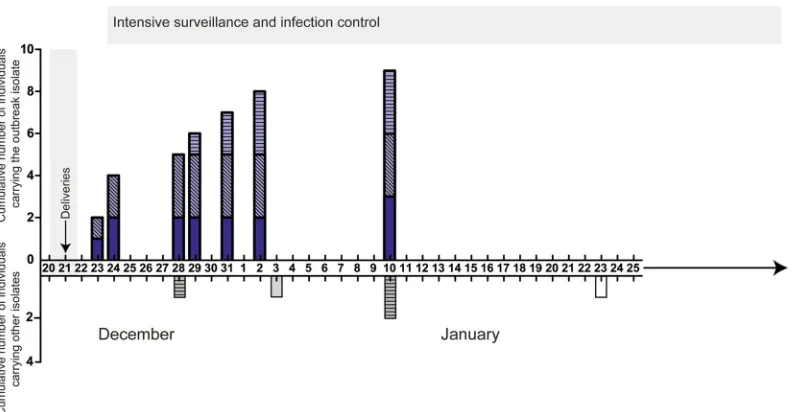
(iii) Other patients.In the immediate aftermath of the out-break, 42 maternity patients were screened, including women who reported symptoms compatible with GAS and had been on the FIG 1Identification of group A streptococcus (GAS) cases. Cumulative number of individuals carrying or infected with the outbreak strain by date (upper axis, blue-shaded bars). Individuals with the outbreak strain included maternity patients (solid shading), household contacts (diagonal crosshatching), and health care workers (HCWs) (horizontal crosshatching). Individuals found to be carrying or infected with other GAS isolates during the period of surveillance are shown on the lower axis (gray-shaded bars). Period of overlapping hospital admissions for three maternity patients denoted by gray shaded area. During the period of intense surveillance, which started on December 24 and lasted for 2 months, two HCWs were identified to be carryingemm12 GAS (gray-shaded horizontally hatched bars) and two nonmaternity patients had community-acquired invasive infections caused by GAS strainsemm89 (gray-shaded bar, January 3) and
[image:2.585.96.492.65.271.2]unit at the same time as the two fatal cases, along with those ad-mitted up to 2 weeks later. One of the 42 had a throat swab that yielded GAS, typed asemm1. This patient reported pharyngitis and sinusitis that developed 1 week postpartum and also recalled meeting and talking with case 1 on the day of her original admis-sion (Fig. 1). This patient was treated with antibiotics in the com-munity.
Two further cases of invasive GAS infection (necrotizing fas-ciitis and pneumonia) were admitted from the community during the month following the maternal fatalities; however, the GAS isolates in these cases wereemm75 andemm89 (Fig. 1).
(iv) Environment.GAS was not isolated from any environ-mental samples.
(v) Enhanced surveillance.A further 4-month period of en-hanced clinical surveillance was instituted within the obstetric and neonatal units, in which HCW were particularly alert for all GAS infections and swabbed throats and skin of patients or HCW if there was any history or indication of inflammation. No further GAS carriage or infection was detected.
MATERIALS AND METHODS
Phenotyping and genotyping of GAS strains.All 15emm1 GAS isolates from the cluster were compared with 14 otheremm1 GAS isolates submit-ted to the national reference laboratory from elsewhere in England in the 3 months preceding and 1 month following the outbreak, including, where available, isolates from the same region (see Table S1 in the supple-mental material). GAS phenotypic comparison,emmtyping, superanti-gen superanti-genotyping, andcovR/Sandsicloci sequencing are described in Sup-plementary Methods in the supplemental material.
GAS whole-genome sequencing.Illumina sequencing was performed on 24 strains, including all 15 strains from the outbreak, and mapped against the complete genome sequence of the U.S.emm1 strain MGAS5005 (3). In excess of 50-fold sequence coverage was achieved for all isolates (see Supple-mentary Methods in the supplemental material).
Measurement of anti-emm1 GAS immunity in pregnant women.As a surrogate for immunity among women of child-bearing age in the United Kingdom, anonymized serum samples from antenatal screening were obtained from the Imperial College Healthcare Trust and used in accordance with approval from the West London Research Ethics Com-mittee.
Enzyme-linked immunosorbent assay-based assays of immunity. Reactivity of antenatal sera to recombinant SIC proteins was tested using an enzyme-linked immunosorbent assay (ELISA)-based method. Diluted antenatal sera were applied to SIC protein-coated wells, and bound IgG was detected with goat anti-human IgG-horseradish peroxidase (HRP). In order to quantify SIC-reactive IgG in each serum sample relative to the human blood product intravenous immunoglobulin (IVIG) (Zenalb 4.5), which has high antistreptococcal activity, the activity of IVIG against SIC protein was also measured using a series of fixed concentrations of IVIG to generate a standard curve of IVIG-equivalent anti-SIC IgG on each plate. A similar protocol was used to detect IgG reactivity of antenatal sera to wholeemm1S. pyogenescells, but the wells were coated with heat-killed
emm1S. pyogenes. Details are provided in the supplemental methods in the supplemental material.
Opsonophagocytosis assays of immunity.Assays were performed us-ing 80 (of 199) representative heat-inactivated antenatal patient sera.
emm1 GAS (strain H584) isolates were stained with fluorescein isothio-cyanate (FITC) (Invitrogen) and opsonized with test serum before being added to 2⫻106fresh human neutrophils and 10% complement (rabbit
serum; Merck Chemicals, United Kingdom). Neutrophils with internal-ized FITC-labeled bacteria were measured by flow cytometry. To exclude the external adherent, FITC-labeledS. pyogenes, samples were quenched with trypan blue. Antenatal serum-opsonized GAS isolates were com-pared with IVIG (2.5 mg/ml)-opsonized GAS isolates, as this has been
shown to provide optimum opsonophagocytosis. Nonopsonized GAS isolates were used as negative controls.
RESULTS
Phenotypic analysis of GAS isolates.To determine whether the 15emm1 GAS isolates from the maternity unit exhibited excessive virulence, they were compared phenotypically with 14 other emm1 strains from the United Kingdom using a wide panel ofin vitroanalyses that included investigations of growth in whole blood, superantigenicity, and the expression of capsule, Strepto-coccus pyogenes cell envelope protease (SpyCEP), and cysteine protease SPEB (see Fig. S1 in the supplemental material). There were no phenotypic differences detected between the outbreak cluster and otheremm1 strains circulating in the United King-dom. Isolates were penicillin sensitive, and the MIC did not differ from otheremm1 isolates (0.03 mg/liter).
Molecular analysis of GAS isolates.Although 5 to 10 different superantigen genotypes have been described in Europeanemm1 strains (4), all 29 outbreak and nonoutbreak strains had the same superantigen genotype (speA,speG,speJ, andsmeZ).
Sequencing of the streptococcal inhibitor of complement (SIC), encoded bysic, identified 13 differentsicalleles among all 29 emm1 isolates studied, the most common allele identified being sic1.02(see Table S1 in the supplemental material). Of the 15 emm1 isolates from the maternity unit, 14 had the samesicgene, which had a unique gene sequence and was designatedsic1.300. One genital tract isolate from case 1 demonstrated another unique sicallele, designatedsic1.301. Assic1.301differed fromsic1.300by an 87-bp deletion (see Fig. S2 in the supplemental material), we surmised thatsic1.301had arisen fromsic1.300during mucosal carriage indicating possible longer duration of infection (5,6).
Despite distinctive sequences, there were no differences in the abilities of the recombinant variant SIC proteins to inhibit com-plement-mediated erythrocyte lysis (SIC1.300, SIC1.301, and the common United Kingdom type SIC1.02) (see Fig. S2 in the sup-plemental material), a finding that is consistent with those re-ported in published structure-function data (5,7).
Whole-genome sequencing.To determine if any features in addition to those revealed by thesicgenotyping distinguished the isolates, we performed whole-genome sequencing of the 15emm1 isolates from the maternity unit and 9 of the 14 unrelatedemm1 isolates from elsewhere in England.
Genome sequencing confirmed that the 15 strains from the maternity unit were unique and differed from the nearest GAS relative by eight core genome single nucleotide polymorphisms (SNPs); six of these SNPs were unique to the outbreak cluster (Fig. 2). Of the six unique SNPs, one was nonsynonymous and oc-curred in thesicgene (consistent with the previously noted unique sicgene sequence). Additionally, outside the core genome, the outbreak cluster had a unique nonsynonymous SNP that mapped to a gene of unknown function in phage 5005.2.
Comparing all 24 sequencedemm1 strains with the genome of MGAS5005, the most recently sequenced U.S.emm1 strain (3), we identified a total of 250 SNP loci within the core genome (exclud-ing phage-related sequences), of which 138 (⬃55%) were nonsyn-onymous substitutions, 80 (⬃32%) were synnonsyn-onymous substitu-tions, and 32 (⬃13%) were within intergenic regions (see Tables S2, S3, and S4 in the supplemental material).
lates from the maternity unit. Unique mutations incovR/Sandrgg were seen in only 2/7 and 1/7 invasiveemm1 strains from else-where in England, respectively, and these were associated with predicted phenotypic changes that included increased SpyCEP and capsule expression but reduced SPEB expression (see Fig. S1 in the supplemental material).
During the short outbreak course, no new SNPs arose in the 15 isolates from the maternity unit. However, in addition to thesic deletion mutation already detected in a genital tract isolate from case 1, a truncation deletion mutation in the capsule biosynthesis gene,hasB, was identified in a pharyngeal isolate cultured from the maternity patient contact with an 11-day history of pharyngitis. This mutation had no measurable impact on capsule production in vitro(see Fig. S1 in the supplemental material).
Immunity toemm1 GAS in antenatal population.To inves-tigate antenatal population immunity toemm1 GAS, 199 sera were tested for reactivity to each SIC allele (sicbeing specific to emm1) and toemm1 GAS surface proteins. Of the 199 sera, 7% showed no recognition of SIC, while a majority showed low reactivity and a small proportion (8%) showed strong reactiv-ity similar to IVIG. SIC allele-specific responses were seen only in a minority (see Fig. S3 in the supplemental material). Only 16% of the 199 antenatal sera had any detectable antibody to GAS surface proteins (see Fig. S4).
The ability of antenatal sera to opsonize FITC-labeledemm1 GAS was determined by coincubation with fresh human
neutro-phils and measurement of subsequent phagocytosis; 5% of sam-ples demonstrated no ability to facilitate opsonophagocytosis of emm1 GAS, while only 4% showed activity equivalent to that seen when we tested human IVIG (Fig. 3).
DISCUSSION
This lethal outbreak ofemm1S. pyogenesin a maternity unit was explosive, commencing with a likely community source. Trans-FIG 2Phylogenetic analysis results demonstrating that the core genomes of the maternity unit isolates were identical to each other but different from contemporaneousemm1 isolates submitted to the reference laboratory. The genomes of all 15 isolates from the maternity unit and 9emm1 isolates from around England were sequenced and mapped to the complete genome sequence of MGAS5005, a contemporary U.S. strain (3). A maximum-likelihood phylogenetic tree was generated from core genome single nucleotide polymorphisms (SNPs) of 24emm1 isolates. The maternity unit isolates clustered in a single clade that differed from the nearest relative by eight SNPs. Thesicallele for each isolate is indicated. The uniquesicallele (sic1.301) that arose within the outbreak cluster is highlighted in red. The scale bar represents substitutions per SNP site. Details of all SNPs are provided in Table S2 in the supplemental material. HCW, health care worker; URT, upper respiratory tract; LRT, lower respiratory tract; GT, genital tract.
[image:4.585.103.487.65.343.2] [image:4.585.301.543.544.647.2]mission events occurred on a single day with apparently transient contacts. Whole-genome sequencing confirmed cases to be clonal, although events were so rapid that a chronological sequence of transmission could be determined only by changes in a virulence gene,sic, rather than conventional analysis of whole-genome data. The rapid and lethal nature of this outbreak highlights a num-ber of learning points. First, GAS infection remains a deadly threat in the puerperium. The transmission risk within the maternity setting is high, not only to HCWs who attend infected patients, but also between social and family contacts and to the newborn infant. Oropharyngeal carriage may be important, and single ep-isodes of contact are sufficient for productive transmission to oc-cur (including both social and health care contact). Recognized risk factors for postpartum sepsis may not always be present, and signs of severe sepsis may be masked or present atypically. Invasive GAS infection in the postpartum period is not limited to classical genital tract (puerperal) sepsis and may present to physicians other than obstetricians, progressing rapidly to irreversible mul-tiorgan failure.
Remarkably, sepsis is now the leading cause of “direct” mater-nal deaths in the United Kingdom. GAS accounted for 13 of 25 sepsis-related maternal deaths reported in 2006 –2008 by the United Kingdom Centre for Maternal and Child Enquiries (CMACE), although the reasons for the increases are unclear (8, 9). Although pairs of GAS infection caused by the sameemmtype have been previously reported in maternity units, such events are rare and may be related to HCW carriage (1,10,11). The outbreak we describe was caused byemm1 GAS, the major lineage associ-ated with invasive disease in Europe and the United States. Al-though GAS puerperal sepsis is most frequently associated with emm28, the mortality of GAS puerperal infection is dispropor-tionately attributable toemm1 andemm3S. pyogenesisolates (12). Environmental factors that influence GAS outbreaks have been well documented (seasonal variation, sharing rooms, hygiene, etc.) (13–16), but the host and the pathogen factors affecting transmission ofS. pyogenesin this setting are unknown.
The affected women were admitted while in labor to the same unit on the same day. It is not possible to definitively determine whether infection was acquired outside the hospital and then transmitted sequentially through person-to-person contact within the unit or whether the maternity patients acquired their infection from a common source. However, one isolate (out of four) from case 1 had undergone allelic change insicat an un-known time point, indicative of longer mucosal colonization. Changes insicwithin individual patients carrying GAS mucosally have been reported previously, as have changes during epidemics within a population (5, 17), but these events may require pro-longed carriage (⬎2 weeks). Indeed, experimental mucosal infec-tion of mice failed to induce allelic change insicover a 9-day period (data not shown), a result consistent with those of other studies (5,6). Due to the high numbers of repeat regions within thesicgene, the mapping software used to analyze whole-genome sequencing cannot identify such changes, necessitating conven-tional sequencing. Although both women delivered within min-utes of one another, symptomatic infection in case 2 arose 2 days later than in case 1. We speculate that the administration of pro-phylactic beta lactam antibiotics for ruptured membranes in case 2 may have delayed the onset of disease, although these antibiotics were insufficient to prevent GAS acquisition or colonization.
The routes of GAS transmission in the puerperium are hard to
investigate, since samples are seldom obtained from women ante-natally or from contacts, except where an outbreak occurs. GAS is rarely present in the genital tract antenatally (18–21); hence, screening or clearance regimens, antenatally or intrapartum, are unlikely to have an impact on postpartum GAS infection. The importance of the respiratory tract as a source of infection in re-cently pregnant women was established in the 1930s, and reiter-ated in the CMACE report (8,18). GAS throat carriage among healthy adults in England is low, around 0.5 to 2% (22), consistent with the 1% carriage rate (of nonoutbreak strains) we detected in HCWs who were not direct contacts of the cases. In contrast, 4.3% of the HCWs identified as contacts were found to be carriers or infected with the outbreak strain, suggesting that it may be espe-cially suited to spreading in this setting. Transmission of the out-break strain to HCWs was, however, limited to those reporting close but single clinical contacts, and this raises the question as to whether mask wearing is advisable for HCWs treating GAS pa-tients. Screening of HCWs was performed using throat or skin swabs and thus excluded those carrying GAS rectally or vaginally. Previous outbreaks have been associated with HCWs asymptom-atically carrying GAS at all mucosal sites (11); therefore, we cannot fully exclude an unknown HCW source of GAS. HCW compliance is essential during outbreak investigation (13,23), and newly is-sued United Kingdom guidance (14) recommends that nasal, rec-tal, and vaginal swabs should be obtained, if other swabs are neg-ative, in situations where an HCW is implicated in transmission. Contact with children in either the home or the workplace has been reported in association with maternal invasive GAS infection (8); coincidentally, both women who died were teachers, although they taught at separate schools. While it is possible that children represented a reservoir for the outbreak strain, the strain was not detected in any of 193 HCWs that were not contacts of the pa-tients, nor was it identified among 100 GAS throat isolates sub-mitted from the affected region in the 1 year following the out-break. We therefore concluded that the outbreak strain was not circulating in the wider community.
Women after delivery are 20 times more susceptible to hemo-lytic streptococcal bacteremia than nonpregnant women; this risk increases to 100-fold if all invasive GAS infections are included (24,25). There is a pressing need to establish a robust assay for GAS immunity that can be applied to large populations. To deter-mine immunity in a similar but unrelated antenatal population, we used an assay that solely measured serum opsonic function that could result in phagocytosis. Five percent of antenatal sera showed no opsonic activity againstemm1 GAS, highlighting an underlying susceptibility to GAS infection in a significant proportion of the antenatal population. Whether immunity to GAS alters during the course of pregnancy is entirely unknown and is a subject of ongo-ing research.
regulatory gene function may have underpinned the rapid transmissibility and adaptability essential for this explosive but short-lived outbreak.
This outbreak illustrates the devastating rapidity and inten-sity with which GAS can spread within a susceptible population and draws attention to the need for urgent infection control intervention, including immediate staff screening in a sus-pected outbreak to prevent consequent additional transmis-sion events. Staff screening should occur prior to the availability of the results of molecular typing and ideally before staff members return to duty, although this may necessitate a flexible approach to the screening process rather than the conventional use of occupa-tional health units.
Theemm1 GAS organism is equipped with a range of virulence factors that allow it to cause the spectrum of disease observed in this outbreak (30), from asymptomatic colonization through to tonsillitis, hemorrhagic pneumonia, and bacteremia toxic shock. Recent guidelines regarding outbreak prevention (14) and man-agement of severe sepsis in the obstetric patient are timely (31). When reviewing maternity patients presenting after childbirth, vigilance is needed with regard to suspicion of sepsis, which may develop in the community, as occurred in both cases reported here.
ACKNOWLEDGMENTS
This work was supported by the United Kingdom Clinical Research Col-laboration, which funds the National Centre for Infection Prevention and Management at Imperial College London. The work was also supported by The Wellcome Trust (C.E.T., R.A.L., S.D.B., and M.T.G.H.). S.S. is grateful for support from the United Kingdom NIHR Biomedical Re-search Centre funding scheme.
We thank the core sequencing and informatics teams at the Sanger Institute for their assistance, Alison Holmes and Theresa Lamagni for helpful comments regarding the manuscript, and the ICHT Department of Virology for access to screened sera.
REFERENCES
1.Chuang I, van Beneden C, Beall B, Schuchat A.2002. Population-based surveillance for postpartum invasive group AStreptococcusinfections, 1995-2000. Clin. Infect. Dis.35:665– 670.
2.Lamagni TL, Neal S, Keshishian C, Alhaddad N, George R, Duck-worth G, Vuopio-Varkila J, Efstratiou A.2008. SevereStreptococcus pyogenesinfections, United Kingdom, 2003-2004. Emerg. Infect. Dis. 14:202–209.
3.Sumby P, Porcella SF, Madrigal AG, Barbian KD, Virtaneva K, Ricklefs SM, Sturdevant DE, Graham MR, Vuopio-Varkila J, Hoe NP, Musser JM.2005. Evolutionary origin and emergence of a highly successful clone of serotype M1 group AStreptococcusinvolved multiple horizontal gene transfer events. J. Infect. Dis.192:771–782.
4.Maripuu L, Eriksson A, Norgren M. 2008. Superantigen gene profile diversity among clinical group A streptococcal isolates. FEMS Immunol. Med. Microbiol.54:236 –244.
5.Hoe NP, Nakashima K, Lukomski S, Grigsby D, Liu M, Kordari P, Dou SJ, Pan X, Vuopio-Varkila J, Salmelinna S, McGeer A, Low DE, Schwartz B, Schuchat A, Naidich S, De Lorenzo D, Fu YX, Musser JM. 1999. Rapid selection of complement-inhibiting protein variants in group AStreptococcusepidemic waves. Nat. Med.5:924 –929.
6.Stockbauer KE, Grigsby D, Pan X, Fu Y, Mejia L, Cravioto A, Musser JM.1998. Hypervariability generated by natural selection in an extracel-lular complement-inhibiting protein of serotype M1 strains of group A
Streptococcus. Proc. Natl. Acad. Sci. U. S. A.95:3128 –3133.
7.Binks MJ, Fernie-King BA, Seilly DJ, Lachmann PJ, Sriprakash KS. 2005. Attribution of the various inhibitory actions of the streptococcal inhibitor of complement (SIC) to regions within the molecule. J. Biol. Chem.280:20120 –20125.
8.Cantwell R, Clutton-Brock T, Cooper G, Dawson A, Drife J, Garrod D,
Harper A, Hulbert D, Lucas S, McClure J, Millward-Sadler H, Neilson J, Nelson-Piercy C, Norman J, O’Herlihy C, Oates M, Shakespeare J, de Swiet M, Williamson C, Beale V, Knight M, Lennox C, Miller A, Parmar D, Rogers J, Springett A.2011. Saving mothers’ lives: reviewing maternal deaths to make motherhood safer: 2006 –2008. The Eighth Report of the Confidential Enquiries into Maternal Deaths in the United Kingdom. BJOG118(Suppl 1):1–203.
9.Sriskandan S.2011. Severe peripartum sepsis. J. R Coll. Physicians Edinb. 41:339 –346.
10. Viglionese A, Nottebart VF, Bodman HA, Platt R.1991. Recurrent group A streptococcal carriage in a health care worker associated with widely separated nosocomial outbreaks. Am. J. Med.91:329S–333S. 11. The Prevention of Invasive Group A Streptococcal Infections
Work-shop Participants.2002. Prevention of invasive group A streptococcal disease among household contacts of case patients and among postpartum and postsurgical patients: recommendations from the Centers for Dis-eases Control and Prevention. Clin. Infect. Dis.35:950 –959.
12. Luca-Harari B, Darenberg J, Neal S, Siljander T, Strakova L, Tanna A, Creti R, Ekelund K, Koliou M, Tassios PT, van der Linden M, Straut M, Vuopio-Varkila J, Bouvet A, Efstratiou A, Schalén C, Henriques-Normark B, Strep-EURO Study Group, Jasir A. 2009. Clinical and microbiological characteristics of severeStreptococcus pyogenesdisease in Europe. J. Clin. Microbiol.47:1155–1165.
13. Deutscher M, Schillie S, Gould C, Baumbach J, Mueller M, Avery C, Van Beneden CA.2011. Investigation of a group A streptococcal outbreak among residents of a long-term acute care hospital. Clin. Infect. Dis.52: 988 –994.
14. Steer JA, Lamagni T, Healy B, Morgan M, Dryden M, Rao B, Sriskan-dan S, George R, Efstratiou A, Baker F, Baker A, Marsden D, Murphy E, Fry C, Irvine N, Hughes R, Wade P, Cordery R, Cummins A, Oliver I, Jokinen M, McMenamin J, Kearney J.2012. Guidelines for prevention and control of group A streptococcal infections in acute healthcare and maternity settings in the UK. J. Infect.64:1–18.
15. Milne LM, Lamagni T, Efstratiou A, Foley C, Gilman J, Lilley M, Guha S, Head F, Han T.2011.Streptococcus pyogenescluster in a care home in England April to June 2010. Euro Surveill. 16:20021. http://www .eurosurveillance.org/ViewArticle.aspx?ArticleId⫽20021.
16. Jordan HT, Richards CL, Jr, Burton DC, Thigpen MC, Van Beneden CA.2007. Group A streptococcal disease in long-term care facilities: de-scriptive epidemiology and potential control measure. Clin. Infect. Dis. 45:742–752.
17. Matsumoto M, Hoe NP, Liu M, Beres SB, Sylva GL, Brandt CM, Haasa G, Musser JM.2003. Intrahost sequence variation in the streptococcal inhibitor of complement gene in patients with human pharyngitis. J. In-fect. Dis.187:604 – 612.
18. Colebrook DC.1936. The source of infection in puerperal fever due to haemolytic streptococci. JAMA106:1683.
19. Mead PB, Winn WC.2000. Vaginal-rectal colonization with group A
Streptococciin late pregnancy. Infect. Dis. Obstet. Gynecol.8:217–219. 20. Mason KL, Aronoff DM.2012. Postpartum group AStreptococcussepsis
and maternal immunology. Am. J. Reprod. Immunol.67:91–100. 21. Hassan IA, Onon TS, Weston D, Isalska B, Wall K, Afshar B, Efstratiou
A.2011. A quantitative descriptive study of the prevalence of carriage (colonisation) of haemolyticStreptococcigroups A, B, C and G in preg-nancy. J. Obstet. Gynaecol.31:207–209.
22. Spitzer J, Hennessy E, Neville L. 2001. High group A streptococcal carriage in the Orthodox Jewish community of north Hackney. Br. J. Gen. Pract.51:101–105.
23. Daneman N, Green KA, Low DE, Simor AE, Willey B, Schwartz B, Toye B, Jessamine P, Tyrrell GJ, Krajden S, Ramage L, Rose D, Schertzberg R, Bragg D, McGeer A, Ontario Group A Streptococal Study Group.2007. Surveillance for hospital outbreaks of invasive group A streptococcal infections in Ontario, Canada, 1992 to 2000. Ann. Intern. Med.147:234 –241.
24. Lamagni T, Efstratiou A, Sriskandan S, Rao B, Guy R, Cordery R, George R, Sheridan E.2011. Excess maternal risk of severe GAS infection, abstr 34, p 31. Abstr. XVIII Lancefield International Symposium on Strep-tococci and Streptococcal diseases, Palermo, Italy.
25. Deutscher M, Lewis M, Zell ER, Taylor TH, Van Beneden C, Schrag S. 2011. Incidence and severity of invasiveStreptococcus pneumoniae, group AStreptococcusand group BStreptococcusinfections among pregnant and postpartum women. Clin. Infect. Dis.52:114 –123.
Genome-wide analysis of group AStreptococcireveals a mutation that modulates global phenotype and disease specificity. PLoS Pathog.2:e5. doi:10.1371/journal.ppat.0020005.
27. Walker MJ, Hollands A, Sanderson-Smith ML, Cole JN, Kirk JK, Henningham A, McArthur JD, Dinkla K, Aziz RK, Kansal RG, Simpson AJ, Buchanan JT, Chhatwal GS, Kotb M, Nizet V.2007. DNase Sda1 provides selection pressure for a switch to invasive group AStreptococcal
infection. Nat. Med.13:981–985.
28. Kansal RG, Datta V, Aziz RK, Abdeltawab NF, Rowe S, Kotb M.2010. Dissection of the molecular basis for hypervirulence of an in vivo-selected phenotype of the widely disseminated M1T1 strain of group A Streptococ-cusbacteria. J. Infect. Dis.201:855– 865.
29. Hollands A, Pence MA, Timmer AM, Osvath SR, Turnbull L, Whitchurch CB, Walker MJ, Nizet V.2010. Genetic switch to hyperviru-lence reduces colonization phenotypes of the globally disseminated group AStreptococcusM1T1 clone. J. Infect. Dis.202:11–19.
30. Nizet V.2007. Understanding how leading bacterial pathogens subvert innate immunity to reveal novel therapeutic targets. J. Allergy Clin. Im-munol.120:13–22.
31. Royal College of Obstetricians and Gynaecologists.2012. Sepsis follow-ing pregnancy. Green-top Guideline No. 64b. Royal College of Obstetri-cians and Gynaecologists, London, United Kingdom.http://www.rcog .org.uk/womens-health/clinical-guidance/sepsis-following-pregnancy -bacterial-green-top-64b.