This is a repository copy of The physics of life : one molecule at a time.
White Rose Research Online URL for this paper:
http://eprints.whiterose.ac.uk/84953/
Version: Accepted Version
Article:
Leake, Mark C orcid.org/0000-0002-1715-1249 (2013) The physics of life : one molecule
at a time. Philosophical Transactions Of The Royal Society Of London Series B - Biological
Sciences. pp. 2012-0248. ISSN 1471-2970
https://doi.org/10.1098/rstb.2012.0248
[email protected] https://eprints.whiterose.ac.uk/ Reuse
Items deposited in White Rose Research Online are protected by copyright, with all rights reserved unless indicated otherwise. They may be downloaded and/or printed for private study, or other acts as permitted by national copyright laws. The publisher or other rights holders may allow further reproduction and re-use of the full text version. This is indicated by the licence information on the White Rose Research Online record for the item.
Takedown
If you consider content in White Rose Research Online to be in breach of UK law, please notify us by
Philosophical Transactions B.
The Physics of Life: one molecule at a time
Mark C. Leake
1, 2*1
Clarendon Laboratory, Dept of Physics, Parks Road, Oxford University,
Oxford OX1 3PU, UK, 2Dept of Biochemistry South Parks Road Oxford, OX1 3QU, UK.
*
Correspondence: [email protected]
Received Philtrans B 28 August, 2012. Accepted Philtrans B 13 September,
2012.
Contributed review to theme issue of PhiltransB “Single molecule cellular
ABSTRACT
The esteemed physicist Erwin Schrödinger, whose name is associated with
the most notorious equation of quantum mechanics, also wrote a brief essay
entitled “What is Life?”, asking: “How can the events in space and time which
take place within the spatial boundary of a living organism be accounted for
by physics and chemistry?” The 60+ years following this seminal work have
seen enormous developments in our understanding of biology on the
molecular scale, physics playing a key role in solving many central problems
through the development and application of new physical science techniques,
biophysical analysis and rigorous intellectual insight. The early days of single
molecule biophysics research was centred around molecular motors and
biopolymers, largely divorced from a real physiological context. The new
generation of single molecule bioscience investigations has much greater
scope, involving robust methods for understanding molecular level details of
the most fundamental biological processes in far more realistic, and
technically challenging, physiological contexts, emerging into a new field of
“single molecule cellular biophysics”. Here, I outline how this new field has
evolved, discuss the key active areas of current research, and speculate on
where this may all lead in the near future.
1. INTRODUCTION
Richard Feynman, celebrated physicist and bongo-drum enthusiast, gave a lecture in 1959 viewed by nanotechnologists of the future as a prophecy imagining perfectly their own field. The title was “There’s plenty of room at the bottom”, and it discussed a potential future to control and manipulate machines and store information and on a length scale tens of thousands times smaller than that of the everyday “macroscopic” world [1]. It was a clarion call to engineers and scientists to establish a new discipline, later coined
nanotechnology [2]. Feynmann alluded to this small scale as relevant to that
composed. The point is, in general, we do not need to refer to length scales smaller than single molecules to understand most biological processes.
Technological developments in experimental biological physics have been the primary driving force in establishing the field of single molecule biophysics, and even though the discipline in its modern form is only a human generation in age it is clear that at the often prickly interfaces between the physical and the life sciences, and at scale of the single biological molecule, many of the most fundamental questions concerning cellular systems are being addressed. This field is evolving into a new discipline of single molecule
cellular biophysics [3]. It is manifest not only in investigations at the single
molecule level using live cells as the test system, i.e. in vivo single molecule studies, but also by some highly ingenious single molecule studies in vitro that, although divorced from the native physiological context, have a very high level of complexity either in the make up of the experimental components studied or in the combinatorial single molecule biophysics methods used, which greatly enhance the physiological relevance of the data obtained.
2. THE ESTABLISHMENT OF SINGLE MOLECULE BIOPHYSICS
(a) Why bother with single molecules?
An experimental method which utilises single molecule biophysics gives us
information on the position of a biomolecule in space at a given time or will
allow the control and/or measurement of forces exerted by/on that molecule
[4], or sometimes both. However, these approaches, despite being
established for over two decades in dedicated scientific research laboratories
around the world, are still technically challenging since they operate in a
regime dominated by stochastic thermal fluctuations of water solvent
molecules whose characteristic energy scale, that of kBT where kB is
Boltzmann’s constant and T the absolute temperature measured in Kelvin, is
comparable to energy transitions involved in molecular processes in biology.
Forces are characterized by the piconewton (pN) scale, and the length scale
of molecules and complexes is of the order of a few nanometres (nm), two
Why should we wish to perform such experiments which, as a rule,
require measurements of tiny signals in environments of significant noise, in
all but rare cases suffering from poor yields and, traditionally, being not
remotely “high-throughput”? There already exist many robust bulk ensemble
average biophysical methods which illuminate several aspects of structure
and function of cellular systems using well-characterized experimental
apparatus [5, 6], with an effect of averaging over copious molecular events,
typically resulting in low measurement noise.
The principal reason for using novel physical methods and analyses for
studying biological processes at the level of single molecules is the
prevalence of molecular heterogeneity. One might suppose that the mean
average property of ~1019 molecules (roughly the number of molecules in 1 µl of water, equivalent to 1/(18x1000)th of a mole), as is the case for most bulk ensemble average techniques, is an adequate representation of the
properties of any given single molecule. In some exceptional biological
systems this is true, however, in general this is not the case. This is because
single biological molecules usually exist in multiple states, intrinsically related
to their biological functions. A state here is a measure of the energy locked in
to that molecule. For example, there are many molecules which exist in
multiple spatial conformations, such as molecular motors, with each
conformation having a characteristic energy state.
Although there may be a single conformation which is more stable than
the others for these tiny molecular machines, several shorter-lived
conformations still exist which are utilized in different stages of motion and
force generation. The mean conformation would look something close to the
most stable of these many different conformations, but this single average
parameter does not tell us a great deal about the behaviour of the other
shorter-lived but essential states. Bulk ensemble average analysis,
irrespective of what experimental property is measured, can not probe
multiple states in a heterogeneous molecular system.
Also, temporal fluctuations in the molecules from a population result in
broadening the distribution of a measured parameter from a bulk ensemble
fluctuations are driven by collisions from the surrounding water molecules
(~109 per second - biological molecules are often described as existing in a
thermal bath) which can drive biological molecules into different states. In an
ensemble experiment this may broaden the measured value, making reliable
inference difficult. In single molecule measurements these states can often be
probed individually.
Furthermore, there is a danger of lack of synchronicity in ensemble
experiments. The issue here is that different molecules within a large
population may be doing different things at different times, molecules may for
example be in different conformations at a given time, so the average
snapshot from the large population encapsulates all such temporal
fluctuations resulting in a broadening of the distribution of any molecular
parameter being investigated. The root cause of molecular asynchrony is that
in most ensemble experiments the population is in steady-state, that is the
rate of change between forward and reverse molecular states is identical. If
the system is briefly taken out of equilibrium then transient molecular
synchrony can be obtained, such as by forcing all molecules into just a single
state, however this by definition is a transient effect so practical
measurements are likely to be short-lived and technically challenging. These
molecular-synchronizing methods include chemical and temperature jumps
such as in stopped-flow reactions, electric and light field methods to align
molecules, as well as freezing a population or causing it to form regular
crystals. A danger with such approaches is that the normal physiological
function may be different. Some biological tissues, for example cell
membranes and muscles, are naturally ordered on a bulk scale and so these
have historically generated the most physiologically relevant ensemble data.
The real strength of single molecule biophysics experiments is that
these sub-populations of molecular states can be investigated. The
importance to biology is that this multiple-state heterogeneity is actually an
essential characteristic of the normal functioning of molecular machines; there
is a fundamental instability in these molecules which allows them to switch
A final point to note is that, although there is a wide range in
concentration of biological molecules inside living cells, the actual number of
molecules that are directly involved in any given biological process at any one
time is generally low. Biological processes at this level can therefore be said
to occur under minimal stoichiometry conditions in which just a few stochastic
molecular events become important. In fact, it can often be these rarer, single
molecule events that may be the most significant to cellular processes, and so
it becomes all the more important to investigate life at the level of single
molecules, and many approaches developed from the physical sciences have
now been established focussed upon using single molecule biophysics
techniques to address fundamental biological questions [7].
(b) The first generation of single molecule biophysics investigations
Single molecule biophysics is still a youthful field, in the context of the
traditional “core” sciences. The first definitive single biological molecule
investigations used pioneering electron microscopy techniques to produce
metallic shadow replicas of large, filamentous molecules including DNA and a
variety proteins [8], using fixed samples in a vacuum. Single particle detection
began in non-biological samples, involving trapping single elementary
particles in a gaseous-phase in the form of a single electron [9], and later as a
single atomic ion [10].
The first single molecule biophysics investigation in which the
surrounding medium included that one compound essential to all known forms
of life, namely water, came with the fluorescence imaging in the lab of Boris
Rotman in 1961 with the detection of single molecules of the enzyme β -galactosidase by chemically modifying one of its substrates to make it
fluorescent, and observing the emergence of these molecules during the
enzyme-catalysed reaction inside microscopic droplets [11] - although the
sensitivity of detection at that time was not sufficiently high to monitor single
fluorescent molecules directly, this particular assay utilised the fact that a
single molecule of the β-galactosidase enzyme could generate several
thousand substrate molecules which could be detected and thereby indicate
Thomas Hirshfeld over a decade later in aqueous solution without the need
for microdroplets using the organic dye fluorescein, similar in structure to the
fluorogenic component in the 1961 Rotman study, attached via antibodies to
single globulin protein molecules, each with 80-100 individual fluorescein
molecules bound [12]. The decade that followed involved marked
developments in measurement sensitivity, including fluorescence detection of
single molecules of a liquid-phase solution of the protein phycoerythrin
labelled with ~25 molecules of the orange organic dye rhodamine [13], as well
as parallel developments in the detection of single molecules in solids using
optical absorption of a non-biological sample [14].
The seminal single molecule biophysics work that came in the
subsequent decade involved in vitro studies, experiments done, in effect, in
the test tube. In the first instance, these investigations were driven by
developments in a newly established technique of optical trapping, also
known as laser or optical tweezers. The ability to trap particles using laser
radiation pressure was reported by Arthur Ashkin, forefather of optical
trapping, as early as 1970 [15], though the modern form which results in a net
optical force on refractile/dielectric particles of higher refractive index than the
surrounding medium roughly towards the intensity maximum of a focussed
laser (figure 2a-c), was developed in the early 1980s by Ashkin and
co-workers [16], and these optical force-transduction devices have since been
applied with great diversity to study single molecule biophysics [17, 18].
Arguably, the key pioneering biophysical investigation involving optical
trapping used only a relatively weak optical trap in combination with a very
sensitive sub-nm-precise detection technique called back focal plane
interferometry [19], with micron-sized beads conjugated to molecules of the
motor protein kinesin to monitor the displacement of single kinesin motors on
a microtubule filament track, which indicated quantized stepping of each
motor of a few nm consistent with the structural periodicity of kinesin binding
sites on the microtubule [20]. This was followed by a study on another
molecular motor of a type of myosin protein which was implicated in the
generation of force during muscle contraction in its interaction with F-actin
filaments [21].This investigation utilised two independent optical traps to tether
had been functionalized with the “motor-active” part of the myosin molecule.
This was the first study to clearly measure both the quantized nature of
displacement and force of a single molecular motor to nm/pN precision.
Biopolymer molecules were also the source of seminal single molecule
biophysics investigations, using optical trapping to measure the mechanical
molecular properties by stretching molecules and observing how the forces
that developed changed with end-to-end displacement. These were applied to
both single and double-stranded DNA [22] and RNA [23] nucleic acids (the
latter study also investigating folding/unfolding transitions in the model RNA
hairpin structural motif), as well as large modular proteins made up of
repeating motifs of either the immunoglobulin or fibronectin family including
many proteins related to the class of giant muscle proteins known as titins
[24, 25, 26, 27].
A complementary technique of AFM force spectroscopy also emerged
at around the same time. Surface probe techniques originated through the
seminal work of Gerd Binning using the scanning tunnelling microscope
(STM) [28] that measured electron tunnelling between a sample surface and
micron-sized probe tip (a quantum mechanical effect whose probability
depended exponentially on the tunnelling distance involved) as a measure of
the surface topography. This developed into atomic force microscopy (AFM)
[29], in which a similar probe tip, typically composed of silicon nitride, detects
primarily Van der Waals forces from a sample surface, allowing imaging of
surface topography to sub-nm precision. AFM force spectroscopy instead of
imaging the surface uses a probe tip as a fishing-rod to clasp ends of
molecules bound to gold-coated surface, and subsequently stretch them in
retracting the tip away from the surface. This approach was used on modular
protein constructs of titin to demonstrate forced unfolding of individual
immunoglobulin modules. In doing so, this seminal paper showed evidence
for a single molecule “signature” - a physical measurement indicating that
there really is a single molecule under investigation, as opposed to multiples
or noise, and in the case of AFM force spectroscopy this signature was a
characteristic “sawtooth” pattern of the molecular force-extension trace that
one of the immunoglobulin modules made a forced transition from folded to
unfolded conformations [30].
Developments in optical imaging, most importantly fluorescence
microscopy, had an enormous impact on pushing single molecule biophysics
forward. These have included molecular interaction methods using single
molecule Förster resonance energy transfer (smFRET) in which energy can
be transferred non-radiatively between differently coloured donor and
acceptor dye molecules, each designed to be attached to biological structures
which transiently interact as part of their biological function. FRET occurs
provided there is suitable spectral overlap between the emission and
absorption spectra, and the two molecules are both oriented appropriately and
within less than ~10 nm of each other. The first clear report of smFRET
measurements involved monitoring single molecule assembly of the DNA
double helix [31].
Fluorescence imaging was also applied to monitor rotation of single
molecules of the rotary motor F1-ATPase by attachment of a
rhodamine-tagged fluorescent filament of F-actin conjugated to the F1-ATPase rotor
subunit, which demonstrated clear rotation of this vital biological machine
responsible for the generation of the universal cellular fuel ATP, but also
showed the motion occurs in quantized angular units mirroring the symmetry
of the enzyme’s atomic structure [32].
In another pioneering study, single molecule fluorescent dye imaging
was used to monitor the movement of tagged myosin molecules to show that
they travelled along F-actin tracks in a hand-over-hand mechanism. This was
the first study to show unconstrained walking of a single molecular motor,
using nm-precise localization in the form of Gaussian fitting of the “point
spread function” image of each single fluorescent dye molecule, which the
investigators denoted as fluorescence imaging with one nanometre accuracy,
or FIONA [33].
A seminal in vitro study which links to several key in vivo investigations
involved the application of high-speed millisecond fluorescence imaging to
monitor real-time diffusion of single lipid molecules labelled with an organic
dye, expressed in an artificial lipid bilayer [34], thus acting as a mimic for real
accuracy better than the optical resolution limit (~200-300 nm) using a method
which estimated the centre of the fuzzy diffraction-limited intensity image of
single dye molecules to within a few tens of nm precision by using Gaussian
fitting to the raw images (a method that was originally applied almost a
decade earlier to determine the centre position of 190 nm diameter
kinesin-coated beads conjugated to microtubules from non-fluorescence brightfield
differential interference contrast (DIC) images to within 1-2 nm precision [35]).
3. THE “GOLDEN AGE” – THE EMERGENCE OF SINGLE MOLECULE
“CELLULAR” BIOPHYSICS
(a) Approaches that investigate living, functional cells
With so much exemplary single molecule biophysics research performed in
the test tube, a question which should be addressed is: why do we care about
studying molecular details in live-cell, or near live-cell, environments? Test
tube environments are significantly more controllable, less contaminated and
come associated with less measurement noise. The best answer is that cells
are not test tubes. A test tube experiment is a much reduced version of the
native biology containing only components which we think/hope are important.
We now know definitively that even the simplest cells are not just bags of
chemicals, but rather have localized processes in both space and time. Also,
the effective numbers of molecules involved in many cellular processes are
often low, sometimes just a few per cell, and these minimal stoichiometry
conditions are not easy to reproduce in the test tube without incurring a
significant reduction in physiological efficiency.
Single molecule biophysics investigations in vivo are, however,
technically very difficult. Here, fluorescence microscopy is an invaluable
biophysical tool. It results in exceptionally high signal-to-noise ratios for
determining the localization of molecules tagged with a fluorescent dye but
does so in a way that is relatively non-invasive compared to other single
molecule biophysics methods. This minimal perturbation to native physiology
makes it a probe of choice in single molecule biophysics studies in the living
cell. Many of the improvements in our ability to detect single molecules have
been driven by developments in the technology that allows photons to be
fluorescent, including both “point” detectors such as the photomultiplier tube
(PMT) to pixel arrays of the next generation high quantum-efficiency cameras
called electron multiply charge-coupled devices (EMCCDs), and these
comparative technologies are reviewed in this Theme Issue [36].
It was only as recently as the year 2000 that the first definitive single
molecule biophysics investigation involving a living sample was performed -
by Sako and others [37] in which the investigators performed single molecule
live-cell imaging on the cell membrane, here the high-contrast imaging
technique of total internal reflection fluorescence microscopy (figure 3a), or
TIRF [38], monitoring fluorescently-labelled EGF ligands binding to membrane
receptor, and by Byassee and others [39] in which the researchers performed
single molecule live-cell imaging inside the centre of a cell using confocal
microscopy to monitor fluorescently-labelled transferrin molecules undergoing
endocytosis.
Significant developments have been made over the past decade in the
field of live-cell super-resolution imaging [40],the ability to perform optical
imaging in vivo at a spatial resolution better than that predicted from the Abbe optical resolution limit of ~0.61λ/NA, where λ is the detected wavelength for imaging and NA is the numerical aperture of the imaging system (typically set
by the objective lens of the optical microscope of ~1.2-1.5), in particular an
ability to monitor functional molecular complexes with such precision [41, 42].
There are several reviews that the reader can seek to discover the
state-of-the-art in regards to various super-resolution technologies, however in this
Theme Issue, super-resolution methods are reviewed in the context of a
relatively new and highly promising technique called optical lock-in detection
(OLID) which permits dramatic improvements to imaging contrast in native
cellular imaging, far in excess of other competing super-resolution methods
[43].
Recent developments in cellular single molecule fluorescence imaging
have include the ability to definitively count molecules that are involved in
functional biological processes integrated in the cell membranes of live cells,
for example to quantify multiple protein subunit components in relatively large
channels [46], and to combine counting with tracking of relatively mobile
components around different spatial locations in the cell, such as molecular
machines involves in protein translocation [47] and ATP fuel generation via
oxidative phosphorylation, or OXPHOS [48, 49]. The state-of-art of our ability
to image molecular components in cell membranes has led to substantial
improvements to our understanding of their complex architecture, reviewed in
two articles in this Theme Issue for model bacterial systems [50] as well
focussing on putative zones of molecular confinement in the membrane,
commonly referred to as lipid rafts [51]. By modifying the modes of
fluorescence illumination, for example using narrow-field [34] or slimfield
imaging [52], it has been possible to increase the excitation intensity in the
vicinity of single cells to allow millisecond single molecule imaging. This has
permitted visualization of native components normally expressed in the
cytoplasm of cells whose viscosity is 100-1,000 times smaller than that of the
cell membrane and so would be expected to diffuse at a faster rate by this
same factor, allowing observation of gene expression bursts [53], regulation of
transcription factors [54] and quantification of functional replisome
components used in bacterial DNA replication machines [55].
Despite the central importance of fluorescence methods for single
molecule cellular imaging there are also non-fluorescence detection
techniques which can generate highly precise. For example, scanning probe
microscopy (SPM) techniques. These cover a range of experimental
approaches allowing topographical detail from the surface of a sample to be
obtained by laterally scanning a probe across the surface. There are more
than 20 different types of SPM methods currently developed which measure a
variety of physical parameters as the probe is placed in proximity to a sample
surface, and the most popular to date has been AFM (figure 3b). In this
Theme Issue, Klenerman et al [56] reviews SPM techniques in the context of
singe molecule precise imaging on the topographical details of live cells,
namely probe-accessible features present on the cell membrane, and
discusses in depth a relatively novel SPM approach of scanning ion
conductance microscopy, or SICM (figure 3c).
Another non-fluorescence technique which shows significant future
scattering (SERS). Raman scattering is an inelastic process such that
scattered photons from a sample have a marginally different frequency to
those of the incident photons due primarily to vibrational energy transfer from
the molecular orbitals in the sample, either resulting in a loss of energy from
the photons (Stokes scattering) or, less commonly, a gain (anti-Stokes
scattering). However, to detect the presence of a single molecule in a sample
using Raman spectroscopy requires significant enhancement to the standard
method used to acquire a scattering spectrum from a bulk, homogeneous
sample. The most effective method utilises surface enhancement, which is
reviewed in this Theme Issue [57], involving placing the sample in a colloidal
substrate of gold or silver nanoparticles tens of nm in diameter. Photons from
a laser will induce surface plasmons in the metallic particles, and in the
vicinity of the surface the local electric field E associated with the photons is
enhanced by a factor E4. The enhancement depends critically on the
size/shape of the nanoparticles, but typically generates a better measurement
sensitivity by a factor ~1014, particularly effective if the molecule itself is conjugated to the nanoparticle surface. This enhancement can be sufficient to
detect single biomolecules.
(b) In vitro methods of high complexity
This is not to say that in vitro experiments are intrinsically bad and in vivo
experiments are definitively good. Rather, they each provide complementary
information.
In vitro experiments are detached from a true physiological setting, but
the level of environmental control is high. In vivo experiments are more
demanding technically and are subject both to greater experimental noise and
intrinsic biological variation - being in a native physiological environment is
appealing at one level but offers difficulty in interpretation since there is a
potential lack of control over other biological processes not directly under
study but which may influence the experimental results.
Next generation in vitro single molecule biophysics approaches are
characterized by a much greater complexity than those involved in the early
days of the field. In this Theme Issue, some of these often highly involved
a particular emphasis on a high-throughput single molecule biophysics
method to investigate the binding of proteins to DNA, called DNA curtains.
One particular focus of recent in vitro single molecule experiments has
been the FoF1-ATPase enzyme, a highly complex machine composed of two
rotary molecular motors of the membrane-integrated Fo motor and the
hydrophilic F1 motor, which are ultimately responsible for the generation of
cellular ATP. In this Theme Issue, recent single molecule biophysics
approaches to investigate this vital, ubiquitous enzyme are reviewed in Sielaff
and Börsch [59], with novel confirmation that the mechanism of nanoscale
stepping of the F1 component elucidated in a thermophilic enzyme at room
temperature, in which molecular rotation has been fuelled by the hydrolysis of
ATP in the opposite direction to that involved during ATP manufacture, is
shared by the mesophilic E. coli F1 enzyme, suggesting that even in markedly
different environments there are common modes of action to this ubiquitous,
essential molecular machine (Bilyard et al [60]).
(c) Novel automated and bio-computational techniques
Single molecule biophysics experiments are often plagued with noise, with the
effective signal-to-noise ratio being sometimes barely in excess of 1 and
generally less than 10. This constitutes an enormous analytical challenge to
reliably detect a true signal and not erroneously measure noise. Molecular
events are often manifest as some form of transient step signal in a noisy
time-series, for example a motor protein might move via stepping along a
molecular track. Thus, the challenge becomes one of reliable step-detection
from noisy data. The aim is to assemble quantitative statistics of such step
events in a fully objective, automated way.
Edge-preserving filtration of the raw, noisy data is often the first tool
employed, which preserves distinct edge event in time-series, such as the
simple median filter, or better still the Chung-Kennedy filter which consists of
two adjacent running windows whose output is the mean from the window
possessing the smallest variance [26, 27] - a step event may then be classed
as “true” on the basis of the change in the mean and variance between the
A significant issue with step-detection from a data time-series is that
detection is sensitive to the level of threshold set. An alternative approach
where all steps in a series are expected to be of the same size is to convert
the time-series into a frequency-domain using a Fast Fourier transform, and
then detect the periodicity in the original trace by looking for a fundamental
peak in the associated power spectrum, which has been used to good effect
for the estimation of molecular stoichiometry using step-wise photobleaching
of fluorescent proteins [44].
A recent improvement to objectifying single molecule biophysics data is
in how the distributions of single molecule properties are rendered. Traditional
approaches used histograms, however these are highly sensitive to histogram
bin size and position. A more general, objective approach uses kernel density
estimation (KDE) - data are convolved with a Gaussian whose width is the measurement error for that property in that particular experiment, and whose
height is normalized so that the area under the Gaussian is precisely one (i.e.
one detected event), used to good effect in studying single molecule
architectures of the bacterial replisome [55].
Spatial dynamics of single molecules and complexes inside living cells
is a feature of biological processes. However, due to the low signal-to-noise
ratio involved in cellular imaging experiments, the analysis of the motions of
molecular complexes is non-trivial. In this Theme Issue, Robson et al [61]
describe a novel method implementing a well-known weapon in the
statistician’s armoury called Bayesian inference to robustly determine the
underlying different modes of molecular diffusion relevant to live-cell imaging
in both an objective and automated manner.
One of the biggest challenges to single molecule biophysics is the
traditionally low-throughput nature of experiments. In this Theme Issue,
Ullman et al [62] describe methods combining automated microfluidics and
novel imaging/analysis to dramatically improve the high-throughput nature.
4. THE CONTRIBUTIONS IN THIS THEME ISSUE
This Theme Issue presents a series of articles from leaders in the field
biophysics research which has now moved towards a far greater physiological
relevance into the regime of addressing real, cellular questions. In summary,
these articles include:
i. A comprehensive review of new approaches in photon detection
technology essential to modern single molecule cellular biophysics
research [36].
ii. Novel insights into super-resolution fluorescence imaging using the
exceptionally high-contrast method of optical lock-in detection,
OLID [43].
iii. An appraisal of the increasing use of model bacteria as
experimental testbeds for addressing fundamental biological
questions using single molecule techniques [50].
iv. A robust comparison of the single molecule biophysics methods
which probe the nanoscale architectures of lipid microdomains in
cell membranes [51].
v. A description of new, exciting single molecule surface probe
technologies for living cells, including surface ion conductance
microscopy, SICM [56].
vi. A discussion of promising new single molecule cellular biophysics
probing techniques using surface enhanced Raman spectroscopy,
SERS [57].
vii. A review of elegant, in vitro approaches to comb out single DNA
tethers for investigating single molecule protein translocation [58].
viii. An exploration of the state-of-the-art in single molecule biophysics
methodologies for experimentally probing the molecular means of
ATP generation in cells [59].
ix. Novel, cutting-edge single molecule biophysics research showing
how the rotary molecular motors used in ATP generation in cell
species which experience markedly different physical environments
share fundamental mechanistic features [60].
x. New research illustrating powerful new bio-computational
approaches to characterize the underlying modes of molecular
xi. A novel investigation demonstrating how single molecule
experiments on live cells can be made substantially more
high-throughput by utilising ingenious engineering developments in
microfluidics and computational improvements to optical
microscope automation [62].
5. THE OUTLOOK - BEYOND THE SINGLE MOLECULE AND THE SINGLE
CELL
The development of single molecule cellular biophysics represents a
coming-of-age of methods using physics to understand life at the molecular level.
There is great potential to now apply these novel technologies into areas that
may have a large future impact on society, including those of
bionanotechnology, systems and synthetic biology, fuel production for
commerical use and single molecule biomedicine.
As a scientific field, single molecule cellular biophysics is undergoing
enormous expansion and is likely to be a key discipline in revealing underlying
mechanistic features of biological processes in cells, with significant
implications for the shape of both biophysical and biomedical research in the
future. The industrial motivation to miniaturize synthetic bio-inspired devices is
already starting to feedback into academic research laboratories in catalysing
a general down-sizing approach for measurement apparatus.
There is a compelling need to push this area of physiologically relevant
interfacial science forward significantly, and this can only be truly facilitated by
future generations of life and physical scientists talking to each other. Folk
from each side of the bioscience fence traditionally blend like oil and water,
such immiscibility often stemming from unfortunately early academic choices
that schoolchildren make. However, what is needed now is an appreciation
that some of the most fundamental concepts in each discipline can be shared
by both camps, once elements of unwieldy language and overly complex
maths have been put aside.
The outlook for single molecule cellular biophysics is highly promising,
but it is fundamentally driven by the enthusiasm of the talented researchers
Preparation of this article was supported by a Royal Society University
Research Fellowship and EPSRC research grant (EP/G061009) to M.C.L.
The help of the anonymous referees who commented on contributions to this
Theme Issue is gratefully acknowledged, as is the diligent assistance and
Figure 1. A schematic representation of the length scale of biological molecules and complexes in the context of larger macroscopic length scale entities.
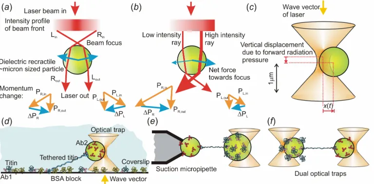
Figure 2. Optical trapping. (a) Ray-optic depiction of the trapping force for an optically trapped particle - a parallel Gaussian-profile laser beam is focussed and refracted by the trapped particle, such that equal and opposite changes in momentum on either side of the particle cancel out resulting in zero net force when the particle is roughly at the laser focus. But, (b) when the particle is laterally displaced from the focus the net momentum change experienced due to the reaction forces when refracted beams of light emerge from the particle are directed back towards the laser focus, illustrated by the momentum vector plots. (c) Displacement of a micron sized bead in an optical trap, the lateral trapping force is proportional to the lateral
[image:20.612.114.498.259.448.2]REFERENCES
1. Feynman, R. P. 1959. There’s Plenty of Room at the Bottom. Lecture
transcript deposited at Caltech Engineering and Science, 23:5, 22-36, and at
www.its.caltech.edu/~feynman/plenty.html
2. Taniguchi, N. 1974. On the basic concept of 'nano-technology'. Proc. Intl.
Conf. Prod. Eng. Tokyo, Part II, Japan Society of Precision Engineering.
3. Leake, M.C. 2012. Singe-molecule cellular biophysics, 1st edn. Cambridge University Press.
4. Harriman, O. L. J. and Leake, M. C. 2011. Single molecule experimentation
in biological physics: exploring the living component of soft condensed matter
one molecule at a time. J. Phys.: Condens. Matter. 23, 503101.
5. Van Holde, K E., Johnson, C. and Shing Ho, Pui. 2005. Principles of
Physical Biochemistry, 1st edn. Pearson Education.
6. Nölting, B. 2009. Methods in Modern Biophysics, 2nd edn. Springer. 7. Lenn, T. and Leake, M. C. 2012. Experimental approaches for addressing
fundamental biological questions in living, functioning cells with single
molecule precision. Open Biol, 2, 120090.
8. Hall, C. E. 1956. Method for the Observation of Macromolecules with the
Electron Microscope Illustrated with Micrographs of DNA. Biophys Biochem
Cytol. 2, 625-628.
9. Wineland, D., Ekstrom, P. and Dehmelt, H. 1973. Monoelectron Oscillator.
Phys. Rev. Lett. 31, 1279.
10. Nagourney, W., Sandberg, J. and Dehmelt, H.. 1986. Shelved optical
electron amplifier: Observation of quantum lumps. Phys Rev Lett. 56, 2797.
11. Rotman, B. 1961. Measurement of activity of single molecules of
beta-D-galactosidase. Proc. Natl. Acad. Sci. USA 47, 1981-1991.
12. Hirschfeld, T. 1976. Optical microscopic observation of single small
molecules. Appl. Opt. 15, 2965-2966.
13. Nguyen, D. C., Keller, R. A., Jett, J. H. and Martin, J. C. 1987. Detection
of single molecules of phycoerythrin in hydrodynamically focused flows by
laser-induced fluorescence. Anal Chem 59, 2158-2161.
14. Moerner, W. E. and Kador, L. 1989. Optical detection and spectroscopy in
15. Ashkin, A. 1970. Acceleration and Trapping of Particles by Radiation
Pressure. Phys. Rev. Lett. 24, 156-159.
16. Ashkin, A., Dziedzic, J. M., Bjorkholm, J. E. and Chu, S. 1986.
Observation of a single-beam gradient force optical trap for dielectric particles.
Opt. Lett. 11, 288-290.
17 . Svoboda, K. and Block, S. M. 1994. Biological applications of optical
forces. Annu. Rev. Biophys. Biomol. Stuct. 23, 247-85.
18. Moffitt, J. R., Chemla, Y. R., Smith, S. B. and Bustamante, C. 2008.
Recent advances in optical tweezers. Annu. Rev. Biochem. 77, 205-28.
19. Gittes, F. and Schmidt, C. F. 1998. Interference model for back-focal-plane displacement detection in optical tweezers. Opt. Lett. 23, 7-9.
20. Svoboda, K., Schmidt, C. F., Schnapp, B. J. and Block, S. M. 1993. Direct
observation of kinesin stepping by optical trapping interferometry. Nature 365:
721-727.
21. Finer, J. T., Simmons, R. M. and Spudich, J. A. 1994. Single myosin
molecule mechanics: piconewton forces and nanometre steps. Nature 368,
113-119.
22. Smith, S. B., Cui, Y. and Bustamante, C. 1996. Overstretching B-DNA: the
elastic response of individual double-stranded and single-stranded DNA
molecules. Science 271, 795-799.
23. Liphardt J., Onoa, B., Smith, B. S., Tinoco Jr., I. and Bustamante, C.
2001. Reversible unfolding of single RNA molecules by mechanical force.
Science 292, 733-7.
24 . Kellermayer, M. S., Smith, S. B., Granzier, H. L. and Bustamante, C.
1997. Folding-unfolding transitions in single titin molecules characterized with
laser-tweezers. Science 276, 1112-1116.
25. Tskhovrebova, L., Trinick, J., Sleep, J. A. and Simmons, R. M. 1997.
Elasticity and unfolding of single molecules of the giant muscle protein titin.
Nature 387, 308-312.
26. Leake, M. C., Wilson, D., Bullard, B. & Simmons R. M. 2003. The elasticity
of single kettin molecules using a two-bead laser-tweezers assay. FEBS Lett.
27. Leake, M. C., Wilson, D., Gautel, M. & Simmons, R. M. 2004. The
elasticity of single titin molecules using a two-bead optical tweezers assay.
Biophys. J. 87, 1112-1135.
28. Binnig, G., Rohrer, H., Gerber, Ch. and Weibel, E. (1982). Tunneling
through a controllable vacuum gap. Applied Phys. Lett. 40, 178-180.
29. Binnig, G., Quate, C. F. and Gerber, C. 1986. Atomic Force Microscope.
Phys. Rev. Lett. 56, 930-933.
30. Rief, M., Gautel, M., Oesterhelt, F., Fernandez, J. M. and Gaub, H. E.
1997. Reversible unfolding of individual titin immunoglobulin domains by AFM
Science 276, 1109-1112.
31. Ha, T., Enderle, Th., Ogletree, D. F., Chemla, D. S., Selvin, P. R. and
Weiss, S. 1996. Probing the interaction between two single molecules:
Fluorescence resonance energy transfer between a single donor and a single
acceptor. Proc. Natl. Acad. Sci. U.S.A. 93, 6264-6268.
32. Noji, H., Yasuda, R., Yoshida, M. and Kinosita, K. J. 1997. Direct
observation of the rotation of F1-ATPase. Nature 386, 299-302.
33. Yildiz, A., Forkey, J. N., McKinney, S. A., Ha, T., Goldman, Y. E. and
Selvin, P. R. 2003. Myosin V walks hand-over-hand: Single fluorophore
imaging with 1.5-nm localization. Science 300, 2061-2065.
34. Schmidt, T., Schütz, G. J., Baumgartner, W., Gruber, H. J. and Schindler,
H. 1996. Imaging of single molecule diffusion. Proc. Natl. Acad. Sci. USA 93,
2926-2829.
35. Gelles, J. Schnapp, B. J. and Sheetz, M. P. 1988. Tracking kinesin-driven
movements with nanometre-scale precision. Nature. 331, 450-453.
36. Michalet X et al. 2013 Development of new photoncounting detectors for
single-molecule fluorescence microscopy. Phil. Trans. R. Soc. B 368.
(doi:10.1098/rstb.2012.0035).
37. Sako, Y., Minoguchi, S. and Yanagida, T. 2000. Single-molecule imaging
of EGFR signalling on the surface of living cells. Nature Cell Biol 2, 168-172.
38. Axelrod, D., Burghardt, T. P. and Thompson, N. L. 1984. Total internal
reflection fluorescence. Ann. Rev. Biophys. Bioeng. 13, 247-268.
39. Byassee, T. A., Chan, W. C. and Nie, S. 2000. Probing single molecules
40. Chiu, S.-W. and Leake, M. C. 2011. Functioning nanomachines seen in
real-time in living bacteria using single-molecule and super-resolution
fluorescence imaging. Int. J. Mol. Sci. 12, 2518-2542.
41. Leake, M. C. 2010. Shining the spotlight on functional molecular
complexes: the new science of single-molecule cell biology. Commun Integr
Biol. 3, 415-418.
42. Dobbie, I. M., Robson, A., Delalez, N. and Leake, M. C. 2009. Visualizing
Single Molecular Complexes In Vivo Using Advanced Fluorescence
Microscopy. J Vis Exp 31, 1508.
43. Yan Y, Petchprayoon C, Mao S, Marriott G. 2013 Reversible optical
control of cyanine fluorescence in fixed and living cells: optical lock-in
detection immunofluorescence imaging microscopy. Phil. Trans. R. Soc. B
368. (doi:10.1098/rstb.2012.0031).
44. Leake, M. C., Chandler, J. H., Wadhams, G. H., Bai, F., Berry, R. M. and
Armitage, J. P. 2006. Stoichiometry and turnover in single, functioning
membrane protein complexes. Nature. 443, 355-8.
45. Delalez, N. J., Wadhams, G. H., Rosser, G., Xue, Q., Brown, M. T.,
Dobbie, I. M., Berry, R. M., Leake, M. C. and Armitage, J. P. 2010.
Signal-dependent turnover of the bacterial flagellar switch protein FliM. Proc Natl
Acad Sci U S A 107, 11347-11351.
46. Ulbrich, M. H. and Isacoff, E. Y. 2007. Subunit counting in
membrane-bound proteins. Nat Meth. 4, 319-21.
47. Leake M. C., Greene, N. P., Godun, R. M., Granjon,,T., Buchanan, G.,
Chen, S., Berry, R. M., Palmer, T. and Berks, B. C. 2008. Variable
stoichiometry of the TatA component of the twin-arginine protein transport
system observed by in vivo single-molecule imaging. Proc Natl Acad Sci U S
A. 105, 15376-15381.
48. Lenn, T., Leake, M. C. and Mullineaux, C. W.2008. Are Escherichia coli
OXPHOS complexes concentrated in specialised zones within the plasma
membrane? Biochem. Soc. Trans., 36, 1032-1036.
49. Lenn T., Leake, M. C. and Mullineaux, C. W. 2008 In vivo clustering and
dynamics of cytochrome bd complexes in the Escherichia coli plasma
50. Ritchie K, Lill Y, Sood C, Lee H, Zhang S. 2013 Single molecule imaging
in live bacteria cells. Phil. Trans. R. Soc. B 368. (doi:10.1098/rstb.2012.0355).
51. Klotzsch E, Schu¨tz G. 2013 A critical survey of methods to detect plasma
membrane rafts. Phil. Trans. R. Soc. B 368. (doi:10.1098/rstb.2012.0033)
52. Plank, M., Wadhams, G. H. and Leake, M. C. 2009. Millisecond timescale
slimfield imaging and automated quantification of single fluorescent protein
molecules for use in probing complex biological processes. Integr. Biol. 1,
602612.
53. Cai, L., Friedman, N. and Xie, X. S. 2006. Stochastic protein expression in
individual cells at the single molecule level. Nature 440, 358-62.
54. Elf, .J, Li, G. W. and Xie X. S. 2007. Probing transcription factor dynamics
at the single-molecule level in a living cell. Science 316, 1191-4.
55. Reyes-Lamothe R., Sherratt D. J. and Leake, M. C. 2010. Stoichiometry
and architecture of active DNA replication machinery in Escherichia coli.
Science, 328, 498-501.
56. Klenerman D, Shevchuk A, Novak P, Korchev Y, Davis S. 2013 Imaging
the cell surface and its organization down to the level of single molecules.
Phil. Trans. R. Soc. B 368. (doi:10.1098/rstb.2012.0027).
57. Wang Y, Irudayaraj J. 2013 Surface enhanced Raman spectroscopy
(SERS) at single molecular scale and its implications in biology. Phil. Trans.
R. Soc. B 368. (doi:10.1098/rstb.2012.0026).
58. Greene E, Duzdevich D. 2013 Towards physiological complexity with in
vitro single-molecule biophysics. Phil. Trans. R. Soc. B 368.
(doi:10.1098/rstb.2012.0271).
59. Boersch M, Sielaff H. 2013 Twisting and subunit rotation in single
FOF1-ATP synthase. Phil. Trans. R. Soc. B 368. (doi:10.1098/rstb.2012.0024).
60. Berry R, Bilyard T, Nakanishi-Matsui M, Steel B, Pilizota T, Nord A,
Hosokawa H, Futai M. 2013 Highresolution single-molecule characterization
of the enzymatic states in Escherichia coli F1-ATPase. Phil. Trans. R. Soc. B
368. (doi:10.1098/rstb.2012.0023).
61. Robson, A, Burrage, K, Leake, MC. 2013. Inferring diuffusion in single
cells at the single molecule level. Phil. Trans. R. Soc. B 368.
62. Ullman G, Walldén M, Marklund E, Mahmutovic A, Razinkov I, Elf J. 2013
Hi-throughput gene expression analysis at the level of single proteins using a
microfluidic turbidostat and automated cell tracking. Phil. Trans. R. Soc. B