FACTOR (LIF) ON BOVINE EMBRYO
DEVELOPMENT IN VITRO
A Thesis presented in partial fulfilment
of the requirements for the degree of
Master of Agricultural Science
in Animal Science
at
Massey University
ENDANG TRI MARGA WATI
ABSTRACT
Margawati, E.T. 1995. The effect of Leukaemia Inhibitory Factor (UFJ on bovine embryo development in vitro. MAgSc thesis, Massey University, Palmerston North, New Zealand, 94 pp.
The aim of the study was to investigate the effect of Leukaemia Inhibitory Factor (LIF) either during in vitro maturation (IVM) or in vitro culture (IVC) on bovine embryo development. Three main experiments were conducted using oocytes aspirated from 2-8 mm diameter follicles collected from cows slaughtered at local abattoirs, Hamilton. The oocytes were matured in a modified TCM-199 containing 10 µg/ml of FSH and LH, and 1 µg/ml
Bi.
fertilised in TALP and cultured in SOF/AA/BSA.Experiment 1 examined the effect of LIF (0, 500, 1000 or 2000 U/ml) and various time periods of IVM ( 18, 22 or 28 h), in a 4 x 3 factorial design on oocyte maturation. Following maturation, oocytes were stripped out of cumulus cells, then denuded oocytes were stained in 1 % lacmoid for determination of maturation stage while the cumulus cells were examined for the incidence of apoptosis by observation of DNA fragmentation using gel electrophoresis procedures.
Experiment 2 comprised two parts, (a) the effect of LIF (0, 500, 1000 or 2000 U/ml) at 24 h IVM in a randomised block design on in vitro development of embryos, (b) comparison of 20 vs 24 h IVM in the presence of LIF (0, 500, 1000 or 2000 U/ml) in a 2 x 4 factorial experiment on embryo development. In the two studies, the proportion of bovine oocytes that cleaved and developed to blastocyst stage was recorded. In addition, cell numbers of blastocysts after Giemsa staining were counted.
hatched blastocyst stages and cell numbers of blastocyst inner cell mass (ICM) and trophectoderm (TE) after differential staining with Hoechst 33342 and propidium iodide were determined.
In Experiment One, an interaction of LIF concentration and duration of IVM was not observed for the proportion of immature oocytes reaching metaphase II (P>0.05). The presence of LIF (500, 1000 or 2000 U/rnl) increased the proportion of oocytes at metaphase II at 18 h (50%, 52% or 58%, respectively, compared to without LIF= 27%), indication that LIF may accelerate the maturation process in vitro. Supplementation of LIF during IVM did not affect the incidence of apoptosis of the cumulus cells.
In Experiment Two, compared to 24 h IVM in the presence of LIF, 20 h IVM significantly increased blastocyst rates (1: blastocysts: 1: cleaved, P<0.05). Cell numbers of blastocysts were not different from oocytes matured for 20 or 24 h in the presence of LIF (P>0.05), however the data show that treatment groups of 20 h IVM in LIF resulted in higher cell numbers of blastocysts than achieved by 24 h IVM.
In Experiment Three, there was a correlation between LIF during IVM and LIF during IVC in the proportion of blastocysts (P<0.05). This finding shows that the proportion of blastocysts decreased when oocytes were matured in the absence of LIF and cultured in LIF. In contrast, more blastocysts developed when the oocytes were matured and then cultured in media containing LIF. There was no effect of addition of LIF during IVM and IVC for cell numbers of blastocysts (P>0.05). However, blastocysts derived from oocytes matured without LIF had significantly increased cell numbers (121 cells) compared to those matured in 1000 U/rnl LIF (109 cells, P<0.05).
Supplementation of LIF both during IVM and IVC did not affect the proportion of blastocyst stages (P>0.05). However, a concentration of 2000 U/rnl LIF during IVC accelerated blastocyst development with more blastocysts hatching (60%, P<0.05).
concentration of 1000 U/ml LIF during IVC resulted in higher cell numbers of ICM (P<0.05).
ACKNOWLEDGEMENTS
I realise that this piece of work could not have been completed without the support, encouragement and enthusiasm of a number of people.
First, I wish to express my sincere gratitude to my main supervisor, Associate Professor M.F. McDonald, who allowed me to pursue my own interests in this study. I also appreciate to his valuable time given in editing this manuscript.
My deepest gratitude to my supervisor, Ms. Anne Pugh (AgResearch Ruakura, Hamilton), who has assisted in developing this work. I appreciate her dedication and enthusiasm for this study and invaluable guidance and advice during the preparation of this manuscript.
I extend my smcere appreciation to Dr. H.R. Tervit and Dr. J.G.E. Thompson (AgResearch Ruakura, Hamilton) for their advisory expertise in planning this study.
A special thank you to a number of people in Animal Physiology Centre, AgResearch Ruakura, Hamilton. They include Dr. Lora Hagemann in helping with DNA analysis, Dr. Allan Nixon in setting-up a fluorescence microscope during crucial time of cell observations, M. Donnison, J. Larsen, B. Shane and several people in the Embryology Laboratory have given technical expertise during my hectic work. Without their assistance, the volume and quality of this work would not have been possible.
I am deeply grateful to Professor D.J. Garrick and Dr. P.C.H. Morel for their patience help and advice on statistical analyses. My thanks to Dr. John Campbell and Dr. Muhammad Anwar who have freely given their comments on drafts of material in this thesis.
A special note -- I would extend my thanks to my friends: Dan Waggoner MAgSc and
Bob Leonard D.V.M who assisted in providing important references related to this study. I am thankful to Dr. Frank and Lydia Weilert MSc for translating a Germany
reference.
Thank you to all the rest of staff and post-graduate colleagues and friends of the Animal
Science Department who were always friendly and ready to lend a hand and made my
stay in New Zealand pleasant and memorable.
My sincerest appreciation to Timothy , Suzanne and Patrick Byrne who in one way or another have lightened the yoke on my shoulders with their concern and understanding.
On a more personal note I would like to dedicate the fruit of these struggles to my
parents, brothers and a sister for their considerable patience, encouragement and support
TABLE OF CONTENTS
ABSTRACT . . . . . . . . . . . . . . . . . . . . . . . . . . . . . . . . . . . . . . . . . . 11
ACKNOWLEDGEMENT . . . . . . . . . . . . . . . . . . . . . . . . . . . . v
LIST OF TABLES . . . . . . . . . . . . . . . . . . . . . . . . . . . . . x
LIST OF FIGURES . . . . . . . . . . . . . . . . . . . . . . . . . . . xi
LIST OF PLATES . . . . . . . . . . . . . . . . . . . . . . . . . . . xii
LIST OF ABBREVIATIONS . . . . . . . . . . . . . . . . . . . . . . . . . . . . . . xiv CHAPTER 1: INTRODUCTION . . . . . . . . . . . . . . . . . . . . . . . . . . 1
CHAPTER 2: REVIEW OF LITERATURE PERTAINING TO THE RESEARCH TOPIC . . . . . . . . . . . . . . . . . . . . . . 4
2.1 MATURATION OF BOVINE OOCYTES . . . . . . . . . . . 4
2.1.1 Meiosis . . . . . . . . . . . . . . . . . . . . . . . . . . . . . . . . 4
2.1.2 Control of Oocyte Maturation In Vitro . . . . . . . . 5
2.2 APOPTOSIS . . . . . . . . . . . . . . . . . . . . . . . . . . . . . . . . 8
2.2.1 What is Apoptosis . . . . . . . . . . . . . . . . . . . . 8
2.2.2 Incidence of Apoptosis . . . . . . . . . . . . . . . . . . . . . . 8
2.2.3 Mechanisms . . . . . . . . . . . . . . . . . . . . . . 9
2.2.4 Agonists and Antagonists of Apoptosis . . . . . . . . . . . . 10
2.3 IN VITRO FERTILISATION . . . . . . . . . . . . . . . . . . . . . . 10
2.3.1 Capacitation of Spermatozoa . . . . . . . . . . . . . 10
2.3.2 Fertilisation Events . . . . . . . . . . . . . . . . . . . . . . . 12
2.3.2.1 2.3.2.2 2.3.2.3 2.3.2.4 2.3.2.5 Attachment and Binding . . . . . . . . . . . 12
Penetration of The Zona Pellucida . . . . . . . 13
Fusion of Sperm and Oocyte . . . . . . . . . . . 13
Oocyte Activation and Formation of Pronuclei . . . . . . . . . . . . . . . . . . . . 14
Syngamy . . . . . . . . . . . . . . . . . . . 15
2.4 EARLY EMBRYO DEVELOPMENT . . .. ... . . 15
2.4.1 In Vivo 2.4.2 In Vitro . . . . . . . . . . . . . . . . . . . . . . . . . . . . 15
2.4.2.1
2.4.2.2
Developmental Block in Early Bovine
Embryos . . . . . . . . . . . . . . . . . . . . . . . . . 17
Media and Culture Systems . . . . . . . . . 17
2.5 ROLE OF GROWTH FACTORS IN EMBRYO DEVELOPMENT . . . . . . . . . . . . . . . . . . . . . . . . . . . . . . . . 20
2.5.1 2.5.2 In Vivo In Vitro 2.5.2.1 2.5.2.2 .... .. . . .. .. . . .. 20
21 Action of Growth Factors . . . . . . . . . . 21
Growth Factors Involved in Embryo Development . . . . . . . . . . . . . . . . . 22
2.6 LEUKAEMIA INHIBITORY FACTOR (LIF) . . .. .. . . 25
2.6.1 Biological Functions . . . . . . . . . . . . . . . . . . . 25
2.6.2 Physiological Roles on Embryo Development . . . . . . . . . . 25
2. 7 EFFECT OF CULTURE SYSTEMS AND GROWTH FACTORS ON EMBRYO SURVIVAL . . . . . . . . . . . . . . . . . . . . . . . . . . . 27
2.7.1 Blastocyst Cell Numbers . . . . . . . . . . . . . . . . . . . . . 27
2. 7 .2 Inner Cell Mass and Trophectoderm . . . . . . . . . . . . . . 28
2.8 OBJECTIVES OF THE STUDY . . . . . . . . . . . . . . . . . . . 29
CHAPTER 3: MATERIALS AND METHODS .... . .. .. . . 31
3.1 OOCYTE COLLECTION AND IN VITRO MATURATION .. .. 31
3.2 SPERM PREPARATION AND IN VITRO FERTILISATION . . . 32
3.3 IN VITRO CULTURE . .. . . 33
3.4 FIXATION AND STAINING OF OOCYTES AND EMBRYOS . . . .. . . 33
3.4.1 Lacmoid Staining of Oocytes . . . . . . . . . . . . . . . . . . . . . 33
3.4.2 Giemsa Staining of Blastocysts . . . . . . . . . . . . . . . . . . . . 34
3.4.3 Differential Staining of Blastocyst Inner Cell Mass and Trophectoderm Cells . . . . . . . . . . . . . . . . . 34
3.5 DNA PREPARATION AND GEL ELECTROPHORESIS . . . 35
3.5.1 Extraction of DNA 3.5.2 Gel Electrophoresis . . . . . . . . . . . . . . . . . . . . . . . 35
3.6 ASSESSMENT . . . . . . . . . . . . . . . . . . . . . . . . . 36
3.6.1 Metaphase II . . . . . . . . . . . . . . . . . . . . . . . . 36
3.6.2 DNA Fragmentation . . . . . . . . . . . . . . . 37
3.6.3 Embryo Development . . . . . . . . . . . . . . . . . . . . . . . . 37
3.6.4 Blastocyst Cell Numbers . . . . . . . . . . . . . . . . . . . . . . 37
3.6.5 Inner Cell Mass and Trophectoderm Cell Numbers . . . . . . 38
3.7 METHODOLOGY OF INDIVIDUAL EXPERIMENTS . . . . . . 38
3.7.1 Experiment 1. 3.7.2 Experiment 2. 3.7.3 Experiment 3. Effect of LIF on the time course of IVM on bovine oocytes . . . . . . . . 38
Effect of LIF during IVM on bovine embryo development following IVF . . . . . . . . . . . 39
Effect of LIF during IVM or IVC on bovine embryo development following IVF . . . . . . . . . . . . . 40
3.8 STATISTICAL ANALYSES . . . . . . . . . . . . . . . . . . 40
CHAPTER 4: RESULTS . . . 46
4.1 MATURATION OF OOCYTES . . . 46
4.2 DNA FRAGMENTATION OF CUMULUS CELLS . . . 48
4.3 DEVELOPMENT OF BOVINE EMBRYOS . . . 49
4.4 CELL NUMBERS OF BLASTOCYSTS . . . . . . . . . . . . . . . . 53
4.5 PROPORTION OF EARLY, EXPANDED AND HATCHED BLASTOCYST ST AGES . . . . . . . . . . . . . . . . . . . . . . . . . . . . . 55
4.6 CELL NUMBERS OF BLASTOCYST INNER CELL MASS AND TROPHECTODERM . . . . . . . . . . . . . . . . . . 57
CHAPTER 5: DISCUSSION . . . . . . . . . . . . . . . . . . . . . . . 60
5.1 MATURATION OF OOCYTES . . . . . . . . . . . . . . . . . . 62
5.2 DNA FRAGMENTATION . . . . . . . . . . . . . . . . . . 62
5.3 DEVELOPMENT OF BOVINE EMBRYOS . . . . . . . . . . . . . 62
5.4 CELL NUMBERS OF BLASTOCYSTS . . . . . . . . . . . . . . . . 65
5.5 ST AGES OF BLASTOCYST DEVELOPMENT . . . . . . . . . 66
5.6 BLASTOCYST INNER CELL MASS AND TROPHECTODERM . . . . . . . . . . . . . . . . . . . . . . . 67
CHAPTER 6: CONCLUSIONS . . . . . . . . . . . . . . . . . . . 69
APPENDICES . . . . . . . . . . . . . . . . . . . . . . . . . . . . . . . . . 71
LIST OF TABLES
Table Page
1. Methods of artificial capacitation of bull sperm . . . . . . . . . . 12
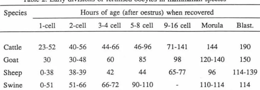
2 Early division of fertilised eggs in mammalian species . . . . . . . . . . 16
3. Duration of the first 4-cell cycles in bovine embryos . . . . . . . . . . . 16
4. The effect of LIF during IVM and the duration of IVM on the percentage
of oocytes reaching Metaphase II . . . . . . . . . . 46
5. Development of bovine embryos derived from 24 h IVM in LIF
following IVF and IVC . . . . . . . . . . . . . . . . . . 50
6. Development of bovine embryos derived from 20 vs 24 h IVM following
IVF and IVC . . . . . . . . . . . . . . . . . . . . . . . . . . . . . . . . 50
7. The effect of LIF during IVM and the duration of IVM on bovine
embryo development . . . . . . . . . . . . . . . . . . . . . . . . . . 51
8. The effect of LIF during IVM and IVC on bovine embryo development
.. . . .. . . .. . .. . .. .. .. .. . . ... . . 52
9. The effect of dose of LIF during IVM on blastocyst cell numbers
following IV C . . . . . . . . . . . . . . . . . . . . . . . . . . . . . 54
10. The effect of LIF during IVM and the duration of IVM on blastocyst cell
numbers following IVC . . . . . . . . . . . . . . . . . . . . . . . 54
11. The effect of LIF during IVM and IVC on blastocyst cell numbers . . . . . 55
12. The effect of LIF during IVM on blastocyst cell numbers following
IVC . . . . . . . . . . . . . . . . . . . . . . . . . . . . . . . . . . . . . . . . . 55
13. The effect of LIF during IVM on the percentage of blastocysts that
reached the early, expanded or hatched stages following IVC . . . . . . 56
14. The effect of LIF during IVM and IVC on the percentage of blastocysts
that reached the early, expanded or hatched stages . . . . . . . . . . . . . . . 57
15. The effect of LIF during IVM and IVC on the numbers of inner cell
LIST OF FIGURES
Figure Page
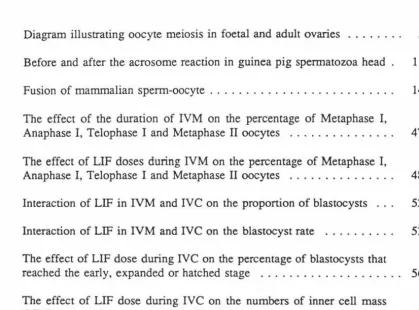
1. Diagram illustrating oocyte meiosis in foetal and adult ovaries . . . . . 5
2. Before and after the acrosome reaction in guinea pig spermatozoa head . 11
3. Fusion of mammalian sperm-oocyte . . . . . . . . . . . . . . . . . . . 14
4. The effect of the duration of IVM on the percentage of Metaphase I,
Anaphase I, Telophase I and Metaphase II oocytes . . . . . . . . . . . . . 47
5. The effect of LIF doses during NM on the percentage of Metaphase I,
Anaphase I, Telophase I and Metaphase II oocytes . . . . . . . . . 48
6. Interaction of LIF in IVM and IVC on the proportion of blastocysts . . . 52
7. Interaction of LIF in IVM and IVC on the blastocyst rate . . . . . . . . . . 53
8. The effect of LIF dose during IVC on the percentage of blastocysts that
reached the early, expanded or hatched stage . . . . . . . . . . . . . . . . 56
9. The effect of LIF dose during IVC on the numbers of inner cell mass
LIST OF PLATES
Plate Page
I. Ovaries collected from abattoirs prior to aspiration . . . . . . . . . . . . . 42 2. Aspirated follicular contents were kept on a warmed (39°C) bench . . . . 42 3. The frozen-thawed sperm layered on the top of the percoll gradient . . . 43
4. Metaphase I of bovine oocyte maturation 43
5. Telophase I of bovine oocyte maturation 44
6. Metaphase II of bovine oocyte maturation . . . . . . . . . . . . . . . . . . . . . 44 7. Bovine blastocyst following Giemsa staining . . . . . . . . . . . . . . . . . 45 8. Bovine blastocyst following differential staining with H33342 and
LIST OF APPENDICES
Plate Page
LIST OF ABBREVIATIONS
The following abbreviations have been used in this text without prior definition:
Units:
oc
degree Celsiusgr gram (s)
h hour (s)
mg milligram (s)
ml millilitre (s)
mm millimetre (s)
ng nanogram (s)
g microgram (s)
1 microlitre (s) w/v weight/volume
Hormones:
E2 oestradiol
FSH follicle stimulating hormone LH luteinizing hormone
Growth Factors:
EGF epidermal growth factor
FGF fibroblast growth factor
IGF insulin-like growth factor type I and II LIF leukaemia inhibitory factor
PDGF platelet-derived growth factor
Solutions or Media:
BSA
B 199 ea as
FCS
H 199 neaas
PBS
PBlPVA
SOF
TALP TCM-199 Others: Ana Blst Rate cAMP conct DNAFAF
ICM IVC IVF IVMMet I
Metil MOET
MQH20
mRNA
rRNA
SAS
bovine serum albumin bicarbonate buffered 199 essential amino acids foetal calf serum hepes buffered 199 non essential amino acids phosphate-buffered saline
enriched phosphate-buffered saline polyvinyl alcohol
synthetic oviduct fluid
Tyrode's medium with albumin, lactate and pyruvate tissue culture medium-199
anaphase blastocyst rate
cyclic adenosine mono phosphate concentration
deoxyribonucleic acid fatty acid free
inner cell mass
in vitro culture in vitro fertilisation in vitro maturation metaphase I
metaphase II
multiple ovulation and embryo transfer
milli Q water
messenger ribonucleic acid
SEM standard error of the mean
TE trophectoderm
Tel I telophase I
CHAPTER 1.
INTRODUCTION
In recent years, off spring of cattle, sheep, goats and pigs have been produced by
techniques involving the in vitro maturation (IVM), in vitro fertilisation (IVF) and in
vitro culture (IVC) of ovarian oocytes that have been recovered from animals
slaughtered at abattoirs. These techniques are termed as in vitro production. In cattle,
this technique was first introduced over 15 years ago and the first IVF calf was born in
1981 (Brackett et al., 1982).
In vitro production technology offers the potential for inexpensive mass production of
oocytes and embryos for both research and commercial purposes. Several commercial
firms are already marketing in vitro produced embryos. In addition to the recovery of
oocytes from abattoir-derived ovaries, oocytes can be recovered from living animals
using ultrasound guided transvaginal oocyte recovery techniques. A similar procedure
has been described for the recovery of oocytes from prepubertal calves. Using these
techniques, oocytes of living animals can be retrieved several times from the same
animal and in vitro embryo production from calves can result in increased genetic gain.
Oocytes can also be recovered from cows that have been culled due to ill health, or
misadventure. For techniques such as pronuclear injection for transgenetics or for
cloning purposes, large numbers of precisely staged zygotes or matured oocytes are
required. The technique of IVM and IVF enables a large number of embryos to be
produced more efficiently than traditional superovulation and surgical oocyte recovery.
Sperm from a number of species can be successfully sorted into X- and Y- bearing
population using a fluorescence activated cell sorter. However, current techniques do not
produce sufficient numbers of sorted sperm for use by conventional A.I. procedures.
This can be overcome by using in vitro fertilisation where as few as 2000 sperm per
matured oocyte is required (Wei
et al.,
1994). In vitro fertilisation techniques can alsocertain deer species) and, further, by using techniques such as intra cytoplasmic sperm injection non-motile sperm can be used for fertilisation.
Other possible commercial application for in vitro produced embryo include the generation of Bos Taurus and Bos Indicus embryos for export, the production of beef embryos for transfer into dairy herds and the use of in vitro derived embryos to produce twins in beef cattle. These applications would result in increased efficiency in the production of desirable quality meat animals.
The development of in vitro production technology has considerably improved in many laboratories. Further improvement in the technique is still required since only 25% to 30% of all oocytes develop into blastocysts. Numerous factors have been identified that affect embryo development in vitro, such as inadequate maturation causing a lower percentage of fertilised eggs and subsequently a lower number of producing transferable embryos. A developmental block was also found in bovine embryos cultured in vitro. Using co-culture systems with somatic cells or conditioned medium have enhanced the development of IVMJIVF of embryos to the blastocyst stage. Similarly, several growth factors have been added to culture media to overcome the developmental block in early bovine embryos (reviewed by Heyner et al., 1993). Leukaemia Inhibitory Factor (LIF) is one of several growth factors that may overcome the problems in embryo development in vitro.
follicles have DNA fragments while healthy cells do not. Apoptosis is evidenced by the
presence of 'a DNA fragment' ladder on an agarose gel. This fragmentation of DNA is
caused by DNA breakage due possibly to free radical generation.
While many growth factors have been examined for their effects on maturation and/ or
development of bovine oocytes and embryos, there are few reports on the effects of and
possible actions of LIF. This present study was therefore conducted to examine whether
LIF in IVM medium alone or in IVC medium would enhance bovine embryo
CHAPTER 2.
REVIEW OF LITERATURE PERTAINING TO THE RESEARCH TOPIC
2.1 MATURATION OF BOVINE OOCYTES
2.1.1 Meiosis
In mammals, meiosis consists of two cell divisions, each including four stages:
Prophase, Metaphase, Anaphase and Telophase (see Figure 1, Tsafriri, 1978). In most
mammals, the first meiotic division occurs in foetal life while the second meiotic
division is completed in the oviduct only following sperm penetration. First meiotic
division (or meiotic prophase I) commences early in development either before or shonly after binh (Baker, 1979; Gondos, 1978). This division is subdivided into 5
subphases: Leptotene, Zygotene, Pachytene, Diplotene and Diakinesis. In rats and mice,
an additional Dyctiate (or Dyctiotene) stage follows immediately after Diplotene (Figure
1). The oocytes in meiotic prophase I are found in the prefollicular ovary. The first
meiotic division is then arrested at a diffuse diplotene or dyctiate stage (Baker, 1979)
and will be completed in the adult at a few hours before ovulation (Gondos, 1978; Tsafriri, 1978). Thus, the duration of the first meiotic arrest of the oocyte persists until
a time prior to ovulation when germinal vesicle breakdown occurs.
Maturation is completed when meiosis is resumed (from no. 7 to 11, Figure 1) and
ovulation takes place usually at the metaphase II stage (No. 11, Figure 1). The
resumption of meiosis in vivo, which occurs after dictyate stage, takes place only in
oocytes that are in maturing antral follicles, and is dependent on the high levels of
gonadotrophic hormones in follicular fluid during proestrus (so-called "LH-surge").
Oocytes removed from antral follicles will progress in vitro from the diplotene (germinal
vesicle) stage to Metaphase II while those from preantral follicles remain at the
diplotene stage. In addition, oocytes from small antral follicles are seemingly "blocked"
without further development at Metaphase I (Erickson and Sorensen, 1974). Thus, only
those oocytes within normal antral follicles will undergo maturation to Metaphase II of
Figure 1.
FIRST MEIOTIC DIVISION SECOND MEIOTIC DIVISION
11 METAPHASE
i
PRO PHASE RESUMPTION OF MEIOSISI LEPTOTENE
'®
G
2 ZYGOTENE OVULATION
@)
12 ANllPllllSE~
8 METAPHASE....
3 PACHYTEM: ;>;
@l
~
~
400~
6
13 TElDPHASE2::
9 ANllMlllSE
e
~0
<: ....g
.... ~ 5 OICTYAT( STAGE@
10 TELOPHAS E 1'1 PRONUCL£AR EGG@
e
'®
..
-...._,
•UCU
Diagram illustrating oocyte meiosis in foetal and adult ovaries, and in the oviduct following ovulation and fertilisation. For simplicity, only three pairs of chromosomes are described (Tsafriri, 1978)
Oocyte maturation includes both morphological changes, as evidenced by nuclear
maturation and expulsion of the first polar body and physiological maturation which is
referred to as "cytoplasmic maturation". In cattle, extrusion of the first polar body occurs
in 80% of oocytes 12 to 18 hours after onset of maturation (Van der Westerlaken et al.,
1994).
2.1.2 Control of Oocyte Maturation In Vitro
The transformation of oogonia to oocytes has been investigated for a variety of species,
but the factors which initiate meiosis remain obscure. Studies on IVM of mammalian
eggs were initiated by Pincus and Enzmann (1935). Later work by Edwards (1965) and
Moor and Trounson ( 1977) has shown that mammalian oocyte meiosis resumes
spontaneously in serum-containing culture media without supplementation of hormones,
[image:24.536.121.478.62.334.2]embryos (Thibault, 1977; Leibfried and Bavister, 1983; Shalgi, 1984; Fleming et al.,
1985). However, recent reports have demonstrated that bovine oocytes can be matured
and fertilised and will develop to blastocysts in serum-free medium (Rose and Bavister,
1992).
Nuclear maturation, cytoplasmic maturation and membrane maturation (or membrane
competence) are critical components of the maturation process for continued viability
of oocytes following fertilisation (Thibault et al., 1987; Younis et al., 1989). In addition,
Rose and Bavister (1992) suggested that cytoplasmic maturation is necessary for
assessing oocyte maturation since nuclear maturation can occur in oocytes that may not
have undergone cytoplasmic maturation.
Many studies have dealt with the role of follicular constituents in the control of oocyte
maturation. Leibfried and First (1979), Fukui and Sukuma (1980) and Dahlhausen et al.
(1981) demonstrated that there was no maturation, or a low maturation rate of bovine
oocytes when the cumulus cells were removed prior to oocyte culture in vitro.
Supplementation of the culture medium with cumulus cells during IVM did not alter the
frequency of completing nuclear maturation (Critser et al., 1986). However, compared
to nude or corona-enclosed oocytes, IVM of cumulus-enclosed bovine oocytes yielded
a significantly higher proportion of embryos following IVF. The presence of intact
cumulus cells for at least 12 hours is necessary for normal cytoplasmic maturation of
bovine oocytes in vitro. Chian and Niwa (1994) demonstrated that the direct
communication of cumulus cells within the oocyte may be important for cytoplasmic
maturation. This direct communication is not necessary in the later stages of IVM since
there was no difference between oocytes that were stripped free of cumulus and intact
cumulus at either 16 or 20 hours after onset of maturation in the fertilisation rate and
subsequent cleavage and development to morulae and blastocysts (Van der Westerlaken
et al.,
1994).Contact between the oocyte and granulosa cells prevents oocyte maturation (reviewed
by Tsafriri, 1978). This contact through gap junctions may play a part in the inhibition
frequency of oocyte maturation (Larsen et al., 1986). The inhibitory effect of granulosa
cells seems to be similar to the effect of follicular fluid on oocyte maturation. It was
suggested by Tsafriri (1978), that resumption of meiosis is triggered by the preovulatory
gonadotrophin surge and Baker et al. (1977) have reported that LH, FSH and oestradiol
are all effective in inducing meiotic division and progression to metaphase 11. It may be
that LH acts by reducing the number of gap junctions.
Growth factors have been proposed to promote oocyte maturation in vitro in the
presence of steroids and gonadotrophins (Dekel and Sherizly, 1985; Feng et al., 1988;
Downs et al., 1988). Several studies have reported that growth factors, their receptors
and localized production of growth factors occur within the ovary and granulosa cells
(Hammond et al., 1985; Feng et al., 1987; Skimmer et al., 1987; Roy and Greenwald,
1990). Growth factors have also been found in the follicular fluid (Hofman et al., 1990)
and they may act as both autocrine and paracrine regulators of ovarian function
(Hammond et al., 1988; Carson et al., 1989), causing granulosa and theca cell
proliferation and differentiation (Harper and Brackett, 1993b).
Several studies of particular growth factors on maturation of oocytes have been made.
Epidermal growth factor (EGF) has been reported to induce maturation in
follicle-enclosed rat oocytes (Dekel and Sherizly, 1985). EGF has also shortened the time
required for germinal vesicle breakdown and increased the proportion of oocytes
undergoing germinal vesicle breakdown in mice (Downs, 1989; Das et al., 1991). Harper
and Brackett (1993a) suggest that there is a physiological role for EGF in regulating
bovine oocyte maturation. This positive influence of EGF during IVM of bovine oocytes
resulted in an increase in the proponion of oocytes that were able to undergo cleavage
and development to the blastocyst stage. Harper and Brackett (1993b) suggested the
possibility of collaborative action of gonadotrophins with growth factors occurring in
oocyte maturation. They found that a combination of platelet-derived growth factor
(PDGF) with FSH increased the proportions of matured and fertilised oocytes developing
to blastocysts which contrast with the results obtained with a combination of POOF and
LH. In addition, Insulin-like Growth Factor-I (IGF-1) both in IVM and IVC media can
2.2 APOPTOSIS
2.2.1 What is Apoptosis ?
In biological investigations, the effect of some treatments on development has been
measured by the incidence of apoptosis. Apoptosis is a type of cell death which is
distinguished from necrosis. Apoptosis is a term to describe the type of cell death
frequently observed when cells are deleted from living tissues (Wyllie, 1988). Necrosis
is characterised by cellular oedema and terminates in rupture of plasma and internal
membrane and leakage of cellular contents into extracellular space, while apoptosis
involves progressive contraction of cellular volume, widespread chromatin condensation
and preservation of the integrity of cytoplasmic organelles (Wyllie, 1981). The affected
cells separate into membrane-bounded fragments which are rapidly phagocytosed by
adjacent cells. Lately, apoptosis has been referred to as programmed cell death (Arends
and Wyllie, 1991).
2.2.2 Incidence of Apoptosis
Programmed cell death occurs in cenain circumstances which include normal cell
turnover, embryonic development, metamorphosis, hormone-induced atrophy and
regression of endocrine-sensitive tumours and is initiated by physiological stimuli
(Wyllie and Morris, 1982). Cell death of these circumstances is morphologically distinct
from that evoked by nonphysiologic stimuli of the environment which is termed as
'coagulative' necrosis.
Apoptosis is the major mode of death observed from the modelling of tissue from the
early blastocyst (Handyside and Hunter, 1986). In mammals, apoptosis occurs in
endometrial (Rotella
et al.,
1989) and ovarian tissue (O'Sheaet al.,
1978; Zelezniket
al.,
1989). Apoptosis occurred in bovine granulosa cells and was most evident in cellsfrom atretic follicles but can also occur in healthy follicles during the luteal phase of the
The effect of growth factors on the incidence of apoptosis has been reviewed by Arends
and Wyllie (1991). Apoptosis can also be caused by physiologic regulatory hormones,
such as glucocorticoids acting on lymphocytes (Wyllie and Morris, 1982). As observed
under the light microscope, apoptotic cells in tissues are inconspicuous, appearing singly
or in small groups and consisting of portions of dark staining cytoplasm, usually with
smooth contour (Wyllie, 1981). These apoptotic cells are frequently digested by
macrophages.
2.2.3 Mechanisms
There have been few studies to elucidate the mechanisms of apoptosis. However, as
reviewed by Arends and Wyllie (1991) at least six major events are known. These
events are: 1) cell density rises sharply, 2) intracellular calcium concentration undergoes
a moderate but sustained rise, 3) total protein and RNA synthesis are discontinued, 4)
chromatin is cleaved at internucleosomal sites, apparently by an endogenous
endonuclease, 5) previously cryptic glycan groups become exposed on the cell
membrane and act as recognition signals, permitting binding and absorbtion by
phagocytes, 6) cytoskeletal elements become less readily deformable, perhaps as a result
of transglutaminase.
Intemucleosomal chromatin cleavage is mostly associated with the morphology of
apoptosis (Arends and Wyllie, 1991). Evidence from glucocorticoid-treated thymocytes
and lymphoid cell lines of rat indicates that the chromatin condensation of apoptosis is
associated with endogenous endonuclease activity which destroys nucleosome chains
from nuclear chromatin (Wyllie, 1980). Cleavage of intemucleosomal linker DNA
generates well-organised chains of oligonucleosomes with lengths that are integer
multiples of 180-200 base pairs (Wyllie, 1981; Arends and Wyllie, 1991; Jolly et al.,
1993) and appear as a ladder after gel electrophoresis (Arends and Wyllie, 1991). This
ladder has now been reported along with morphologic chromatin condensation of
apoptosis in many cell systems (Rotello et al., 1989; Zeleznik et al., 1989). Evidence
has been demonstrated that the characteristic morphologic condensation of chromatin in
shown that DNA cleavage in apoptosis occurs selectively without associated chromatin
proteolysis (Arends et al., 1990).
2.2.4 Agonists and Antagonists of Apoptosis
Cyclic adenosine mono phosphate (cAMP) has a role in cell death (Wyllie, 1981).
Dibutyryl cAMP also causes premature deletion of embryonic palatal shelf tissue with
the morphology of apoptosis (Pratt and Martin, 1975). Epidermal growth factor (EGF)
blocks this deletion which is thought to exert its effect through depletion of endogenous
cAMP (Hassell and Pratt, 1977). In some species a decrease in cAMP content of oocytes
triggers resumption of meiosis (Thibault et al., 1987)
There have been few studies on the effect of hormones or growth factors on the
incidence of apoptosis. Withdrawal of specific growth factors from lymphoid cell lines
in culture, or serum withdrawal from fibroblasts initiates apoptosis even though the
factors do not necessarily stimulate proliferation (Vaux et al., 1988). It appears that
growth factors have important roles in the regulation of cell survival and death. The
addition of stem cell factor and Leukaemia Inhibitory Factor (LIF) promoted primordial
germ cell survival by suppressing programmed cell death or apoptosis (Pesce et al.,
1993). This study suggested that the two cytokines (stem cell factor and LIF) may affect
the in vitro and possibly in vivo development of mammalian primordial germ cells.
2.3 IN VITRO FERTILISATION
2.3.1 Capacitation of Spermatozoa
Mammalian spermatozoa must undergo capacitation and the acrosome reaction before
they can penetrate the oocyte. Capacitation is a process involving the spermatozoa in a
series of biochemical and physiological reactions (Gordon, 1990). In mammalian
spermatozoa, the acrosome reaction is characterised by the dispersion of the acrosomal
acrosomal membrane at multiple sites (Y anagimachi, 1988). This reaction promotes
movement of the spermatozoon through the zona pellucida and fusion with the egg
plasma membrane. The process of acrosome reaction is illustrated from a study of
guinea pig sperm (Figure 2).
Figure 2.
ACROSOME REACTION
}- Equatorial
Re9<><>
Before and after the acrosome reaction in guinea pig spermatozoa head. During the acrosome reaction, the anterior head plasma membrane (except for the posterior-most equatorial region) is lost after fusion with the outer acrosomal membrane. The equatorial and/or posterior head region initiates fusion with the egg plasma membrane (Myles and Primakoff, 1984).
Numerous capacitation systems have been developed for inducing capacitation of bovine
spermatozoa. Several methods of bull sperm capacitation are presented in Table 1. Based
on the observations of First and Parrish ( 1988), sperm capacitation can be achieved by
agents which cause Ca2• entry into the sperm acrosome and a pH increase within the
spermatozoon. Heparin has been proven as a potent glycosarninoglycan in its ability to
induce the acrosorne reaction in bovine epididymal spermatozoa (Handrow et al., 1982)
[image:30.533.158.415.199.311.2]Table 1. Methods of artificial capacitation of bull sperm
Method Investigator (s)
High Ionic Strength Medium Brackett et al., 1982
Bovine Follicular Fluid
Standard Ionic Strength Medium
Ionophore A23187
Heparin
Liposomes
Percoll Gradien ts/H ypotaurine
Caffeine
TEST-Yolk
2.3.2 Fertilisation Events
Fukui et al., 1983
lritani et al., 1984
Hanada, 1985
Parrish et al., 1985a
Graham et al., 1986
Utsumi et al., 1988
Niwa et al., 1988
Ijaz and Hunter, 1989
Union of the sperm and oocyte normally occurs when spermatozoa have undergone
capacitation and the acrosome reaction and the oocyte has matured to metaphase II. The
fertilisation process involves a series of sequential events. As described by Parrish and
First (1993), the events involve sperm attachment to the zona pellucida of the egg,
binding to the zona pellucida, the acrosome reaction, penetration of the zona pellucida,
fusion of sperm and egg plasma membranes, activation of the egg to complete meiosis
II, the cortical granule reaction to block polyspermy, the zona reaction of hardening the
zona in response to cortical granule exudate, sperm head swelling, sperm chromatin
decondensation in synchrony with oocyte chromatin decondensation, deposition of a
pronuclear envelope around the sperm chromatin and finally syngamy of the two
pronuclei and entry into first mitotic cell cycle.
2.3.2.1 Attachment and Binding
Initial sperm attachment to the zona pellucida is loose and sperm may easily be removed
by pipetting. After a period of interaction with the zona, sperm form a finner
[image:31.542.32.462.70.297.2]with a receptor protein of the zona. In the mouse, the receptor protein is called ZP3
(Bleil and Wassannan, 1983) and is one of three glycoproteins found in the zona
pellucida. Similar zona proteins also exist for cattle (Florman and First, 1988). Binding
Of spermatozoa to the oocyte is mediated by the presence of a binding substance on the
surface of the spermatozoa (Parrish and First, 1993). As a result of the interaction of the
binding substance on spermatozoa, a specific zona protein causes the acrosome reaction
to be induced (Bleil and Wassannan, 1983).
2.3.2.2
Penetration of The Zona PellucidaBased on the review of penetration of mouse oocytes, Parrish and First (1993) described
a possible model for mammalian sperm penetration. This includes binding of a
capacitated sperm to the zona, induction of the acrosome reaction, rebinding of sperm
to a different zona protein (i.e., ZP2 in mouse) by a sperm protease (perhaps acrosin),
cleavage of that zona protein, sperm advancement due to its motility, rebinding of sperm
protease to another molecule of the secondary binding site, and the cycle repeated until
the sperm reaches the perivitelline space. Only acrosome reacted sperm that are motile
cross the zona pellucida.
2.3.2.3
Fusion of Sperm and OocyteAfter a spermatozoon penetrates to the zona and crosses the perivitelline space, it
becomes attached to the vitellus and can then fuse with the oocyte. This occurs at the
plasma membrane over the equatorial region of the sperm head after it contacts the
correct region on the vitellus (Parrish and First, 1993; see Figure 3). The contact place
usually contains microvilli and the contact rarely occurs over the meiotic spindle (i.e.,
a region devoid of microvilli). There are two distinct sites of the mature oocyte plasma
membrane: 1) the cortical granule-free domain that lies over the metaphase
chromosomes and where the second polar body formation occurs, 2) the rest of the
oocyte. A complete fusion of the sperm and oocyte plasma membranes occurs when the
2.3.2.4
Figure 3. Fusion of mammalian sperm-oocyte. The cortical granule free region lies over the metaphase-arrested chromosomes. After sperm and oocyte fusion this region will produce a second polar body.
The first polar body is not shown (Myles, 1993).
Oocyte Activation and Formation of Pronuclei
Immediately after contact of the sperm with the oocyte plasma membrane (vitelline membrane), a series of changes in the egg is initiated. Contact of the sperm and the oocyte causes an increase of cytosolic Ca2
• (reviewed by Parrish and First, 1993). The elevation of Ca2•
is
sufficient to trigger a block to polyspermy, to increase cellularmetabolism and to initiate the resumption of meiosis. In mammalian oocytes, the elevation of Ca2+ can be stimulated by Ca2• ionophore, ethanol and electric voltage
(Parrish and First, 1993).
Fusion of cortical granules in the oocyte with the oocyte plasma membrane and
deposition of the cortical granule contents in the perivitelline space causes the zona
block to polyspermy (Parrish and First, 1993). A block to polyspermy is a crucial early
event during oocyte activation. Fertilisation of an immature oocyte (metaphase I) or an
ageing oocyte results in an incomplete cortical granule reaction resulting in polyspermy
(Soon-Chye et al., 1990). This may be because cortical granules of the oocytes are not
correctly distributed around the oocyte periphery .
Following activation, meiosis resumes in the oocyte. Female chromosomes are released
uneven cytoplasmic division and results in exousion of the second polar body.
Chromosomes decondense during prophase IT and formation of the female pronucleus
then occurs at the same time as the formation of the female pronucleus, the sperm
nucleus undergoes a second expansion then condenses again into a male pronucleus.
This pronuclear development is completed when the membrane forms around each
pronucleus (Parrish and First, 1993).
2.3.2.5 Syngamy
Syngamy is referred as 'union of genetic material from the male and female' and
represents the final event in the fertilisation process. After formation of male and female
pronuclei, they migrate to the centre of the oocyte and remain for the next 12-18 hours
in some mammals (Parrish and First, 1993). The membranes of the pronuclei
disintegrate, chromatin recondenses and intermixes. The chromosomes then replicate to
restore the diploid state of the zygote in prophase of the first cleavage division.
2.4 EARLY EMBRYO DEVELOPMENT
2.4.1 In Vivo
Early embryo development begins soon after fenilisation. In cattle, fenilisation occurs
in the upper oviduct within a few hours after ovulation (Salisbury and V anDemark,
1961) or almost immediately after ovulation (Barnes and Eyestone, 1990). The time of
ovulation and fenilisation is believed to be nearly the same (Hyttel et al., 1988). After
fertilisation, the oocyte undergoes a series of cell divisions, beginning with nuclear
division and progressing to cytoplasmic division. At first, a 1-cell zygote divides into
two cells of approximately the same size, then, another synchronous division, resulting
in four cells. Cellular divisions continue, however they become less synchronous (Salisbury and VanDemark, 1961; Barnes and Eyestone, 1990). The timing of these
Table 2. Early divisions of fertilised oocytes in mammalian species*
Species Hours of age (after oestrus) when recovered
1-cell 2-cell 3-4 cell 5-8 cell 9-16 cell Morula Blast.
Cattle 23-52 40-56 44-66 46-96 71-141 144 190
Goat 30 30-48 60 85 98 120-140 150
Sheep 0-38 38-39 42 44 65-77 96 114-139
Swine 0-51 51-66 66-72 90-110 110-114 114
* Salisbury and VanDemark (1961)
Barnes and Eyestone (1990) have characterised development of early bovine embryos
through four cell-cycles (Table 3). Each cell cycle consists of G 1, S, G2 and M phases
and the length of each phase is different. Extrusion of second polar body and subsequent
division into 2-cell stage takes place during first cell cycle. The first cell-cycle takes the
longest period (28-32 hours) with extrusion of second polar body taking place at 20-24
hours of first cell-cycle. The zygote continues to cleave into 4- and 8-cell stages that
occur at approximately 36-50 and 56-64 hours post fertilisation, respectively. These
cleavages occur during the second and the third cell-cycles which last 13 and 14 hours,
respectively. Asynchronous cleavage of the embryo may be due to a longer G2 phase
(4-6 hours in bovine embryos) at the third cell cycle. Subsequently, cleavage into 16-cell
stage (fourth cell-cycle) occurs at approximately 80-86 hours after fenilisation and is
21-30 hours long.
Table 3. Duration of the first four cell-cycles in bovine embryos (h)
Cell Cycle Gl
s
G2 M TotalDuration
First 6 8 4 2 28-32
Second 8 0-2 2 12-13
Third• 8 4-6 2 13-14
Fourthb ? ? ? ? 21-30
• First observed zygotic transcription
[image:35.539.33.471.75.227.2] [image:35.539.38.451.552.689.2]Early development in bovine embryos is governed by mRNA' s and proteins that are
produced in the oocyte during oocyte growth phase and maturation. This is a period of
maternal control of development (Barnes and Eyestone, 1990). Furthermore, changes in
protein synthetic patterns begin to appear during the 4-cell stage. A major change in
protein pattern occurs in the fourth cell-cycle that is presumed to reflect maternal/zygotic
transition for the control of development. During development from the 1-cell to the
8-cell stages, the synthesis of proteins declines while ribosomal RNA synthesis is active
from the 8-cell stage. The protein production is believed to correspond to an early
developmental block in bovine embryos cultured in vitro. Activation of the embryonic
genome fully occurs during this transition of the fourth cell-cycle.
At approximately 7 days after fertilisation in the cow, the embryo begins to form a
hollow sphere (Salisbury and VanDemark, 1961) resulting in formation of the blastula
or blastocyst. During this stage, an inner mass of cells (ICM) accumulates at one side
of the hollow sphere. This ICM is destined to become the body of the embryo. The thin
layer of trophectoderm cells around the outside of the blastocyst functions primarily to
nourish the embryo.
2.4.2
In Vitro2.4.2.1
Developmental Block in Early Bovine EmbryosThe birth of calves derived from IVF of oocytes matured both in vivo (Brackett et al.,
1982, 1984; Sirard and Lambert, 1985) and in vitro (Critser et al., 1986) has
demonstrated that IVF could be applied in the breeding of domestic animals. However,
the efficiency of producing offspring was still less than achieved using Multiple
Ovulation and Embryo Transfer (MOET) procedures. One of many problems
encountered is the existence of a developmental block following cleavage in vitro. For
example, bovine and ovine embryos cultured in vitro from 1- to 4- cell stages rarely
cleaved further than 8- to 16-cells while embryos cultured from the 8- to 16-cell stages
frequently develop into morulae and blastocysts (Thibault, 1966; Eyestone and First,
The causes of the early developmental block in bovine embryos include improper oocyte
maturation (Van de Sandt et al., 1990) or an inadequate IVC medium for early
embryonic development in vitro (First and Parrish, 1987, Flood et al., 1993).
Farell and Bavister (1984) have observed that the block occurred within a few minutes
of exposure of 2-cell hamster embryos to artificial conditions, as evidenced by loss of
viability. Deficiencies in the conventional culture system compared with the in vivo
environment were the cause of early developmental block in most species of animals
since neither growth retardation nor developmental blockage were observed in vivo
(Natsuyama et al., 1992). Barnes and Eyestone (1990) have stressed that in bovine
embryos cleavage past the 8- to 16-cell stage is very sensitive to environmental
conditions.
First and Parrish (1987) in a review have suggested that there are two causes of the
block in development in early cleavage of mammalian embryos in vitro: IVC media are
lacking in factors that are conducive to early embryo development and that the oviduct
contains specific factors that are required for developmental events that occur during
early cleavage (between 1- and 16- cell stage). The time of blocked development is
characterised by a prolonged cell-cycle, increased rates of DNA, RNA and protein
syntheses, concomitant with a transition from maternal to embryonic control of
development for the cow (King et al., 1985) and the sheep (Calarco and McLaren,
1976).
2.4.2.2 Media and Culture Systems
In the past, the embryonic developmental block in vitro has been a serious problem
encountered for IVF experiments. Recent advances in embryo culture systems using
somatic cell co-culture have allowed the continuous development of cow zygotes
f crtilised either in vitro or in vivo past the problematic 8- to 16- cell stage to morulae
and blastocysts, and has resulted in the birth of calves following non-surgical transfer
to recipients (Eyestone and First. 1989; Ellington et al., 1990). Oviduct epithelial cells
are generally used for co-culture. Some studies have reported that these epithelial cells
(Gandolfi and Moor, 1987; Rexroad and Powel, 1988).
Culture medium conditioned with oviduct cells also promoted development of bovine
embryos. This conditioned medium resulted in an approximately normal pregnancy rate
(50%) after transfer into recipient cows (Eyestone and First, 1989). Gandolfi et al.
(1990) have suggested that the oviduct epithelium of all examined species secretes
specific proteins during the passage of the embryo along its lumen and these proteins
bind to the embryos exactly as in vivo actions. As previously mentioned a variety of cell
types and media will enable embryo development to continue. An unidentified bovine
serum albumin contaminant and co-culturing embryos with trophoblastic vesicles or with
granulosa cells have shown some positive results (Gandolfi et al., 1990).
Besides successful co-culture systems, non co-culture systems have been developed that
will overcome the problem of a developmental block in early sheep and cow embryos.
Under low oxygen (5%) concentration, Tervit et al. (1972) demonstrated that synthetic
oviduct fluid medium (SOF) without somatic cell support enabled early cleavage stage
(1- to 8- cell) embryos derived in vivo, to develop to morulae with the birth of calves
after transfer. Further, Thompson et al. (1990) demonstrated that under reduced oxygen
(4-8%), 2-cell sheep and 2- to 4-cell and 8-cell cow embryos reached morulae stages
after culture in a simple medium of SOF containing Bovine Serum Albumin (BSA)
without somatic cell support. In recent years, a number of non co-culture media for
bovine embryo development has been developed, such as CRl salts medium containing
sodium chloride, potassium chloride, sodium bicarbonate, hemicalcium lactate and
sodium pyruvate (Rosenkrans and First, 1991 ). From various reports of using co-culture
and non co-culture systems in embryo development, Bavister (1992) has suggested that
the presence of somatic cells in culture media may not be necessary and that since
embryos produce their own growth factors (Paria and Dey, 1990), these growth factors
2.5 ROLE OF GROWTH FACTORS IN EMBRYO DEVELOPMENT
2.5.1 In Vivo
Studies of preimplantation embryos and the oviduct indicates that there are several
factors involved in the cooperative interaction between the embryos and the female
genital tract. Gandolfi (1994) has divided these factors into three groups: autocrine,
paracrine and environmental factors. Growth factors derived from the preimplantation
embryos participate in an autocrine pathway while growth factors of the oviduct and
uterus act in a paracrine manner. Complex interactions may occur, and as reviewed by
Simmen et al. (1993), the autocrine and paracrine modes of action of insulin-like growth
factors (IGFs) are regulated within the uterine microenvironment by IGF-1 receptors and
IGF binding proteins which are themselves exposed to local control within the uterus
and conceptus.
Growth factors are also located within the ovary (Hammond et al., 1985; Skimmer et
al., 1987). These growth factors may act as both autocrine and paracrine regulators of
ovarian function (Hammond et al., 1988; Carson et al., 1989). For example, epidermal
growth factor (EGF) has been found in small and medium preantral follicles (Harper and
Brackett, 1993a) and has a function in bovine oocyte maturation.
Growth factor is present in the early developing embryo as it passes through the
reproductive tract. It was speculated that uterine secretions (histotrophe) are involved in
regulating the attachment, implantation, nutrition and growth of the conceptus (Biggers,
1988; Roberts and Bazer, 1988). The uterine environment with its capacity as a source
of nutrition, can provide growth factor and other regulatory proteins critical to growth
and survival of preimplantation conceptuses (Roben and Bazer, 1988). As reviewed by
Brigstock et al. (1989) the involvement of peptide and polypeptide growth factors have
been established in the control of cell proliferation. However, recent work has shown
that a range of growth factors influence not only proliferation but in some cases also
functional differentiation of ovarian follicle cells (Carson
et al.,
1989) andis cooperative interaction among preimplantation embryos and growth factors in embryo
development and growth.
The physiological role of growth factors in mammals has been considered by
Gospodarowicz and Moran (1976) and involves controlling normal development,
maintaining homeostasis and wound healing and regeneration.
2.5.2 In Vitro
2
.
5
.
2.1
Action of Growth FactorsSome growth factors have been shown to influence embryo development both in vivo
and in vitro. As reviewed by Brigstock et al. (1989), peptide and polypeptide growth
factors occur in uterine tissues and fluids. In addition, steroid hormones (oestrogen and
progesterone) have a role in the control of growth factor synthesis. These growth factors
include Insulin-like growth factors/ somatomedins (IGF), Epidermal growth factor
(EGF), Transforming growth factors (TGF-a, -B), Fibroblast growth factors (Acidic,
Basic FGF) and Platelet-derived growth factor (PDGF).
Serum, notably foetal calf serum, is thought to contain a variety of mitogens and growth
factors (Carson et al., 1989). Mitogens can make the cells responsive to growth factors.
Growth factors in the bloodstream can be extracted from plasma, platelets and serum
(Gospodarowicz and Moran, 1976).
Many investigators have reported that growth factors regulate differentiation and
morphogenesis. In a review, Mercola and Stiles (1988) concluded that growth factors
can function as a mitogen in one context and promote differentiation in another.
Furthermore, they suggested that growth factors, like other hormones, regulate gene
expression within their target cells. The initial event in growth factor action is binding
to specific receptors on embryonic cells (Mercola and Stiles, 1988; Heyner et al., 1993).
This is usually a high-affinity receptor which spans the outer cell membrane. Generally,
the binding of a growth factor by its receptor induces soluble, intra cellular second
messengers which transmit a signal to the nucleus. These second messengers may be
phosphoproteins, inositol phosphates, diacylglycerol, cyclic nucleotides, monovalent or
divalent ions (Mercola and Stiles, 1988). Within minutes, after formation of the growth
factor-receptor complex, changes in gene expression can be detected. There appears to
be an association of genes which are expressed across tissue boundaries in response to
numerous growth factors.
2.5.2.2
Growth Factors Involved in Embryo DevelopmentEpidermal Growth Factor. There have been a number of studies investigating the role
of EGF either in maturation of oocytes or early embryonic development in vitro. The
EGF was usually provided either alone in a defined medium or in combination with
gonadotrophins. The addition of EGF in a defined medium with combination of low
concentrations of gonadotrophins during IVM enabled subsequent blastocyst
development at a rate comparable to that produced by media with high concentrations
of FSH and LH (Harper and Brackett, 1993a). Their findings suggested a possible
physiological role of EGF in regulating bovine oocyte maturation. Flood et al. (1993)
found that compared to chemically-defined medium alone, supplementation with EGF
tended to increase the percentage of blastocysts. They concluded that certain growth
factors such as EGF, TGF-a,-Bl, IGF-I,-II, PDGF, bFGF, did not improve the
development of in vitro matured and in vitro fertilised bovine embryos.
EGF and other growth factors (TGF-a, PDGF, CSF-I = Colony stimulating factor-n
significantly enhanced trophoblast outgrowth (Haimovici and Anderson, 1993). They
suggested, these growth factors may play an important role in the preimplantation
process.
Insulin-like Growth Factor. The insulin-like growth factors (i.e., IGF-1 and IGF-m
possess a high degree of amino acid sequence homology with insulin (Heyner et al.,
1993). Like other growth factors, insulin and IGFs bind to cell-receptors and their
observed on the cells of inner cell mass and trophectoderm of mouse blastocysts
(Mattson et al., 1988). An effect of IGF-I was observed for bovine embryo development
following IVM or culture in the presence of IGF-I, however this IGF-I could not replace
granulosa cell co-culture (Herrler et al., 1992).
The potential applications of peptide growth factors in particular the IGF family of
growth factors have been studied for their biological actions in the maternal uterus and
the developing conceptus. These include improved embryo viability, "rescue" of poor
embryos, synchronisation of the maternal uterine environment for receptivity, increase
embryo survival, and increase pregnancy rates and reproductive efficiency (Pope et al.,
1990).
There is little information of IGF-11 in domestic animal species. However, IGF-11 is an
imprinted gene in mice and is involved in the regulation of post implantation growth of
mouse embryos (Rappolee et al., 1992).
Transforming Growth Factor. There are two types of Transforming Growth Factors,
TGF-cx. and TGF-B. TGF-cx. is structurally analogous to Epidermal Growth Factor (EGF)
and others of the TGF-B family. TGF-cx. is expressed in bovine thecal cells and has been
localised in the thecal cell layer during follicular growth (Lobb and Dorrington, 1992).
In addition, a decrease in immunoreactive TGF-cx. has been found in preovulatory
follicles which corresponded to a decrease in mitotic activity of granulosa cells. TGF-B
is also produced by bovine thecal cell culture, however, TGF-B has different actions to
those of TGF-cx. on bovine granulosa cell proliferation.
TGF-cx. has the ability to stimulate the rate of blastocoel expansion and it was observed
in 70% of mouse embryos (Dardik and Schultz, 1991). It seems that TGF-cx. could
support embryo implantation. Flood et al. (1993) have investigated nine growth factors,
and demonstrated that TGF-cx. did not improve the development of bovine IVMJIVF
In mouse, addition of TGF-Bl increased the proportion of 2-cell mouse embryos
developing to blastocysts when cultured singly (Paria and Dey, 1990), but in groups,
TGF-Bl did not affect blastocyst development, hatching or the cell number of bovine
embryos (Keefer et al., 1994). However, Marquant-LeGuinne et al. (1989) reported that
supplementation of TGF-B to a co-culture system stimulated the growth of inner cell
mass cells of bovine embryos. These studies show that TGF-Bl may be necessary for
embryo development in vitro.
Platelet-derived Growth Factor. Stimulatory effects of endometrial platelet-activating
factor (PAF) or embryo-derived PAF (EDPAF) activate platelets resulting in a release
of Platelet-Derived Growth Factor (PDGF). Platelets are isolated from within oviduct
and uterus (Thibodeaux et al., 1993).
PDGF, added to serum-free medium, promoted development of 2-cell bovine embryos
beyond the 16-cell stage (Larson et al., 1992b). However, when added to media for
16-cell embryos, PDGF reduced the proportion of embryos blastulating from 12.7% to
5.8%. In contrast, at the same time, TGF-a increased the proportion of embryos
blastulating from 8.6 to 40.6%. Larson et al. (1992a) concluded that TGF-a and perhaps
basic fibroblast growth factor (FGF) promoted blastulation of the 16-cell embryos during
subsequent culture.
Certain growth factors and gonadotrophins may act synergistically on bovine embryo
development. Harper and Brackett (1993b) found that addition of the combined PDGF
and FSH in IVM medium enhanced bovine oocyte maturation with more matured and
fertilised oocytes developing to blastocysts in vitro than PDGF or FSH alone.
Furthermore, combinations of growth factors such as EGF, IGF-I and PDGF or EGF