R E S E A R C H A R T I C L E
Open Access
Comparison of the effects of 7.2%
hypertonic saline and 20% mannitol on
whole blood coagulation and platelet
function in dogs with suspected
intracranial hypertension - a pilot study
Ivayla D. Yozova
1, Judith Howard
2, Diana Henke
3, Daniel Dirkmann
4and Katja N. Adamik
5*Abstract
Background:Hyperosmolar therapy with either mannitol or hypertonic saline (HTS) is commonly used in the treatment of intracranial hypertension (ICH). In vitro data indicate that both mannitol and HTS affect coagulation and platelet function in dogs. The aim of this study was to compare the effects of 20% mannitol and 7.2% HTS on whole blood coagulation using rotational thromboelastometry (ROTEM®) and platelet function using a platelet function analyzer (PFA®) in dogs with suspected ICH. Thirty client-owned dogs with suspected ICH needing osmotherapy were randomized to receive either 20% mannitol (5 ml/kg IV over 15 min) or 7.2% HTS (4 ml/kg IV over 5 min). ROTEM® (EXTEM® and FIBTEM® assays) and PFA® analyses (collagen/ADP cartridges) were performed before (T0), as well as 5 (T5), 60 (T60) and 120 (T120) minutes after administration of HTS or mannitol. Data at T5, T60 and T120were analyzed as a percentage of values at T0for comparison between groups, and as absolute values for comparison between time points, respectively.
Results:No significant difference was found between the groups for the percentage change of any parameter at any time point except for FIBTEM® clotting time. Within each group, no significant difference was found between time points for any parameter except for FIBTEM® clotting time in the HTS group, and EXTEM® and FIBTEM® maximum clot firmness in the mannitol group. Median ROTEM® values lay within institutional reference intervals in both groups at all time points, whereas median PFA® values were above the reference intervals at T5(both groups) and T60(HTS group).
Conclusions:Using currently recommended doses, mannitol and HTS do not differ in their effects on whole blood coagulation and platelet function in dogs with suspected ICH. Moreover, no relevant impairment of whole blood coagulation was found following treatment with either solution, whereas a short-lived impairment of platelet function was found after both solutions.
Keywords:Intracranial hypertension, Osmotherapy, Hypertonic saline, Mannitol, Hemostasis, Thromboelastometry, Platelet function
* Correspondence:katja.adamik@vetsuisse.unibe.ch
5Division of Small Animal Emergency and Critical Care, Department of
Clinical Veterinary Medicine, Vetsuisse Faculty, University of Bern, Laenggassstrasse 128, 3012 Bern, Switzerland
Full list of author information is available at the end of the article
Background
Osmotherapy is commonly used in the treatment of intracranial hypertension (ICH) due to a variety of causes, including head trauma, intracranial neoplasia, in-fection or hemorrhage, and status epilepticus [1]. The principle goal of osmotherapy is to shift fluid from the intracellular into the extracellular compartment using intravenous hyperosmolar agents, thereby reducing brain edema and improving cerebral perfusion pressure [2]. Although 10–20% mannitol is considered the gold standard hyperosmolar agent in the treatment of ICH [3], mannitol-induced osmotic diuresis may cause hypo-volemia and reduction in cerebral perfusion pressure [1]. In recent years, 3.0–7.5% hypertonic saline (HTS) has gained popularity in the treatment of ICH [4, 5] as it has less pronounced diuretic effects and therefore does not cause hypovolemia [1, 6]. Indeed, in the face of hypovol-emic shock and traumatic brain injury, HTS provides the advantage of volume expansion, restoring adequate cerebral perfusion pressures, and reducing brain edema, which makes it superior to mannitol in trauma patients with shock [7, 8].
Both mannitol and HTS have been shown to interfere with whole blood coagulation and platelet function [9]. This is in part due to dilutional coagulopathy. Further-more, 7.2% HTS may directly disturb both fibrin forma-tion and platelet funcforma-tion [10], and mannitol may interfere with coagulation by reducing clot strength [11]. In addition, hyperosmolarity is supposed to lead to im-pairment of both whole blood coagulation and platelet function [9, 12]. In consequence, the safety of using these agents in patients with ICH and intracranial hemorrhage remains unclear [4, 11, 13]. Previous in vitro studies in humans have demonstrated anticoagulant effects of both mannitol and HTS [9, 14], although one clinical study failed to demonstrate any negative effect on hemostasis using either solution in patients undergoing elective intra-cranial surgery [15]. Similarly, in vitro studies in dogs demonstrated that both mannitol and HTS have negative effects on coagulation in a dose-dependent fashion, al-though in vivo studies in dogs in a clinical setting are lacking [16, 17]. As a result, current guidelines for osmotherapy in dogs with ICH are largely extrapolated from experimental data and human literature [18, 19].
The aim of this study was to compare the effects of clinically recommended doses of 20% mannitol and 7.2% HTS on whole blood coagulation using rotational thromboelastometry (ROTEM®) and on platelet function using a platelet function analyzer (PFA®) in dogs with suspected ICH. Based on a previous canine in vitro study, we expected that mannitol would have a greater impact on coagulation than HTS [17]. A second aim was to compare plasma osmolarity after either osmothera-peutic solution. Knowledge of the extent to which these
solutions may impact global hemostasis may influence clinical decision-making in the selection of solutions for individual dogs.
Methods
The study was designed as a prospective, randomized, non-blinded cohort study using client-owned dogs with suspected ICH. The trial was approved the Animal Experi-ment Committee of the Swiss Federal Veterinary Office (registration number BE 90/13), and informed owner con-sent was obtained for all dogs.
Animals
Dogs with suspected ICH requiring osmotherapy were enrolled between March 2013 and March 2016. A tenta-tive diagnosis of ICH was based on history, neurologic examination (performed by a board-certified neurologist) and/or magnetic resonance or computed tomographic imaging studies (evaluated by a board-certified radiolo-gist or neuroloradiolo-gist). Criteria to substantiate suspicion of ICH were a forebrain or multifocal localization with severely reduced consciousness and miotic pupils on neurological exam, Cushing triad (irregular respiration, bradycardia, and systolic hypertension), brain herniation or other shifts of brain parenchyma on MRI or CT [20], an elevated resistive index [21], and deterioration of modified Glasgow coma scale scores [22]. The decision to administer osmotherapy was at the clinicians’discretion. Exclusion criteria were azo-temia (plasma creatinine concentration > 140μmol/L), body weight < 7 kg, age < 6 months or >12 years, presence of dis-eases known to affect coagulation (hyperadrenocorticism, protein losing enteropathy or nephropathy, and hepatic insufficiency) and administration of non-steroidal anti-inflammatory drugs, osmotherapeutics (mannitol, HTS), artificial colloids, or blood products within two weeks prior to study enrollment. In addition, dogs were excluded if their initial hematocrit was below 0.25 L/L or if platelet counts were below 100 × 109/L, as this may influence re-sults PFA assays [23]. Clinical data collected included pa-tient characteristics (breed, age, gender, and body weight), the underlying disease causing ICH, and any additional crystalloid fluid therapy administered within the measure-ment period.
Treatment groups
given during the measurement period was at the clini-cian’s discretion. Animals were excluded if additional osmotherapy was administered during the study measure-ment period.
Measurements
Whole blood coagulation was analyzed using ROTEM® (ROTEM®, TEM Innovations GmbH, Munich, Germany), according to the manufacturer’s instructions using methods previously described for canine samples [24]. Two ROTEM® machines were available to enable parallel measurements of T0and T5 samples. Clotting activators
used were EXTEM® (re-calcification and tissue factor activation) for assessment of the extrinsic pathway and FIBTEM® (tissue factor activation and platelet inhib-ition with cytochalasin D) for analysis of the extrinsic pathway and qualitative assessment of fibrinogen status (star-tem®, ex-tem®, and fib-tem® reagents, TEM Inno-vations GmbH, Munich, Germany). Data collected were clotting time (the time from the start of the measure-ment until the onset of clotting, CT), clot formation time (the time between the onset of clotting and a clot firmness of 20 mm amplitude, CFT), alpha angle (angle between the baseline and a tangent to the clotting curve through the 2 mm CT,α), and amplitude 10 (clot firmness in mm at the amplitude time point of 10 min after CT, A10), and maximum clot firmness (the max-imum amplitude of the curve measured in millimeters, MCF). Platelet function was assessed using a PFA-100® analyzer (PFA-100®; Siemens Healthcare Diagnostics AG, Zurich, Switzerland), according to the manufac-turer’s instructions using methods previously described for canine samples [23] and collagen/ADP cartridges (Dade PFA Collagen/ADP Test Cartridge, Siemens Healthcare Diagnostics AG, Zurich, Switzerland). Briefly, the time until occlusion of the aperture by platelet plug formation (closure time, CtPFA) was measured.
Institu-tional reference intervals (RI) for ROTEM® and PFA® used in this study were previously established from jugular blood samples of 37 healthy dogs. Hematocrits and plate-let counts were measured using a hematology analyzer (Advia® 120, Siemens Healthcare Diagnostics AG, Zurich, Switzerland) and plasma osmolarity was measured using a freezing-point osmometer (Osmometer Type 1, Loeser Messtechnik, Germany).
Blood sampling
Blood samples were obtained prior to (T0), and five (T5),
60 (T60), and 120 min (T120) after the end of
osmother-apy administration. At each time point, blood was ob-tained by careful puncture of the lateral saphenous vein using a 21G butterfly needle connected to a vacutainer system. To minimize contamination of the whole blood samples with tissue factor, the first and third blood
samples were collected from the right saphenous vein and the second and fourth from the left saphenous vein. Blood was collected in the following order: one serum vacutainer tube (discard tube), two 3.2% buffered sodium citrate vacutainer tubes (BD Vacutainer 1.8 ml coagula-tion tube, buffered trisodium citrate 3.2%, BD, Plymouth, United Kingdom) for PFA® and ROTEM® analyses, one heparin tube (Li-Heparin LH/1.3, 1.3 ml tubes, Sarstedt AG, Sevelen, Switzerland) for osmolarity and one K2-EDTA
tube (K2-EDTA 2 ml tubes, Sarstedt AG, Sevelen, Switzerland) for hematocrit and platelet counts. Blood samples for ROTEM® and PFA® analyses were maintained at room temperature and analyses were started after 30-60 min. Hematocrits, platelet counts and osmolarity were measured within 4 h of sampling. All blood sam-ples were collected and analyzed by the same investiga-tor (IY).
Statistical analysis
Descriptive statistics were evaluated and continuous var-iables were assessed for normality using the D’ Agostino-Pearson test and by examining normal plots. Differences between groups (mannitol vs HTS) were evaluated using independent samples t-tests and Mann-Whitney tests for normal and non-normal distributed continuous data, re-spectively, and chi-squared tests were used for categorical data. For comparison between the groups for ROTEM® and PFA® assays, data at T5, T60and T120 were evaluated
as a percentage of the values measured at T0in each dog.
Repeated measures analyses within each group for osmolarity were performed using a Friedman test and post-hoc pair-wise analysis was applied using Wilcoxon tests with Bonferroni correction. Repeated measures analyses within each group for ROTEM® and PFA® mea-surements were performed using a Skillings-Mack test, which is a generalization of the Friedman test for incom-plete blocks when observations are missing arbitrarily. All data were analyzed using statistical software (MedCalc® version 16.4.3, MedCalc® Software bvba, Ostend, Belgium and Statext version 2.7.17, Statext LLC, Wayne NJ, USA) and significance was set atP< 0.05 throughout.
Results
shepherd, great Dane, Jack Russel terrier, Keeshond, Malinois, mudi, Saint Bernard, Tervuren, and white Swiss shepherd. Of the 30 dogs, 24 underwent magnet resonance imaging and 1 dog underwent computed tomography. Underlying diseases were intracranial neo-plasia (n = 8, mannitol group; n = 8, HTS group), in-toxication (n = 3, mannitol group; n = 1, HTS group), head trauma (n= 2, mannitol group;n = 1, HTS group), meningoencephalitis (n = 2, mannitol group), hydroceph-alus internus (n= 1, HTS group), presumptive hypertensive encephalopathy (n = 1, HTS group), and undetermined in the remaining 3 dogs (HTS group).
In some samples, the anticipated analyses could not be carried out due to technical constraints (sample clotting or measurement error). In addition, blood samples of 7 dogs at T60and a further 3 dogs at T120could not be
ob-tained due to euthanasia prior to the measurement time point. The number of samples used for each analysis is presented in Tables 1 and 2. During the two-hour study period, 19 of 30 dogs received intravenous isotonic crys-talloid therapy (median, 5 ml/kg; range, 2–20 ml/kg).
Osmolarity, hematocrit and platelet counts
No significant difference was found in osmolarity, hematocrit or platelet counts between dogs that received mannitol and those that received HTS at any time point (Table 1). Within group comparisons revealed a significant increase in osmolarity at T5, T60and T120 after HTS and
at T5and T60after mannitol (Table 1).
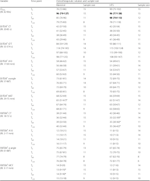
Platelet function analysis
At T0, CtPFA was significantly longer in the HTS group
than in the mannitol group (P= 0.03) but median values were within the RI (Table 2). After treatment, median
values were increased above the RI at T5 (both groups)
and T60(HTS group). However, no significant difference
in percentage relative to values at T0was found between
the groups at T5, T60, or T120(Table 3) and no difference
was found between measurement times within either group (Table 2).
ROTEM® analysis
No significant difference was found for any parameter between the groups at T0 (Table 2) and no significant
difference between the groups in percentage relative to T0was found for any parameter except for a shortening
of FIBTEM® CT at T5and T60 after HTS (Table 3). No
significant difference was found between measurement times within either group except for a decrease of EXTEM® MCF at T5 and FIBTEM® MCF at T5 and
T60compared to T0in dogs receiving mannitol, and a
de-crease of FIBTEM® CT at T5, T60, and T120compared to
T0 in dogs receiving HTS (P < 0.05). Median values
where within RI at all time points in both groups.
Discussion
The present study evaluated the effects on coagulation of intravenous 20% mannitol and 7.2% HTS in a cohort of dogs with suspected ICH using ROTEM® and PFA® analyses. Only minimal differences in coagulation parame-ters were found between dogs treated with mannitol and those receiving HTS at currently recommended doses. In-deed, a significant difference between the groups in per-cent of values relative to baseline (T0) was only found for
FIBTEM® CT, which showed a shorter time until clot de-tection at T5 and T60 in dogs receiving HTS. This was
mirrored by a shorter FIBTEM® CT at T5, T60, and T120
[image:4.595.60.540.529.724.2]compared to T0 in the HTS group. Furthermore, a
Table 1Osmolarity, hematocrit and platelet counts prior to (T0), and at 5 (T5), 60 (T60) and 120 (T120) minutes after administration of mannitol and hypertonic saline (HTS)
Variable Time point Osmotherapeutic solution and sample size Pvalue
Sample size Mannitol Sample size HTS
Osmolarity
(RI: 300-310 mOsm/L)
T0 15 312 (308–327) 15 311 (309–320) 0.89
T5 15 329 (321–339)* 15 336 (324–340)* 0.96
T60 11 326 (318–341)* 12 324 (317–336)* 0.62
T120 8 327 (316-334) 11 326 (319–334)* 0.59
Hematocrit (RI: 0.39-0.57 L/L)
T0 15 0.44 (0.35–0.49) 15 0.41 (0.37–0.49) 0.97
T5 15 0.37 (0.31–0.44) 15 0.37 (0.34–0.42) 0.68
T60 11 0.37 (0.35–0.41) 12 0.38 (0.37–0.42) 0.42
T120 8 0.42 (0.35–0.44) 11 0.40 (0.36–0.42) 0.70
Platelet count
(RI: 150-400 × 109/L) T0 15 221 (167–375) 15 237 (173–312) 0.91
T5 15 202 (157–314) 15 202 (179–287) 0.82
T60 11 207 (169–344) 12 177 (134–284) 0.56
T120 8 214 (188–393) 11 205 (141–269) 0.31
Table 2Rotational thromboelastometry and platelet function analysis closure time prior to (T0), and at 5 (T5), 60 (T60) and 120 (T120) minutes after administration of mannitol and hypertonic saline (HTS)
Variable Time point Osmotherapeutic solution and sample size
Mannitol Sample size HTS Sample size
CtPFA
(RI: 42-90s)
T0 76 (72-86) 15 90 (75-102) 13
T5 96 (79-127) 15 96 (77-138) 13
T60 85 (76-96) 11 98 (70-113) 12
T120 79 (75-83) 8 78 (71-118) 11
EXTEM®CT (RI: 20-85 s)
T0 42 (37-50) 15 42 (35-54) 15
T5 41 (32-45) 15 38 (35-50) 15
T60 38 (36-49) 11 40 (34-45) 12
T120 43 (29-46) 9 41 (36-49) 11
EXTEM®CFT (RI: 55-374 s)
T0 84 (59-129) 14 93 (86-111) 15
T5 114 (74-145) 14 115 (102-124) 14
T60 97 (68-105) 10 115 (99-190) 12
T120 98 (77-125) 8 108 (93-147) 11
EXTEM®A10 (RI: 17-69 mm)
T0 58 (46-62) 15 54 (49-61) 15
T5 56 (46-58) 15 51 (39-61) 14
T60 57 (53-67) 11 54 (53-67) 12
T120 60 (52-63) 9 55 (44-56) 11
EXTEM®α-angle (RI: 57-86°)
T0 73 (61-81) 14 72 (69-75) 15
T5 76 (65-71) 14 67 (64-71) 14
T60 72 (69-79) 10 69 (64-77) 12
T120 69 (65-81) 8 70 (65-75) 11
EXTEM®MCF (RI: 29-75 mm)
T0 68 (52-69) 15 66 (59-69) 15
T5 65 (51-67)* 15 62 (51-67) 14
T60 67 (64-74) 11 63 (50-67) 12
T120 68 (61-71) 9 63 (58-65) 11
FIBTEM®CT (RI: 18-72 s)
T0 38 (31-44) 15 42 (36-46) 14
T5 36 (32-44) 15 33 (32-39)* 14
T60 39 (33-50) 11 35 (30-36)* 11
T120 40 (32-44) 9 35 (36-42)* 10
FIBTEM®A10 (RI: 2-17 mm)
T0 12 (10-21) 15 11 (6-15) 14
T5 11 (10-17) 15 10 (7-13) 14
T60 14 (10-21) 11 10 (9-15) 11
T120 16 (11-17) 9 11 (8-15) 10
FIBTEM®α-angle (RI: 61-88°)
T0 75 (65-79) 13 67 (65-74) 10
T5 75 (67-81) 11 72 (70-75) 10
T60 77 (74-79) 8 67 (62-76) 8
T120 76 (64-78) 8 72 (61-77) 6
FIBTEM®MCF (RI: 3-17 mm)
T0 14 (9-20) 15 12 (7-16) 14
T5 13 (9-19)* 15 11 (8-13) 13
T60 14 (9-18)* 11 10 (9-15) 11
T120 15 (13-18) 9 12 (9-15) 10
short-lived decrease in EXTEM® and FIBTEM® MCF was observed after mannitol. However, median ROTEM® values remained within institutional RIs at all time points in both groups. In contrast, PFA® values in-creased above institutional RIs after mannitol (T5) and
HTS (T5 and T60), although no significant differences
were found between groups or time points. In terms of the importance of the detected changes, alterations in FIBTEM® CT are most likely of no relevance, as in hu-man medicine only the clot firmness variables (A5, A10, etc., and MCF) of the FIBTEM® assay are used (eg., for deciding on the replacement of fibrinogen
sources) [25]. The very mild changes in MCF and CtPFA
[image:6.595.58.537.111.560.2]may be signs for a mannitol induced, short-lived fibrin polymerization defect (decrease in MCF FIBTEM®) as well as short lived platelet-fibrin interaction defect, such as impairment of GPIIb/IIIa receptor mediated binding (decrease in MCF EXTEM® combined with im-paired platelet function) which might become more prominent after higher doses. Furthermore, plasma osmolarity was significantly increased by 15–25 mOsm/ L for up to 1 h (mannitol group) and up to 2 h (HTS group), respectively, but no difference between the groups was detected.
Table 3Rotational thromboelastometry and platelet function analysis closure time (CtPFA) prior to (T0), and at 5 (T5), 60 (T60) and 120 (T120) minutes after administration of mannitol and hypertonic saline (HTS) shown as the percentage of values relative to T0
Osmotherapeutic solution
Variable Time point Mannitol (% from baseline) HTS (% from baseline) Pvalue
CtPFA T5 130 (103-171) 116 (89-151) 0.31
T60 109 (95-126) 112 (89-149) 1.00
T120 100 (93-109) 91 (87-107) 0.32
EXTEM®CT T5 92 (51-113) 91 (60-239) 0.35
T60 107 (70-128) 95 (60-1299 0.21
T120 113 (40-159) 98 (48-143) 0.65
EXTEM®CFT T5 115 (73-246) 118 (84-216) 0.98
T60 111 (62-194) 113 (83-359) 0.97
T120 115 (80-182) 109 (70-162) 0.62
EXTEM®A10 T5 97 (61-122) 98 (16-112) 0.44
T60 103 (86-2014) 102 (88-260) 0.68
T120 97 (92-127) 100 (84-215) 0.37
EXTEM®α-angle T5 98 (65-273) 96 (20-104) 0.48
T60 99 (71-119) 98 (71-109) 0.97
T120 100 (71-113) 100 (93-111) 0.87
EXTEM®MCF T5 97 (70-300) 97 (28-104) 0.86
T60 100 (91-469) 98 (46-106) 0.12
T120 101 (90-446) 97 (89-111) 0.22
FIBTEM®CT T5 100 (70-138) 86 (56-108) 0.01
T60 111 (72-163) 81 (55-110) 0.01
T120 94 (75-142) 85 (25-126) 0.11
FIBTEM®A10 T5 85 (62-167) 103 (15-167) 0.05
T60 114 (82-146) 100 (33-300) 0.76
T120 106 (81-135) 100 (64-200) 0.76
FIBTEM®α-angle T5 101 (80-109) 106 (97-119) 0.09
T60 99 (92-107) 101 (22 (124) 0.60
T120 100 (93-104) 96 (83-112) 0.56
FIBTEM®MCF T5 89 (58-188) 100 (19-171) 0.11
T60 88 (59-104) 100 (14-143) 0.07
T120 88 (62-188) 104 (19-143) 0.60
Previous in vitro studies in humans and dogs indicate that both mannitol and HTS affect primary and second-ary hemostasis in a dose-dependent fashion by delayed clot formation and impairment of fibrin clot firmness [12, 14, 16, 17]. Indeed, a 1:22 dilution of whole blood with 7.2% HTS (mimicking a dose of 4 ml/kg) signifi-cantly affected CtPFA and ROTEM® EXTEM® CFT and
MCF in dogs [16]. Moreover, 20% mannitol affected CtPFA and ROTEM® EXTEM® variables to a greater
ex-tent than equimolar 3% HTS in dilutions mimicking rec-ommended clinical doses [17]. However, given the in vitro nature of these studies and absence of the endothe-lium and compensatory mechanisms, such as buffering, electrolyte homeostasis, and metabolic degradation and excretion of the drug, results may not reflect in vivo conditions [26]. Moreover, in vitro dilution of blood may result in a more significant dilution of coagulation fac-tors than the corresponding in vivo dose, impacting the kinetics of clot formation. Finally, effects of transen-dothelial fluid shifts, which are a crucial effect of osmotherapy, are essentially eliminated by in vitro studies.
The findings of the present study only partially con-firm recent in vitro findings [17]. The less pronounced effect of mannitol in the present study may to some ex-tent be due to disparate osmolarities of the HTS solu-tions evaluated (3% HTS in the previous in vitro study compared to 7.2% in the present study). Indeed, hyper-osmotic stress may result in impaired enzymatic func-tion in the clotting cascade, slower clot formafunc-tion and a weaker clot [9, 10, 12]. In the present study no signifi-cant difference in plasma osmolarity was found between the two groups despite the higher osmolarity and more rapid administration of HTS. Likewise, no significant dif-ference in plasma osmolarity was found between 7.2% NaCl/HES 200/0.5 and 15% mannitol in a previous study in 40 adult neurosurgical patients [27]. This may, in part, explain the lack of difference in ROTEM® and PFA® pa-rameters between the two osmotherapeutic groups. However, the extent to which the changes observed in ROTEM® and PFA® parameters were due to increased osmolarity, increased sodium load, or to additional hemostatic disturbances from each hyperosmolar mol-ecule itself remains unclear. Lastly, the volume adminis-tered in the present study was lower than dilutions used in some previous in vitro studies, which may have masked potential coagulation impairing effects at higher doses.
Similar to the current findings, in two recent studies in people undergoing elective craniotomy or suffering from traumatic brain injury, respectively, no difference in standard coagulation tests and ROTEM® analysis was found between patients administered 20% mannitol and those receiving 3% HTS [15, 28].
To the authors’ knowledge, the present study is the first investigation evaluating platelet function with PFA® after intravenous administration of mannitol or HTS in dogs. Despite the 30% increase in CtPFA after mannitol
at T5and some PFA values above the RI in both groups,
the changes did not reach statistical significance and returned to normal within the two-hour study period. A clear advantage of one of the osmotherapeutics to avert platelet dysfunction was therefore not evident, although mannitol may have more pronounced but shorter lived effects compared to HTS.
In contrast to the aforementioned in vitro studies [16, 17], dogs in the present study were not healthy as they all were affected by conditions causing suspected ICH. Of the in-cluded dogs, 53% had intracranial neoplasia, albeit equally distributed between both treatment groups. Furthermore, 5 dogs (n = 3, HTS group; n = 2, mannitol group) in-cluded in the study had received one dose of glucocor-ticoids (≤1 mg/kg) within 7 days prior to the study, which may be expected to increase clot strength and decrease clot lysis in thromboelastography [29]. How-ever, effects on thromboelastographic amplitude and clot lysis in the respective study were found after two 2 and 4 days of treatment, respectively, with an immuno-suppressive dose of prednisone (median dose 2.07 mg/kg per 24 h). In contrast, the dogs in the present study re-ceived prednisolone in a lower dose and only once. Never-theless, given heterogeneous diseases and previous treatments, some dogs included in this study were ex-pected to have some abnormal initial PFA® or ROTEM® measurements. Indeed, MCF values were above the RI in EXTEM® (n= 1) and FIBTEM® (n = 5). This confounder was cancelled out in the evaluation of differences between time points by using the percentage of parameters relative to those measured at T0instead of absolute values,
allow-ing evaluation of a cohort that truly represents the target population of dogs receiving osmotherapy. Given the small numbers of dogs affected by different diseases, no analyses of associations between disease and coagulation parameters was performed and further studies are needed to establish whether certain conditions leading the ICH are associated with coagulation abnormalities in dogs.
The current guidelines for viscoelastic coagulation testing recommend blood sampling for ROTEM® by atraumatic puncture of the jugular vein [30]. However, as compression of the jugular vein is not recommended in dogs with ICH, samples were taken from the lateral saphenous veins in the present study [18]. The extent to which this may have affected results is not clear.
administration of osmotherapeutics but hematocrits did not fall below 0.30 L/L and platelet counts not fall below 100 × 109/L in any dog at any time point, which is within the current recommendations for accurate PFA® testing [23].
The current study has potential limitations. Firstly, simultaneous standard coagulation testing (prothrombin time (PT), partial thromboplastin time (aPTT), and fi-brinogen concentration) was not assessed. Although ROTEM® analysis enables dynamic assessment of whole blood coagulation and platelet function, standard coagu-lation testing may have helped in the interpretation of the clinical relevance of findings. However, both PT and aPTT are essentially limited to answering the question whether any intervention exerts effects on thrombin generation, depending on the coagulation factors reflected in the respective assay. As we were not expecting the in-terventions in our study to exert impacts on thrombin generation, the information obtainable by additional PT and aPTT assays would be limited to detect potential ef-fects induced by dilution. Furthermore, both poor and good associations between standard coagulation tests and ROTEM® analyses have been reported [33, 34], and infor-mation about platelet function, fibrin polymerization, and platelet interaction with fibrin is not provided by plas-matic coagulation tests. Further, although there are no re-sults of any conventional fibrinogen assay available, variables of clot firmness (MCF and A10) of ROTEM® FIBTEM® assays are highly correlated with the fibrinogen concentration [31] and adequately provide information about potential fibrin-polymerization defects that are not detectable using standard lab tests. Even if potentially ele-vated fibrinogen concentrations in some of the dogs might have alleviated coagulation impairment, importantly no relevant alteration of fibrin polymerization was found after osmotherapy in the present study.
Another limitation was that some dogs received add-itional crystalloid fluid therapy during the study period as treatment was largely at the discretion of the clini-cians. Previous in vitro studies in dogs demonstrated a dilutional coagulopathy caused by 0.9% saline on visco-elastic coagulation and PFA® testing [16, 35]. Contrariwise, a recent in vivo study in healthy anesthetized dogs admin-istered with a 15 ml/kg-bolus of isotonic buffered crystal-loids did not led to relevant ROTEM® and PFA® abnormalities [36]. Nevertheless, simultaneous crystalloid fluid therapy might have impacted results in individual dogs in the present study. Lastly, although the previous in vitro study evaluated a 3% HTS solution, dogs in the present study were given a 7.2% HTS solution as this was the established institutional treatment protocol for dogs with ICH. It is likely that more significant differences be-tween groups would have been found had mannitol been compared with 3% HTS instead of 7.2% HTS.
Conclusion
In conclusion, results of this pilot study suggest that mannitol and HTS do not differ in their effects on coagu-lation when administered in the currently recommended doses in dogs with suspected ICH. Moreover, no clinically relevant impairment of whole blood coagulation was found following treatment with either solution, whereas a short-lived impairment of platelet function was found after both solutions. Further studies are warranted before recommendations can be made with regards to treatment of individual animals.
Abbreviations
A10:Amplitude after 10 min; CFT: Clot formation time; CT: Clotting time; CtPFA: Platelet function analysis closure time; HTS: Hypertonic saline;
ICH: Intracranial hypertension; MCF: Maximum clot firmness; PFA®: Platelet function analyzer; RI: Reference intervals; ROTEM®: Rotational thromboelastometry
Acknowledgements
The authors would like to thank Axon Lab AG, Täfernstrasse 15, 5405 Baden, Switzerland for provision and service of the ROTEM®. The authors would like to thank the Specialization Commission (SPEZKO) of the Vetsuisse Faculty of Bern, Switzerland for partial funding of the study.
Funding
Supported in part by the Specialization Commission (SPEZKO) of the Vetsuisse Faculty of Bern, Switzerland.
Availability of data and materials
The datasets supporting the conclusions of this article are included within the article. The raw data are available from the corresponding author on reasonable request.
Authors’contributions
IDY participated in the design of the study, performed blood samplings, ROTEM and PFA analyses, and wrote the manuscript; DH assisted with acquisition of patients and revised the manuscript; JH performed the statistical analyses and substantially assisted in writing the manuscript; DD helped with data interpretation and assisted with writing the manuscript; KNA conceived and designed the study, interpreted the data, and wrote the manuscript. All authors read and approved the final manuscript.
Competing interests
DD has received honoraria for scientific lectures and traveling support from CSL Behring GmbH, Marburg Germany; TEM international GmbH, Munich, Germany; Abbot Point-of-Care, Princeton, NJ, USA. DD serves as an advisory board member for Werfen GmbH, Kirchheim bei München, Germany.
Consent for publication
Consent from the owners was obtained.
Ethics approval and consent to participate
The study protocol was approved by University of Bern and the Animal Experiment Committee of the Swiss Federal Veterinary Office (No. BE 90/13). Informed owner consent was obtained before study enrollment.
Publisher’s Note
Springer Nature remains neutral with regard to jurisdictional claims in published maps and institutional affiliations.
Author details
1Institute of Veterinary, Animal and Biomedical Sciences, Massey University,
Private Bag 11-222, Palmerston North 4442, New Zealand.2Clinical Diagnostic
Laboratory, Department of Clinical Veterinary Medicine, Vetsuisse Faculty, University of Bern, Laenggassstrasse 124, 3012 Bern, Switzerland.3Division of
Essen University Hospital, Hufelandstraße 55, 45122 Essen, Germany.5Division
of Small Animal Emergency and Critical Care, Department of Clinical Veterinary Medicine, Vetsuisse Faculty, University of Bern, Laenggassstrasse 128, 3012 Bern, Switzerland.
Received: 2 October 2016 Accepted: 12 June 2017
References
1. Grape S, Ravussin P. PRO: osmotherapy for the treatment of acute intracranial hypertension. J Neurosurg Anest. 2012;24(4):402–6. 2. Fink ME. Osmotherapy for intracranial hypertension: mannitol versus
hypertonic saline. Continuum. 2012;18(3):640–54.
3. Carney N, Totten AM, O'Reilly C, Ullman JS, Hawryluk GW, Bell MJ, Bratton SL, Chesnut R, Harris OA, Kissoon N, et al. Guidelines for the Management of Severe Traumatic Brain Injury, Fourth Edition. Neurosurg. 2017;80(1):1–15. 4. Rickard AC, Smith JE, Newell P, Bailey A, Kehoe A, Mann C. Salt or sugar for
your injured brain? A meta-analysis of randomised controlled trials of mannitol versus hypertonic sodium solutions to manage raised intracranial pressure in traumatic brain injury. Emerg Med J. 2014;31(8):679–83. 5. Broderick J, Connolly S, Feldmann E, Hanley D, Kase C, Krieger D, et al.
Guidelines for the management of spontaneous intracerebral hemorrhage in adults: 2007 update: a guideline from the American Heart Association/ American Stroke Association stroke council, high blood pressure research council, and the quality of care and outcomes in research interdisciplinary working group. Circulation. 2007;116(16):e391–413.
6. Goldberg M, McCurdy DK, Ramirez MA. Differences between saline and Mannitol Diuresis in Hydropenic man. J Clin Invest. 1965;44:182–92. 7. Prough DS, Whitley JM, Taylor CL, Deal DD, DeWitt DS. Regional cerebral
blood flow following resuscitation from hemorrhagic shock with hypertonic saline. Influence of a subdural mass. Anesthesiology. 1991;75(2):319–27. 8. Palmer L. Fluid Management in Patients with trauma: restrictive versus liberal approach. Vet Clin North Am Small Anim Pract. 2017;47(2):397–410. 9. Wilder DM, Reid TJ, Bakaltcheva IB. Hypertonic resuscitation and blood
coagulation: in vitro comparison of several hypertonic solutions for their action on platelets and plasma coagulation. Thromb Res. 2002;107(5):255–61. 10. Tan TS, Tan KH, Ng HP, Loh MW. The effects of hypertonic saline solution
(7.5%) on coagulation and fibrinolysis: an in vitro assessment using thromboelastography. Anaesthesia. 2002;57(7):644–8.
11. Marko NF. Hypertonic saline, not mannitol, should be considered gold-standard medical therapy for intracranial hypertension. Crit Care. 2012;16(1):113. 12. Hanke AA, Maschler S, Schochl H, Floricke F, Gorlinger K, Zanger K, et al. In
vitro impairment of whole blood coagulation and platelet function by hypertonic saline hydroxyethyl starch. Scand J Trauma Resusc Emerg Med. 2011;19:12.
13. Thongrong C, Kong N, Govindarajan B, Allen D, Mendel E, Bergese SD. Current purpose and practice of hypertonic saline in neurosurgery: a review of the literature. World Neurosurg. 2014;82(6):1307–18.
14. Luostarinen T, Niiya T, Schramko A, Rosenberg P, Niemi T. Comparison of hypertonic saline and mannitol on whole blood coagulation in vitro assessed by thromboelastometry. Neurocrit Care. 2011;14(2):238–43. 15. Hernandez-Palazon J, Fuentes-Garcia D, Domenech-Asensi P, Piqueras-Perez C,
Falcon-Arana L, Burguillos-Lopez S. Equiosmolar solutions of hypertonic saline and Mannitol do not impair blood coagulation during elective intracranial surgery. J Neurosurg Anesthes. 2015;29(1):8–13.
16. Wurlod VA, Howard J, Francey T, Schweighauser A, Adamik KN. Comparison of the in vitro effects of saline, hypertonic hydroxyethyl starch, hypertonic saline, and two forms of hydroxyethyl starch on whole blood coagulation and platelet function in dogs. J Vet Emerg Crit Care. 2015;25(4):474–87. 17. Adamik KN, Butty E, Howard J. In vitro effects of 3% hypertonic saline and
20% mannitol on canine whole blood coagulation and platelet function. BMC Vet Res. 2015;11:242.
18. Sande A, West C. Traumatic brain injury: a review of pathophysiology and management. J Vet Emerg Crit Care. 2010;20(2):177–90.
19. Driessen B, Brainard B. Fluid therapy for the traumatized patient. J Vet Emerg Crit Care. 2006;16(4):276–99.
20. Bittermann S, Lang J, Henke D, Howard J, Gorgas D. Magnetic resonance imaging signs of presumed elevated intracranial pressure in dogs. Vet J. 2014;201(1):101–8.
21. Fukushima U, Sasaki S, Okano S, Oyamada T, Yoshikawa T, Hagio M, et al. Non-invasive diagnosis of ischemic brain damage after cardiopulmonary
resuscitation in dogs by using transcranial Doppler ultrasonography. Vet Radiol Ultrasound. 2000;41(2):172–7.
22. Platt SR, Radaelli ST, McDonnell JJ. The prognostic value of the modified Glasgow coma scale in head trauma in dogs. J Vet Intern Med. 2001;15(6): 581–4.
23. Callan MB, Giger U. Assessment of a point-of-care instrument for identification of primary hemostatic disorders in dogs. Am J Vet Res. 2001; 62(5):652–8.
24. Smith SA, McMichael M, Galligan A, Gilor S, Hoh CM. Clot formation in canine whole blood as measured by rotational thromboelastometry is influenced by sample handling and coagulation activator. Blood Coagul Fibrinolysis. 2010;21(7):692–702.
25. Gorlinger K, Dirkmann D, Hanke AA, Kamler M, Kottenberg E, Thielmann M, et al. First-line therapy with coagulation factor concentrates combined with point-of-care coagulation testing is associated with decreased allogeneic blood transfusion in cardiovascular surgery: a retrospective, single-center cohort study. Anesthesiology. 2011;115(6):1179–91.
26. Scharbert G, Kozek-Langenecker S. Limitations of in vitro experiments on hydroxyethyl starch solutions. Anesth Analg. 2007;105(3):885. author reply 885-886
27. Harutjunyan L, Holz C, Rieger A, Menzel M, Grond S, Soukup J. Efficiency of 7.2% hypertonic saline hydroxyethyl starch 200/0.5 versus mannitol 15% in the treatment of increased intracranial pressure in neurosurgical patients - a randomized clinical trial. Crit Care. 2005;9(5):R530–40.
28. Wang H, Cao H, Zhang X, Ge L, Bie L. The effect of hypertonic saline and mannitol on coagulation in moderate traumatic brain injury patients. Am J Emerg Med. 2017; (Epub ahead of print)
29. Flint SK, Abrams-Ogg AC, Kruth SA, Bersenas AM, Wood RD. Independent and combined effects of prednisone and acetylsalicylic acid on thromboelastography variables in healthy dogs. Am J Vet Res. 2011;72(10): 1325–32.
30. Flatland B, Koenigshof AM, Rozanski EA, Goggs R, Wiinberg B. Systematic evaluation of evidence on veterinary viscoelastic testing part 2: sample acquisition and handling. J Vet Emerg Crit Care. 2014;24(1):30–6. 31. McMichael MA, Smith SA, Galligan A, Swanson KS. In vitro
hypercoagulability on whole blood thromboelastometry associated with in vivo reduction of circulating red cell mass in dogs. Vet Clin Path. 2014;43(2): 154–63.
32. Smith SA, McMichael MA, Gilor S, Galligan AJ, Hoh CM. Correlation of hematocrit, platelet concentration, and plasma coagulation factors with results of thromboelastometry in canine whole blood samples. Am J Vet Res. 2012;73(6):789–98.
33. Haas T, Spielmann N, Mauch J, Madjdpour C, Speer O, Schmugge M, et al. Comparison of thromboelastometry (ROTEM(R)) with standard plasmatic coagulation testing in paediatric surgery. Br J Anaesth. 2012;108(1):36–41. 34. Hagemo JS, Christiaans SC, Stanworth SJ, Brohi K, Johansson PI, Goslings JC,
et al. Detection of acute traumatic coagulopathy and massive transfusion requirements by means of rotational thromboelastometry: an international prospective validation study. Crit Care. 2015;19:97.
35. Morris BR, de Laforcade A, Lee J, Palmisano J, Meola D, Rozanski E. Effects of in vitro hemodilution with crystalloids, colloids, and plasma on canine whole blood coagulation as determined by kaolin-activated thromboelastography. J Vet Emerg Crit Care. 2016;26(1):58–63. 36. Reuteler A, Axiak-Flammer S, Howard J, Adamik KN. Comparison of the