A Dissertation on
PATTERN OF GLOMERULONEPHRITIS AND
CHANGING TRENDS AT A TERTIARY
CARE CENTRE
Submitted for
D.M. DEGREE EXAMINATION
Branch No. III, Nephrology
Madras Medical College
Chennai – 600 003
The Tamil Nadu Dr. M. G. R. Medical University
Chennai
CERTIFICATE
This is to certify that the Dissertation entitled “PATTERN OF
GLOMERULO NEPHRITIS AND CHANGING TRENDS AT A
TERTIARY CARE CENTRE” presented here is the original work
done by Dr. A. EZHILARASI in the Department of Nephrology,
Government General Hospital, Madras Medical College,
Chennai-600003, in partial fulfillment of the University rules and regulations for
the award of D.M. Nephrology Degree under our guidance and
supervision during the academic period from 2006-2009.
The Dean
Madras Medical College Chennai-600 003
Professor & Head
ACKNOWLEDGEMENT
I would like to express my sincere gratitude to my
beloved Professor and Head of Nephrology Department,
Prof. M. Jayakumar, M.D., D.M., for his motivation, advice, guidance
and constructive criticism, which enabled me to complete this work.
I am extremely grateful to our Assistant Professors Dr. M. Edwin
Fernando, M.D., D.M., Dr. R. Venkataraman, M.D., D.M., and
Dr.T. Balasubramaniam, M.D., D.M. for their valuable guidance and
co-operation.
My sincere thanks are due to the Staff, post graduate colleagues
and Technicians of the Nephrology Department for their cooperation.
I thank Mr. Venkatesan, M.Sc., Medical Statistician of the
Central Unit, for the statistical guidance rendered.
I am immensely thankful to the patients who participated in this
CONTENTS
SI. No. Particulars Page No.
1 Introduction 1
2 Review of Literature 3
3 Objectives of the Study 41
4 Materials and Methods 42
5 Statistical Methods 44
6 Results 45
7 Discussion 65
8 Trial 70
9 Conclusions 74
10 Bibliography 75
INTRODUCTION
Numerous inflammatory and non-inflammatory diseases affect
the glomerular and lead to alteration in glomerular permeability,
structure and function. Many glomerular diseases come under the
genomic title glomerular nephritis (GN) which implies that there is
immune pathogenesis. Not all glomerular diseases are caused by
immune pathogenesis and here to be considered in its differential
diagnosis, particularly important are diabetic nephropathy and
amyloidosis as well as hereditary nephropathy, most commonly Alport
syndrome.
Glomerular nephritis may be primary, restricted in clinical
manifestations to the kidney or it may be part of multisystem disease,
most frequently systemic lupus erythematosis and vasculitis. While the
likelihood of a patient having glomerulo nephritis can be estimated with
varying degree of confidence from the clinical setting and laboratory test
it cannot ultimately be diagnosed without histological evidence of renal
The underlying cause of most glomerular diseases reminds
unknown since knowledge of etiology and pathogenesis is rather
limited. The most reliable classification of glomerular diseases based on
clinical pathological and laboratory evidence of the disease.
Different studies by various authors in western countries as well
as in India have shown varying prevalence of glomerular diseases [1].
In this study of pattern of glomerular diseases (primary and
secondary) an attempt has been made to study the distributions, clinical,
histopathalogical profile of various glomerular diseases as they occurred
in this centre (Madras Medical College, Chennai-3) between two study
periods (i.e., 2000-2006 and 2007-2008).
This data has been compared with other centre in south India
REVIEW OF LITERATURE
Most glomerular diseases are immune-mediated, and described by
the generic term glomerulonephritis (GN). Although the glomerulus is
the primary site of damage, subsequent injury to the tubulointerstitium
plays a major role in the overall outcome of glomerular disease.
Many forms of GN are characterized by the deposition of immune
reactants, particularly immunoglobulin and complements, in the
glomerulus which is accompanied by varying degrees of glomerular
inflammation and injury. However, both cellular and humoral immune
mechanisms may provoke glomerular injury in the absence of such
deposits [2].
The underlying cause of most glomerular diseases remains an
enigma. Infectious agents, autoimmunity, drugs, inherited disorders, and
environmental agents have been implicated as causes of certain
glomerular diseases. Until the precise etiology and pathogenesis of
glomerular disorders is unraveled, we continue in the tradition of
Richard Bright—studying the relationship of clinical, pathological, and
laboratory signs and symptoms of disease, and basing our diagnostic
Glomerular diseases may be categorized into those that primarily
involve the kidney (primary glomerular diseases), and those in which
kidney involvement is part of a systemic disorder (secondary glomerular
diseases) [4-5].
Overview of mechanisms of glomerular injury
Minimal Change Disease
MCD, sometimes known as nil lesion, causes 70–90% of
nephrotic syndrome in childhood but only 10–15% of nephrotic
MCD usually presents as a primary renal disease but can be
associated with several other conditions, including Hodgkin's disease,
allergies, or use of nonsteroidal anti-inflammatory agents; significant
interstitial nephritis often accompanies cases associated with use of
nonsteroidal anti-inflammatory drugs.
MCD on renal biopsy shows no obvious glomerular lesion by
light microscopy and is negative for deposits by immunofluorescent
microscopy, or occasionally shows small amounts of IgM in the
mesangium .
Electron microscopy, however, consistently demonstrates an
effacement of the foot process supporting the epithelial podocytes with
weakening of slit-pore membranes.
The pathophysiology of this lesion is uncertain. Most agree there
is a circulating cytokine, perhaps related to a T cell response that alters
capillary charge and podocyte integrity. The evidence for
cytokine-related immune injury is circumstantial and is suggested by the presence
of preceding allergies, altered cell-mediated immunity during viral
infections, and the high frequency of remissions with steroids.
MCD presents clinically with the abrupt onset of edema and
In children, the abnormal urine principally contains albumin with
minimal amounts of higher molecular weight proteins, and is sometimes
called selective proteinuria.
Although up to 30% of children have a spontaneous remission, all
children today are treated with steroids; only children who are non
responders are biopsied in this setting [5].
Ninety to 95% of children will develop a complete remission after
8 weeks of steroid therapy, and 80–85% of adults will achieve complete
remission, but only after a longer course of 20–24 weeks
Relapses occur in 70–75% of children after the first remission,
and early relapse predicts multiple subsequent relapses. The frequency
of relapses decreases after puberty. Relapses are less common in adults
but are more resistant to subsequent therapy.
Predinisolone is first-line therapy, and other immunosuppressive
drugs, such as Cyclophosphamide, Chlorambucil, and Mycophenolate
Mofetil, Cyclosporine are saved for frequent relapses,
steroid-dependent, or steroid-resistant patients.
Focal Segmental Glomerulosclerosis
FSGS refers to a pattern of renal injury characterized by
clinical findings of FSGS largely manifest as proteinuria. The incidence
of this disease is increasing, and it now represents up to one-third of
cases of nephrotic syndrome in adults and one-half of cases of nephrotic
syndrome in African Americans, in whom it is seen more commonly
[7, 9].
The pathogenesis of FSGS is probably multifactorial. Possible
mechanisms include a T cell–mediated circulating permeability factor,
TGF-–mediated cellular proliferation and matrix synthesis, and
podocyte abnormalities associated with genetic mutations.
The pathologic changes of FSGS are most prominent in glomeruli
located at the corticomedullary junction, so if the renal biopsy specimen
is from superficial tissue, the lesions can be missed, which sometimes
leads to a misdiagnosis of MCD.
In addition to focal and segmental scarring, other variants have
been described, including cellular lesions with endocapillary
hypercellularity and heavy proteinuria; collapsing glomerulopathy
with segmental or global glomerular collapse and a rapid decline in
renal function; or the glomerular tip lesion, which seems to have a better
Table 1. Columbia Classification of the Morphologic Variants of
FSGS
Variant Positive Criteria Negative Criteria
FSGS, Not otherwise
specified
• At least one glomerulus with
segmental increase Exclude other defined variants
below
• There may be segmental GBM collapse without podocyte hyperplasia
Perihilar variant
• At lease one glomerulus with perihilar hyalinosis, with or without
with hyalinosis Exclude cellular, tip, and collapsing
variants
• Perihilar sclerosis and hyalinosis involving >50% of segmental sclerotic glomerulous
Cellular variant
• At least one glomerulus with segmental endocapillary
hypercellularity occluding Lumina, with or without foam cells and karyorrhexis
Exclude tip and collapsing variants
Tip variant
• At least one segmental lesion involving the tip domain (outer 25% of the tuft next to the origin of
the proximal tubule) Exclude collapsing variant exclude if any glomeruli show perihilar sclerosis
• The tubular pole must be identified in the defining lesion
• The lesion must have either an adhesion or confluence of
podocytes with parietal or tubular cells at the tubular lumen or neck
Collapsing variant
• The tip lesion may be sclerosing or cellular
No exclusions
[image:13.612.119.507.157.704.2]Secondary FSGS
With IV drug abuse
With HIV disease
With other drugs (e.g., pamidronate, interferon)
With identified genetic abnormalities (e.g., in podocin, alpha-
actinin-4, TRPC6)
With glomerulomegaly
Morbid obesity
Sickle cell disease
Cyanotic congenital heart disease
Hypoxic pulmonary disease
With reduced nephron numbers
Unilateral renal agenesis
Oligomeganephronemia
Reflux-interstitial nephritis
Post-focal cortical necrosis
FSGS can present with any level of proteinuria, hematuria,
hypertension, or renal insufficiency.
FSGS rarely remits spontaneously, but treatment-induced
remission of proteinuria significantly improves prognosis.
Treatment of patients with primary FSGS should include
inhibitors of the renin-angiotensin system. Based on retrospective
studies, patients with nephrotic range proteinuria can be treated with
steroids but respond far less often than patients with MCD. Proteinuria
remits in only 20–45% of patients receiving a course of steroids over
6–9 months. Use of cyclosporine in steroid-responsive patients helps
ensure remissions, while other cytotoxic agents confer little added
benefit over steroid therapy.
Primary FSGS recurs in 25–40% of patients given allografts at
end-stage disease, leading to graft loss in half of those cases. The
treatment of secondary FSGS typically involves treating the underlying
cause and controlling proteinuria. There is no role for steroids or other
immunosuppressive agents in secondary FSGS.
Membranous Glomerulonephritis
MGN, or membranous nephropathy as it is sometimes called,
adults, with a peak incidence between the ages of 30–50 years and a
male to female ratio of 2:1. It is rare in childhood and by far the most
common cause of nephrotic syndrome in the elderly [9].
In 25–30% of cases, MGN is secondary to malignancy (solid
tumors of the breast, lung, colon), infection (Hepatitis B, Malaria,
Schistosomiasis), or Rheumatologic disorders like lupus or rarely
rheumatoid arthritis.
It is rare in childhood and by far the most common cause of
nephrotic syndrome in the elderly.
Primary/idiopathic membranous glomerulonephritis
Secondary membranous glomerulonephritis
Infection: Hepatitis B and C, Syphilis, Malaria, Schistosomiasis,
Leprosy, Filariasis.
Cancer: Breast, colon, lung, stomach, kidney, esophagus,
neuroblastoma.
Drugs: gold, mercury, penicillamine, nonsteroidal
anti-inflammatory agents, probenecid.
Autoimmune diseases: Systemic lupus erythematosus,
Uniform thickening of the basement membrane along the
peripheral capillary loops is seen by light microscopy on renal biopsy;
this thickening needs to be distinguished from that seen in diabetes and
amyloidosis.
Immunofluorescence demonstrates diffuse granular deposits of
IgG and C3, and electron microscopy typically reveals electron-dense
subepithelial deposits. The presence of subendothelial deposits or the
presence of tubuloreticular inclusions strongly points to a diagnosis of
membranous lupus nephritis, which may precede the extrarenal
[image:17.612.123.501.433.664.2]manifestations of lupus.
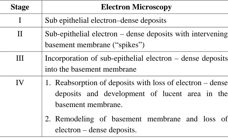
Table 2. Pathological Staging of Membranous Nephropathy
Stage Electron Microscopy
I Sub epithelial electron–dense deposits
II Sub-epithelial electron – dense deposits with intervening basement membrane (“spikes”)
III Incorporation of sub-epithelial electron – dense deposits into the basement membrane
IV 1. Reabsorption of deposits with loss of electron – dense deposits and development of lucent area in the basement membrane.
Heyman’s nephritis, an animal model of MGN, suggests that
glomerular lesions result from in situ formation of immune complexes
with megalin receptor–associated protein as the putative antigen. This
antigen is not found in human podocytes, but human antibodies have
been described against neutral endopeptidase expressed by podocytes,
hepatitis antigens B/C, tumor antigens, and thyroglobulin.
Eighty percent of patients with MGN present with nephrotic
syndrome and nonselective proteinuria. Microscopic hematuria is seen
in up to 50% of patients. Spontaneous remissions occur in 20–33% of
patients and often occur late in the course after years of nephrotic
syndrome. One-third of patients continue to have relapsing nephrotic
syndrome but maintain normal renal function, and approximately
another third of patients develop renal failure or die from the
complications of nephrotic syndrome [10, 11].
Male gender, older age, hypertension, and the persistence of
proteinuria are associated with worse prognosis. Although thrombotic
complications are a feature of all nephrotic syndrome.
MGN has the highest reported incidences of renal vein
thrombosis, pulmonary embolism, and deep vein thrombosis.
for patients with severe or prolonged proteinuria in the absence of risk
factors for bleeding.
In addition to the treatment of edema, dyslipidemia, and
hypertension, inhibition of the renin-angiotensin system is
recommended. Therapy with immunosuppressive drugs is also
recommended for patients with primary MGN and persistent proteinuria
(>4.0 g/24h) and in renal failure.
IgA Nephropathy
Berger first described the glomerulonephritis termed IgA
nephropathy. It is classically characterized by episodic hematuria
associated with the deposition of IgA in the mesangium. IgA
nephropathy is one of the most common forms of glomerulonephritis
worldwide. There is a male preponderance, a peak incidence in the
second and third decades of life, and rare familial clustering [12].
Henoch-Schönlein purpura is distinguished clinically from IgA
nephropathy by prominent systemic symptoms, a younger age
(<20 years old), preceding infection, and abdominal complaints.
Deposits of IgA are also found in the glomerular mesangium in a variety
of systemic diseases, including chronic liver disease, Crohn's disease,
gastrointestinal adenocarcinoma, chronic obstructive bronchiectasis,
fungiodes, leprosy, ankylosing spondylitis, relapsing polychondritis, and
Sjögren's syndrome. IgA deposition in these entities is not usually
associated with clinically significant glomerular inflammation or renal
dysfunction and thus is not called IgA nephropathy.
IgA nephropathy is an immune complex-mediated
glomerulonephritis defined by the presence of diffuse mesangial IgA
deposits often associated with mesangial hypercellularity. IgM, IgG, C3,
or immunoglobulin light chains may be co-distributed with IgA. IgA
deposited in the mesangium is typically polymeric and of the IgA1
subclass, the pathogenic significance of which is not clear.
Abnormalities have been described in IgA production by plasma
cells, particularly secretory IgA; in IgA O-glycosylation; in IgA
clearance, predominately by the liver; in mesangial IgA clearance and
receptors for IgA; and in growth factor and cytokine-mediated events.
Despite the presence of elevated serum IgA levels in 20–50% of
patients, IgA deposition in skin biopsies in 15–55% of patients, or
elevated levels of secretory IgA and IgA-fibronectin complexes
Although the immunofluorescent pattern of IgA on renal biopsy
defines IgA nephropathy in the proper clinical context, a variety of
segmental sclerosis; and, rarely, segmental necrosis with cellular
crescent formation, which typically presents as RPGN.
The two most common presentations of IgA nephropathy are
recurrent episodes of macroscopic hematuria during or immediately
following an upper respiratory infection in children (Henoch-Schönlein
purpura) or asymptomatic microscopic hematuria most often seen in
adults. Between episodes, the urinalysis is normal. When the hematuria
persists, one finds increasing amounts of proteinuria; nephrotic
syndrome, however, is uncommon.. Rarely, patients can present with
acute renal failure and a rapidly progressive clinical picture.
IgA nephropathy is a benign disease for the majority of patients,
with progression to renal failure seen in only 25–30% over 20–25 years;
in fact, 5–30% of patients go into complete remission. Risk factors for
the loss of renal function include the presence of hypertension or
proteinuria, the absence of episodes of macroscopic hematuria, male
age, older age of onset, and more severe changes on renal biopsy [13].
There is no agreement on optimal treatment. Both large studies
that include patients with multiple glomerular diseases or small studies
of patients with IgA nephropathy support the use of
angiotensin-converting enzyme (ACE) inhibitors in patients with proteinuria or
have all been suggested in small studies to benefit select patients with
IgA nephropathy. When presenting as RPGN, patients typically receive
steroids, cytotoxic agents, and plasmapheresis.
Post streptococcal Glomerulonephritis
Post streptococcal glomerulonephritis is prototypical for acute
endocapillary proliferative glomerulonephritis. Acute post streptococcal
glomerulonephritis typically affects children between the ages of 2 and
14 years, but 10% of cases are patients older than 40. It is more common
in males, and the familial or cohabitant incidence is as high as 40%.
Skin and throat infections with particular M types of streptococci
(nephritogenic strains) antedate glomerular disease; M types 47, 49, 55,
2, 60, and 57 are seen following impetigo and M types 1, 2, 4, 3, 25, 49,
and 12 with pharyngitis. Post streptococcal glomerulonephritis due to
impetigo develops 2–6 weeks after skin infection and 1–3 weeks after
streptococcal pharyngitis.
The renal biopsy in post streptococcal glomerulonephritis
demonstrates hypercellularity of mesangial and endothelial cells,
glomerular infiltrates of polymorphonuclear leukocytes, granular
subendothelial immune deposits of IgG, IgM, C3, and subepithelial
Post streptococcal glomerulonephritis is an immune-mediated
disease involving putative streptococcal antigens, circulating immune
complexes, and activation of complement in association with
cell-mediated injury. Many candidate antigens have been proposed over the
years; three such candidates from nephritogenic streptococci are
zymogen, a precursor of exotoxin B; glyceraldehyde phosphate
dehydrogenase, also known as prescribing antigen (PA-Ag); and
streptokinase. All have a biochemical affinity for GBMs, and in this
location they may act as a target for antibodies.
The classic presentation is an acute nephritic picture with
hematuria, pyuria, red blood cell casts, edema, hypertension, and
oliguric renal failure, which may be severe enough to appear as RPGN.
Systemic symptoms of headache, malaise, anorexia, and flank pain (due
to swelling of the renal capsule) are reported in as many as 50% of
cases.
Five percent of children and 20% of adults have proteinuria in the
nephrotic range. In the first week of symptoms, 90% of patients will
have a depressed CH50 and decreased levels of C3 with normal levels of
C4.
Positive cultures for streptococcal infection are inconsistently
(70%) or antihyaluronidase antibodies (40%) can help confirm the
diagnosis.
Treatment is supportive, with control of hypertension, edema, and
dialysis as needed. Antibiotic treatment for streptococcal infection
should be given to all patients and their cohabitants. There is no role for
immunosuppressive therapy, even in the setting of crescents. Recurrent
post streptococcal glomerulonephritis is rare despite repeated
streptococcal infections.
Overall, the prognosis is good, with permanent renal failure being
very uncommon (1–3%), and even less so in children. Complete
resolution of the hematuria and proteinuria in children occurs within 3–6
weeks of the onset of nephritis.
Membranoproliferative Glomerulonephritis
MPGN is sometimes called mesangiocapillary glomerulonephritis
or lobar glomerulonephritis. It is an immune-mediated
glomerulonephritis characterized by thickening of the GBM with
mesangioproliferative changes; 70% of patients have
hypocomplementemia. MPGN is rare in African Americans, and
idiopathic disease usually presents in childhood or young adulthood.
MPGN is subdivided pathologically into Type I, Type II, and Type III
hepatitis C infections, autoimmune diseases like lupus or
cryoglobulinemia, or neoplastic diseases. Types II and III MPGN are
usually idiopathic, except in the presence of C3 nephritic factor and/or in
partial lipodystrophy producing Type II disease or complement receptor
deficiency in Type III disease.
Type I Disease (Most Common)
• Idiopathic
• Subacute bacterial endocarditis
• Systemic lupus erythematosus
• Hepatitis C ± cryoglobulinemia
• Mixed cryoglobulinemia
• Hepatitis B
• Cancer: Lung, breast, and ovary (germinal)
Type II Disease (Dense Deposit Disease)
• Idiopathic
• C3 nephritic factor-associated
• Partial lipodystrophy
Type III Disease
• Idiopathic
Type I MPGN, the most proliferative of the three types, shows
mesangial proliferation with lobular segmentation on renal biopsy and
mesangial interposition between the capillary basement membrane and
endothelial cells, producing a double contour sometimes called
tram-tracking. Subendothelial deposits with low serum levels of C3 are
typical, although 50% of patients have normal levels of C3 and
occasional intra-mesangial deposits.
Low serum C3 and a dense thickening of the GBM containing
ribbons of dense deposits and C3 characterize Type II MPGN,
sometimes called dense deposit disease. Classically, the glomerular tuft
has a lobular appearance; intramesangial deposits are rarely present and
subendothelial deposits are generally absent.
Proliferation in Type III MPGN is less common than the other
two types and is often focal; mesangial interposition is rare, and
subepithelial deposits can occur along widened segments of the GBM
that appear laminated and disrupted.
Type I MPGN is secondary to glomerular deposition of
circulating immune complexes or their in situ formation. Types II and
III MPGN may be related to "nephritic factors," which are
autoantibodies that stabilize C3 convertase and allow it to activate
Patients with MPGN present with proteinuria, hematuria, and
pyuria (30%), systemic symptoms of fatigue and malaise that are most
common in children with Type I disease, or an acute nephritic picture
with RPGN and a speedy deterioration in renal function in up to 25% of
patients. Low serum C3 levels are common. Fifty percent of patients
with MPGN develop end-stage disease 10 years after diagnosis, and
90% have renal insufficiency after 20 years. Nephrotic syndrome,
hypertension, and renal insufficiency all predict poor outcome.
Although all primary renal diseases can recur over time in
transplanted renal allografts, patients with MPGN are well known to be
at risk for this adverse event.
Diabetic Nephropathy
Diabetic nephropathy is the single most common cause of chronic
renal failure in the United States, accounting for 45% of patients
receiving renal replacement therapy, and is a rapidly growing problem
worldwide.
The dramatic increase in the number of patients with diabetic
nephropathy reflects the epidemic increase in obesity, metabolic
syndrome, and Type 2 diabetes mellitus. Approximately 40% of patients
higher prevalence of Type 2 diabetes (90%) compared to Type 1 (10%),
the majority of patients with diabetic nephropathy have Type 2 disease.
Risk factors for the development of diabetic nephropathy include
hyperglycemia, hypertension, dyslipidemia, smoking, a family history of
diabetic nephropathy, and gene polymorphisms affecting the activity of
the renin-angiotensin-aldosterone axis.
Within 1–2 years after the onset of clinical diabetes, morphologic
changes appear in the kidney. Thickening of the GBM is a sensitive
indicator for the presence of diabetes but correlates poorly with the
presence or absence of clinically significant nephropathy. The
composition of the GBM is altered notably with a loss of heparan sulfate
moieties that form the negatively charged filtration barrier. This change
results in increased filtration of serum proteins into the urine,
predominately negatively charged albumin. The expansion of the
mesangium due to the accumulation of extracellular matrix correlates
with the clinical manifestations of diabetic nephropathy. This expansion
in mesangial matrix can be associated with the development of
mesangial sclerosis. Some patients also develop eosinophilic, PAS+
nodules called nodular glomerulosclerosis or Kimmelstiel-Wilson
nodules. Immunofluorescence microscopy often reveals the nonspecific
deposition of IgG (at times in a linear pattern) or complement staining
changes are frequently seen with hyaline and hypertensive
arteriosclerosis. This is associated with varying degrees of chronic
glomerulosclerosis and tubulointerstitial changes. Renal biopsies from
patients with Types 1 and Type 2 diabetes are largely indistinguishable.
Since the onset of Type 1 diabetes is readily identifiable and the
onset of Type 2 diabetes is not, a patient newly diagnosed with Type 2
diabetes may have renal disease for many years before nephropathy is
discovered and presents as advanced diabetic nephropathy. At the onset
of diabetes, renal hypertrophy and glomerular hyperfiltration are
present. The degree of glomerular hyperfiltration correlates with the
subsequent risk of clinically significant nephropathy.
In the approximately 40% of patients with diabetes who develop
diabetic nephropathy, the earliest manifestation is an increase in
albuminuria detected by sensitive radioimmunoassay. Albuminuria in
the range of 30–300 mg/24 h is called microalbuminuria. In patients
with Types 1 or Type 2 diabetes, microalbuminuria appears 5–10 years
after the onset of diabetes. It is currently recommended to test patients
with Type1 disease for microalbuminuria 5 years after diagnosis of
diabetes and yearly thereafter, and, because the time of onset of Type 2
diabetes is often unknown, to test Type 2 patients at the time of
Microalbuminuria is a potent risk factor for cardiovascular events
and death in patients with Type2 diabetes. Many patients with Type2
diabetes and microalbuminuria succumb to cardiovascular events before
they progress to proteinuria or renal failure. Proteinuria in frank diabetic
nephropathy can be variable, ranging from 500 mg to 25 g/24 h, and is
often associated with nephrotic syndrome.
More than 90% of patients with Type1 diabetes and nephropathy
have diabetic retinopathy, so the absence of retinopathy in Type 1
patients with proteinuria should prompt consideration of a diagnosis
other than diabetic nephropathy; only 60% of patients with Type 2
diabetes with nephropathy have diabetic retinopathy. There is a highly
significant correlation between the presence of retinopathy and the
presence of Kimmelstiel-Wilson nodules [15].
Also, characteristically, patients with advanced diabetic
nephropathy have normal to enlarged kidneys, in contrast to other
glomerular diseases where kidney size is usually decreased. Using the
above epidemiologic and clinical data, and in the absence of other
clinical or serologic data suggesting another disease, diabetic
nephropathy is usually diagnosed without a renal biopsy [16].
After the onset of proteinuria >500 mg/24 h, renal function
5–10 years; thus, from the earliest stages of microalbuminuria, it usually
takes 10–20 years to reach end-stage renal disease.
Hypertension may predict which patients develop diabetic
nephropathy, as the presence of hypertension accelerates the rate of
decline in renal function.
Good evidence supports the benefits of blood sugar and blood
pressure control as well as inhibition of the renin-angiotensin system in
retarding the progression of diabetic nephropathy. In patients with
Type1 diabetes, intensive control of blood sugar clearly prevents the
development or progression of diabetic nephropathy. The evidence in
patients with Type 2 disease, although less compelling, also supports
intensive control of blood sugar. Controlling systemic blood pressure to
levels of 130/80 mmHg or less decreases renal and cardiovascular
adverse events in this high-risk population. The vast majority of patients
with diabetic nephropathy require three or more antihypertensive drugs
to achieve this goal. Drugs that inhibit the renin-angiotensin system,
independent of their effects on systemic blood pressure, have been
repeatedly shown to slow the progression of diabetic nephropathy at
early (microalbuminuria) and late (proteinuria with reduced glomerular
filtration) stages, independent of any effect they may have on systemic
Since angiotensin II increases efferent arteriolar resistance and,
hence, glomerular capillary pressure, one key mechanism for the
efficacy of ACE inhibitors or angiotensin receptor blockers (ARBs) is
reducing glomerular hypertension. Patients with Type1 diabetes for 5
years who develop albuminuria or declining renal function should be
treated with ACE inhibitors. Patients with Type2 diabetes and
microalbuminuria or proteinuria may be treated with ACE inhibitors or
ARBs [17].
Renal Amyloidosis
Most renal amyloidosis is either the result of primary fibrillar
deposits of immunoglobulin light chains [amyloid L (AL)], or secondary
to fibrillar deposits of serum amyloid A (AA) protein fragments. Even
though both occur for different reasons, their clinicopathophysiology is
quite similar and will be discussed together. Amyloid infiltrates the
liver, heart, peripheral nerves, carpal tunnel, upper pharynx, and kidney,
producing restrictive cardiomyopathy, hepatomegaly, macroglossia, and
heavy proteinuria sometimes associated with renal vein thrombosis.
In systemic AL amyloidosis, also called primary amyloidosis,
light chains produced in excess by clonal plasma cell dyscrasias are
made into fragments by macrophages so they can self-aggregate at acid
lambda class. About 10% of these patients have overt myeloma with
lytic bone lesions and infiltration of the bone marrow with >30% plasma
cells; nephrotic syndrome is common, and about 20% of patients
progress to dialysis.
AA amyloidosis is sometimes called secondary amyloidosis and
also affects the kidney with nephrotic syndrome. It is due to deposition
of -pleated sheets of serum amyloid A protein, an acute phase reactant
whose physiologic function is unknown. Forty percent of patients with
AA amyloid have rheumatoid arthritis, and another 10% have
ankylosing spondylitis or psoriatic arthritis; the rest derive from other
lesser causes.
Fragments of serum amyloid A protein increase and
self-aggregate by attaching to receptors for advanced glycation end products
in the extracellular environment; nephrotic syndrome is common, and
about 40–60% of patients progress to dialysis.
AA and AL amyloid fibrils are detectable with Congo red or in
more detail with electron microscopy. Biopsy of involved liver or
kidney is diagnostic 90% of the time when the pretest probability is
high; abdominal fat pad aspirates are positive about 70% of the time, but
distributed along blood vessels and in the mesangial regions of the
kidney.
The treatment for primary amyloidosis is not particularly
effective; Melphalan and autologous hematopoietic stem cell
transplantation can delay the course of disease in about 30% of patients.
Secondary amyloidosis is also relentless unless the primary disease can
be controlled. Some new drugs in development that disrupt the
formation of fibrils may be available in the future [18].
Lupus Nephritis
Lupus nephritis is a common and serious complication of
systemic lupus erythematosus (SLE) and most severe in
African-American female adolescents. Thirty to fifty percent of patients will
have clinical manifestations of renal disease at the time of diagnosis, and
60% of adults and 80% of children develop renal abnormalities at some
point in the course of their disease. Lupus nephritis results from the
deposition of circulating immune complexes, which activate the
complement cascade leading to complement-mediated damage,
leukocyte infiltration, activation of procoagulant factors, and release of
various cytokines. In situ immune complex formation following
injury. The presence of antiphospholipid antibodies may trigger a
[image:35.612.131.495.226.439.2]thrombotic microangiopathy in a minority of patients.
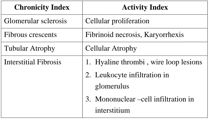
Table 3. Pathological Indices of Activity and Tonicity in Lupus
Nephritis
Chronicity Index Activity Index
Glomerular sclerosis Cellular proliferation
Fibrous crescents Fibrinoid necrosis, Karyorrhexis
Tubular Atrophy Cellular Atrophy
Interstitial Fibrosis 1. Hyaline thrombi , wire loop lesions 2. Leukocyte infiltration in
glomerulus
3. Mononuclear –cell infiltration in interstitium
Importance of biopsy in Lupus
Border Suggested > 6 RBC / HPF and urine protein
> 200mg / 24hrs. Abnormal serum creatinine [18]. Mahajan et al.
described 12 patients with diffused proliferative lupus nephritis but
without clinical or laboratory evidence of renal involvement so biopsy
recommended in patients with SLE even in the absence of overt clinical
Histological scoring system for lupus nephritis is an effort to
accurately predict the renal outcome and to help to determine which
patients are likely to benefit from aggressive therapy.
The clinical manifestations, course of disease, and treatment of
lupus nephritis are closely linked to the renal pathology. The most
common clinical sign of renal disease is proteinuria, but hematuria,
hypertension, varying degrees of renal failure, and active urine sediment
with red blood cell casts can all be present.
The extrarenal manifestations of lupus are important in
establishing a firm diagnosis of systemic lupus because, while serologic
abnormalities are common in lupus nephritis, they are not diagnostic.
Anti-dsDNA antibodies that fix complement correlate best with the
presence of renal disease. Hypocomplementemia is common in patients
with acute lupus nephritis (70–90%) and declining complement levels
may herald a flare. Renal biopsy, however, is the only reliable method
of identifying the morphologic variants of lupus nephritis.
The World Health Organization (WHO) workshop in 1974 first
outlined several distinct patterns of lupus-related glomerular injury;
these were modified in 1982. In 2004 the International Society of
Nephrology in conjunction with the Renal Pathology Society again
best defines clinicopathologic correlations, provides valuable prognostic
information, and forms the basis for modern treatment
recommendations.
Class I nephritis describes normal glomerular histology by any
technique or normal light microscopy with minimal mesangial deposits
on immunofluorescent or electron microscopy. Class II designates
mesangial immune complexes with mesangial proliferation. Both Class I
and II lesions are typically associated with minimal renal manifestation
and normal renal function; nephrotic syndrome is rare. Patients with
lesions limited to the renal mesangium have an excellent prognosis and
generally do not need therapy for their lupus nephritis.
Table 4. Revised in 2004 by the International Society of
Nephrology-Renal Pathology Society Study Group.
Class I Minimal mesangial
Normal histology with mesangial deposits
Class II Mesangial proliferation
Mesangial hypercellularity with expansion of the mesangial matrix
Class III Focal nephritis
Focal endocapillary ± extracapillary proliferation with focal subendothelial immune deposits and mild mesangial expansion
Class IV Diffuse nephritis
Class V Membranous nephritis
Thickened basement membranes with diffuse subepithelial immune deposits; may occur with Class III or IV lesions and is sometimes called mixed membranous and proliferative nephritis
Class VI Sclerotic nephritis
Global sclerosis of nearly all glomerular capillaries
The subject of lupus nephritis is presented under acute nephritic
syndromes because of the aggressive and important proliferative lesions
seen in Class III–V renal disease.
Class III describes focal lesions with proliferation or scarring,
often involving only a segment of the glomerulus Class III lesions have
the most varied course. Hypertension, an active urinary sediment, and
proteinuria are common with nephrotic-range proteinuria in 25–33% of
patients. Elevated serum creatinine is present in 25% of patients.
Patients with mild proliferation involving a small percentage of
glomeruli respond well to therapy with steroids alone, and fewer than
5% progress to renal failure over 5 years. Patients with more severe
proliferation involving a greater percentage of glomeruli have a far
worse prognosis and may have lower remission rates. Treatment of
those patients is the same as that for Class IV lesions, as some
nephrologists believe that Class III lesions are simply an early
Class IV describes global, diffuse proliferative lesions involving
the vast majority of glomeruli. Patients with Class IV lesions commonly
have high anti-DNA antibody titers, low serum complement, hematuria,
red blood cell casts, proteinuria, hypertension, and decreased renal
function; 50% of patients have nephrotic-range proteinuria. Patients
with crescents on biopsy may have a rapidly progressive decline in renal
function. Without treatment, this aggressive lesion has the worst renal
prognosis. However, if a remission is achieved with treatment, renal
outcomes are excellent. Treatment must combine high-dose steroids
with either Cyclophosphamide or Mycophenolate Mofetil. Current
evidence suggests that inducing a remission with administration of
steroids and either Cyclophosphamide or Mycophenolate Mofetil for
2–6 months, followed by maintenance therapy with lower doses of
steroids and Mycophenolate Mofetil, may best balance the likelihood of
successful remission with the side effects of therapy.
The Class V lesion describes subepithelial immune deposits
producing a membranous pattern; a subcategory of Class V lesions is
associated with proliferative lesions and is sometimes called mixed
membranous and proliferative disease. Sixty percent of patients present
with nephrotic syndrome or lesser amounts of proteinuria. Patients with
lupus nephritis Class V, like patients with idiopathic membranous
thrombolic complications. A minority of patients with Class V will
present with hypertension and renal dysfunction.. Patients with severe
nephrotic syndrome, elevated serum creatinine, and a progressive course
will probably benefit from therapy with steroids in combination with
other immunosuppressive agents. Therapy with inhibitors of the
renin-angiotensin system also may attenuate the proteinuria.
Patients with any of the above lesions also can transform to
another lesion; hence patients often require reevaluation, including
repeat renal biopsy. Lupus patients with Class VI lesions have greater
than 90% sclerotic glomeruli and end-stage renal disease with interstitial
fibrosis. As a group, approximately 20% of patients with lupus nephritis
will reach end-stage disease, requiring dialysis or transplantation.
Systemic lupus tends to become quiescent once there is renal failure,
perhaps due to the immunosuppressant effects of uremia. Renal
transplantation in renal failure from lupus, usually performed after
approximately 6 months of inactive disease, results in allograft survival
rates comparable to patients transplanted for other reasons.
Antiglomerular Basement Membrane Disease
Patients who develop autoantibodies directed against glomerular
basement antigens frequently develop a glomerulonephritis termed
present with lung hemorrhage and glomerulonephritis, they have a
pulmonary-renal syndrome called Good pasture's syndrome.
The target epitopes for this autoimmune disease lie in the
quaternary structure of 3 NC1 domain of collagen IV. MHC-restricted T
cells initiate the autoantibody response because humans are not tolerant
to the epitopes created by this quaternary structure. The epitopes are
normally sequestered in the collagen IV hexamer and can be exposed by
infection, smoking, oxidants, or solvents. Good pasture's syndrome
appears in two age groups: in young men in their late 20s and in men
and women in their 60–70s.
Disease in the younger age group is usually explosive, with
hemoptysis, a sudden fall in hemoglobin, fever, dyspnoea, and
hematuria. Hemoptysis is largely confined to smokers, and those who
present with lung hemorrhage as a group do better than older
populations who have prolonged, asymptomatic renal injury;
presentation with oliguria is often associated with a particularly bad
outcome [20].
The performance of an urgent kidney biopsy is important in
suspected cases of Good pasture's syndrome to confirm the diagnosis
and assess prognosis. Renal biopsies typically show focal or segmental
cellular proliferation, leads to crescent formation in Bowman's space.
As these lesions progress, there is concomitant interstitial nephritis with
fibrosis and tubular atrophy. The presence of anti-GBM antibodies and
complement is recognized on biopsy by linear immunofluorescent
staining for IgG .
Between 10–15% of sera from patients with Good pasture's
syndrome also contain ANCA antibodies against myeloperoxidase. This
subset of patients has a vasculitis-associated variant, which has a
surprisingly good prognosis with treatment.
Prognosis at presentation is worse if there are >50% crescents on
renal biopsy with advanced fibrosis, if serum creatinine is >5–6 mg/dL,
if oliguria is present, or if there is a need for acute dialysis. Patients with
advanced renal failure who present with hemoptysis should still be
treated for their lung hemorrhage, as it responds to plasmapheresis and
can be lifesaving. Treated patients with less severe disease typically
respond to 8–10 treatments of plasmapheresis accompanied by oral
prednisone and Cyclophosphamide in the first 2 weeks.
Kidney transplantation is possible, but because there is risk of
recurrence, patients should wait for 6 months and until serum antibodies
ANCA Small Vessel Vasculitis
A group of patients with small-vessel vasculitis (arterioles,
capillaries, and venules; rarely small arteries) and glomerulonephritis
have serum ANCA; the antibodies are of two types, anti-proteinase 3
(PR3) or anti-myeloperoxidase (MPO). ANCA are produced with the
help of T cells and activate leukocytes and monocytes, which together
damage the walls of small vessels. Endothelial injury also attracts more
leukocytes and extends the inflammation. Wegener's granulomatosis,
microscopic polyangiitis, and Churg-Strauss syndrome belong to this
group because they are ANCA-positive and have a pauci-immune
glomerulonephritis with few immune complexes in small vessels and
glomerular capillaries. Patients with any of these three diseases can have
any combination of the above serum antibodies, but anti-PR3 antibodies
are more common in Wegener's and anti-MPO antibodies are more
common in microscopic polyangiitis or Churg-Strauss. While each of
these diseases have some unique clinical features, most features do not
predict relapse or progression, and as a group they are generally treated
in the same way. Only the presence of upper-airway involvement,
persistent pulmonary injury, and anti-PR3 antibodies suggests that the
course of disease will be more difficult. Induction therapy usually
Cyclophosphamide. The benefit of Plasmapheresis in this setting is
uncertain.
The steroids are tapered soon after acute inflammation subsides,
and patients are maintained on Cyclophosphamide or Azathioprine for
up to a year to minimize the risk of relapse.
Wegener's Granulomatosis
Patients with this disease classically present with fever, purulent
rhinorrhea, nasal ulcers, sinus pain, polyarthralgias/arthritis, cough,
hemoptysis, shortness of breath, microscopic hematuria, and 0.5–1 g /
24 h of proteinuria; occasionally there may be cutaneous purpura and
mononeuritis multiplex. Presentation without renal involvement is
termed limited Wegener's granulomatosis, although some of these
patients will show signs of renal injury later. Chest x-ray often reveals
nodules and persistent infiltrates, sometimes with cavities. Biopsy of
involved tissue will show a small-vessel vasculitis and adjacent
noncaseating granulomas. Renal biopsies during active disease
demonstrate segmental necrotizing glomerulonephritis without immune
Microscopic Polyangiitis
Clinically, these patients look somewhat similar to those with
Wegener's granulomatosis, except they rarely have significant lung
disease or destructive sinusitis. The distinction is made on biopsy where
the vasculitis in microscopic polyangiitis is without granulomas. Some
patients will also have injury limited to the capillaries and venules.
Churg-Strauss Syndrome
When small-vessel vasculitis is associated with peripheral
eosinophilia, cutaneous purpura, mononeuritis, asthma, and allergic
rhinitis, a diagnosis of Churg-Strauss syndrome is considered.
Hypergammaglobulinemia, elevated levels of serum IgE, or the
presence of rheumatoid factor sometimes accompanies the allergic state.
Lung inflammation, including fleeting cough and pulmonary infiltrates,
often precedes the systemic manifestations of disease by years; lung
manifestations are rarely absent. A third of patients may have exudative
pleural effusions associated with eosinophils. Small-vessel vasculitis
and focal segmental necrotizing glomerulonephritis can be seen on renal
biopsy, usually absent eosinophils or granuloma. The cause of
Churg-Strauss syndrome is autoimmune, but the inciting factors are unknown.
Interestingly, some asthma patients treated with leukotriene receptor
OBJECTIVES OF THE STUDY
1. To study and analyse the clinical pattern of Glomerulonephritis
and to observe the changing pattern of Glomerulonephritis in
our centre during two study periods, i.e. 2000-2006 and
2007-2008.
MATERIALS AND METHODS
• The study was done as prospectively from Jan 2007- Dec 2008.
Retrospective data from Jan 2000-Dec 2006 were retrieved from
case record.
• Study centre : Department of Nephrology , Government General
Hospital , Madras Medical College , Chennai – 600 003
• Plan of study: All patients who had clinical, laboratory and
histopathological features of Glomerular disease were analysed.
• Detailed history and Clinical examination were done to find out
evidence of volume status, BP measurements and Funduscopic
examination followed by basic laboratory investigation like
urine analysis complete blood count, coagulation profile, renal
function test, liver function test, Chest X–Ray, Ultrasonographic
examination of the Abdomen with done and basic serelogical
and immunological workup like HBsAg, anti HCV, HIV, ASO
Titre and ANA were done.
• After informed consent Ultrasound guided Renal Biopsy was
• All biopsies were evaluated by Light microscopy and
immunoflorescence.
• Patients were grouped according to age, gender, presence of
STATISTICAL ANALYSIS
Demographic variables like age, sex and histology categories
were given in frequencies with their percentages.
Sex-wise and year wise difference on histological categories was
analysed using Two Sample Binomial Proportion Test.
Sex-wise, renal biopsy in diabetes was analyzed using Pearson
Chi Square Test.
Decade-wise clinical syndrome was analyzed using Two Sample
Binomial Proportion Test.
Comparison with other studies was analysed using Pearson Chi
Square Test.
RESULTS
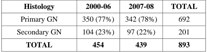
Distribution of Patients
• Total number of biopsies done from 2000-2008 is 893 are
included in this study.
• Datas were collected from the case records for the years
2000-2006 and between 2007-2008 were studied prospectively.
• Total number of biopsies done from 2000-2006 was 454 out of
which 350(77%) was due to primary glomeluar disease and
104(23%) was due to secondary gomerular disease.
• Number of biopsies in 2007-2008 was 439 out of which 342
(78%) was primary glomeluar disease and 97 (22%) was
[image:50.612.132.493.610.701.2]contributed secondary glomeluar disease.
Table 5. Distribution of patients
Histology 2000-06 2007-08 TOTAL
Primary GN 350 (77%) 342 (78%) 692 Secondary GN 104 (23%) 97 (22%) 201
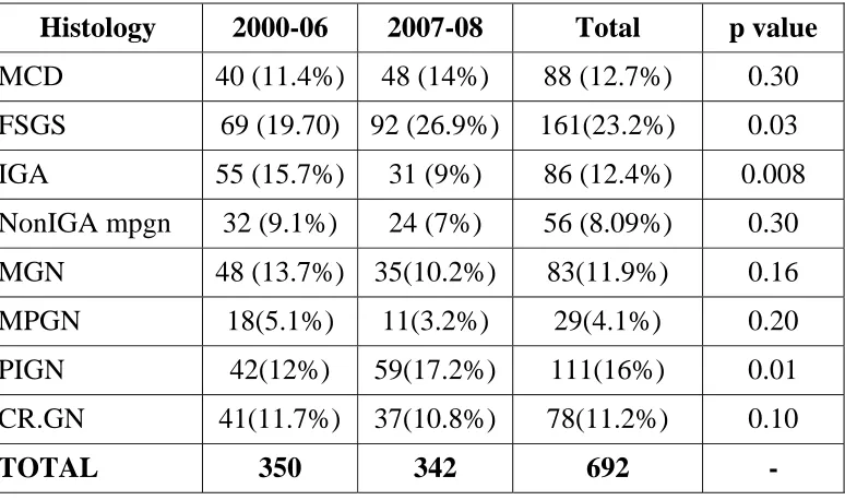
The distribution of various primary glomerular diseases are given
[image:51.612.121.508.241.470.2]below:
Table 6. Distribution of Primary Glomerular Disease
Histology 2000-06 2007-08 Total p value
MCD 40 (11.4%) 48 (14%) 88 (12.7%) 0.30 FSGS 69 (19.70) 92 (26.9%) 161(23.2%) 0.03 IGA 55 (15.7%) 31 (9%) 86 (12.4%) 0.008 NonIGA mpgn 32 (9.1%) 24 (7%) 56 (8.09%) 0.30
MGN 48 (13.7%) 35(10.2%) 83(11.9%) 0.16 MPGN 18(5.1%) 11(3.2%) 29(4.1%) 0.20 PIGN 42(12%) 59(17.2%) 111(16%) 0.01 CR.GN 41(11.7%) 37(10.8%) 78(11.2%) 0.10
TOTAL 350 342 692 -
P=0.05 significant
Incidence of FSGS and PIGN has increased significantly from 2000-06 to 2007-08.
Distribution of Primary Glomerular Disease 2000-2006
MCD 40 FSGS 69 IGA 55 nonIGA 32 MGN 48 MPGN 18 PIGN 42 CR.GN 41
Distribution of Primary Glomerular Disease 2007-2008
Distribution of Secondary Glomerular Disease
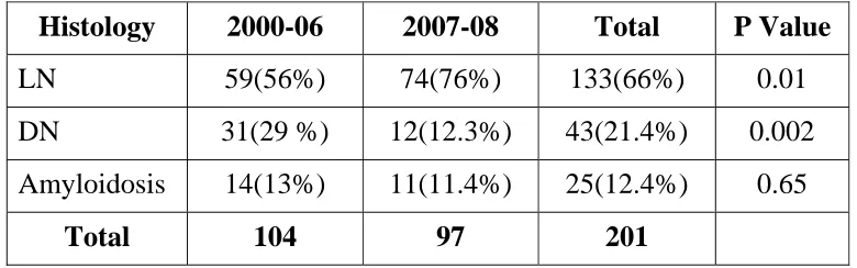
Number of biopsies done between 2000 and 2006 was 104
whereas the number of biopsies done between 2007 and 2008 was
97.The incidence of secondary Glomerular disease –Lupus Nephritis
was 59(56%), Diabetic nephropathy(Where non diabetic kidney disease
was suspected) was 31(29%) and Amyloidosis 14(11.4%) respectively
.In second group between 2007-2008 the number of biopsies done was
201, Lupus Nephritis was 74(76%), Diabetic nephropathy was
[image:53.612.118.508.390.512.2]12(12.3%) and Amyloidosis 11(145%) respectively.
Table 7. Distribution of Secondary Glomerular Disease
Histology 2000-06 2007-08 Total P Value
LN 59(56%) 74(76%) 133(66%) 0.01 DN 31(29 %) 12(12.3%) 43(21.4%) 0.002
Amyloidosis 14(13%) 11(11.4%) 25(12.4%) 0.65
Total 104 97 201
P= 0.05 significant
Incidence of diabetic nephropathy has decreased in our study because of our biopsy protocol.
This is probably of change in biopsy policy in patients with
diabetic mellitus with proteinuria. We do biopsy only if patient had
active urine sediment, micro hematuria, rapidly progressive renal
Distribution of Secondary Glomerular Disease 2000-2006
LN 59 DN 31
amyloidosis 14
Distribution of Secondary Glomerular Disease 2007-2008
LN 74 DN 12
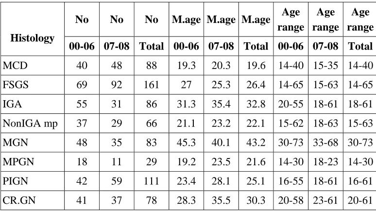
The Distribution of Age in various Primary Glomerular Disease
[image:55.612.118.505.198.416.2]are given below
Table 8. Distribution of Age in Primary Glomerular Disease
Histology
No No No M.age M.age M.age Age
range Age range
Age range
00-06 07-08 Total 00-06 07-08 Total 00-06 07-08 Total
MCD 40 48 88 19.3 20.3 19.6 14-40 15-35 14-40 FSGS 69 92 161 27 25.3 26.4 14-65 15-63 14-65 IGA 55 31 86 31.3 35.4 32.8 20-55 18-61 18-61 NonIGA mp 37 29 66 21.1 23.2 22.1 15-62 18-63 15-63 MGN 48 35 83 45.3 40.1 43.2 30-73 33-68 30-73 MPGN 18 11 29 19.2 23.5 21.6 14-30 18-23 14-30 PIGN 42 59 111 23.4 28.1 25.1 16-55 18-61 16-61 CR.GN 41 37 78 28.3 35.5 30.3 20-58 23-61 20-61
Mean age is higher in PIGN & Cr.GN .As for FSGS & MGN are concerned mean age is lower.
Table 9. Distribution of Age in Secondary Glomerular Disease
Histology
No No No M.age M.age M.age Age
range Age range
Age range
00-06 07-08 Total 00-06 07-08 Total 00-06 07-08 Total
LN 59 74 113 24.3 26.3 25 18-62 20-58 18-62 DN 31 12 43 43 39 41.8 33-58 30-49 30-58 Anyloidosis 11 14 25 46 42 45.6 39-63 36-65 36-65
The increasing trend observed for mean age in Lupus Nephritis.
[image:55.612.117.509.530.649.2]The Distribution of Gender in various Primary Glomerluar
[image:56.612.155.471.197.426.2]Diseases is given below:
Table 10. Distribution of Gender in Primary Glomerluar Disease
Histology M/F Ratio Total
00-06 07-08
MCD 1.4 1.4 1.4
FSGS 2.2 2.4 2.3
IGA 2.2 2.5 2.3
nonIGA mp 1.3 1.6 1.4
MGN 1.6 1.8 1.7
MPGN 1.4 1 1.3
PIGN 2.1 2.6 2.3
CR.GN 0.8 0.9 0.8
Male predominance was observed in FSGS, IgAN, MGN and PIGN,
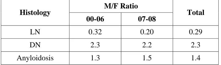
The Distribution of Gender in various secondary Glomerluar
[image:57.612.124.504.201.313.2]Diseases is given below.
Table 11. Distribution of Gender in Secondary Glomerular Disease
Histology M/F Ratio Total
00-06 07-08
LN 0.32 0.20 0.29
DN 2.3 2.2 2.3
Anyloidosis 1.3 1.5 1.4
Female predominance was observed in Lupus nephritis whereas male dominance was observed in Diabetic nephropathy.
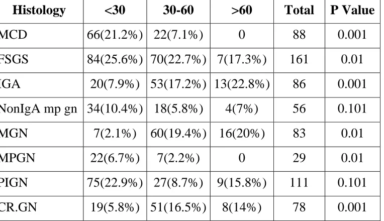
The distribution of various glomerular diseases with age group is
given below.
Table 12. Distribution of Glomerular Disease with Age Group
Histology <30 30-60 >60 Total
Primary GN 327 308 57 692 Secondary GN 124 71 6 201
Total 451 379 63 893
Incidence of primary glomerular disease was similar between 30
[image:57.612.155.474.524.623.2]The Distribution of various Primary Glomerular Diseases with
Age group is given below.
Table 13. Distribution of Primary Glomerular Disease with Age
Group
Histology <30 30-60 >60 Total P Value
MCD 66(21.2%) 22(7.1%) 0 88 0.001
FSGS 84(25.6%) 70(22.7%) 7(17.3%) 161 0.01
IGA 20(7.9%) 53(17.2%) 13(22.8%) 86 0.001
NonIgA mp gn 34(10.4%) 18(5.8%) 4(7%) 56 0.101
MGN 7(2.1%) 60(19.4%) 16(20%) 83 0.01
MPGN 22(6.7%) 7(2.2%) 0 29 0.01
PIGN 75(22.9%) 27(8.7%) 9(15.8%) 111 0.101
CR.GN 19(5.8%) 51(16.5%) 8(14%) 78 0.001
P=0.05 significant
Incidence of MCD, PIGN and FSGS was higher < 30 years of age
group. Incidence of IgA, CrGN & MGN nephropathy was more in the
[image:58.612.126.508.271.491.2]The Distribution of various Secondary glomerular diseases with
Age Group is given below.
Table 14. Distribution of Secondary glomerular disease with Age
Group
Histology <30 30-60 >60 Total P Value
LN 117(94.3%) 14(19.7%) 2(33.3%) 133 0.10
DN 4(3.2%) 38(53.5%) 1(16.6%) 43 0.10
Amyloidosis 3(2.4%) 19(26.7%) 3(50%) 25 0.001
Total 124 71 6 201
P=0.05
Incidence of lupus nephritis was higher in less than 30 years of
[image:59.612.119.504.267.409.2]The Clinical Presentation of various Glomerular Diseases at
initial presentations is given below.
Table 15. Clinical Presentation of Glomerular Disease on
Presentation
Histology Edema Oliguria
Macrohe-maturia Fever Anorexia Dyspnoea Total
MCD 80 6 0 12 6 6 88
FSGS 98 26 3 18 23 31 161
IGA 21 23 25 8 21 19 86
nonIGA mp 15 13 2 7 3 7 56
MGN 40 7 3 3 7 8 83
MPGN 12 2 3 7 4 3 29
PIGN 60 71 24 25 19 23 111
CR.GN 60 55 21 23 33 35 78
LN 31 23 17 43 23 20 133
DN 36 21 1 7 8 10 43
Amyloidosis 12 4 0 3 3 3 25
MCD (90.9%), FSGS (60.8%) & MGN (48.1%) are most commonly presented with edema.
In IgA nephropathy, 29% had Macrohaematuria.
In PIGN 63.96 % had Oliguria 18% had Marohaematuria.
In CrGN 76.92% had edema, 70.5% had oliguria 26.92% had macrohaematuria.
In Lupus nephritis 32.3% had fever 17.29 had oliguria.
[image:60.612.117.508.196.500.2]The Examination Findings of various Glomerular Diseases at
initial presentations are given below.
Table 16. Examination Findings of Glomerular Disease on
Presentation
Histology Edema Ht Retinopathy Pleural
effusion Ascitis Total
MCD 84 10 0 32 33 88
FSGS 106 41 31 31 32 161
IGA 25 34 12 9 8 86
nonIGA mp 18 16 3 2 3 56
MGN 45 27 13 5 4 83
MPGN 15 12 5 3 2 29
PIGN 73 65 4 12 11 111 CR.GN 66 53 8 19 7 78
LN 36 70 23 33 21 133
Incidence of hypertension in MCD (11.36%), FSGS (25.46%), MGN (32.53%).
Incidence of hypertension in IgA nephropathy (39.53%), PIGN (28.55%).
Incidence of hypertension in CrGN (67.94%).
[image:61.612.121.512.180.477.2]The lab features of various primary and secondary diseases are
[image:62.612.119.509.196.530.2]given below:
Table 17. Lab Features of Glomerular Disease
Histology
Microhe-maturia Blood urea >7 Mmol Sr.creat >130 micromo Chol >200 mg/dl Avg. Urine pcr Avg.sr. Albumin <3g/dl Total
MCD 12 3 2 52 6.2 43 88 FSGS 36 53 48 73 2.8 73 161 IGA 41 34 31 10 2.1 2.2 86 nonIGA mp 11 13 11 8 1.3 7 56 MGN 37 13 10 12 5.8 22 83 MPGN 8 7 6 7 3.1 8 29 PIGN 73 73 69 9 3.1 9 111 CR.GN 69 61 63 5 3.3 13 78 LN 101 33 30 8 2.1 12 133 DN 13 29 20 21 4.3 16 43
Amyloidosis 2 6 4 7 3.6 10 25
In MCD 2 of them presented with renal failure. One had sepsis
(22 yrs) due to spontaneous bacterial peritonitis and recovered with
antibiotics. Another person (41 yrs) presented with renal vein
more than 200 mg/dl with average urine PCR 6.2 and 43 of them
presented with hypo Albuminemia.
In FSGS 48 of them presented with renal failure and 73 of them
presented with hyper lipidemia.
In IgA nephropathy 41 of them presented with micro hematuria
and 31 of them presented with renal failure.
In PIGN 69 of them presented with renal failure.
In CrGN 69 of them presented with micro hematuria and 63 of
them presented with renal failure.
In lupus nephritis 30 of them presented with renal failure and 8 of
them had hyper lipidemia.
Table 18. Comparison with Other Studies Histological Categories MMC Study (n=893) CMC, Vellore (n=4035) Minnesota (n=195) Italian Study (n=13835) Pearson Chi Square Test Comparison between Studies
n % n % n % n %
FSGS 161 11.79 677 16.8 33 21.5 1730 12.5
χ2 =181.6 P=0.001
1 Vs 2,3
PIGN 111 7.70 543 13.5 7 3.6 360 2.6
χ2 =760.1 P=0.001
1 Vs 2,3,4
MCD 88 6.48 433 10.8 8 4.1 1065 7.7
χ2 =162.2 P=0.001
1 Vs 2
Mesangial
PGN 56 5.09 293 7.3 1231 8.9
χ2 =36.75 P=0.001
1 Vs 2,4
MN 83 5.92 384 9.5 20 10.2 3127 22.6. χ2 =572
P=0.001 1 Vs 2,3,4
IgAN 86 6.04 338 8.4 42 21.5 5188 37.5
χ2 =173.1 P=0.001
1 Vs 2,3,4
Lupus
Nephritis 133 8.97 279 6.9 25 12.8
χ2 =14.71 P=0.001
1 Vs 2
Crescentic
GN 78 4.21 140 3.5 10 5.1 941 6.8
χ2 =72.64 P=0.001
1 Vs 2,4
Diabetic
Nephropathy 43 3.77 111 2.8
χ2 =4.32
P=0.04 1 Vs 2 Amyloidosis 75 4.15 41 1.0 5 2.6 χ2 =62.8
The results of this study correlate significantly with south Indian
study (CMC, Vellore) for FSGS, MCD, MN, IgAN and Lupus
Nephritis.
The results of this study significantly correlate with Italian study
DISTRIBUTION OF PRIMARY GLOMERULAR DISEASE 40 69 55 32 48 18 42 41 48 92 31 24 35 11 59 37 0 10 20 30 40 50 60 70 80 90 100
MCD FSGS IGA nonIGA mp MGN MPGN PIGN CR.GN
N o . o f p a ti e n ts 2000-06 2007-08 1.4 2.3 2.3 1.4 1.7 1.3 2.3 0.8 0.21 2.3 1.4 0 0.5 1 1.5 2 2.5 M/ F r a ti o
MCD FSGS IGA nonIGA m p
MGN MPGN PIGN CR.GN LN DN Anyloid DISTRIBUTION OF GENDER RATIO AMONG
19.6 26.4 32.8 22.1 43.2 21.6 25.1 30.3 25 41.8 45.6 0 5 10 15 20 25 30 35 40 45 50 % o f p a ti e n ts
MCD FSGS IGA nonIGA mp MGN MPGN PIGN CR.GN LN DN Amyloid
DISTRIBUTION OF MEAN AGE OF GLOMERULAR DISEASE
DISTRIBUTION OF AGEWISE PRIMARY GLOMERULAR DISEASE
0 10 20 30 40 50 60 70 80 90 100
MCD FSGS IGA nonIGA
mp
MGN MPGN PIGN CR.GN LN DN Amyloid
DISTRIBUTION OF GLOMERULAR DISEASE 0 20 40 60 80 100 120
MCD FSGS IGA nonIGA mp MGN MPGN PIGN CR.GN LN DN Amyloid
N o . o f p a ti e n ts micro bu scr>130 chol>200 sr.alb<3
CLINICAL PRESENTATION OF GLOMERULAR PRESENTATION OF GLOMERULAR DISEASE AT PRESENTATION
0 20 40 60 80 100 120
MCD FSGS IGA nonIGA
mp
MGN MPGN PIGN CR.GN LN DN Amyloid
EXAMINATION FINDINGS OF GLOMERULAR DISEASE AT PRESENTATION
0 20 40 60 80 100 120
MCD FSGS IGA nonIGA mp MGN MPGN PIGN CR.GN LN DN Amyloid
N
o
. of
pa
ti
e
n
ts
Edem a
HT
Ratiopathy
pl.effusion