THE TAMILNADU
Dr. M.G.R. MEDICAL UNIVERSITY
CHENNAI
STUDY OF TUBERCULOUS CERVICAL
LYMPHADENOPATHY
DISSERTATION SUBMITTED FOR M.S. DEGREE BRANCH 1
(GENERAL SURGERY)
DEPARTMENT OF GENERAL SURGERY
MADURAI MEDICAL COLLEGE
MADURAI.
CERTIFICATE
This is to certify that the dissertation entitled “Study of Tuberculous
Cervical Lymphadenopathy” is a bonafide record of work done by
Dr. M. Gnaanasekar, in the Department of Surgery, Government Rajaji
Hospital, Madurai Medical College, Madurai.
Prof. Dr. M. Kalyanasundaram, Prof. Dr. V.Seetharaman, M.S., FICS., M.S.,
Professor and H.O.D of Surgery Additional Professor of Surgery
Govt. Rajaji Hospital and Govt. Rajaji Hospital and
Madurai Medical College Madurai Medical College
Madurai Madurai
Dean
ACKNOWLEDGEMENT
I wish to express my sincere gratitude to my Chief
Prof.V.SEETHARAMAN, MS., Additional Professor of Surgery for having
helped me at every step in successfully completing this study.
I wish to express my sincere thanks to
Prof.M.KALYANASUNDARAM, MS., Professor and Head of the
Department of Surgery for his sincere advice and co-operation for completing
this study.
I also thank my Professor and Head of the Department of Pathology
and to all teachers who helped me to complete this study in time.
I am also very grateful to our DeanDr.M.VASANTHA,MD., Her kind
permission to utilize the clinical material for this study.
I also extend my thanks to all my patients for their full co-operation in
TABLE OF CONTENTS
Sl. No. Titles Page
1. Introduction 1
2. Historical Review 4
3. Embryology of Lymphatic System 7
4. Anatomy of Lymphatic System 8
5. Lymph Drainage of Head and Neck 10
6. Etiology of Lymphadenopathy 19
7. Clinical Features 36
8. Investigations 38
9. Differential Diagnosis 43
10. Treatment 44
11. Materials and Methods 54
12. Analysis of Data 57
13. Summary 73
14. Conclusions 74
15. Bibliography
16. Proforma
INTRODUCTION
Neck consists of 300 lymph nodes nearly 1/3 of total lymph nodes of
the body. The enlargement of these nodes is significant because of many
etiologic factors.
Any infection of the upper respiratory tract can be associated with
cervical adenitis. In adolescents infections Mononucleosis may begin with
diffuse adenopathy.
Chronic granulomatous diseases, particularly cervical lymph node
tuberculosis, are endemic in various parts of the world. Sarcoidosis often
affects mediastinal and tracheal lymph nodes but cervical adenopathy is also
common.
Histroplasmosis, Coccidoidomycosis and Actionmycosis can also
produce cervical lymphadenopathy.
Salivary gland infections can also produce cervical lymphadenopathy,
so also any infection in the oral cavity, ear nose, throat and scalp can also
produce cervical lymphadeopathy.
Massive lymphadenopathy in young adults and children is seen in
Malignant metastasis can also be the cause of cervical lymph node
enlargement. Lymphomas also present as cervical lymphadenopathy
Among the different infective and inflammatory conditions of cercical
lymphadenopathy Tunerculosis is the most commonly found because of the
high prevalence of the disease in our country.
Cervical lymph node involvement is one of the common
extra-pulmonary manifestations of tuberculosis. It is commonly encountered in daily
surgical out patient departments in our country.
Tuberculosis is a disease of great antiquity and has even been found in
Egyptian mummies. It is remains a major disease on a worldwide basis.
Fortunately by effective host defence mechanisms and small number of
infecting bacilli most people overcome the primary infection. Better nutrition
and improved social conditions have brought down the disease to low levels in
developed countries. It is still common in developing countries like India.
Estimates suggest that worldwide 10 million people develop tuberculosis
annually. The risk is greatly increased in immunocompromised patients.
Tuberculous lymphadenopathy commonly adolescents and young
adults. Children are also affected. Common age of affected children is 0-5
Mycobacterium bovis was considered to be the cause of tuberculous
lymphadenopathy in the past. But now mycobacterium tunerculosis is shown
to be responsible for most of the tuberculous lymphadenopathy and
mycobacterium bovis in a few cases.
This study comprises of 50 cases of cervical lymphadenopathy taken
from Government Rajaji Hospital, Madurai during the period Mach 2002 to
March.2005.
This study was done to know the incidence and etiological factors of
cervical lymphadenopathy; the distribution according to age, sex, urban –rural
population, socioeconomic conditions of patients.
This study is mainly on inflammatory and infective causes of cervical
lymphadenopathy. Other causes of cervical lymphadenopathy are excluded
HISTORICAL REVIEW
The lymphatic system was first described Erasistratus in Alexandria
more than 2000 years ago.
The development of lymphatic system from class Pisces to class
Mammalia has grown in complexity. Inspite of tremendous knowledge
explosion occurring all over the world there remains much to be understood
about the structure and functions of the lymphatic system.
After understanding the anatomy of blood vessels and circulation,
medical men started to learn about lymphatic vessels. Herophilos 300 B.C. of
Alexandria described the lymphatic vessels. In 1062 Gasparo Asselli,
Professor of surgery in Milan, described the lymphatic vessels in the
mesentery of the dog.
Galen postulated that these vessels enter the liver and empty their
contents into blood vessels. Kean in 1651, in France described the
recepticulum chili and the thoracic duct. Thomas Bartholin coined the term
“lymphatics” in 1653.
William Hunter in 1718 said that the lymphatic vessels are the
Malphigi (1661) described in detail the structure of blood capillaries.
William Hawson made extensive dissections of the lymphatic system in fishes
and mammals. He found that in fishes lymphatic glands were absent, but well
developed in man. Karl Ludwig (1816) pioneered the study of lymphatic
system by collecting lymph from the lymphatic vessels in different parts of the
body. Lumberg thought that lymph was a filtrate derived from blood. Ernest
Starling (1866-1927) point out the fundamental relationship of plasma proteins
and proteins and colloidal osmotic pressure of blood. An account of structure
of lymph node and lymphatic vessels appeared in Watson’s Ademographica
(Leipiz, 1861 & 1863). Toldt was the first to advocate the name lymph node.
In 1955, at the “Naminia Anatomica” of Paris all adopted the term.
TUBERCULOUS LYMPHADENOPATHY: Tuberculous lesions have
been found in vertebrae of Neolithic man in Europe and Egyptian mummies
perhaps as early as 3700 B.C
Dele Boe Sylvius. (1614-22) had discovered tuberculosis after studying
postmortem specimens. Tuberculosis was so named because it produces
tubercles. Sylvius described tuberculosis as many small glands enlarging and
producing large caseating glandular masses in lymph nodes of neck. Scrofula
is the old name for tuberculous cervical lymphadenopathy.(Scrofula in Latin
means ‘sow’) Neck of the affected patients resembled neck of swine and hence
Laennac was the first to consider chronic cervical lymphadenopathy as
the later development of initial tubercular infection.
In 1865 Williams demonstrated that materials from the diseased tissue
produced tuberculosis in animals.
In 1867 J.A.Villemin injected tuberculous material into rabbit and
demonstrated nodules in lymphatic system.
1868-Langhans described characteristic giant cells of tubercle.
1880-Weigert described coagulation coagulation necrosis and its
relation to caseation
1882-Robert Koch identified the organism Mycobacterium
tuberculosis.
1894-Bald Smith separated and studied human and bovine strains of
tubercle bacilli.
Albert established that the tubercular origin of the lymph node
enlargement and advised hygienic surroundings, good food, fresh air, and
sunshine.
In 1954 Walksman discovered streptomycin and its use in tuberculosis
treatment has revolutionsed the management of tuberculosis. Various new
drugs are introduced since then and along with corticosteroids are used in the
EMBRYOLOGY OF LYMPHATIC SYSTEM
There are two views regarding the initial stages in the development of
the lymphatic system. According to Hunington Chere and Butler lymphatic
spaces commence as clefts in the mesenchyme and their lining cells take on
the character of endothelium. These spaces form capillary plexuses from
which certain lymph sacs are derived.
Another view (Sabin) is that the earliest lymph vessels arise as capillary
offshoots from the endothelium of veins as capillary plexuses loose their
connection with the venous system and form lymph sacs.
In the human embryo the lymph sacs are 6 in number from which the
lymphatic vessels are derived.
Two paired –Jugular and posterior lymph sacs
Two unpaired- Retroperitioneal lymph sacs and the cisterna chili
All the lymphatic sacs except the cisterna chili at a later stage are
divided by a number of slender connective tissue bridges. Later they are
invaded by lymphocytes and transformed into groups of lymph nodes, the
ANATOMY OF LYMPHATIC SYSTEM
The lymphatic system is composed of lymphatic capillaries that collect
interstitiual fluid, transporting vessels and lymph nodes. Lymphatic capillaries
are the site fo interstitial fluid absorption throughout the body. These empty
into the transporting vessels that traverse the extremities and body cavities to
eventually empty into the venous system via the thoracic ducts. Lymph nodes
periodically interrupt these transporting vessels. In the lymph nodes the lymph
is filtered and lymph nodes serve a primary immunologic function.
Lymph capillaries are similar to blood capillaries but the basement
membrane is much less distinct. Large gaps exist between adjacent lymphatic
endothelial cells that allow particles as large as bacteria, red blood cells and
even lymphocytes to pass through the vessel walls. The capillaries are without
valves and they communicate directly with collecting vessels at the dermal
subcutaneous junction. The superficial lymphatic channels of the extremities
consist of several valved channels that pass primarily on the medial aspect of
he limb toward the groin or axilla where they end in one or more lymph nodes.
Three is a separate deep lymphatic system that runs deep to the muscular
fascia coursing along the neurovascular bundles.
Lymph vessels have a well-defined adventitia, the tunica media with
smooth muscle cells and the thin intima. Interposed throughout the course of
has several afferent channels that enter the capsule and lymph drains into
sinuses that bathe the cortical and medullary areas of the lymph node. The
lymph then exits by a single efferent channel. The cortical areas of the lymph
node. The lymph then exits by a single efferent channel. The cortical areas of
the lymph node contain predominantly lymphocytes that are arranged in
follicles separated by extensions of the capsule. Within the follicles are
discrete germinal centers. The medulla may contain macrophages and plasma
cells as well as lymphocytes, and these cells are believed to be in dynamic
equilibrium within the lymph node. Each node has a separate vascular and
nervous supply, and lymphatic vascular interactions may occur within the
node itself.
LYMPHNODES:
Lymph nodes are small oval or reniform bodies 0.1-2.5 cms long lying
in the course of the lymphatic vessels. Each has an indentation on one side, the
hilum, through which blood vessels enter and leave and an efferent lymphatic
vessel also emerges. Several afferent lymphatics enter peripherally. Lymph
nodes have a highly cellular cortex and medulla contains poorly demarcated
cavities. The cortex is deficient at the hilum where the medulla reaches the
hilum. The efferent vessel emerges from the hilum while afferent vessels
LYMPH DRAINAGE OF HEAD AND NECK
With the outstanding exception of the Tongue, the lymph drainage if
the head and neck tends to follow the general rule of superficial lymphatics
accompanying venis and deep lymphatics accompanying arteries. All the
lymph for the head and neck drains ultimately in to the deep cervical lymph
nodes, a chain of lymph nodes surrounding the whole of the internal jugular
Vein. Most of this lymph has already filtered through outlying nodes that are
arranged in two circles, the inner circle and the outer circle. The outer is made
up superficial nodes from chin to occiput. The inner circle lies within it,
surrounding the upper air and alimentary passages, comprising the pretracheal
and retropharyngeal nodes. With few exceptions, lymph from a node in either
of theses circles drains into the nearest deep cervical nodes. The internal
jugular veins, surrounded by deep cervical nodes, lie vertically between the
inner and outer circles.
Lymph from deep cervical nodes is collected in to jugular lymph trunk.
This joins the Thoracic duct on the left side, but on the right side usually opens
independently in to the internal jugular or Brachiocephalic Vein.
THE OUTER CIRCLE OF SUPERFICIAL NODES:
• Sub mental
• Sub auricular
• Post auricular
• Facial
• Parotid
• Occipital
• Superior cervical
• Anterior cervical
Submental
3 or 4 small nodes lie beneath the chin. They drain the tissues opposite
the premaxilla of the upper jaw i.e. tip of the tongue, lingual and labial gum
opposite the incisor teeth and the same extent of lower lip. The drainage is
bilateral. The sub mental nodes in to sub mandibular group, but a few efferents
pass directly to the jugulo Omohyoid node (posterior Inferior group).
Sub Mandibular
Besides receiving lymph from the sub mental nodes, this group drains a
wide area extending from the center of forehead, nose and nearby cheek, upper
lip and anterior two thirds of tongue. Also from
• Upper teeth
• Most of the lower teeth
• Anterior half of nasal cavity
• Frontal, maxillary middle and anterior ethmoidal sinuses
The nodes lie beneath the deep fascia on the surface of sub mandibular
salivary gland. Most of them drain into the jugulo omohyoid node; a few of
Buccal and Mandibular Nodes
A small node often lies on Buccinator muscle, another at the anterior
border of masseter. They drain part of the cheek and lower eyelid. Their
efferents pass to the anterior superior group of deep cervical nodes.
Preauricular Nodes
They are situated in front of the tragus. They lie superficial to the
parotid fascia. They drain the outer surface of pinna and side of the scalp.
They drain to upper group of deep cervical nodes.
Parotid Nodes
These nodes are situated both in the substance of the parotid and deep
to it i.e. between parotid and sidewall of the pharynx. The deep nodes drain
1. Nasopharynx
2. Back of the nose
The more superficial receive lymph from
1. The eyelids
2. Front of the scalp
3. External auditory meatus
4. Tympanic cavity
Facial Nodes
Consist of superficial and deep groups
• Superficial group consists of
• Infraorbital
• Buccinator
• Supremandibular
Deep nodes lie around maxillary vessels in relation to external pterygoid
muscle.
These drain
• The temporal fossa
• Infratemporal fossa
• Back of the nose
• Pharynx
Post-Auricular Nodes
Situated on the mastoid process behind the pinna. They drain the
temporal region of scalp, back of the pinna and external auditory meatus.
Superficial External Nodes
Situated on the outer surface of the sternomastoid around the external
Occipital Nodes
A few lie at the apex of the posterior triangle midway between the
mastoid process and external occipital protuberance. These drain the back of
the scalp and auricle. Their efferents pass to supraclavicular nodes.
Anterior Cervical Nodes
These lie near the midline of the neck, in front of the larynx and
trachea. They consist of superficial and deep set of nodes. Superficial set lie in
relation to the anterior jugular vein and drain the skin of the neck. Deep set
consists of
Infrahyoid Nodes: these lie on the thyrohyoid membrane and drain the front
of the larynx.
Pre Laryngeal Nodes: These are lie on the cricothyroid ligament and drain
the larynx. These modes are the first to become enlarged in cancer of the
larynx. These nodes also assist in the drainage of thyroid.
Pre Tracheal Nodes: these lie in relation to the inferior thyroid veins in front
Efferents of the Circular Chain: the deep cervical chain receives all the
lymph from the above group of nodes except facial & sub mental. The
effernets from these two groups pass first to the sub mandibular group.
Vertical Chain of Deep Cervical Nodes: this consists of a large number of
nodes lying in relation to carotid sheath. A few members of this group occupy
on outlying position behind the pharynx and are called retro pharyngeal nodes.
They drain the back of he nose & pharynix and the auditory tube.
The vertical chain of deep cervical nodes lie along side the pharynx,
trachea and esophagus and extends from the base of the skull to the root of the
neck, they are arbitrarily divided in to super deep cervical and infero deep
cervical groups by the point of bifurcation of the common carotid. (or by
omohyoid). They are in close relationship with the internal jugular vein.
Some of the nodes of inferior group project beyond the posterior border
of the sternomastoid, in to posterior triangle of neck.
There are a few small nodes, which lie I the groove between the trachea
and oesophagus alongside the recurrent laryngeal nerve. They are called the
paratracheal nodes and assist in the drainage of the thyroid
Two of the deep cervical groups are named
• Jugulo digastric node (main node of tonsil)
For convenience of description deep cervical nodes are classified as follows
ANTERO SUPERIOR CHAIN
This consists of the jugulodigastric node and the primary lymph node
of the tongue they are most commonly affected by direct spread of
tuberculosis from mucus membrane of nasopharynix.
Jugulo digastric node –lies below the posterior belly of the digastric
between the angle of the mandible and the anterior border of sternomastoid.
Principal lymph node of the tongue is situated at the bifurcation of common
carotid i.e. just below the greater cornu of hyoid bone. Transmitted pulsations
of the vessel is felt and seen over the node when enlarged. A swelling situated
in the anterior border of the under surface of the sternomastoid is another
indication of antero superior group of nodes. The mass may be adherent to the
superficial layer of the deep fascia or to the common facial vein and internal
jugular vein deeply.
POSTERO SUPERIOR CHAIN: This is situated under the posterior part of
the sternomastoid muscle and behind the internal jugular vein. They receive
afferents from
• Retro pharyngeal
• Parotid
• Posterior auricular
• Superficial vessels directly from the mucous membrane of nasopharynx, pharyngeal tonsil, Eustachian tube, middle ear and nasal
fossa.
Lymphatics from adenoids pass directly to a group of nodes lying deep to
stern mastoid muscle, below the tip of mastoid process on the rectus capitalis
and lateralis muscle. Spinal accessory nerve is in intimate relation with these
nodes soon after it has crossed the internal jugular vein.
ANTERO INFERIOR CHAIN: This lies on the outer side of the internal
jugular vein on the scalenus anticus muscle in intimate relation with the
phrenic nerve and the thoracic duct on the left and right lymphatic duct on
right side. They receive afferents from antero superior group of nodes and
directly form larynx, oesophagus, trachea and thyroid. Their efferents
communicate directly with superior mediastinal lymph nodes.
POSTERIOR INFERIOR CHAIN: This lies behind the internal jugular vein
on the levator anguli scapularis, scalenus medius and upper part of serratus
magnus muscle in intimate relation with the branches of brachial plexus.
Afferent vessels to this group from postero superior chain and from axillary
nodes. Their efferents communicate with anterior group and mediastinal
RETROPHARYNGEAL NODES: They lie behind the upper part of
pharynx in front of prevertebral fascia and rectus capitus major muscle. They
receive afferent vessels from nasal fossa, nasopharynx and Eustachina tube.
Their efferents pass to upper deep cervical nodes. When suppurating they form
swellings on either side of midline, as seen from the mouth. The
post-pharyngeal abscess resulting from caries of the cervical spine is usually
median in position and behind the prevertebral facia. In either case an abscess
may be of a size that it extends into the neck and present at the upper part of
posterior border of stern mastoid.
SUPRASTERNAL NODES: These lie in the space of burns, just above the
manubrium sterni. The term “VIRCHOW’S NODE” is applied to the inferior
cervical node on the left side, when infiltrated by carcinoma secondary to
primary growth in the abdomen, they lie in proximity to the junction of the
internal jugular vein with the subclavian vein and felt just behind the lowest
ETIOLOGY OF LYMPHADENOPATHY
CLASSIFICATION
A. INFLAMMATORY
• Acute lymphadenitis
• Chronic lymphadeitis
• Granulomatous lymphadenitis-Tuberculosis, Syphilis, Cat scratch
disease, infectious mononucleosis, toxoplasmosis, histoplasmosis,
sarcoidosis
B. NEOPLASTIC
Benign-almost nonexistent
MALIGNANT-PRIMARY MALIGNANCY: Lymphoma (Hodgkin’s and
Non Hodgkin’s)
METASTATIC MALIGNANCY:
Skin – Melanoma, Squamous cell carcinoma
Nasopharynx – Nose, Sinus, Pharynx, Larynx
Mouth – Oral cavity, Tongue, Lips
Oesophagus
Thyroid
Infraclavicular-Lung, Bronchus, GI tract, Seminoma,
Breast, Cervix
C. LYMPHATIC LEUKAEMIA
D. AUTO IMMUNE DISORDERS
Juvenile rheumatoid arthritis
Other collagen disorders like SLE, PAIN and Scleroderma
Lymphadenopathy is a rather common finding in clinical practice. It
can be primary or secondary manifestation of numerous disorders. More than
two their have nonspecific causes or upper respiratory illnesses (viral or
bacterial) and less than 1% have malignancy. In one – study researchers
reported that 186 out of 220 patients (84%) referred for evaluation of
lymphadenopathy had a benign disease. Of the 186benign lymphadenopathy
patients 112 (63%) had a nonspecific or reactive etiology (nocausative agent
found) and the remainder had a specific cause most commonly infectious
monouncleoasis, toxoplasmosis or tuberculosis. Thus the vast majority of
patients with lymphadenopathy will have a nonspecific etiology requiring few
diagnostic tests.
REACTIVE DISORDERS OF LYMPHNODES: the lymphoreticular
system is remarkably labile and quickly responds to the presence of the
presence of infective agents of foreign material in the activation of an immune
There are two main patterns of immune response:
The cell mediated response-this involves the activity of T lymphocytes,
which are either directly or indirectly cytotoxic
The humoral response –this involves the activation of B-lymphocytes,
which transform into antibody secreting plasma cells; interaction of antibody
with antigen leads to destruction of the antigen.
Following tissue damage, particularly infection, lymph nodes in the
drainage area become particularly active and enlarged; this is termed reactive
hyperplasia. This may involve one or more of the principal cellular
constituents of the node, depending on the nature of the foreign material
encountered:
In a predominantly humoral response, there is hyperplasia of the
cortical follicles [mainly composed of B lymphocytes] with development of
large B cell germinal centres [follicular hyperplasia]
In a predominantly cell mediated response, there is hyperplasia of the
para cortical [para follicular] region of the node which is mainly occupied by
T lymphocytes [para follicular or para cortical hyperplasia]
Certain stimuli evoke intense phagocytic activity leading to dilatation
of sub capsular and medullary sinuses with increased numbers and activity of
Certain foreign agents stimulate characteristic patterns of reaction in
lymphnodes, which allow a diagnosis of disease of disease to be made on
lymph node biopsy e.g., multiple minute granulomas in toxoplasmosis. In
addition, lymph nodes are classically involved by specific chronic
granulomatous infections such as tuberculosis, sarcoidosis, crohn’s disease
and syphilis
Damage or inflammation of any tissue may excite a reactive response
in the draining lymph nodes. The three basic patterns of response, follicular
hyperplasia, Para cortical hyperplasia and sinus hyperplasia, may be seen
separately or in combination according to the nature of stimulus.
AIDS: The human immunodeficiency virus type 1 (HIV-1) is a lymphotropic
virus which gains access to cells by way of the CD4 surface protein normally
found on T helpers cells as well as most monocytes and other macrophages.
Infection with HIV- 1 is associated with several clinical and pathological
syndromes. Some patients develop fever, weight loss, diarrhea and generalixed
lymph node enlargement (lymphadenopathy) in which there is generalized
follicular hyperplasia. In patients with full-blown immunodeficient state of
AIDS, lymph nodes commonly show loss of follicles, lymphocyte depletion,
vascular proliferation and fibrosis.
The main consequences of the immunodeficient state seen in AIDS are:
toxoplasmosis, mycobacterial infections (tuberculous as well as atypical
mycobacteria), mucocutaneous and other fungal infections, including
Cryptococcus. Neoplasis-development of Kaposi’s Sarcoma as well as
Non-Hodgkin’s lymphomas.
INFLAMMATORY OR HYPER PLASTIC DISEASES: Acute nonspecific
lymphadenitis: rarely biopsied microscopically, the earliest change is sinus
dialation resulting from increased flow of lymph, followed by accumulation of
neutrophils, vascular dialation and edema of capsule. Suppurative
lymphadenitis is a feature of staphylococcal infections, mesenrteric
lymphadenitis. Necrotizing features may be seen in bubonic plague, anthrax,
typhoid fever. The necrotizing lymphadenitis is most commonly seen in Asian
countries. Patients are most commonly young women with a persistent,
painless cervical lymphdenopathey of modest dimensions that may be
accompanied by fever. Microscopically, affected nodes show focal, well
circumscribed, para cortical necrotizing lesions. Plasma cells and neutrophils
are very scanty. Plasmacytoid monocytes are often numerous.
The evolution is generally benign and self-limited. However
differential diagnosis is malignant lymphoma with secondary necrosis.
CHRONIC NONSPECIFIC LYMPHADENITIS: The morphologic
features merge with those of hyperplasia. The general features are follicular
immunoblasts, plasma cells and histiocytes and fibrosis. The capsule may
appear inflamed and /or fibrotic, and the process may extend in to the
immediate perinodal tissues. In some cases predominance in the number of
eosinophils, foamy macrophages and /or mast cells may be seen.
TUBERCULOSIS: Lymph nodes involved by tuberculosis may become
adherent to each other and form a large multinodular mass that can be
confused clinically with metastatic carcinoma. The most common location of
clinically apparent lymphadenopathy is the cervical region (scrofula) where a
draining sinus that communicates with the skin (scrofuloderma) may form.
Microscopically the appearance ranges from multiple small epitheloid
granuloms reminiscent of sarcoidosis to huge caseous masses surrounded by
Langhan’s giant cells, epitheloid cells and lymphocytes. Demonstration of
organisms by special stains or cultures is necessary to clinch the diagnosis.
ATYPICAL MYCOBACTERIOSIS: Atypical mycobacteria are a common
cause of granulomatous lymphadenitis. A caseating granulomatous disease in
a cervical lymph node of a child unaccompanied by pulmonary involvement
is more likely to be caused by an atypical organism than by mycobacterium
tuberculosis. The process typically involves the lateral nodes in the mid
portion of the neck. Drainage may continue for months or years in the absence
of specific therapy, and healing may result in scarring and contractures.
tuberculosis, but often the granulomatous response is overshadowed by
suppurative changes. Acid –fast stain and culture will prove the diagnosis.
Tuberculosis is a disease of great antiquity. Tuberculous lesions have
been found in the vertebrae of Neolithic man in Europe and Egyptian
mummies perhaps as early as 3700 B.C.
Robert Koch discovered the infectious agent, the tubercle bacillus, in
1882. the W.H.O. estimates that 1722 million people are infected with tubercle
bacilli. In 1990 eight million new cases of tuberculosis occurred, 95% of them
in the developing word.
Tuberculosis, like other infections, when introduced in to susceptible
population takes the form of an epidemic wave. The peak was reached in
European and American countries, but not yet in developing countries.
Deterioration of public health services, socioeconomic conditions and
impoverishment are the main factors for the prevalence. Increased number of
patients harbour resistant tubercle bacilli. The largest numbers of new cases
are in Asia. The problem is multiple when co infection with HIV exists.
The two main species recognized are mycobacterium tuberculosis and
mycobacterium bovis. M. Tuberculosis has the ability to be dormant for many
Human tubercle bacilli are transmitted by inhalation of droplets
coughed or sneezed in to air by a patient with lesions. Most infections are got
by sustained exposure rather than casual contact.
Bovine tubercle bacilli are transmitted by milk from diseased cows and
first produce intestinal or tonsillar lesions.
Tuberculosis flourishes whenever there is poverty, malnourishment and
lack of medical care.
The tuberculosis has decreased in develop countries because of
improvement in standard of living conditions and quality of life of the people.
AGE AND SEX: Infants and young children of both sexes have weak
defences and are prone to suffer from tuberculosis. Vast majority of cases in
developing countries occur between 15 and 59 years.
MALNUTRITION: There is good evidence that malnutrition reduces
resistance.
TOXIC FACTORS: Tobacco smoking, high alcohol consumption,
corticosteroid and immunosuppressive drugs also reduce body defences.
OTHER DISEASES: Infection with HIV, diabetes, leukaemia, measles and
whooping cough, chronic malaria and worm infestation are less important
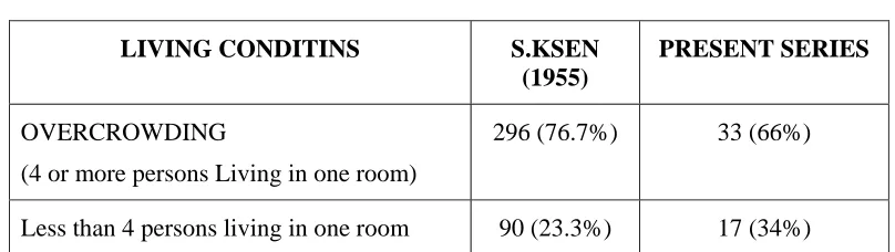
POVERTY: Overcrowding significantly increases the risk of transmission of
tubercle bacilli.
TUBERCULOUS CERVICAL LYMPHADENOPATHY: Tuberculous
lymph node involvement in the neck is an inflammatory reaction, usually not
resulting in any clinical evidence of tuberculosis of upper respiratory tract.
Lumph node involvement is an extra pulmonary primary complex. Primary
lesion is rarely identified and the condition may be blood borne.
Routes of entry of the organism in to cervical lymph node:
1. Cutaneous implantation and mucosal inoculation
2. Tuberculosis following dental ulcer with cervical lymphadenitis is
reported in children. The scars disappear as the permanent teeth erupt.
Bacilli are cultured in a few cases. In some cases cutaneous or mucosal
inoculation can be identified.
3. Retrograde lymphatic spread, from hilar and mediastinal nodes
infection spreading upwards in to lower cervical nodes have been
reported: typically bilateral.
OROPHARYNGEAL PRIMARY COMPLEX: Trough the tonsils,
adenoids, carious tooth, nasopharynx and middle ear where the primary focus
may exist tuberculosis can affect cervical lymph nodes. Primary may difficult
Adenoids: In lymph nodes of posterior triangle about 22% have adenoids as portal of entry. Scalp is an unlikely site of entry of tubercle bacilli. Some
lymphatics from adenoids enter the posterior triangle directly.
Caries tooth : This is the second frequent route of entry (12%). Sub maxillary nodes are involved. Tubercle bacilli have been demonstrated in decayed root
pulp of extracted tooth.
Tonsils : The tonsils are the important portal of entry for tubercle bacilli and are the commonest source of primary complex. Cervical lymphadenitis is the
outward visible sign.
Tubercle bacilli reaching these nodes subsequently also reactivate the
lymph node tuberculosis. Role of tonsillectomy in the treatment of cervical
lymphadenitis is controversial. Primary complex is not in the tonsil in at least
50% of cases of cervical lymphadenitis.
PRIMARY PULMONARY WITH HAEMATOGENOUS SPREAD:
Children under 15 years may have primary complex ad pulmonary
tuberculosis with cervical lymphadenitis. Cervical lymphadenitis may be a
local manifestation of generalized disease and organisms reaching cervical
lymph nodes through blood stream.
MULTIPLE SIMULTANEOUS INFECTIONS: Multiple primary
cervical and pulmonary complexes simultaneously. Pulmonary disease can
occur in children already having cervical lymphadenitis. Heavy primary
infection causing cervical lymphadenitis can produce additional pulmonary or
mesenteric disease.
TRANS PLACENTAL INFECTION: Jones and Campbell recorded a case
of tuberculous mother delivering a baby at 34 weeks of gestation. Baby was
not in contact with mother after delivery but fount montoux positive at 4th
week and cervical nodes showed evidence of tuberculosis on biopsy. The
infection might have occurred via umbilical vein or amniotic fluid infection. If
by inhalation, nasopharynx is infected then cervical nodes can ge5t involved.
BACTERIOLOGY: Disease form Mycobacteria has been documented in the
bones or pre historic humans and animals. In the 18th and 19th centuries it was
known as white plague, decimating the population of Europe.
According to WHO, tuberculosis kills 3 million people yearly in
underdeveloped countries, Mycobacterium tuberculosis the cause of
tuberculosis is one of the 54 recognized species of mycobacteria. Of these 14
are known to cause disease in human beings. A remarkable increase in
infections caused by M. Avium complex during 1990 can also be attributed to
Infection is spread from person to person by inhalation of airborne
droplet nuclei 1 to 5 micrometre in diameter. They are not trapped by nasal
turbinates or mucociliary membranes and may reach the alveolar walls to be
taken up by alveolar macrophages.
The genus mycobacterium comprises the acid-fast bacilli, organisms
that share the property of acid fastness, i.e resistance to decolorization by
weak mineral acids after staining with one of the arylmethane dyes. The name
mycobacterium (fungus like growing bacterium) is derived from the mould
like appearance of M.tuberculosis when growing in liquid media. The
pathogens of this genus include organisms responsible for human and bovine
tuberculosis and for leprosy.
Organisms of M.tuberculosis complex are always pathogenic for man
and their isolation from clinical sample implies infection. Person to person
spread is common, especially with M.tuberculosis. infection with M.bovis, a
well-recognized animal pathogen, can be acquired zoonotically, via infected
cow’s milk or from other animals such as deer.
MORPHOLOGY AND STAINING: Mycobacterium are complex
unicellular organisms with a wide range of antigenic determinants. They are
They are Gm +ve but many species stain poorly with this stain because of the
characteristics of the cell wall, rich in diverse lipids.
CULTURAL CHARACTERS: Many Mycobacteria produce carotenoid
pigments under appropriate conditions. Some organisms produce pigment in
the dark (scotochromogens) and others only on exposure to light
(photochromogens). This pigment production can be a useful guide to
identifications.
MYCOBACTERIUM TUBERCULOSIS COMPLEX: Organisms of this
group show classical ‘cording’ (bacilli lie parallel to each other) on Z N
stained smears, usually as slightly curved bacilli, which clump together.
M.TUBERCULOSIS: M.Tunerculosis gives eugenic (rough tough and buff)
growth on glycerol and pyruvate egg. It is an aerobe that grows only at 37
degree Celsius and is usually sensitive to standard antimycobacterial therapy.
M.BOVIS: Responsible for bovine tuberculosis but also pathogenic to
humans. Growth is enhanced by sodium pyruvate in culture media, giving
eugenic colonies rather than dysgenic (small smooth) colonies in glycerol egg.
It is microaerophilic and resistant to pyrazinamide.
MYCOBACTERIUM AVIUM COMPLEX (MAC): The M.avium complex
disease in elderly patients and localized infection (notably cervical adenitis) in
children.
The complex includes m.avium, M.intracellulare and M.scrofulaceum.
They are deeply staining acid fast small coccobacilli, which may show
beading although long bacilli occur in some stains. Growth on egg media is
dysgenic and some strains may producer a yellow orange pigment.
Other slow growing mycobacteria are M, Kansasii, M.Malmonese,
M.Ulcerans, M Xenopi, and M.Gordone.
The main atypical mycobacteria causing cervical lymphadenopathy are
M. avium, m. intracellulare and M.scrofulaceum.
PATHOGENESIS: While the majority of inhaled bacilli are trapped in the
upper airways a fraction reach the alveoli. There, activated macrophages
ingest the bacilli. The virulence and number of bacilli is important in the
pathogensis. If the bacilli multiply their growth quickly kills macrophage. No
activated monocytes attracted to the site ingest the bacilli released from lysed
macrophage.
Once specific immunity develops large numberof activated
macrophages accumulate at the site of primary lesion and granulomatous
activated macrophages such as epitheloid cells and giant cells. Initially tissue –
damaging response destroys macrophages and produces early solid necrosis at
the center of the tubercle.
Tuberculous lymphadenitis is a regular feature of the primary complex
of tuberculosis. Lymph node tuberculosis is one of the commonest
presentations of extra pulmonary tuberculosis. Widespread nodal disease may
be seen in individuals with increased susceptibility to infections.
Before the stringent control of tuberculosis in the cattle and the
universal pasteurization of milk the primary lesion was often in the tonsils
(with cervical lymphadenitis) or in the gut (with mesenteric lymphadenitis).
Most of the cases were of bovine strain infection.
The lymph node lesions are slowly evolving granulomas with necrosis
at the center of the focal granulomas. The individual tubercles coalesce to
form necrotic chessy material, but the process is halted before this and the
granulomas become calcified.
The rapid spread of caseation and multiplication of.Tuberculosis in the
nodes result in ulceration through the walls of adjacent blood vessels and
spread of infection through blood stream leading to military tuberculosis. The
development of caseation is linked with appearance of hypersensitivity to
The stage of infection are seen clearly in the neck nodes. From the
tonsillar portal of entry the infection spreads by the lymphatics to the nearest
lymph node. When the disease spreads many lymph nodes are affected.
In the event the patient develop natural resistance, fibrosis and
calcification of lymph nodes occur. In other circumstances the caseating
material liquefies and breaks through the capsule of lymph node and a cold
abscess forms. The pus is at first confined to deep cervical fascia. Within a
few weeks the fascia is eroded at some point and the pus flows through this
small opening in to the more accommodating space beneath the deep fascia.
This is known as the collar stud abscess. Unless proper treatment is given at
this stage the skin gets indurate, breaks down to form a sinus, which without
treatment may remain for years. From each of these stages resolution may
occur with calcification if caseation has occurred and with much scarring if
sinuses have formed.
ATYPICAL OR OPPORTUNISTIC MYCOBACTERIAL INFECTIONS:
Nontuberculous mycobacteria involve the lymphatic system most
commonly in the cervical nodes in the children between 1 and 5 years. Most of
them are due to M. avium, M.intracellulare and M.scrofulaceum. the nodes are
almost always unilateral and rarely any systemic symptoms. The site is usually
occur and may be fatal. Localized cervical nodes are best treated by total
excision.
HIV ASSOCIATED TUBERCULOSIS: Tuberculosis is the most common
opportunistic disease associated with AIDS in many developing countries.
90% have pulmonary disease. Of those who have extra pulmonary disease
85% have lymphadenopathy (mainly cervical). Also common is mediastinal
lymphadenopathy. Sputum is often smear negative. Because of poor cellular
immunity Tuberculin skin test is usually nonreactive. Tubercle bacilli may be
obtained by blood culture.
M.avium, M. intracellulare infections are common in AIDS.
Histologically affected tissues are infiltrated with foamy histiocytes with
CLINICAL FEATURES
Mostly young adults children are affected by cervical
lymphadenopathy. It can occur at any age.
The human strain of M.tuberculosis is the cause of 70% of cases, and
not the bovine strain as believed earlier.
In 80% of cases the pathology is confined to the clinically affected
group of lymph nodes. There may be coexisting pulmonary or renal
tuberculosis. Appropriate investigations are done in such cases. Mediastinal
and mesenteric lymph nodes may be involved in a small percentage of
patients. So also the axillary lymph nodes. In miller’s series (1956)
tuberculous cervical lymphadenitis was found to be a localized disease in 120
out of 160 children.
Patel (1960) studied 100 cases of cervical lymphadenitis out of which
60 were of tuberculous etiology.
Jones and Campbell have described the following stages of tuberculous
cervical lymphadenopathy.
Stage I
Enlarged, firm, mobile, discrete, slightly tender nodes showing
Stage II
Larger nodes, rubbery, fixed to surrounding tissue due to periadenitis.
Histologically caseation present. Glands matted together.
Stage III Central abscess (cold absences) formation
Stage IV Collar stud abscess formation
Stage V Sinus formation
In stage I, nodes are mistaken for chronic nonspecific adenitis. Gland
biopsy will clinch the diagnosis.
In Stage III, differential diagnosis include infected branchial cyst or
thyroglossal cyst.“cold abscess” can be distinguished by the normal skin
temperature and absence of inflammation.
Stage IV Collar stud abscess may present as
1. Orthodox type
2. Long stemmed type
3. Dumb bell type
Stage V if untreated collar stud abscess bursts open through the skin
leading to sinus (scrofuloderma) discharging the necrotic material beneath the
INVESTIGATIONS
A careful history with stress on contact with tuberculosis is important.
Detailed clinical examination of the patient and the description of the group of
lymph nodes are to be recorded.
Clinical features suggesting tuberculous lymphadenopathy include
multiplicity, matting and caseation, but these parameters are not specific.
Blood: Hypochromic microytic anaemia may be present. Hb percentage maybe decreased
WBC count may not be altered much
ESR increased. It is nonspecific. But it is a prognostic indicator.
Tuberculin testing:
In children a positive tuberculin test is taken as evidence of infection. It
is mainly used for cantact tracing. It is rarely of any value in diagnosis of
tuberculosis.
Patients are tested with purified protein derivative (PPD) of
Mycobacterium tuberculosis or old tuberculin. The test based on cell mediated
immunity with the development of in duration and inflammation at the site of
THE MANTOUX TEST
This is used for individual patients.
0.1ml of 1:1000 strength PPD equivalent to 10 tuberculin units is injected
intradermally.
The induration is measured after 72 hours. This test is positive if the
induration is 10mm or more in diameter.
CHEST X-RAY
If pulmonary lesion is present the radiological evidence of patchy or
nodular shadows in the upper zones, loss of volume and fibrosis with or
without cavitation or typical calcification may be present.
The mediastinal nodes enlargement can be detected by X ray chest.
There may be evidence of pleural effusion.
SMEAR FOR ACID FAST BACILLUS:
A smear from a specimen of sputum, pus, discharge from sinus
willdemonstrate the acid-fast bacilli if present. The stain used is zeihl-neelsen
stain.
The tubercle bacilli resist decolorizing by both acid and alcohol. It will
stain. The presence of acid-fast bacilli from cervical node abscess is variable.
It may be demonstrable in countries where the prevalence is high.
CULTURE
It is the process of propagating organisms by providing the proper
environmental conditions-nutrients, pH, temperature, aeration, salt, osmotic
pressure of the medium and light for photosynthetic organisms. Lowenstein
jensen medium is the most widely used for the culture of M.tuberculosis.
visible growth is seen 2 weeks on primary isolation from clinical material.
Colonies may take up to 8 weeks to appear. Colonies are buff colored and
appear like dry breadcrums.
FNAC
It is the study of cells obtained by aspiration using fine needle under
vacuum. The needle gauze of 21 to 22 is used. Minute quantity of tissue fluid
is got. Greig and Cray first described it in 1904.
Microscopically presence of epitheloid cells, Langhan’s giant cells
along with caseating material are suggestive of tuberculosis. Definitive
diagnosis is by staining is by staining or culture of the tubercle bacilli.
The advantages of FNAC are that it is a simple procedure done on Op
disadvantage is that the histologic interpretation depends on the experience of
the histologist in the field.
BIOPSY:
Surgical excision of the affected lymph node is mandatory. Ideally it
should be done under general anaesthesia. But excision of small superficial
lymph node may be done under local anaesthesia.
Finding of histopathological examination of a tuberculous lymphnode
consist of graunlomatous lesion with epitheloid and giant cells of langerhans
with or without caseation. Action –fast bacilli may be demonstrated by zeihl
Neelson technique. Coalescence of the tubercles may lead to central necrotic
cheesy material but this process may halt much before and walled of y fibrosis
and calcification.
A sound knowledge of anatomy is important while doing lymph node
biopsy. Otherwise the spinal accessory nerve may be injured while removing
lymph node from the posterior triandle of neck. Phrenic nerve may get injured
while removing supraclavicular lymph node. Injury to transverse cervical
veins and inferior cervical veins also must be recognized and managed in such
caesbefore closure. The cut section of the node should be examined
macroscopically before sending the specimen for histopathological
ANIMAL INOCULATION:
Guinea pig inoculation is a sensitive method and may detect as few as
10 bacilli/ ml. But results are got only after several weeks because of the slow
growth. Modern culture methods are very yielding that animal inoculation
DIFFERENTIAL DIAGNOSIS
1. Acute lymphadenitis
2. Chronic lymphadenitis
3. Syphilitic lymphadenitis
4. Cat scratch disease
5. Infectious mononucleosis (glandular fever)
6. Tularemia
7. Brucellosis
8. Sarcoidosis
9. Toxoplasmosis
10.Fungal diseases
11.Neoplasms
12.Hodgkins lymphoma
TREATMENT
CHEMOTHERAPY: Modern chemotherapy is so effective that proper drug
regimens are prescribed and if the patient is wholly co operative in treatment
than cure rate is 100% patients with initially drug sensitive tubercle bacilli and
in majority of patients having drug resistant organisms. The concept of
short-term chemotherapy is to select combinations of drugs that are rapidly
sterilizing. Treatment with 3 or 4 drugs in the initial phage will almost always
prevent the emergence of ‘persisters’
The short-term regimens have the advantage of reducing chronic
toxicity, making patient compliance easier, making relapse highly unlikely.
DRUGS USED IN PREVIOUSLY UNTREATED PATIENTS
ISONIAZID (H) PYRAZINAMIDE (Z)
RIFAMPICIN (R) STREPTOMYCIN (S)
ETHAMBUTOL (E) THIACETAZONE (T)
DRUGS USED IF PRIMARY TREATMENT FAILS
CYCLOSERINE (CYC) CAPREOMYCIN (CAP)
ETHIONAMIDE (ETH) VIOMYCIN (VIO)
PROTHIONAMIDE (PRO) KANAMYCIN (KAN)
ISONIAZID
It is a very powerful bactericidal drug; has very few side effects; it is
very cheap. Highly effective concentrations are achieved in all tissues. There
is no cross resistance with other drugs.
Adverse effects Rarely generalized skin rashes can occur. Peripheral
neuropathy is the main adverse effect.
It can be treated by giving 100-200 mg pyridoxine daily or can be prevented
by giving 10mg pyridoxine daily.
DOSAGE:
Tablets – Adults:300mg daily
Syrup – Children :10mg/kg daily
RIFAMPICIN
Highly effective concentrations are obtained in all tissues. No cross
resistance with other drugs. Cost is higher. Taken ½ hour before breakfast. It
colours the sweat, urine and tears orange. Mild flushing, itching of skin, rah
can occur occasionally. Hepatitis is extremely uncommon unless patient has a
history of liver disease or alcoholism.
DOSAGE
Capsules –Adults:450-600mg/daily
STREPTOMYCIN
Not absorbed by the intestine. So it is given by intra muscular
injections. Diffuse readily into body tissues. Ethambutol can be substituted for
streptomycin if giving injections daily is not practicable, only in adults.
Damage to vestibular apparatus is manifested by giddiness, especially in old
patients. Treatment must be stopped immediately. Anaphylaxis is occasionally
seen. It should be avoided in pregnancy.
DOSAGE
Vials: Adults: 0.75-1gm daily
Children: 20mg/kg daily
Ethambutol
It is a bacteriostatic drug. It is mainly used to prevent the emergence of
drug resistance to main bactericidal drugs (INH, Rifampicin, Streptomycin).
Not given to young children who are unlikely to tell they are losing their sight.
Also avoided in renal failure. The main adverse reaction is progressive loss of
vision due to retro bulbar neuritis. Drug must be stopped in that case.
DOSAGE
Tablets: Adults:1.5-2.0 gm daily
THIACETAZONE:
It is very weak drug but valuable as a companion drug in preventing the
development of isoniazid resistance. It should be used only in communities
where it has been shown to be effective and to be of low toxicity. Adverse
reactions are cutaneous rashes (often severe) and conjunctivits.
SECOND LINE DRUGS
These drugs are to be used in retreatment when there is resistance to the
drugs used in standard treatment. The drugs are Ethionamide / Prothionamide,
PAS, Cycloserine, Kanamycin and Capreomycin.
Drugs Dosage Side effects
Ethionamide /
Prothionamide 0.75-1 gm daily Metallic taste in mouth
PAS 10-12gm daily in 2 divided
doses Fever and rash
Cycloserine 250mg 2×daily up to 250mg
3×daily Confusion, convulsion
Capreomycin
Kanamycin,
Viomycin
0.75-1gm daily
Vestibular toxicity
Hypocalcaemia
Hypomagnesemia
CHEMOTHERAPY FOR NEWLY DIAGNOSED PATIENTS
6 MONTHS REGIMEN – APPROVED BY WHO
Isoniazid (H) plus Rifampicin (R ) plus Pyrzinamide (Z) for the two
months, followed by isoniazid and Rifampicin for 4 months. The short hand
The recent development is the fixed dose combination of isoniazid,
rifampicin and pyrazinamide. This simplifies the regimen. It is essential to
ensure fixed drug combinations do not alter serum drug concentrations.
FOUR DRUG REGIMEN
In populations with a higher rate of initial drug resistance add a fourth
drug for the first 2 months, either Streptomycin (S) or Ethambutol (E)
These regimens are written as 2SHRZ/4HR or 2EHRZ /4HR
It can also be used as a retreament regime (where a patient has missed
more than 1 month of routine treatment)
These regimens are recommended for both pulmonary and
non-pulmonary tuberculosis in both adults and children.
SUPERVISORY TREATMENT
If possible make sure that each dose is fully supervised for the first 2
months of treatment.
If possible make sure that each dose is fully supervised for the first 2
months of treatment.
EIGHT – MONTH REGIMEN
First 2 months
Add a fourth drug if high rate of initial drug resistance is prevalent.
Next 6 months.
1. Isoniazid +Thiacetazone
2. If side effects to thiacetazone develop give Isoniazid +Ethambutol or
Isoniazid alone to complete 8 months.
This regimen is also recommended by WHO, summarized as 2 HRZ/6HT
Or
EHRZ /6 HT.
INTERMITTENT OR PARTLY INTERMITTENT SHORT COURSE
REGIMENS
Dose of drugs are different than daily short course chemotherapy.
Alternate regimens used are 2HRZ/4H3 R3
2HRZ/4H2 R2, 2E3 H3 R3 Z3
Or
2S3 H3 R3 Z3/ 4 H3 R3 (drugs given thrice weekly)
MANAGEMENT OF PATIENTS WITH DRUG RESISTANCE
It is very difficult and expensive
GENERAL PRINCIPLES
1. Obtain accurate chemotherapy history
2. Obtain information regarding resistance pattern, if possible
3. Reserve drugs are costly
4. Treatment failure is often due to noncompliance
Factors that influence compliance include anxiety, alcohol, stigma of
disease and ignorance.
Chemotherapy is very effective. If the patient is cooperative and proper
regimens are given cure rates are 100%.
FAILURE OF CHEMOTHERAPY
It is due to several reasons.
Failure to use 2 drugs for which organisms are sensitive
Noncompliance by patients
Drug resistance
Coexisting tuberculosis with other diseases like lymphoma
TREATMENT OF CERVICAL LYMPHNOES OF ATYPICAL
MYCOBACTERIAL CAUSE
Nontuberculous mycobacteria involve the lymphatic system most
commonly in the cervical nodes in children between 1 and 5 years. The large
majorities are due to MAIS complex but M.Kansasii may be responsible. The
nodes are always unilateral and rarely any systemic manifestations are there.
The site is usually submandibular or sub maxillary; very rarely the pre
auricular, axillary or inguinal nodes may be affected. Lymph node
Both M. scrofulaceum and M.avium intracellulare show marked drug
resistance to anti tubercular drugs. Hence antimycobacterial drugs are not
helpful. However complete healing without recurrence is reported when
treatment with clarithromycin as a monotherapy is used in children with
cervical lymphadenitis caused by M.avium complex.
Total surgical excision of the involved lymph node is the treatment of
choice. The advantages of surgical excision in the hands of experienced
surgeons are high cure rate (95%) low complication rate and excellent
cosmetic results.
If surgery cannot be performed or when complete excision is not
possible, Rifampicin can be used as chemotherapy. Alternative nonexcisional
approaches include needle aspiration, incision and drainage, incision and
curettage.
SURGICAL TREATMENT
First line of treatment for tuberculous lymphadenitis is medical. Under
chemotherapy with antituberculous drugs the glands gradually shrink.
Occasionally the swellings become large with center fluctuant (cold abscess),
then the capsule of lymphnode is broken involving the deep fascia forming
collar stud abscess or involve skin forming discharging sinus. In such cases
TREATMENT ABSCESS OF NECK
The incision and drainage is done; collar stud abscesses should be
recognized. The drainage should be adequate and complete. Otherwise it will
lead to persistent sinus.
OPERATION
Incision is made along the skin crease. Ideally anaesthesia should be
used. Making skin and deep fascia incision will drain pus. Deeper extensions
and granulations will be revealed. These should be explored ad curetted out.
The pus should be sent for microscopy, culture and sensitivity for pyogenic
bacteria and acid fast bacilli. Histological examination of lymphnode is useful.
When the report comes as tuberculous lymphadenitis antituberculous
chemotherapy is started. The drainage wound is lightly approximated around a
soft rubber tube with few stitches. Drain is removed after 24-48 hours.
EXCISION OF LYMPHNODE
INDICATIONS
• When no response to chemotherapy
• Persisting sinus
An oblique incision gives good access. If nodes are related to internal
jugular vein, it should be safeguarded. If nodes are large and matted they will
be adherent and removal is difficult. One may injure the vein causing
bleeding. If injured the segment of the vein can be dissected after ligating both
EXCISION OF SINUSES
The sinuses should be excised along with the underlying nodes. If the
sinuses are not excised they may persist for many months or years. The
sinuses should be excised early under cover of antibiotics and antituberculous
drugs. Blunt and sharp dissections are used for excision of sinus and the
lymphnodes.
In 50% of tuberculous lymphadenopathy the primary focus is in
oropharynx. Even if the focus is in tonsil, it may appear normal.
Tonsillectomy is not of much help and is not practiced.
Associated pulmonary tuberculosis is not a contraindication for surgical
MATERIAL AND METHODS
The material for this study consists of patients attending the surgical
OPD of Govt. Rajaji Hospital, Madurai. This study comprises of 50 cases
studied during March 2002-March 2005.
In this series 50 cases were studied clinical history, Physical
examination and investigations were done. After physical examination and
arriving at clinical diagnosis, confirmation was done by special investigations.
Lymphnode biopsy was the most important of these. Cases were taken at
random and only cases who gave consent for lymph node biopsy were taken
for study. Only inflammatory and infective cases were taken. Cases of other
etiology were not included in this study. Name, Age, Sex, Religion Address,
Occupation of the patients were noted. Patients were divided in to 3 income
groups according to their monthly income.
1. Up to Rs. 2000 Low income group
2. 2000-6000 Middle income group
3. Above Rs. 6000 Higher income group
In the history particular emphasis was given to the type of
accommodation, the nutritional value of food, history of contact with
tuberculosis, any consumption of raw milk. Also history of recurrent
After clinical diagnosis was made investigations were done to confirm
the diagnosis, Routine blood examination was done like Erythrocyte
sedimentation rate (ESR), Total white cell count (TC), Differential count (DC)
and haemoglobin percentage (Hb%) Montoux test was done. Chest X-ray was
taken. Sputum examination was done.
FNAC was done in all cases.
Lymph node biopsy was done in all cases. Macroscopic appearance of
the specimen noted down and sent for hostopathological examination.
After the confirmation of diagnosis, for tunerculous lymphadenopathy
treatment with antituberculous chemotherapy ranging from 6 to 9 months was
started, depending on the severity of the node involvement and the patient’s
response to chemotherapy.
The chemotherapy regimens used were as follows
Four drugs regimen was used. INH plus Rifampicin plus Ethambutol
plus pyrazinamide were used for the first 2 months.
INH 300 mg daily
Rifampicin 450 mg daily
Ethambutol 800 mg daily
Pyridoxine was supplemented with these drugs to prevent peripheral
neuritis. In the continuation phase of 4 months only 2 drugs were given –INH
and Rifampicin along with pyridoxine 10 mg daily
For those patients in whom glands persisted the continuation phase was
for 7 more months i.e.9 months regimen.
Repeated aspirations were done on patients having cold abscess on
weekly basis till pus stopped collecting. Excisions of discharging sinuses were
done. All these were done while patient was put on anti tuberculous
chemotherapy.
Along with this, advice was given to take nutritious food.
Most of the patients were followed up for a period of 6 to 9 months. A
few patients were lost for follow up once the treatment was started. A few
were being followed up for 3rd or 4th month at the time of preparing this
ANALYSIS OF DATA
The total number of cases studied is 50. The patients were attending the
[image:61.612.115.514.245.344.2]surgicalout-patient departments of Govt. Rajaji Hospital, Madurai
Table - 1
AETIOLOGY OF CERVICAL LYMPHADENOPATHY
Studied series (50 cases) Aetiology
No %
Tuberculous lymphadenopathy 34 68
Nonspecific Lymphadenopathy 16 32
Sarcoidosis - -
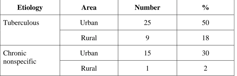
From the above table it can be seen that tuberculous lymphadenopathy
is the commonest cause of cervical lympadenopathy with 68% followed by
chronic nonspecific lymphadenopathy with 32%. There was no case of
INCIDENCE
AGE
The incidence of cervical lymphadenopathy in different age groups is
shown in table - 2.
Wilson series
(100 cases)
P.B.Trivedi series (322 cases)
S.P.Pamra series (322 cases)
Study series (50 cases) Age
(in years)
No % No % No % No %
0-1 9` 9 25 10 55 17 3 6
11-20 25 25 101 44 82 25 19 38
21-30 32 32 83 35 109 35 18 36
31-40 13 13 18 8 37 11.45 2 4
41-50 9 9 8 3 24 7.45 4 8
51-60 2 2 - - 15 4.65 4 8
>60 7 7 - - - - - -
In this series of 50 cases the disease commonly affected the 2nd and 3rd
decades with 19% and 18% respectively. Next common age group in which
tuberculous lymphadenopathy presented is 5th and 6th decades. 4% of cases
In wilson’s series of 100 cases the common age group of patients was
in the 2nd and 3rd decade followed by the 4th decade with 25%,32%,and 13%
respectively.
In B.PTrivedi’s series of 235 cases also the commonest age group of
presentation was in the 2nd and 3rd decade with 44%, and 35%.Next common
age groups affected were 1st and 4th decade with 10% and 8% respectively.
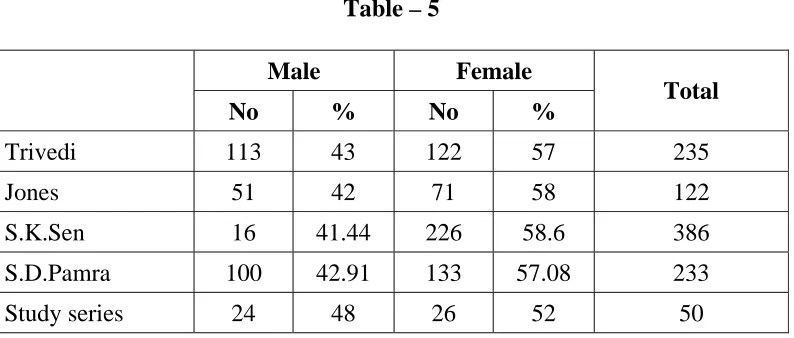
In S.P. Pamra series of 322 cases the commonest age group affected
were 2nd and 3rd decades with 25% and 35%. Next common age group were
the 1st and 4th decade with 17% and 11.45%.
In the preset study chronic nonspecific adenopathy affected most
commonly the age group of 2nd and 3rd decades with 18% and 8% respectively.
In our country the tuberculous lymphadenopathy commonly affects the
younger age group. Commonest age group affected is between 11 and
20,21and 30 closely followed by 31 and 40 years. Nonspecific
lymphadenopathy commonly affects the age group of 11 to 20, 21 to 30 and
less commonly o to 10.
But in western counties the pattern is different. Common age group
affected is 0 to 10 years. The causative organism in this age group is atypical
mycobacterium. In adults the causative agent is most commonly the