A STUDY ON
ACUTE LARGE BOWEL
OBSTRUCTION
Dissertation submitted for
M.S., GENERAL SURGERY
BRANCH - I DEGREE EXAMINATION
THE TAMIL NADU DR. M.G.R. MEDICAL UNIVERSITY
CHENNAICertificate
This is to certify that the dissertation entitled
“A STUDY ONACUTE LARGE BOWEL OBSTRUCTION”
presented here is the
original
work
done by
Dr. K. RAMESH,post-graduate at Madras Medical
College,
Govt. General Hospital,
Chennai-3in partial fulfillment of the
University rules and regulations for award of
M.S., Degree Branch-I(General Surgery)
under my guidance and supervision during the
academic
period
from 2003-2006.
Prof. P.G. Kolandaivelu M.S.,
Professor and HOD,
Department of General Surgery, Madras Medical College,
Chennai - 600 003.
Prof. Kalavathy Ponniraivan
DECLARATION
I hereby declare that the dissertation entitled “A STUDY ON ACUTE LARGE BOWEL OBSTRUCTION” was done by me at Government General Hospital, Madras Medical College, Chennai-3
during the period of my post graduate study for M.S. Degree Branch-I
(General Surgery) from 2003 to 2006.
This dissertation is submitted to the Tamil Nadu Dr. M.G.R.
Medical University in partial fulfillment of the University regulations for
award of M.S., Degree in General Surgery.
Chennai Dr. K. RAMESH
Date: M.S. postgraduate in General
Surgery,
ACKNOWLEDGEMENT
I wish to express my sincere thanks to our Dean Dr. Kalavathy
Ponniraivan, B.Sc., M.D, Madras Medical College for having allowed
me to conduct this study.
I am extremely grateful to Prof. P.G. Kolandaivelu, M.S.,
Professor and Head of the Department of General Surgery, Madras
Medical College for his encouragement and guidance throughout the
study and the prompt help rendered whenever approached.
I wish to express my gratitude towards all my assistant
professors, for helping me with their timely advice during the study.
Last but not the least, I sincerely thank all the patients involved
in this study. Their cooperation and endurance has made the study a
CONTENTS
SL.NO TITLE PAGE NO
1. INTRODUCTION 1
2. AIM OF THE STUDY 2
3. REVIEW OF LITERATURE 3
4. MATERIALS AND METHODS 34
5. OBSERVATION 35
6. DISCUSSION 49
7. CONCLUSION 51
INTRODUCTION
Acute obstruction of the large bowel is an important cause of the
acute abdomen in any emergency.
Large bowel obstruction accounts for 6.3% of all intestinal
obstruction (Maingot 10th ed). Although the obstruction may be the
result of mechanical, inflammatory or neoplastic process, diagnosis
often is made and treatment initiated on clinical findings with limited
radiographic or other information.
In many ways the wisdom of adage “Never let the sun rise or set
on an intestinal obstruction” remains a pure practical guideline.
Whenever any uncertainty exists, surgical options are dictated by the
nature of disease, patient’s status and experience of the operating
surgeon. Improvements in the fields of anesthesiology, knowledge of
fluid and electrolyte balance and availability of broad-spectrum
antibiotics have greatly complemented the outcome of the surgical
AIM OF THE STUDY
1. To study the epidemiology, aetiology, pattern of intestinal
obstruction and various operative procedures.
2. To determine the incidence of malignant and non-malignant
causes producing obstruction.
3. To compare the variables like age distribution, sex incidence
and complications.
4. To determine the outcome of surgery.
REVIEW OF LITERATURE
Large intestine extends from the end of ileum to the anus and is
comprised of caecum, appendix, colon, rectum, and anal canal. It is
135cm in length with maximum dilatation of 7.5cm at caecum. It is
made up of circular muscle layer that is continuous and longitudinal
layer arranged in three bands, the taenia coli as far as the rectum
where they fuse to form a continuous layer. The voluminous large
bowel is gathered by taenia coli to give the characteristic sacculated
appearance.
Large intestine comprises of the following:
Caecum is a blind sac at the commencement of large intestine.
It measures 6cm long 7.5cm broad and situated in Right Iliac Fossa.
Ascending colon is 15cm long and fixed posteriorly.
Hepatic flexure lies between Ascending colon and Transverse
Colon.
Transverse colon is about 45-50cm long with its mesentery (i.e.)
Splenic flexure is fixed by phrenicocolic ligament that contains
blood vessels, which need to be ligated during mobilization of this
part.
Descending colon is about 20cm long and fixed posteriorly.
SIGMOID COLON:
This forms a loop of 20-60cm in length with its own mesentery.
This has an omega (ω) shaped attachment to the posterior wall. It
extends from descending colon at the pelvic brim to commencement
of rectum at S3 level.
The taenia coli of sigmoid are wider than in other parts of colon
and have well developed appendices epiploicae. It lies in the pelvic
cavity coiled in front of rectum lying on the peritoneal surface of
bladder. It partially fuses with parietal peritoneum along an inverted
‘V’ shaped base. The linings of the inverted ‘V’ diverge from the
bifurcation of the common iliac artery over the sacro - iliac joint at the
pelvic brim. Base of the sigmoid mesocolon thus measures around
RECTUM:
It proceeds downwards & forwards closely applied to the
concavity of sacrum and coccyx, measuring around 12-15cm. It ends
2-3cm in front and below the tip of the coccyx by turning abruptly
backwards and downwards through the levator ani to form the anal
canal. It is devoid of taenie coli, appendices epiploicae, haustrations
and sacculations.
ANAL CANAL:
It is the terminal portion of large intestine measuring about
3.8cm and develops partly from endoderm (dorsal division of cloaca)
and partly from ectoderm (proctodeum).
Mainly through,
1. Superior Mesenteric Artery
2. Inferior Mesenteric Artery
3. Branches of internal iliac
Middle Rectal Artery
Inferior Rectal Artery
1. SUPERIOR MESENTERIC ARTERY:
Direct branch of aorta at the level of L1 gives 3 colic arteries
apart from a number of jejunal and ileal branches.
The 3 colic arteries are:
• Ileo colic
• Right colic
• Middle colic
o Left branch
Left branch take part in the (arch of Riolan) meandering artery,
an important collateral channel anastomosing with Inferior Mesenteric
Artery.
2. INFERIOR MESENTERIC ARTERY:
Arises from aorta at the L3 level and supplies the colon from
splenic flexure to the rectum.
It gives off
o Left colic artery
o Sigmoidal arteries (3-4 in number)
o Superior rectal Artery
Marginal Artery of Drummond: It is a paracolic vessel of
anastomosis between colic arteries from which the terminal arteries to
the colon (vasa recta) arises.
This vessel extending from ascending colon to the pelvic colon
lies about 2.5-3.8cm from the bowel wall. This artery is less consistent
at the splenic flexure and lower sigmoid, at critical segments called
VASA RECTA:
They are the terminal arteries to the colon. They arise from the
marginal artery and penetrate the bowel wall. The long vasa
recta encircle the bowel wall and anastomose with each other on the
anti mesocolic border of the bowel. The short vasa recta supply the
mesocolic half of bowel circumference.
RECTAL ANASTOMOSIS:
There is abundant and constant anastomosis between the
superior and middle rectal Artery, a branch of internal iliac artery.
This is responsible for the blood supply of terminal colon after ligature
of Inferior Mesenteric Artery. However most part of rectum and anal
canal are supplied by inferior rectal Artery, a branch of internal
pudendal Artery.
VENOUS DRAINAGE:
Corresponds to that of arteries. Veins from the right side of
colon flow into Superior Mesenteric Vein, which drains the midgut.
This lies to the right of the artery and joins the splenic Vein to form
the portal vein. Veins from the left side of colon flow into the Inferior
superior rectal vein. It continues vertically anterior to left renal vein to
the left of duodeno jejunal flexure and joins the splenic vein. The
Inferior Mesenteric Vein can sometimes drain into Superior
mesenteric vein.
LYMPHATIC DRAINAGE:
Lymphatic of large intestine accompany the vascular pedicle
ultimately draining into four tiers of lymph nodes.
• Epicolic lymph nodes – along side of colonic wall.
• Paracoloic lymph nodes – along the marginal vessel.
• Intermediate group of lymph nodes – along the larger branches of arteries supplying the colon.
Principal nodes situated along the superior & inferior mesenteric
trunks. They then pass through efferent lymph vessels to the coeliac
INNERVATION:
The colon is innervated by both the sympathetic (T11 T12 L1 L2)
and para sympathetic (right vagus, S2, S3, S4) system. Sympathetic
nerves have an inhibitory effect on colonic peristalsis and secretion
while parasympathetic nerves are secretomotor and inhibit the
sphincteric musculature.
Colonic pain may be referred to a site distant to the organic
insult.
Caecum: Mc burney’s point extending to epigastrium.
Ascending colon: Right upper quadrant
Hepatic flexure: Right upper quadrant.
Descending colon: Midline and to the left.
Recto Sigmoid: Suprapubic and coccygeal areas.
PATHOPHYSIOLOGY OF LARGE BOWEL OBSTRUCTION
a. Dynamic Obstruction:
Dynamic Obstruction is more commonly encountered in surgical
practice. Here peristalsis is present against the obstructing agent,
which may be In the lumen: e.g .: Inspissated faecal material.
In the wall: e.g .: Malignant stricture.
Outside the wall: e.g .: Adhesions, volvulus.
b. Adynamic Obstruction:
Here peristalsis ceases and no true propulsive waves occur.
E.g .: Paralytic ileus, mesenteric vascular occlusion.
Causes of Mechanical Large Bowel Obstruction in the Adult
WITHIN THE LUMEN
WITHIN THE WALL OUTSIDE THE
WALL Faecal impaction Inspissated barium Gallstone Foreign body
Tumors especially carcinoma Inflammation
o Diverticulitis
o Crohn’s disease
o Tuberculosis
o Lymphogranuloma venereum
o Schistosomiasis Congenital causes
o Adult hirschsprung’s disease Ischaemia Bands and adhesions External hernias Internal hernias Tumors in
Radiation Miscellaneous
o Anastomotic stricture
o Intussusception.
ACUTE OBSTRUCTION:
Usually seen in small bowel with immediate central severe
colicky pain, early vomiting, central abdomen distension and
constipation.
CHRONIC OBSTRUCTION:
This is mainly seen in large bowel with abdomen distension
colicky pain, progressive constipation and later with absolute
constipation.
ACUTE ON CHRONIC OBSTRUCTION:
Spreads from large bowel to involve small bowel and give rise to
pain and constipation of a variable time scale followed by abdominal
distension and vomiting.
EVENTS IN OBSTRUCTION:
Initially due to the stretch reflex, increased peristalsis occurs in
become flaccid and paralysed. The cessation has a protective effect
in preventing vascular damage due to increasing intraluminal
pressure, unless the obstruction is of closed loop type where
ischaemic necrosis of bowel wall ensues if treatment is delayed.
Distension occurs proximal to obstruction due to:
1. Gas:
Swallowed air – 68%
Diffusion from bowel lumen – 22%
Products of digestion & bacterial activity – 10%
Oxygen & Carbon dioxide get absorbed and resultant
mixture is made up of
Nitrogen – 90%
Hydrogen Sulphide – 8%
2. Fluid:
This is made up of whatever fluid this patient has ingested
before surgery as well as various digestive juices.
About 8000ml is secreted in 24 hrs.
Above pylorus: 2500ml
Saliva: 1000ml
Gastric juices: 1500ml
Below pylorus: 4000ml
Bile & pancreatic juice: 2000ml
Succus entericus: 2000ml
In obstruction, absorption of water & electrolytes from gut is
retarded but secretion of digestive juices into the lumen persists or is
even increased.
Negative balance of fluid & electrolytes in obstruction is due to:
1. Diminished intake by mouth.
2. Defective intestinal absorption.
4. Sequestration into bowel lumen.
EFFECT OF BOWEL DISTENSION:
As a result of distension, the diaphragm is pushed up into the
chest and moves inadequately. Ventilation becomes shallow, and
compression of lung bases may cause right to left shunt. Oxygen
tension is reduced. Hypoxia and possibly hypercarbia and acidosis
from under-ventilation contribute to multiple organ failure.
EFFECT OF OBSTRUCTION ON GUT FLORA:
Normal faecal flora swims in symbiosis with the human host and
support several physiological process.
E.g .: Degradation of bile pigments.
Degradation of several toxic products.
Vitamin-K production.
Alteration of colonic motility & absorption.
Normally 99% of faecal flora is anaerobic of which bacteroides
is the most abundant along with Lactobacillus bifidus, Clostridium of
Aerobes are mainly Escherichia coli present in counts of
10ⁿ(n=7)/ gm-wet faeces. Streptococcus faecalis is the principal
enterococcus.
Unrelieved obstruction results in mucosal ischaemia especially
when there is strangulation and gangrene. This results in
transmigration of bacteria from lumen to systemic circulation as well
as absorption of potentially lethal metabolic products of bacteria
leading to severe systemic inflammatory response syndrome (SIRS)
and multi system organ failure (MSOF).
STRANGULATING OBSTRUCTION:
In addition to the changes in the simple obstruction, viability of
bowel is threatened due to impairment of blood supply either by
internal compression or by interruption of mesenteric blood flow or by
rising intraluminal pressure as in closed loop obstruction.
In all these, except mesenteric artery thrombosis, venous return
is affected before arterial supply leading to increased capillary
pressure. This causes escape of intravascular fluid and diapedesis of
RBC’s into the bowel wall, lumen and peritoneal cavity leading to
length of the bowel affected. In addition, there is transmigration of
bacteria and its products leading to endotoxemia.
COLONIC PSEUDO OBSTRUCTION:
There is controversy over whether the disease is due to primary
abnormalities of intramural nerve plexus or intestinal smooth muscle.
The aetiology may be:
1. Idiopathic – Diabetes Mellitus, Hypokalemia, Uraemia,
Myxoedema.
2. Severe trauma / shock.
3. Retro Peritoneal irritation – Blood, Tumour
4. Drugs, Tricyclic antidepressants, Phenothiazines,
L-dopa.
DIAGNOSIS OF LARGE BOWEL OBSTRUCTION:
Diagnosis of Large Bowel Obstruction is commonly based on
history and physical examination.
Essentially all patients with non perforating colonic obstruction
Constipation
Distension
Colicky pain is due to increased wall tension in the distended
obstructed colon, while progressive continuous pain is suggestive of
strangulation and gangrene. Distension is the main feature in volvulus
but pain and other features eventually become apparent. Fever is
more common if there is diverticulitis or perforation secondary to
obstruction or when there is ischaemic gangrene. Blood in the stool is
more common in inflammatory bowel disease and sometimes in
obstructive colonic carcinoma. Large bowel ischaemia can also
CLINICAL PRESENTATIONS OF COMMON CAUSES OF LARGE BOWEL OBSTRUCTION INCLUDES
Cancer Volvulus
Pain 81-85 67-100
Nausea/ Vomiting 65 50-87
Constipation 35 43-66
Obstipation 47 XX
Diarrhoea XX 24
Distension 69-80 80-100
Tenderness 44 70
Fever Rare 11
Peritonitis 30 10-19
SIGMOID VOLVULUS:
‘Volvulus’ is defined as the twisting of a hollow viscus either
around an axis passing longitudinally to it (organo - axial) or passing
through its mesentery at right angles to the first (mesenterio - axial).
In the case of sigmoid colon, a portion of the large intestine whose
proximal and distal ends (descending colon and rectum respectively)
are plastered to the parietes close to each other, allowing it to hang
out like a large loop, twisting occurs only along the mesentery. The
twist compresses the mesentery, which carries the vasculature to the
organ and hence compromises the same leading to strangulation and
Laurell (1926) defined volvulus for the first time. Studies in the
20th century by Obalansky (1984), Gibson and Ballantyne (1982)
showed the distribution of this condition predominantly in the
non-English speaking countries.
AGE AND SEX:
In Africa, India, Iran and Brazil and Eastern Europe about
70-90% of cases are seen in the middle ages (40-60) whereas in the
USA, UK, Australia and Israel, it afflicts the older age group of 60-70
years.
There is a marked overall preponderance of male patients
comprising approximately 90% of reported patients (Ballantyne 1982).
The consumption of high fibre diet by the male produces bulky stools
and flatus and their irregular bowel habits produce over- loading of the
colon and possibly leading to volvulus.
PATHOGENESIS:
The different theories of pathogenesis include:
1. Cullen’s theory of spasmodic constriction.
3. Long and Loose mesentery as a predisposing factor by
Von Rokitansky in 1949.
4. Kuttner’ s theory of long, redundant colon resulting from
coarse vegetable fibre diet consumption; this is the
commonly accepted view by various authors such as
Anderson (1968), Sinha (1969) and Riedl (1978).
Neurological diseases such as Parkinson’s disease have been
associated in the evolvement of sigmoid volvulus probably by way of
contributing to their being bedridden and thus contributing to chronic
constipation in these institutionalized patients (Dean 1952).
Sigmoid volvulus is said to be the most common cause of
intestinal obstruction in pregnant women. Harper (quoted by
Ballantyne 1982) speculated that the enlarging uterus might cause
kinking in the colon, where it is fixed to the pelvic walls. The ensuring
proximal distension raises a redundant or abnormally mobile sigmoid
PREDISPOSING FACTORS:
1. Narrow attachment of the mesocolon.
2. Long pelvic mesocolon.
3. Overloaded colon thereby providing the torting force to the
limbs of the bowel. Hence, the dietary and bowel habits of
the person play a major role in its causation.
4. Adhesions act as organic axis around which the volvulus
takes place.
CLINICAL DIAGNOSIS:
The triad of abdominal pain, distension and constipation are the
predominant signs and symptoms of sigmoid volvulus. The duration of
symptoms will be characteristically short (Bolt 1956). Its characteristic
1. Distended flanks.
2. Sometimes visible large bowel loops.
3. Empty and ballooned rectum on per rectal
examination.
4. The characteristic “Frimann – Dahl” signs on X-ray
make the diagnosis unmistakable.
Clinical features that suggest the presence of gangrene include
severe pain, deterioration in the general condition of the patients with
tachycardia and hypotension and marked abdominal tenderness with
absent intestinal sounds.
The characteristic radiological features include:
1. ‘Inverted U sign’: Inverted ‘U’ shaped loop massively
distended and devoid of haustra.
2. ‘Liver overlap sign’: Haustral margin overlapping the
lower border of the liver shadow.
3. ‘Left flank overlap sign’: Haustral margin overlaps the
4. ‘Frimann – Dahl sign’: the two limbs of the loops
converge interiorly giving rise to three white lines,
representing the outer walls and the two adjacent inner
wall of volvulus. It is usually on the left side of the pelvis.
5. Usually a huge amount of air accumulates in the loop
giving an air fluid ratio greater than 2:1.
TREATMENT:
The initial management of the patient involves the treatment of
shock with intravenous fluids supported by parenteral antibiotics while
the patient being put on nil oral and insertion of a nasogastric tube.
The presence or absence of the gangrenous bowel is an
important factor in the management.
Flexible colonoscopy has been successfully employed in the
non-operative reduction of volvulus with viable bowel (Ghazi 1976,
Arigabu 1985).
No controversy exists in the management of sigmoid volvulus
with gangrenous bowel and surgery is the treatment. However,
controversy exists in cases with viable bowel regarding the choice of
only a temporary procedure as the recurrence has been reported to
be 33-100% (Botsford et al 1967,Hines et al 1967). Therefore elective
resection of the sigmoid loop should be done preferably during the
same hospital admission.
PRIMARY RESECTION AND ANASTOMOSIS:
In the presence of viable gut, primary resection of the redundant
sigmoid loop with end-to-end anastomosis with proximal colostomy is
an effective procedure.
Gurel advocated primary resection and anastomosis in the
management of viable sigmoid volvulus together with “on table”
lavage technique. This technique of intra operative ante-grade
irrigation of the colon allows the large bowel to be prepared intra
operatively to be followed by primary resection and anastomosis, and
in rare cases with protective colostomy.
Resection and anastomosis in an emergency setting without the
bowel preparation has been done by recent surgeons without
PROCEDURES FOR GANGRENOUS SIGMOID COLON:
The presence of gangrenous bowel demands emergency
laparotomy and resection of gangrenous loop. The following options
are available, depending upon the patient’s age, extent of peritonitis
and shock.
Hartmann’s procedure involves a resection of the gangrenous
loop with proximal colostomy and closure of the distal stump.
The advantage is it can be done in patients with extensive
peritonitis in whom primary anastomosis is at risk.
The disadvantage is second stage closure of colostomy is
required which is difficult due to dense adhesions between the rectal
stump and bowel loops.
Thus Hartmann’s procedure should be reserved for cases with
extensive gangrene where distal stump cannot be brought out.
Primary resection and anastomosis has been done in the
presence of gangrenous bowel with due consideration to patient’s
age, duration of symptoms, extent of peritonitis and shock (Kuzu
MALIGNANT LARGE BOWEL OBSTRUCTION:
In the West up to 90% of the patients suffer obstruction
secondary to carcinoma. But the converse is not true. Only about 15%
of large bowel malignancies present with obstruction (Philips et al
1985, Kyllonen 1987). The risk of obstruction by a colorectal
malignancy varies with the site of malignancy.
In decreasing order of risk:
1. Splenic flexure – up to one half go in for obstruction
(Philips et al 1985, Kyllonen 1987, Waldron and Donovan
1986).
2. The rest of the colon except the rectum – around one fifth
risk.
3. Rectal carcinoma – one-tenth the risk.
In clinical practice, approximately three-fourth of all malignant
large bowel obstruction cases are situated in the left colon (i.e.) at or
DIAGNOSIS:
Is based on:
1. Clinical features: Classically abdominal pain, distension,
absolute constipation and vomiting. Abdominal pain is
seen in 90% of cases (Umpelby and Williamson 1984,
Serpell et al 1988).
2. Plain abdominal radiography: Gaseous dilatation of the
large bowel proximal to the site of obstruction and a distal
cut-off.
3. Per-op findings: The proximal large bowel is dilated and
edematous.
MANAGEMENT OF MALIGNANT LARGE BOWEL OBSTRUCTION:
D e c o m p r e s s i v e c o l o s t o m y S t a g e d r e s e c t i o n H i g h - r i s k p a t i e n t L a r g e f a e c a l l o a d M a s s i v e d i l a t i o n
R e s e c t i o n w i t h d e l a y e d a n a s t o m o s i s D i l a t i o n
I s c h a e m i a P e r i t o n i t i s L a r g e f a e c a l l o a d
P r i m a r y r e s e c t i o n a n d a n a s t o m o s i s M o d e r a t e d i l a t i o n a n d f a e c a l l o a d
The aim in the management of patients with malignant
obstruction is to relieve the obstruction with low mortality and
morbidity, to ensure adequate clearance where possible to ensure
long-term survival, but also to provide good palliation in the
remainder. It is now generally accepted that for obstruction proximal
to the splenic flexure, resection and primary anastomosis is optimal
therapy (Phillips et al 1985). An internal bypass will be justified in
patients with an irresectable tumor and in high-risk patients with
extensive distant spread of disease.
The controversy arises in the more common, more distal
lesions. Most surgeons have been reluctant to ignore the traditional
wisdom that it is unwise to anastomose dilated, edematous,
unprepared bowel and so therapy has been to initially decompress the
bowel (Pain and Cahill 1991). At a second operation the obstruction is
resected with anastomosis and then the colostomy closed. Presently,
concerns that staged tumor resection may result in a worsening of
long term prognosis has had led on to increased performance of
I. METHODS OF DECOMPRESSION:
1. Laser Luminisation: Laser can be used to relieve the
intraluminal obstruction, help in bowel preparation for
future elective surgery or might be palliation enough for
highly advanced malignancy. Any level of obstruction can
be dealt with this way.
2. Trans-tumoral stents: Lesions in the rectum or sigmoid
can be stented as a decompression procedure (Keen and
Orsay 1992, Tejero et al 1995).
II. RESECTION AND IMMEDIATE ANASTOMOSIS:
Conceptually, the ideal management of malignant obstructive
pathology would be to remove the tumor and restore bowel continuity
in one sitting. However, segmental resection of left sides lesions
involve anastomosis, in unprepared, dilated and edematous bowel
which would give a mortality rate of up to 50% from anastomotic
III. RESECTION AND DELAYED ANASTOMOSIS:
The tumor is resected and the proximal bowel is brought to the
surface as end colostomy, while the distal stump is closed intra
abdominally (Hartmann’s procedure).
DISADVANTAGES:
1. In contrast to the deaths following initial decompression,
mortality here is secondary to intra abdominal sepsis
(Waldron and Donovan 1986).
2. The stoma is a potential source of complications –
necrosis and retraction occurs in up to 20% of patients
(Stephen et al 1990, Allen-Mersh 1993).
3. Reversal of the procedure is a major task with higher
morbidity and mortality (Mosdell and Doberneck 1991).
4. The same problems of disease progression and
To minimize these risks, Per-op bowel preparation is employed:
1. On-Table Lavage: A large Foley catheter is introduced into
the caecum either through an appendix stump or through
the terminal ileum across the Ileo-Caecal valve. Another
scavenger tube is fixed proximal to obstruction, but distal
to the site of anastomosis intended. Warm saline is run in
an ante grade fashion and faeces removed distally. This
method is useful if faecal load is a viscous fluid.
2. Simple decompression of flatus and extrusion of solid
faeces is better than lavage, if load is solid as the lavage
would make the faeces fluid and difficult to manage.
(Amsterdam & Krispin 1985, White & Macfie 1985, Mealy
et al 1998, Dorudi et al 1990).
Using the above procedures, resection and immediate
anastomosis is found to have morbidity and mortality rates at least
ADVANTAGES:
1. Stoma and its associated problems avoided.
2. Decreased hospital stay.
SUB TOTAL COLECTOMY:
Advantages:
1. The entire unprepared proximal bowel is removed.
2. Ileo – colic anastomosis has lesser leak rate than
colo-colic anastomosis – (10% vs 18% - Philips et al 1985).
3. Obstructing carcinoma has increased risk of
synchronous malignancy in proximal bowel.
Disadvantages:
1. Diarrhoea is more common.
2. Major procedure.
INDICATIONS:
1. <50 years of age.
2. Positive family history.
3. Obstructing carcinoma.
MATERIALS AND METHODS
This is a prospective study, which comprises of 38 patients
treated for acute large bowel obstruction from August 2003 – January
2006 at Government General Hospital, Chennai-3.
The patients on admission were subjected to thorough physical
examination and available investigation. They were treated with IV
fluids, antibiotics, and blood transfusion when required in the pre and
postoperative period and were subjected to appropriate surgical
procedure.
The postoperative period was monitored for complications. After
discharge an attempt was made to follow up the cases. Patients who
underwent colostomy were followed till the bowel continuity was
OBSERVATION
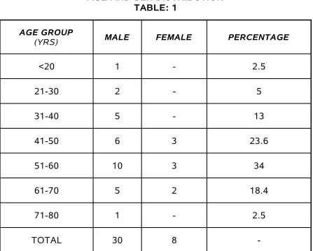
[image:41.612.84.531.161.520.2]AGE AND SEX DISTRIBUTION TABLE: 1
AGE GROUP
(YRS) MALE FEMALE PERCENTAGE
<20 1 - 2.5
21-30 2 - 5
31-40 5 - 13
41-50 6 3 23.6
51-60 10 3 34
61-70 5 2 18.4
71-80 1 - 2.5
TOTAL 30 8
-Of the 38 cases 30 were males and 8 were females.
Male: Female = 3.75:1
AGE AND SEX DISTRIBUTION
0 1 2 3 4 5 6 7 8 9 10 11 12
<20 21-30 31-40 41-50 51-60 61-70 71-80
AGE GROUP
N
O
.O
F
P
A
T
IE
N
T
S
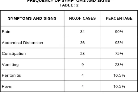
FREQUENCY OF SYMPTOMS AND SIGNS TABLE: 2
SYMPTOMS AND SIGNS NO.OF CASES PERCENTAGE
Pain 34 90%
Abdominal Distension 36 95%
Constipation 28 75%
Vomiting 9 23%
Peritonitis 4 10.5%
Fever 4 10.5%
In our study abdominal distension was the most common
PLAIN X-RAY FEATURES OF ACUTE LARGE BOWEL OBSTRUCTION
TABLE: 3
X-RAY FINDING NO.OF CASES Percentage
Bent inner tube
sign 15 75%
Distended colon with irregularly spaced haustral
folds
COFFEE BEAN SIGN
SIGMOID VOLVULUS
ETIOLOGY
MALIGNANT OBSTRUCTION SIGMOID VOLVULUS
SIGMOID VOLVULUS – GANGRENOUS BOWEL
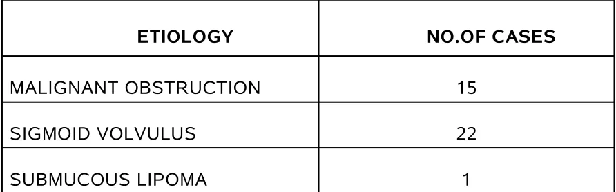
ETIOLOGY: TABLE: 4
ETIOLOGY NO.OF CASES
MALIGNANT OBSTRUCTION 15
SIGMOID VOLVULUS 22
SUBMUCOUS LIPOMA 1
In our study non-malignant causes account for 60.5% of cases
SIGMOID VOLVULUS – RESECTED SPECIMEN
NATURE OF OBSTRUCTION TABLE: 5
BENIGN 23
MALIGNANT 15
BENIGN MALIGNANT
Sigmoid volvulus and one case of submucous lipoma out
SIGMOID COLON GROWTH AGE GROUP:
TABLE: 6
<20 21-30 31-40 41-50 51-60 61-70 71-80
BENIGN - 2 4 6 7 4
-MALIGNANT 2 - 1 3 4 4 1
The most common benign cause of obstruction is sigmoid
volvulus. This occurred a decade earlier when compared to the
literature (Arnold and Nance, 1973; Anderson and Lee, 1981;
0 1 2 3 4 5 6 7 8
<20 21-30 31-40 41-50 51-60 61-70 71-80
AGE GROUP
BENIGN MALIGNANT
SITE OF OBSTRUCTION (MALIGNANT): TABLE: 7
In our study, the site of malignant obstruction is more common
on the left side.
CAECUM 1
ASCENDING COLON 2
TRANSVERSE COLON 3
DESCENDING COLON
-SIGMOID 4
SUBMUCOUS LIPOMA - SIGMOID COLON
SITE OF OBSTRUCTION (MALIGNANT)
CAECUM ASCENDING COLON
TRANSVERSE COLON SIGMOID
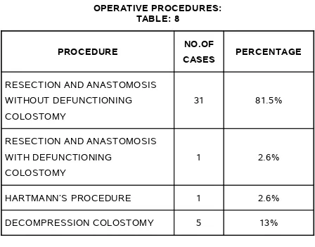
OPERATIVE PROCEDURES: TABLE: 8
PROCEDURE NO.OF
CASES PERCENTAGE
RESECTION AND ANASTOMOSIS WITHOUT DEFUNCTIONING
COLOSTOMY
31 81.5%
RESECTION AND ANASTOMOSIS WITH DEFUNCTIONING
COLOSTOMY
1 2.6%
HARTMANN’S PROCEDURE 1 2.6%
COMPLICATIONS: TABLE: 9
Wound infection was the most common complication, which
occurred in thirteen patients. They were treated with appropriate
antibiotics. Four of them were subjected for secondary suturing.
Anastomotic leak occurred in four patients and were managed
conservatively. One patient who was on treatment for Parkinsonism
with sigmoid volvulus died in the postoperative period due to medical
complication. All the other deaths occurred in the malignant patients
due to factors such as advanced disease and late presentation.
WOUND INFECTION 13
PULMONARY INFECTION 4
ANASTOMOTIC LEAK 4
COMPLICATIONS
WOUND INFECTION PULMONARY INFECTION
DISCUSSION
In this study of 38 cases of acute large bowel obstruction, the
mean age of presentation was 50.1 years. This is slightly on the lower
side, when compared to the relevant literature where it is 64 years
(Maingot 10th ed). Moreover almost same frequency in sex incidence
occur according to the same studies earlier but here, almost
three-fourth of the cases are male.
Abdominal distension was the most common presenting feature
(95%), which is correlating with the earlier studies on this (Anderson
1991, 92.5%, Ballantyne 1982, 92.5%, Kell 1990, 95.3%).
Sigmoid volvulus was the single most common condition
(57.8%), which was responsible for the obstruction. This was mainly
due to the increased incidence found in this part of the world (Gill and
Eggleston, 1965;Basu and Misra 1991). Volvulus was common as
usual as in the 6th decade. Twenty out of Twenty two patients
underwent primary resection and anastomosis. One patient who had
gangrenous bowel underwent primary resection and anastomosis with
proximal transverse loop colostomy, which was closed later. Another
and definitive surgery later. One death occurred due to associated
medical illness along with Parkinsonism.
One case was submucous lipoma of the sigmoid colon, which
presented as colo-colic intusucception for which primary resection and
anastomosis was done.
Malignant conditions accounted for 15 cases out of 38 (39.4%).
Recto sigmoid growth is the most common site. This correlates well
with the earlier studies on this. Obstruction is more likely to occur with
the neoplasms of the left colon as they are of stenotic variety
(Goligher and Smiddy, 1957).
Definitive procedures such as Hemicolectomy, Segmental
resection and anastomosis were done in three-fourth of the cases.
Decompression procedures such as Transverse loop colostomy and
sigmoid loop colostomy were done in the rest followed by definitive
treatment at a later date.
Three deaths occurred mainly due to preoperative cachexia, late
CONCLUSION
In this series of 38 patients with the diagnosis of acute large bowel
obstruction the following are the conclusion:
Men out number the Women with the male: female ratio
(3.75:1).
The age ranged from 14-75 (mean 50.1 years) with the
majority of them in the 51- 60 age group.
Abdominal distension was the most common symptom.
Sigmoid volvulus was the single most common condition,
which accounted for 57.8% of the total.
Among malignant obstruction recto sigmoid growth was the
common cause.
Primary resection and anastomosis was done in most of the
conditions.
Decompression procedures were done in cases that presented
late with advanced disease thereby adding to the mortality.
Wound infection was the commonest postoperative
BIBLIOGRAPHY
1. Addison NV (1983) Pseudo obstruction of the large bowel. J
R Soc Med 76:252-255.
2. Adolff M, Arnaud JP & Ollier JC (1984) Emergency one stage
subtotal colectomy with anastomosis for obstructing carcinoma
of the left colon. Dig Surg 1: 37-40.
3. Agrez M, Cameron D. Radiology of sigmoid volvulus. Dis
Colon Rectum 1981; 24: 510-514.
4. Anderson JR, Lee D, Taylor TV, Ross AH. The management
of acute sigmoid volvulus. Br J Surg 1981; 68: 117-120.
5. Arigabu AO, Badejo OA, Akinola DO. Colonoscopy in the
emergency treatment of colonic volvulus in Nigeria. Dis Colon
Rectum 1985; 28:795-798.
6. Arnaud JP & Bergamaschi R (1994) Emergency subtotal/
total colectomy with anastomosis for acutely obstructed
7. Arnaud JP, Casa C, Georgeac C, Ronceray J, Serra- Maudet
V & Kanane S (1994) Intraoperative colonic irrigation in the
emergency treatment of occlusive lesions of the left colon [in
French]. J Chir 131:538-540.
8. Arnold GJ, Nance FC. Volvulus of the sigmoid colon. Ann
Surg 1973; 177:527-531.
9. Asbun HH, Castllanos H, Balderrma B, et al. Sigmoid
volvulus in the high attitude of the Andes: review of 230 cases.
Dis Colon Rectum 1992; 35:350.
10. Bagarani M, Conde AS, Longo R, Italiano A, Terenzi A,
Venuto G Sigmoid volvulus in West Africa: a prospective study
on surgical treatments. Dis Colon Rectum 1993 Feb; 36(2):
186-90.
11. Ballantyne GH (1981) Volvulus of the splenic flexure: a report
of a case and review of the literature. Dis Colon Rectum 24:
630-632.
12. Ballantyne GH (1982) Review of Sigmoid Volvulus: history
13. Ballantyne GH, Brandner MD, Beart RW, Illstrup DM.
Volvulus of the colon. Incidence and mortality. Ann Surg 1985;
202:83-92.
14. Balslev I, Jensen H-E & Nielsen J (1971) Carcinoma of the
colon. Acta Chir Scand 137: 175-179.
15. Baronofsky ID (1950) Primary resection and aseptic
end-to-end anastomosis for acute or subacute large bowel
obstructions. Surgery (St Louis) 27:664.
16. Basu PK & Mishra VK (1991) Volvulus of the sigmoid colon: a
study of 105 cases in northeastern UP. J Indian Med Assoc
89:340-341.
17. Boggs HW, Ratcliffe HH. Volvulus of the sigmoid colon.
South Med J 1960; 53:1039-1042.
18. Boley JJ, Agarwal GP, Warren AR. Pathophysiologic effects
of bowel distension on intestinal blood flow. Am J Surg 1969;
117: 228.
19. Bruusgaard C. Volvulus of the sigmoid colon and its
20. Buffin RP, Dabrowski A, Kaskas M, Helfrich P, Sabbah M.
Volvulus of the sigmoid colon. Emergency resection and
anastomosis J Surg. 1992 May:129 (5):254-256.
21. Carden, ABG. Acute volvulus of the sigmoid colon. Aust N Z
J Surg 1966; 35:307-312.
22. Corman ML. Volvulus. IN: Corman ML. Colon and rectal
surgery. Philadelphia, Lippincott, 1998, p. 1066-1073..
23. Diaz-Plasencia J, Rebaza-Iparraguirre H. An index of the
severity of intestinal gangrene due to colonic volvulus. Rev
Gastroenterol Peru. 1993 May-Aug; 13(2): 96-104.
24. Diaz-Plasencia J, Sanchez C, Bardales M, Rebaza H,
Calipuy W. Operative mortality in sigmoid volvulus . Rev
Gastroenterol Peru. 1993; 13(1): 37-44.
25. Drapanas T, Stewart JD. Acute sigmoid volvulus: concept in
surgical treatment. Am J Surg 1961; 101: 70-77.
26. Dudley HAF, Radcliff AG, McGeehan, D. Intraoperative
irrigation of the colon to permit primary anastomosis. Br J Surg
27. Friedlander E. The surgical treatment of acute volvulus of the
megasigmoid by primary resection. J Int Coll Surg 1961; 35:
296-301.
28. Frimann-Dahl J. Roentgen Examinations in acute abdominal
diseases. Springfield, C.C Thomas, 1960, p.285.
29. Gabriel LT, Campbell DA, Musselman MM. Volvulus of the
sigmoid colon. Gastroenterology 1953; 24:379-384.
30. Gibney EJ, Volvulus of the sigmoid colon. Surg Gynecol
Obstet 1991; 173:243.
31. Griffin WD, Barton GR, Volvulus of the sigmoid colon. Gynec
Obstet 1945; 51:287-294.
32. Gurel M, Alic B, Bac B, Keles C, Akgun Y, Boylu S.
Intraoperative colonic irrigation in the treatment of acute sigmoid
volvulus. Br J Surg 1989; 76: 957-8.
33. Habr Gama A, Haddad J, Simonson O, Warde P, Manzione
A, Da Silva JH, Ioshimoto M, Cutait D, Raia A. Volvulus of the
sigmoid colon in Brazil. A report of 230 cases. Dis Colon
34. Hall-Craggs ECB. Sigmoid volvulus in an African population.
Br Med J 1960; 1: 1015-1017.
35. Hamilin CH, Palermino DA. Volvulus associated with
pregnancy: report of five cases. Am J Obstet Gynecol 1966;
94:1147-1148.
36. Harer WB Jr, Harer WB Sr,. Volvulus complicating pregnancy
and puerpuerium. Obstet Gynecol 1958; 12:399.
37. Hilton HD, Waugh JM. Volvulus of the sigmoid colon. Arch
Surg 1951; 62:437-442.
38. Hines JR, Guerkink RE, Bass RT. Recurrence and mortality
rates in sigmoid volvulus. Surg Gynecol Obstet 1967;
124:567-570.
39. Hinshaw DB, Carter R. Surgical Management of acute
volvulus of the sigmoid colon: A study of 65 cases. Ann Surg
1957; 146:52-60.
40. Horwitz A, Smith DF, Rosensweig J. The acutely obstructed
41. Jagetia A, Verma S, Mittal D, Agarwal PD, Jain S, Prasad P.
Sigmoidopexy (tube sigmoidostomy) as definitive procedure for
volvulus. Indian society of Gastroenterology 1998 Vol 17.
42. Keller A, Aeberhard P. Emergency resection and primary
anastomosis for sigmoid volvulus in an African population. Int J
Colorectal Dis.1990 Dec; 5(4): 209-12.
43. Khoury GA, Pickard R, Knight H. Volvulus of the sigmoid
colon. Br J Surg 1977; 64:587-589.
44. Kuzu MA, Aslar AK, Soran A, Polat A, Topcu O, Hengirmen
S. Emergent resection for acute sigmoid volvulus: results of 106
consecutive cases. Dis Colon Rectum. 2002 Aug; 45(8);
1085-90.
45. Lord SA, Boswell WC, Hungerpiller JC, Sigmoid volvulus in
pregnancy. Am Surg 1996; 63: 380.
46. Mangiante EC, Croce MA. Sigmoid Volvulus: a four-decade
experience. Am J Surg 1989; 55:41- 44.
47. Mokoena TR, Madiba TE. Sigmoid volvulus among Africans
48. Morissey TB, Deitch EA. Recurrence of sigmoid volvulus
after surgical intervention. Am Surg 1994; 60; 329.
49. Northeast ADR, Dennison AR, Lee EG. Sigmoid volvulus:
New thoughts on epidemiology. Dis Colon Rectum 1984; 27:
260.
50. Pasch AR, Adams JT. Acute volvulus of the sigmoid colon:
Current management. Cont Surg 1985; 26: 65.
51. Peoples JB, McCafferty JC, Scher KS. Operative therapy for
sigmoid volvulus: Identification of risk factors affecting outcome.
Dis Colon Rectum. 1990 Aug; 33(8): 643-646.
52. Pool RM, Dunavant WD. Volvulus of sigmoid colon. Ann Surg
1951; 133:719-725.
53. Procaccino J, Labow SB. Trancolonic decompression of
sigmoid volvulus. Dis Colon Rectum 1989; 32: 349.
54. Raveenthiran V. Restorative resection of unprepared
left-colon in gangrenous vs. viable sigmoid volvulus: Int J Colorectal
55. Rennie JA. Peptic ulceration and sigmoid volvulus in India.
Ann R Coll Surg Engl. 1981 Mar; 63(2): 105-7.
56. Robert Bruce Sawyer, Kenneth C. Sawyer. Volvulus of the
colon. The Lancet, Volume 347, Issue 9014, 1 June 1996, Page
1528.
57. Ryan P. Sigmoid volvulus with and without megacolon. Dis
Colon Rectum 1982; 25:673.
58. Sankaran V. Volvulus in south India. Ind J Surg. 1962;
24:784-790.
59. Shepherd JJ. The epidemiology and clinical presentation of
sigmoid volvulus. Br J Surg 1969; 56:353-359.
60. Shields R. The absorption and secretion of fluid and
electrolytes by the obstructed bowel. Br J Surg 1965; 52:774.
61. Sule AZ, Iya D, Obekpa PO, Ogbonna B, Momoh JT, Ugwu
BT. One-stage procedure in the management of acute sigmoid
62. Tanga MR. Sigmoid volvulus: A new concept in treatment
The American Journal of Surgery. Volume 128, Issue 1, July
1974, Pages 119-121.
63. Tegegne A. Cultural bowel patterns and sex difference in
sigmoid volvulus morbidity in an Ethiopian hospital. Trop
Geograph Med 1995; 47:212-215.
64. Waldeyes A, Jhonson O, Mengistu M. Management of
sigmoid volvulus with special reference to primary resection.
Ethiop Med J 1976; 14:143-150.
65. Welch GH, Anderson JR. Acute volvulus of the sigmoid
colon. World J Surg 1987; 11:258.
66. Welch JP, Donaldson GA. Management of severe obstruction
of the large bowel due to malignant disease. Am J Surg 1974;
127: 492.
67. Wuepper KD, Otteman MG, Stahlgren LH. An appraisal of the
operative and nonoperative treatment of sigmoid volvulus. Surg
68. Wyman A, Zeiderman MR. Maintaining decompression of
sigmoid volvulus. Surg Gynec Obstet 1989; 169:265.s.
69. Young WS, Engle HE, Stoker B. Plain film analysis of
No. NAME AGE/SEX IP.NO
P
A
IN NA
U S E A / V O M IT IN G C O N S T IP A T IO N D IS T E N S IO N T E N D E R N E S S F E V E R P E R IT O N IT IS DIAGNOSIS V IA B L E B O W E L G A N G R E N O U S B O W E L R A W IT H O U T D E F U N C T IO N IN G C O L O S T O M Y R A W IT H D E F U N C T IO N IN G C O L O S T O M Y H A R T M A N N ’S P R O C E D U R E D E C O M P R E S S IO N C O L O S T O M Y
1. KUMAR 40/M 615874 + + + VOLVULUSSIGMOID + +
2. SUBRAMANI 75/M 627158 + + + GROWTHSIGMOID +
3. SURESH 34/M 628512 + + + SIGMOID
VOLVULUS + +
4. PALANIAPPAN 14/M 612851 + + + RECTAL CA +
5. DHANAMMAL 60/F 638517 + + + RECTOVAGINAL FISTULA
+
6. MANICKAM 28/M 639542 + + + VOLVULUSSIGMOID + +
7. KRISHNAN 50/M 779693 + RECTAL CA +
8. SUNDARAMURTHY 60/M 776558 + + CA.CAECUM +
9. ALLIMUTHU 50/F 734281 + + + SIGMOID
VOLVULUS + +
10. RAGHU 29/M 694282 + + + SUBMUCOUS
LIPOMA + +
11. CHANDRA IYER 45/M 762927 + + + CA.ASCENDING COLON +
12. SAROJA 61/F 684986 + + + GROWTHSIGMOID +
13. PERUMAL 54/M 684534 + + + SIGMOID
VOLVULUS + +
14. RAJAMMAL 48/F 701585 + + + SIGMOID
VOLVULUS + +
15. VENKATESAN 63/M 694597 + + + TRANSVERSE
No. NAME AGE/
SEX IP.NO PA IN NA
U S E A / V O M IT IN G C O N S T IP A T IO N D IS T E N S IO N T E N D E R N E S S F E V E R P E R IT O N IT IS DIAGNOSIS V IA B L E B O W E L G A N G R E N O U S B O W E L R A W IT H O U T D E F U N C T IO N IN G C O L O S T O M Y R A W IT H D E F U N C T IO N IN G C O L O S T O M Y H A R T M A N N ’S P R O C E D U R E D E C O M P R E S S IO N C O L O S T O M Y
1. BALAJI 35/M 714589 + + + SIGMOID
VOLVULUS + +
2. EZHUMALAI 47/M 704583 + + + SIGMOID
VOLVULUS + +
3. AMSA 64/F 674210 + + + + TRANSVERSE COLON GROWTH
+ +
4. CHINNATHAMBI 64/M 654351 + + + + SIGMOID
VOLVULUS + +
5. KANAGAVALLI 47/F 645948 + + + SIGMOID
VOLVULUS + +
6. IYYANAR 69/M 704586 + + + + VOLVULUSSIGMOID + +
7. DHANALAKSHMI 59/F 714596 + + + RECTAL CA +
8. NARASIMAN 69/M 705148 + + + + VOLVULUSSIGMOID + +
9. KALA 64/F 724988 + + + TRANSVERSE COLON GROWTH
+ +
10. NAGABOOSHANAM 53/F 674296 + + + + SIGMOID
VOLVULUS + +
11. YESUPATHAM 49/M 684923 + + + + SIGMOID CA +
12. PONNAMMAL 63/F 697419 + + + SIGMOID
VOLVULUS + +
13. MOHAMMED 45/M 714289 + + + + SIGMOID
VOLVULUS + +
14. CHINNATHAMBI 57/M 679716 + + + VOLVULUSSIGMOID + +
15. MATHIAZHAGAN 35/M 724216 + + + + ASCENDING CA COLON
No. NAME AGE/SEX IP.NO
P
A
IN NA
U S E A / V O M IT IN G C O N S T IP A T IO N D IS T E N S IO N T E N D E R N E S S F E V E R P E R IT O N IT IS DIAGNOSI S V IA B L E B O W E L G A N G R E N O U S B O W E L R A W IT H O U T D E F U N C T IO N IN G C O L O S T O M Y R A W IT H D E F U N C T IO N IN G C O L O S T O M Y H A R T M A N N ’S P R O C E D U R E D E C O M P R E S S IO N C O L O S T O M Y
16. SURESH 20/M 693528 + + + + + RECTAL CA +
17. CHINNATHAI 53/F 654921 + + + SIGMOID
VOLVULUS + +
18. GOVINDAMMAL 54/F 664129 + + + + + SIGMOID CA + +
19. SHANMUGAM 56/M 764219 + + + SIGMOID VOLVULUS + +
20. JOTHIMANI 36/M 721738 + + + + SIGMOID VOLVULUS + +
21. ALWAR 44/M 681920 + + + SIGMOID
VOLVULUS + +
22. NAGARAJAN 59/M 691740 + + + SIGMOID
VOLVULUS + +