Copyright © 1997, American Society for Microbiology
pp60
c-src
Binding to Polyomavirus Middle T-Antigen (MT)
Requires Residues 185 to 210 of the MT Sequence
CHARLOTTE E. P. BREWSTER, HILARY R. GLOVER,ANDSTEPHEN M. DILWORTH*
Department of Metabolic Medicine, Royal Postgraduate Medical School, Hammersmith Hospital, London W12 0NN, United Kingdom
Received 7 February 1997/Accepted 4 April 1997
Interaction with the src family of tyrosine kinases is crucial to the transforming action of polyomavirus middle T-antigen (MT). Association with MT activates the tyrosine kinase activity of pp60c-srcand, through subsequent MT phosphorylation, creates binding sites for signalling molecules whose stimulation culminates in cell transformation. Despite this importance, and many studies, little is known of the mechanisms by which pp60c-srcbinds to MT. We report here isolation of the first MT mutants that disrupt pp60c-srcbinding without affecting the interaction between MT and protein phosphatase 2A (PP2A). Through deletion analysis we established that interaction with pp60c-srcrequires the sequences between amino acids 185 and 210 of MT, but these residues have no effect on PP2A binding. Cells expressing these mutants showed few altered properties, indicating that the PP2A-MT interaction alone has little influence on cell phenotype. Subcellular location of these mutant MT molecules was indistinguishable by immunofluorescence analysis from that of wild-type MT but was altered markedly on loss of PP2A binding. This suggests a possible role for PP2A in specifying subcellular distribution.
Expression of the middle T-antigen (MT) encoded by poly-omavirus is sufficient to alter established rodent fibroblasts to a fully transformed and tumorigenic phenotype (43). MT is a protein of approximately 55 kDa that associates with cellular membranes and has no known enzymatic properties. Instead, MT promotes its effects by interacting with molecules that regulate cell growth and division (13, 24). So far, MT is known to associate with the 60-kDa regulatory (A) and 35-kDa cata-lytic (C) components of protein phosphatase 2A (PP2A) (34,
47), the src family tyrosine kinases, pp60c-src(12), pp62c-yes(26)
and pp59c-fyn(7, 23, 28), the 85-kDa regulatory component of
phosphatidylinositol 39OH kinase (PI3K) (10, 25), the 52- and
66-kDa forms of Shc (4, 14), Grb2 (4, 14), phospholipase C-g1
(PLC-g1) (40), and some members of the 14-3-3 family of
proteins (32).
The 60-kDa A component of PP2A probably binds directly to MT, leading to an indirect association between MT and the 35-kDa C component but not the B subunits (3, 36). As PP2A reacts with both MT and small t, the binding site must lie within their shared N-terminal 191 amino acids. Mutations in a number of sites within this sequence, notably a cluster of cys-teine residues, disrupt the interaction (3, 18, 19, 29).
Association between MT and PI3K is mediated via the SH2 domains of the PI3K 85-kDa subunit binding to phosphory-lated tyrosine 315 of MT (42, 48). As a consequence, the concentration of PI3K products increases within cells (20, 39).
PLC-g1 binding to MT is also mediated by the SH2 domain
binding to a phosphotyrosine residue in MT, in this case phos-phorylated tyrosine 322, and this can cause an increase in the
tyrosine phosphorylation state of PLC-g1 in MT-expressing
cells (40). Shc association occurs through a related mechanism: the novel PTB domain of Shc binds to phosphorylated tyrosine 250 of MT (2a, 4, 14). Once bound, Shc itself becomes tyrosine phosphorylated, providing a binding site for the SH2 domain
of the adapter molecule Grb2 (4, 14). This, via the Grb2-associated guanine nucleotide exchange factor mSos, may be
sufficient to promote accumulation of p21rasin its GTP-bound
active state and so stimulate the mitogen-activated protein kinase pathway (38). The similarity of these interactions to those occurring during growth factor receptor signalling has led us to propose that MT be considered as a permanently active homolog of a growth factor receptor (13). Disrupting the binding to MT of either PI3K, Shc, or, to a lesser extent,
PLC-g1, reduces MT’s transforming ability (4, 10, 14, 25, 40).
MT binding to either PI3K, PLC-g1, or Shc, then, requires
prior tyrosine phosphorylation of MT. This phosphorylation is probably catalyzed by the src family tyrosine kinases also
as-sociated with MT. Therefore, MT binding to pp60c-src is a
prerequisite to the formation of any further signalling com-plexes. Despite this importance, little is known of the
molec-ular processes involved in forming the MT-pp60c-src
interac-tion. It has been shown to be extremely robust, dissociating only under strong denaturing conditions (11), and only a single
src family kinase is thought to be bound to each molecule of
MT (6). Within the MT-pp60c-src complex the regulatory
ty-rosine residue of pp60c-src, Tyr 527, is not phosphorylated and
kinase activity is consequently activated (2, 5, 9). It is still not
clear whether this dephosphorylation of pp60c-srcoccurs before
interaction with MT or as a consequence of binding, perhaps by the coassociated PP2A. Similar to the association with PP2A, the sequences in MT required for the interaction with
pp60c-srclie in the N-terminal region of the molecule. All of the
MT mutations studied that disrupt PP2A binding also prevent
pp60c-srcassociation (3, 18, 19, 29), suggesting that these
inter-actions may be linked.
We have reported previously that monoclonal antibodies which react with amino acids 200 to 219 of MT fail to bind MT
associated with pp60c-src, yet do react with the MT-PP2A
com-plex (16). To study this further, we have now constructed several mutants within this region. Using a series of overlap-ping deletions, we demonstrated that the region of MT
be-tween amino acids 185 and 210 is required for pp60c-srcbinding
but does not affect association with PP2A. This sequence does
* Corresponding author. Mailing address: Department of Metabolic Medicine, Royal Postgraduate Medical School, Hammersmith Hospi-tal, Du Cane Rd., London W12 0NN, United Kingdom. Phone: 44-181-383-2155. Fax: 44-181-746-1159. E-mail: [email protected].
5512
on November 9, 2019 by guest
http://jvi.asm.org/
not appear to have similarities with any known regulators of
pp60c-src, and as such, probably represents a novel means of
controlling its kinase activity. Disrupting the interaction with
pp60c-srcseemed to have little gross effect on MT’s subcellular
distribution, but lack of binding to PP2A caused a marked alteration in the location of mutant MT molecules. This sug-gests that association with PP2A may control the subcellular distribution of the MT signalling complex.
MATERIALS AND METHODS
Deletion mutagenesis. All mutations were made to one of two plasmids. pEMT is vector pEMBL8 containing the BamHI-EcoRI fragment of polyoma-virus lacking the MT intron. pUCMT consists of the same fragment of MT cloned into the vector pUC19. All plasmid preparations were isolated by Qiagen columns (Qiagen) according to the manufacturer’s protocols. DNA manipula-tions were achieved by standard techniques (37). MT deletion mutants were made using a PCR-based technique (22). Two PCR fragments were isolated initially. The first was made with a primer 59to the MT region and an oligonu-cleotide containing the deletion. The other fragment was generated using a deletion-containing primer on the opposite strand and a 39oligonucleotide. These DNA pieces were then combined by PCR using the two external primers. An SphI-CelII restriction enzyme fragment containing the deletion was then isolated from the resulting DNA and used to replace the same segment excised from pEMT. The whole region was then sequenced with a Sequenase 2 plasmid sequencing kit (U.S. Biochemicals) to confirm both the presence of the deletion and the absence of any other mutations.
Cell culture, focus assays, and cell line isolation.All cell culturing was carried out by standard methods, using Dulbecco’s modified Eagle’s medium (DMEM) supplemented with 10% fetal calf serum, followed by incubation at 37°C in the presence of 10% CO2.
The ability to form foci was assayed on the Rat2 cell line. Barely subconfluent cells on 10-cm-diameter petri dishes were transfected overnight with 10mg of plasmid DNA, using the calcium phosphate technique. The cells were then kept in culture for 14 days, changing the medium every 3 days. After this time the medium was removed and the cells were stained with Leishmann’s solution (Merck).
Cell lines expressing each mutant MT were isolated by cotransfecting 5mg of mutant DNA into Rat2 cells together with 1mg of the plasmid pSVneo. At 24 h after transfection, the cells were subcultured into medium containing 700mg of G418 (Gibco) per ml. After 7 days, individual colonies were isolated, checked by Western blotting for MT synthesis, and then recloned by limiting dilution.
Lysate preparation, in vitro kinase assays, and immunoprecipitations.Lysates were prepared, immunoprecipitated, labelled by an in vitro kinase reactions, separated on sodium dodecyl sulfate (SDS)-containing polyacrylamide gels, and subjected to autoradiography as described previously (16). Monoclonal antibody PAb 762 (2a) was used to precipitate MT, and [g-33P]ATP was used to label proteins. Immunoprecipitations for Western blotting were also performed in the same manner, using PAb 762. For each mutant, the amount of lysate used was adjusted to provide equivalent amounts of MT. The resulting precipitates were then separated by SDS-polyacrylamide gel electrophoresis and transferred onto 0.2-mm nitrocellulose (Merck). Unreacted protein binding sites were blocked by incubation for 1 h in 5% powdered skimmed milk in phosphate-buffered saline (PBS). MT-associated proteins were then detected with specific antisera and horseradish peroxidase-labelled protein G (Zymed) and revealed by enhanced chemiluminescence reagent (Amersham) according to the manufacturer’s in-structions. The antibodies used were: for detection of pp60c-src, an antipeptide serum raised in sheep (Affiniti Labs), an anti-Shc SH2 domain raised in rabbits (Transduction Labs), and an anti-PI3K SH2 domain raised in rabbits (Transduc-tion Labs); and for detec(Transduc-tion of PP2A, antipeptide sera raised in rabbits (45) (a kind gift of Emin Ulug). For MT detection, PAb 762 antibody was purified on Sepharose-protein A columns and biotinylated with a commercially available biotinylation kit (Amersham). This was then used to probe blots and detected with horseradish peroxidase-labelled streptavidin (Amersham).
Immunofluorescence assays. Cells expressing mutant MT molecules were
seeded onto glass coverslips and allowed to grow for 24 h. The medium was then removed, and the coverslips were washed with DMEM at 37°C. The DMEM was then removed, and the cells were fixed by the addition of 3.7% formaldehyde in PBS, followed by incubation for 10 min at room temperature. The cells were then washed twice with PBS and permeabilized by incubation with 1% Nonidet P-40 in PBS for 5 min. MT was detected by incubation with tissue culture fluid containing PAb 762 for 1 h at room temperature, followed by washing with PBS for 30 min. Biotinylated antimouse antibody (Amersham) was then added and incubated for a further 1 h, followed by PBS washes for 30 min. MT location was then revealed by incubation with Cy3-labelled streptavidin (Amersham) and incubation for 45 min, followed again by 30-min washes with PBS, and the coverslips were mounted on Aquamount (Merck). Photomicrographs were taken through a Nikon Optiphot microscope with a340 objective, using TMax 400 film (Kodak). All exposure times were similar.
RESULTS
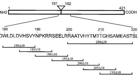
Amino acids 180 to 210 are required for MT-induced trans-formation.Our finding that monoclonal antibodies which bind to amino acids 200 to 219 of MT do not immunoprecipitate the
MT-pp60c-srccomplex suggested that this region was involved
in the interaction with pp60c-src (16). As no mutations have
been isolated in this area previously, we initially constructed a mutant DNA encoding an MT species lacking the whole se-quence between amino acids 200 and 219 inclusive (Fig. 1;
200D20). This mutant DNA was then transfected into Rat2
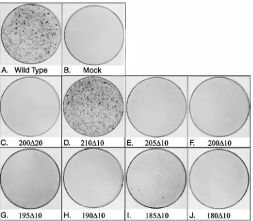
fibroblasts to determine whether recipient cells could form foci on adherent cell monolayers (Fig. 2). Figure 2C shows that the
200D20 mutant was completely deficient in this assay. To
ex-amine whether the entire 20 amino acids is required to induce transformation, we constructed a series of overlapping 10-ami-no-acid deletion mutants (Fig. 1) and transfected them into
Rat2 cells. Removing amino acids 210 to 219 (mutant 210D10)
had no effect on the transforming action of MT (Fig. 2D), but
205D10 (panel E) induces less than 5% of the colonies formed
by the same amount of wild-type DNA. However, MT with the 10 residues between amino acids 200 and 210 deleted was completely transformation defective (Fig. 2F). Therefore, the sequences of MT required to induce transformation extend only as far as amino acid 210 on the C-terminal side, and those between 210 and 220 are not necessary.
To determine how far this region extends in an N-terminal direction, more overlapping deletion mutants were constructed back to amino acid 180, well within the sequence common to MT and small t antigen (Fig. 1), and the DNA was again
assayed for the ability to form foci (Fig. 2). Mutants 195D10
and 190D10 (Fig. 2G and H) showed no transforming activity
in this assay. 185D10 induced a small number of colonies (Fig.
2I), suggesting a limit may have been approached, similar to
the behavior of mutant 205D10. However, mutant 180D10,
which extends further yet toward the N terminus, had no trans-forming activity (Fig. 2J). The whole region between amino acids 180 and 210 is therefore required for MT to induce transformation.
pp60c-src, PI3K, and Shc binding to MT correlates with transformation. To examine the biochemical properties of these mutant MT species, we isolated stable cell lines express-ing each protein by cotransfection of the mutant DNA with a
neo gene-containing plasmid, followed by selection with G418.
[image:2.612.319.555.69.200.2]Each line was characterized to ensure expression of MT, and then lysates were prepared for immunoprecipitation studies.
FIG. 1. Amino acids deleted by MT mutants. MT is represented schemati-cally at the top, and the regions deleted from MT mutants are indicated under-neath. The amino acid numbers of the N- and the C-terminal ends and the splice position are indicated. The amino acid sequence encoded by RNA sequences surrounding the splice point is shown in the single-letter code in the center.
VOL. 71, 1997 pp60c-src BINDING TO POLYOMAVIRUS MT 5513
on November 9, 2019 by guest
http://jvi.asm.org/
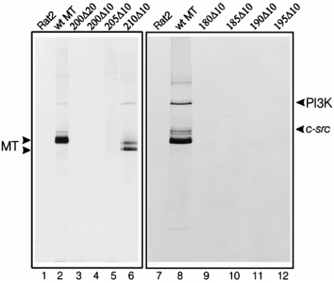
To detect src family kinase activity associated with MT, an anti-MT monoclonal antibody, PAb 762, was used to immuno-precipitate MT and the resulting complex was incubated with
[g-33P]ATP. Figure 3 shows the polypeptides in each MT
mu-tant immunoprecipitate that became phosphorylated during this reaction. The src family kinases associated with wild-type MT phosphorylated MT, themselves, and the 85-kDa PI3K component (lanes 2 and 8), as reported previously. Mutant
210D10, which has wild-type levels of transforming activity,
also exhibited similar phosphorylation patterns (lane 6). All of the mutants that have no focus-inducing capacity showed no MT-associated kinase activity (lanes 3, 4, 9, 11, and 12). The
mutants that generate low numbers of foci (205D10 and
185D10) exhibited small amounts of kinase activity on longer
exposures of the autoradiograph (data not shown). This may provide an explanation for their ability to induce only low numbers of foci. During calcium phosphate-mediated DNA transfection, the amount of DNA integrated into the host genome and expressed varies between individual cells. If
pp60c-srchas to be activated above a certain threshold to
trans-form recipient cells, mutants that poorly activate pp60c-srcwill
transform only those cells that express high levels of MT. This means that the number of foci observed reflects the proportion of cells expressing this large amount of protein. Therefore, there is an exact correlation between transforming capacity and MT-bound src family kinase activity in these mutants.
To identify the other proteins that are bound to each mutant MT, we employed an approach involving immunoprecipitation
with the monoclonal antibody PAb 762, followed by Western blotting and finally detection of the associated proteins using other antibodies. Figure 4 shows the results of these experi-ments. We first probed the MT immunoprecipitates with a
polyclonal anti-pp60c-srcantibody (panel A). (As it is likely that
pp60c-src, pp62c-yes, and pp59c-fynbind to MT in an analogous
manner, only pp60c-srcwas examined in these studies). pp60c-src
was easily detected in wild-type MT immunoprecipitates (lanes
2 and 8) and was observed associated with 210D10 MT (lane 6),
which also transforms cells and shows in vitro kinase activity.
However, no pp60c-srcwas observed associated with the
trans-formation-deficient mutant MT polypeptides (lanes 3, 4, 5, 9, 10, 11, and 12). The absence of kinase activity in immunopre-cipitates containing defective MT protein is, therefore, due to
a lack of binding to pp60c-src, rather than to an inability to
activate the kinase. Interaction of MT with both Shc and PI3K
is dependent on MT’s prior association with pp60c-src. Probing
duplicate blots with anti-Shc (panel B) or anti-PI3K 85-kDa subunit antibodies (panel C), therefore, showed similar results.
Both proteins bound to wild-type (lanes 2 and 8) and 210D10
(lane 6) MT but did not associate with MT molecules that fail
to bind pp60c-src (lanes 3, 4, 5, 9, 10, 11, and 12). Mutants
205D10 and 185D10, which exhibit a low level of focus-inducing
capacity, were found to bind very small amounts of pp60c-src,
Shc, and PI3K when more sensitive experiments were per-formed (data not shown). There is, then, a complete
correla-tion between the ability to bind pp60c-src, Shc, and PI3K and
[image:3.612.125.494.69.392.2]transformation.
FIG. 2. The focus-inducing properties of MT deletion mutants. Ten micrograms of an MT cDNA-containing plasmid was transfected into Rat2 cells. After 14 days the cells were stained with Leishmann’s reagent. (A) Foci induced by a plasmid containing wild-type MT cDNA. (B) Foci induced by a control plasmid. The deletion mutant used is indicated for the other panels.
on November 9, 2019 by guest
http://jvi.asm.org/
When similar immunoprecipitates were probed with anti-bodies against the A and C components of PP2A, however, a different result was obtained (Fig. 4D and E). All of the MT polypeptides bound normal levels of both PP2A components,
except the most N-terminal mutant, 180D10, which associated
with neither (lane 9). This may explain why 185D10 retains
some transforming activity, but 180D10 does not. The 185
position probably delineates the end of the region required to
bind pp60c-srconly, whereas the sequences between 180 and
184 are part of a region involved in associating with both PP2A
and pp60c-src. Probing similar blots with an anti-MT antibody
demonstrated that similar levels of MT were expressed in each line (Fig. 4F), and the polypeptides were truncated relative to wild-type MT as predicted by the size of the deletion mutant
used. Thus, the MT region required to bind pp60c-srclies
be-tween amino acids 185 and 210. To determine whether this
region binds to pp60c-srcdirectly, we have constructed a
gluta-thione S-transferase fusion protein containing amino acids 180 to 220 of MT, but so far, we cannot detect any interaction with
pp60c-srcin cell lysates (2). This suggests that the association
may also depend on additional factors, such as posttransla-tional modifications.
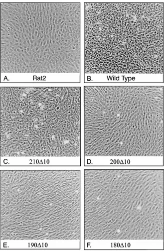
Phenotypic properties of cell lines expressing MT mutants.
Deletion mutants 190D10, 195D10, 200D10, and 200D20 are
the first MT species to be isolated that bind PP2A but not
pp60c-src. Cells expressing these mutants do not form foci
(Fig. 2) or grow in soft agar (data not shown). To examine whether any other phenotypic changes in cells expressing these mutant MTs could be observed, cell lines expressing each polypeptide were allowed to grow to similar levels of confluency and then photographed under phase-contrast conditions (Fig. 5). The parental Rat2 cell line exhibited a flattened, fibroblast morphology, with no tendency to over-grow other cells, and a clear contact inhibition of over-growth (Fig. 5A). Expression of wild-type MT caused these cells to
lose this contact inhibition; the cells overgrew one another in a random fashion, and individual cells appeared more rounded and refractile (Fig. 5B). Cells expressing
focus-inducing mutant 210D10 showed a phenotype similar to that
of wild-type MT-expressing cells (Fig. 5C), whereas those synthesizing nontransforming mutants exhibited few, if any, changes to the parental cells (Fig. 5D, E, and F). Therefore,
the ability to bind PP2A in the absence of pp60c-srcand any
[image:4.612.57.298.69.273.2]effects this may have on phosphatase activity have few overt
FIG. 3. In vitro kinase assay of mutant MT polypeptides from G418-selected cell lines. Lysates were prepared from cell lines expressing each MT deletion mutant, immunoprecipitated with monoclonal antibody PAb 762, incubated with [g-32P]ATP, and then separated on a polyacrylamide gel containing SDS. The
[image:4.612.333.538.69.525.2]deletion mutant expressed is indicated above each lane. The separate panels contain results from two different experiments, run on separate gels. Each ex-periment included a control using a lysate from the parental Rat2 cell line (lanes 1 and 7) and a wild-type MT-containing cell lines (lanes 2 and 8). The migration positions of wild-type and mutant MT polypeptides are indicated on the left, and those of the 85-kDa component of PI3K and pp60c-srcare on the right.
FIG. 4. Western blot analysis of immunoprecipitated proteins associated with deletion mutants of MT. Lysates of cell lines expressing mutant MTs were immunoprecipitated with PAb 762, electrophoretically separated, and then transferred onto nitrocellulose filters. These were then probed with the appro-priate antibodies to detect MT-associated pp60c-src(A), Shc (B), the PI3K
85-kDa component (C), the PP2A A subunit (D), the PP2A C subunit (E), and MT itself (F). The deletion mutant analyzed in each case is indicated above the lanes in panel A. A negative (Rat2) and a positive (wt MT) control is included in each gel. The migration positions of the associated polypeptides detected are shown to the right of each panel.
VOL. 71, 1997 pp60c-src BINDING TO POLYOMAVIRUS MT 5515
on November 9, 2019 by guest
http://jvi.asm.org/
effects on cell phenotype. Mutant cells expressing the
par-tially transforming mutants (185D10 and 205D10) exhibited
phenotypes between these two extremes, with the degree of alteration depending on the amount of MT expressed (data not shown).
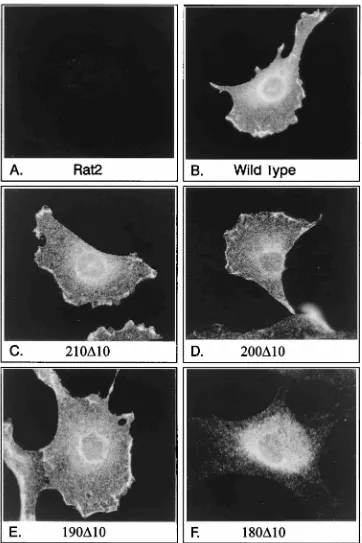
Mutant MT subcellular location.It has been suggested that
MT mutants that are defective in both PP2A and pp60c-src
binding have an altered subcellular distribution (27, 31). To
determine whether pp60c-srcassociation alters the location of
MT, mutant-expressing cells were subjected to
immunofluo-rescence analysis using PAb 762 to detect MT (Fig. 6). Figure 6B shows the wild-type distribution found in transformed Rat2 cells, in which MT was located in grainy structures throughout the cytoplasm but was concentrated around the nucleus and in discrete patches at the cell periphery. An
identical MT distribution was observed in mutant 210D
10-transformed cells (Fig. 6C) and in cells expressing MT
mu-tants that fail to associate with pp60c-src(Fig. 6D and E) (2a).
The deletion mutant that lacks pp60c-srcand PP2A
[image:5.612.143.472.69.573.2]interac-tion, however, showed a dramatic alteration in location (Fig.
FIG. 5. Phase-contrast photomicrographs of Rat2 cell lines expressing selected mutants of MT. The parental Rat2 cell line (A), a wild-type MT-expressing cell line (B), and MT deletion mutant-expressing cell lines were grown in DMEM with 10% fetal calf serum until just confluent and then photographed under phase-contrast illumination. The mutant expressed is indicated under each panel.
on November 9, 2019 by guest
http://jvi.asm.org/
6F). 180D10 MT was still found in grainy cytoplasmic struc-tures, but the accumulation in a perinuclear location and at the plasma membrane appeared to be absent. This suggests strongly that the subcellular distribution of MT observed by immunofluorescence is influenced by binding to PP2A
rather than by association with pp60c-src. It is not clear at
present whether this is a consequence of the phosphatase activity or whether PP2A itself relocates the MT complex to a precise subcellular location. Therefore, the subcellular distribution of MT seen in immunofluorescence studies is
grossly unaffected by association with pp60c-srcbut is
[image:6.612.125.487.68.611.2]influ-enced by binding to PP2A.
FIG. 6. Immunofluorescence analysis of MT present in Rat2 cell lines. Cell lines expressing various mutants of MT were seeded onto glass coverslips and stained with fluorescently labelled anti-MT antibodies. Representative cells are shown in each panel, together with a control Rat2 cell and a wild-type-expressing cell treated in a similar manner. The mutant expressed in each cell is indicated beneath each panel.
VOL. 71, 1997 pp60c-src BINDING TO POLYOMAVIRUS MT 5517
on November 9, 2019 by guest
http://jvi.asm.org/
DISCUSSION
The association between MT and pp60c-srchas been known
for some time to be crucial to the transforming properties of MT. Despite this, it has proven impossible to establish the molecular basis for the interaction, due in part to difficulties in recreating the association in vitro. Nearly all knowledge con-cerning the interaction, therefore, has been derived from stud-ies of mutant molecules and by using monoclonal antibodstud-ies. It appears that only the kinase domain and a few C-terminal
amino acids of pp60c-srcare required for interaction with MT
(8, 17), so the SH2 and SH3 domains normally responsible for
pp60c-srcregulation (41) are probably not involved in this case.
This makes the mechanism behind this interaction particularly interesting, because it currently represents a unique means of influencing src family kinase activity.
The interaction with pp60c-srcrequires the N-terminal region
of MT, as monoclonal antibodies that bind in this area fail to precipitate the complex and all previously isolated transforma-tion-defective MT mutants with alterations in this region
dis-rupt both MT-PP2A and MT-pp60c-srcinteractions. Here, for
the first time, we have separated these associations and have
isolated mutants that bind PP2A but not pp60c-src. No mutant
has so far been isolated that does not bind PP2A yet still
associates with pp60c-src. As others have suggested (3, 18, 19),
then, it seems likely that MT, pp60c-src, and PP2A are linked in
some way. The mutants reported here eliminate the possibility
that pp60c-srcbinds directly only to PP2A, as this is unlikely to
be affected by MT mutants outside the PP2A binding region,
and suggest a contact between MT and pp60c-srcat one
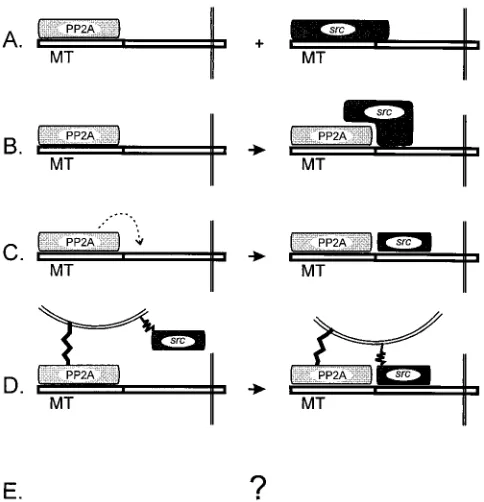
loca-tion, at least. This still leaves a number of possible models for these interactions, however (Fig. 7). Model A proposes that
pp60c-srcand PP2A recognize similar regions of MT. The
ob-servation that PP2A and pp60c-srccan be detected in the same
complex (45) suggests that this model is unlikely to be correct.
Model B suggests that pp60c-srcbinding is a bipartite
mecha-nism, with pp60c-srccontacting both MT and PP2A. Model C
offers the idea that the phosphatase action of PP2A is required
for pp60c-srcbinding. Model D suggests the intriguing
possibil-ity that PP2A is required to alter the subcellular location of
MT to a membrane site at which pp60c-srcbinding can occur.
Finally, model E acknowledges that other, as yet unknown, mechanisms may be involved. Of course, all of these models are not mutually exclusive, and a combination of any of the suggestions may actually occur. The isolation of these new mutants that identify a region which is required, although
possibly not sufficient, for interaction with pp60c-src at last
provides us with some means of distinguishing between these possibilities.
Through the use of deletion analysis, we have limited the
area of MT required to bind pp60c-src, but not PP2A, to amino
acids 185 to 210. Within this sequence are a number of putative phosphorylation sites (Fig. 1). Previous studies have suggested that phosphorylation may be required to promote the
MT-pp60c-srcinteraction (30), so these sites are of particular
inter-est. Analyzing this region with the Prosite database suggested that Ser195 is a strong candidate for a cyclic AMP-dependent protein kinase phosphorylation site but produced no strong matches for any of the other residues. Point mutagenesis is now being performed to determine whether any of these
res-idues are required for binding to pp60c-src.
The new MT mutants that lack pp60c-srcbinding ability, but
still associate with PP2A, had no transforming activity (Fig. 2), and cell lines expressing the mutated polypeptides exhibited few altered morphological properties on initial observations (Fig. 5), although we cannot yet exclude more subtle effects on cell characteristics. The MT-PP2A interaction alone, then, seems to have little effect on cell phenotype. However, a dif-ferent picture emerged when the subcellular locations of the mutant MT molecules were studied by immunofluorescence (Fig. 6). MT in Rat2-transformed cells exhibited a reticular staining pattern that stretches throughout the cytoplasm, with a strong accumulation in a juxtanuclear location and at discrete patches at the cell periphery. Much of this is similar to the distribution observed in lytically infected cells (15), with the exception of the plasma membrane location. This difference is probably a consequence of the use in the current study of transformed cells which contain a much higher percentage of complexed, rather than free, MT than do lytically infected cells (2a) and of the availability of more sensitive techniques and reagents. This distribution, observed by immunofluorescence,
is unaffected by pp60c-srcassociation but is altered dramatically
by the loss of PP2A binding (Fig. 6F). In the absence of a PP2A interaction, MT no longer accumulates around the nucleus and, interestingly, is no longer associated with the cell periph-ery. These plasma membrane patches resemble areas of mem-brane ruffling observed in motile fibroblasts, so it will be inter-esting to examine whether these regions colocalize with polymerized actin molecules. Related observations confirming these results were reported previously (31), whereby MT
mu-tants lacking both pp60c-srcand PP2A binding were shown to
exhibit a more diffuse MT location. Mutants that still associate with PP2A were not available to those authors, however, so they were unable to assess the contribution of each interaction. All of the MT mutants described here retain a functional membrane binding sequence at the C-terminal end of the pro-tein, and the immunofluorescent staining patterns are still con-sistent with a membrane-bound state. The results shown
sug-FIG. 7. Potential models for the relationship between pp60c-srcand PP2A binding to MT. MT is represented in a linear fashion, and the sequences poten-tially recognized by PP2A or pp60c-srcare represented schematically above. Fine double lines at the C-terminal end of MT represent membrane binding, and the rounded lines in model D signify a specific membrane bilayer to which PP2A and pp60c-srcare attached. The broken arrow in model C represents the enzymatic action of PP2A.
on November 9, 2019 by guest
http://jvi.asm.org/
[image:7.612.59.301.72.323.2]gest that PP2A influences the final location of the MT complex rather than MT controlling the distribution of its associated proteins, as is usually envisaged. However, we cannot yet for-mally exclude a more indirect correlation between PP2A asso-ciation and MT distribution. For example, the MT-PP2A as-sociation may stimulate membrane ruffling in cells, thus increasing the amount of MT detected in such structures. Al-ternately, in the absence of PP2A binding, another molecule may associate with MT, such as HSP70 (33, 46), and this may be responsible for the more diffuse location of MT. In this case,
the diffuse 180D10 MT staining represents an altered location
and the usual distribution of MT is an accumulation in jux-tanuclear and cell periphery regions. Studies are now under way to investigate these and other possibilities. If, as we feel is more likely, PP2A binding does cause an MT location change, it could exert this effect through its phosphatase activity expos-ing sites in MT or through PP2A itself beexpos-ing targeted to this subcellular site. In this regard, it is interesting to note that although PP2A is generally considered to be a soluble, cyto-plasmic protein, a number of recent reports have identified alternative locations, including membrane sites (1, 21, 35, 44, 45). Clearly, redistribution of MT may influence greatly how signals produced by MT complexes during cell transformation are generated and act and could also alter the PP2A activity at this specific location. Further experiments are under way to study these exciting possibilities. Finally, it has been suggested
from biochemical studies that pp60c-srcmay control other
as-pects of MT’s subcellular distribution (27). Although we have observed no evidence for this in our immunofluorescence anal-ysis, this may merely reflect the fact that not all subcellular changes can be observed using one technique and some cy-toskeleton association-mediated relocations are beyond the resolving power of immunofluorescence.
The interactions between MT and other signalling molecules are analogous to those occurring on activated tyrosine kinase-associated growth factor receptors. However, there appears to
be no analogy between the interaction of MT and pp60c-srcwith
any other pp60c-src-binding molecules. A search of the protein
database with the 185 to 210 region of MT produced no clear homologies, but it seems unlikely that MT is employing a
totally unique mechanism of regulating pp60c-src. Instead, MT
might mimic the action of an as-yet-undiscovered regulator of
pp60c-srcactivity. Such a regulator is now actively being sought.
ACKNOWLEDGMENTS
We are grateful to Emin Ulug at Kansas State University for the generous gift of antipeptide antisera to the PP2A components. We also thank all the members of the RPMS who provided valuable advice during the course of this work, particularly Mick Jones, Nick Dibb, and Nina Krauzewicz, and finally, we thank Victoria Horner for technical assistance.
This work was generously supported by grants from the Cancer Research Campaign (UK).
REFERENCES
1. Alexander, D. R., M. H. Brown, A. L. Tutt, M. J. Crumpton, and E. Shivnan. 1992. CD3 and CD2 antigen-mediated CD3g-chain phosphorylation in permeabilized human T cells. Biochem. J. 288:69–77.
2. Bolen, J. B., C. J. Thiele, M. A. Israel, W. Yonemoto, L. A. Lipsich, and J. S.
Brugge.1984. Enhancement of cellular src gene product associated tyrosyl kinase activity following polyoma virus infection and transformation. Cell
38:767–777.
2a.Brewster, C. E. P., H. R. Glover, and S. M. Dilworth. Unpublished data. 3. Campbell, K. S., K. R. Auger, B. A. Hemmings, T. M. Roberts, and D. C.
Pallas.1995. Identification of regions in polyomavirus middle T and small t antigens important for association with protein phosphatase 2A. J. Virol.
69:3721–3728.
4. Campbell, K. S., E. Ogris, B. Burke, W. Su, K. R. Auger, B. J. Druker, B. S.
Schaffhausen, T. M. Roberts, and D. C. Pallas.1994. Polyoma middle tumor
antigen interacts with SHC protein via the NPTY (Asn-Pro-Thr-Tyr) motif in middle tumor antigen. Proc. Natl. Acad. Sci. USA 91:6344–6348. 5. Cartwright, C. A., P. L. Kaplan, J. A. Cooper, T. Hunter, and W. Eckhart.
1986. Altered sites of tyrosine phosphorylation in pp60c-srcassociated with polyomavirus middle tumor antigen. Mol. Cell. Biol. 6:1562–1570. 6. Cheng, S. H., P. C. Espino, J. Marshall, R. Harvey, and A. E. Smith. 1990.
Stoichiometry of cellular and viral components in the polyomavirus middle-T antigen-tyrosine kinase complex. Mol. Cell. Biol. 10:5569–5574.
7. Cheng, S. H., R. Harvey, P. C. Espino, K. Semba, T. Yamamoto, K.
Toyo-shima, and A. E. Smith.1988. Peptide antibodies to the human c-fyn gene product demonstrate pp59c-fyn is capable of complex formation with the middle-T antigen of polyomavirus. EMBO J. 7:3845–3855.
8. Cheng, S. H., H. Piwnica-Worms, R. W. Harvey, T. M. Roberts, and A. E.
Smith.1988. The carboxy terminus of pp60c-srcis a regulatory domain and is involved in complex formation with the middle-T antigen of polyomavirus. Mol. Cell. Biol. 8:1736–1747.
9. Courtneidge, S. A. 1985. Activation of the pp60c-src kinase by middle T antigen binding or by dephosphorylation. EMBO J. 4:1471–1477. 10. Courtneidge, S. A., and A. Heber. 1987. An 81 kd protein complexed with
middle T antigen and pp60c-src: a possible phosphatidylinositol kinase. Cell 50:1031–1037.
11. Courtneidge, S. A., and A. E. Smith. 1984. The complex of polyoma virus middle-T antigen and pp60c-src. EMBO J. 3:585–591.
12. Courtneidge, S. A., and A. E. Smith. 1983. Polyoma virus transforming protein associates with the product of the c-src cellular gene. Nature 303: 435–439.
13. Dilworth, S. M. 1995. Polyoma virus middle T antigen: meddler or mimic? Trends Microbiol. 3:31–34.
14. Dilworth, S. M., C. E. P. Brewster, M. D. Jones, L. Lanfrancone, G. Pelicci,
and P. G. Pelicci.1994. Transformation by polyoma virus middle T-antigen involves the binding and tyrosine phosphorylation of Shc. Nature 367:87–90. 15. Dilworth, S. M., H. A. Hansson, C. Darnfors, G. Bjursell, C. H. Streuli, and
B. E. Griffin.1986. Subcellular localisation of the middle and large T-antigens of polyoma virus. EMBO J. 5:491–499.
16. Dilworth, S. M., and V. P. Horner. 1993. Novel monoclonal antibodies that differentiate between the binding of pp60c-srcor protein phosphatase 2A by polyomavirus middle T antigen. J. Virol. 67:2235–2244.
17. Dunant, N. M., M. Senften, and K. Ballmer-Hofer. 1996. Polyomavirus middle-T antigen associates with the kinase domain of Src-related tyrosine kinases. J. Virol. 70:1323–1330.
18. Glenn, G. M., and W. Eckhart. 1995. Amino-terminal regions of polyoma-virus middle T antigen are required for interactions with protein phospha-tase 2A. J. Virol. 69:3729–3736.
19. Glenn, G. M., and W. Eckhart. 1993. Mutation of a cysteine residue in polyomavirus middle T antigen abolishes interactions with protein phospha-tase 2A, pp60c-src, and phosphatidylinositol-3 kinase, activation of c-fos ex-pression, and cellular transformation. J. Virol. 67:1945–1952.
20. Gorga, F. R., C. E. Riney, and T. L. Benjamin. 1990. Inositol trisphosphate levels in cells expressing wild-type and mutant polyomavirus middle T anti-gens: evidence for activation of phospholipase C via activation of pp60c-src. J. Virol. 64:105–112.
21. Hansra, G., F. Bornancin, R. Whelan, B. A. Hemmings, and P. J. Parker. 1996. 12-O-tetradecanoylphorbol-13-acetate-induced dephosphorylation of protein kinase Cacorrelates with the presence of a membrane-associated protein phosphatase 2A heterotrimer. J. Biol. Chem. 271:32785–32788. 22. Higuchi, R., B. Krummel, and R. K. Saiki. 1988. A general method of in vitro
preparation and specific mutagenesis of DNA fragments: study of protein and DNA interactions. Nucleic Acids Res. 16:7351–7367.
23. Horak, I. D., T. Kawakami, F. Gregory, K. C. Robbins, and J. B. Bolen. 1989. Association of p60fynwith middle tumor antigen in murine
polyomavirus-transformed rat cells. J. Virol. 63:2343–2347.
24. Kaplan, D. R., D. C. Pallas, W. Morgan, B. Schaffhausen, and T. M. Roberts. 1989. Mechanisms of transformation by polyoma virus middle T antigen. Biochim. Biophys. Acta 948:345–364.
25. Kaplan, D. R., M. Whitman, B. Schaffhausen, D. C. Pallas, M. White, and L.
Cantley.1987. Common elements in growth factor stimulation and onco-genic transformation: 85kd phosphoprotein and phosphatidylinositol kinase activity. Cell 50:1021–1029.
26. Kornbluth, S., M. Sudol, and H. Hanafusa. 1987. Association of the poly-omavirus middle-T antigen with c-yes protein. Nature 325:171–173. 27. Krauzewicz, N., J. Elliot, and B. E. Griffin. 1994. Cell fractionation in
non-ionic detergents distinguishes sub-populations of polyoma virus middle T antigen and reveals a novel form. Oncogene 9:2283–2291.
28. Kypta, R. M., A. Hemming, and S. A. Courtneidge. 1988. Identification and characterization of p59fyn (a src-like protein tyrosine kinase) in normal and polyoma virus transformed cells. EMBO J. 7:3837–3844.
29. Markland, W., and A. E. Smith. 1987. Mutants of polyomavirus middle-T antigen. Biochim. Biophys. Acta 907:299–321.
30. Matthews, J. T., and T. L. Benjamin. 1986. 12-O-tetradecanoylphorbol-13-acetate stimulates phosphorylation of the 58,000-Mrform of polyomavirus middle T antigen in vivo: implications for a possible role of protein kinase C in middle T function. J. Virol. 58:239–246.
VOL. 71, 1997 pp60c-src BINDING TO POLYOMAVIRUS MT 5519
on November 9, 2019 by guest
http://jvi.asm.org/
31. Messerschmitt, A., C. Disela, S. Dilworth, A. G. Marti, and K.
Ballmer-Hofer.1996. Polyomavirus middle-T antigen lacking a membrane anchor sequence accumulates in the nucleus. J. Gen. Virol. 77:17–26.
32. Pallas, D. C., H. Fu, L. C. Haehnel, W. Weller, R. J. Collier, and T. M.
Roberts.1994. Association of polyomavirus middle tumor antigen with 14-3-3 proteins. Science 265:535–537.
33. Pallas, D. C., W. Morgan, and T. M. Roberts. 1989. The cellular proteins which can associate specifically with polyomavirus middle T antigen in hu-man 293 cells include the major huhu-man 70-kilodalton heat shock proteins. J. Virol. 63:4533–4539.
34. Pallas, D. C., L. K. Shahrik, B. L. Martin, S. Jaspers, T. B. Miller, D. L.
Brautigan, and T. M. Roberts.1990. Polyoma small and middle T antigens and SV40 small t antigen form stable complexes with protein phosphatase 2A. Cell 60:167–176.
35. Pitcher, J. A., E. S. Payne, C. Csortos, A. A. DePaoli-Roach, and R. J.
Lefkowitz.1995. The G-protein-coupled receptor phosphatase: a protein phosphatase type 2A with a distinct subcellular distribution and substrate specificity. Proc. Natl. Acad. Sci. USA 92:8343–8347.
36. Ruediger, R., D. Roeckel, J. Fait, A. Bergqvist, G. Magnusson, and G.
Walter.1992. Identification of binding sites on the regulatory A subunit of protein phosphatase 2A for the catalytic C subunit and for tumor antigens of simian virus 40 and polyomavirus. Mol. Cell. Biol. 12:4872–4882. 37. Sambrook, J., E. F. Fritsch, and T. Maniatis. 1989. Molecular cloning: a
laboratory manual, 2nd ed. Cold Spring Harbor Laboratory Press, Cold Spring Harbor, N.Y.
38. Schlessinger, J. 1993. How receptor tyrosine kinases activate Ras. Trends Biochem. 18:273–275.
39. Serunian, L. A., K. R. Auger, T. M. Roberts, and L. C. Cantley. 1990. Production of novel polyphosphoinositides in vivo is linked to cell transfor-mation by polyomavirus middle T antigen. J. Virol. 64:4718–4725.
40. Su, W., W. Liu, B. S. Schaffhausen, and T. M. Roberts. 1995. Association of polyomavirus middle tumor antigen with phospholipase C-g1. J. Biol. Chem.
270:12331–12334.
41. Superti-Furga, G., and S. A. Courtneidge. 1995. Structure-function relation-ships in Src family and related protein tyrosine kinases. Bioessays 17:321– 330.
42. Talmage, D. A., R. Freund, A. T. Young, J. Dahl, C. J. Dawe, and T. L.
Benjamin.1989. Phosphorylation of middle T by pp60c-src: a switch for binding of phosphatidylinositol 3-kinase and optimal tumorigenesis. Cell
59:55–65.
43. Treisman, R., U. Novak, J. Favaloro, and R. Kamen. 1981. Transformation of rat cells by an altered polyoma virus genome expressing only the middle-T protein. Nature 292:595–600.
44. Turowski, P., A. Fernandez, B. Favre, N. C. J. Lamb, and B. A. Hemmings. 1995. Differential methylation and altered conformation of cytoplasmic and nuclear forms of protein phosphatase 2A during cell cycle progression. J. Cell Biol. 129:1400–1413.
45. Ulug, E. T., A. J. Cartwright, and S. A. Courtneidge. 1992. Characterization of the interaction of polyomavirus middle T antigen with type 2A protein phosphatase. J. Virol. 66:1458–1467.
46. Walter, G., A. Carbone, and W. J. Welch. 1987. Medium tumor antigen of polyomavirus transformation-defective mutant NG59 is associated with 73-kilodalton heat shock protein. J. Virol. 61:405–410.
47. Walter, G., R. Ruediger, C. Slaughter, and M. Mumby. 1990. Association of protein phosphatase 2A with polyoma virus medium tumor antigen. Proc. Natl. Acad. Sci. USA 87:2521–2525.
48. Yoakim, M., W. Hou, Y. Liu, C. L. Carpenter, R. Kapeller, and B. S.
Schaffhausen.1992. Interactions of polyomavirus middle T with the SH2 domains of the pp85 subunit of phosphatidylinositol-3-kinase. J. Virol. 66: 5485–5491.