0022-538X/95/$04.0010
Copyrightq1995, American Society for Microbiology
Sequence Comparison of Porcine Respiratory Coronavirus Isolates
Reveals Heterogeneity in the S, 3, and 3-1 Genes
ERIC M. VAUGHN,1,2PATRICK G. HALBUR,1
ANDPREM S. PAUL1,2*
Veterinary Medical Research Institute1and Department of Microbiology, Immunology, and Preventive Medicine,2Iowa State University, Ames, Iowa 50011
Received 16 September 1994/Accepted 24 January 1995
Four new porcine respiratory coronavirus (PRCV) isolates were genetically characterized. Subgenomic mRNA patterns and the nucleotide sequences of the 5*ends of the S genes, the open reading frame (ORF) 3/3a genes, and the ORF 3-1/3b genes of these PRCV isolates were determined and compared with those of other PRCV and transmissible gastroenteritis virus (TGEV) isolates. The S, ORF 3/3a, and ORF 3-1/3b genes are under intense study because of their possible roles in determining tissue tropism and virulence. Northern (RNA) blot analysis of subgenomic mRNAs revealed that mRNA 2, which encodes for the S gene, of the PRCV isolates migrated faster than the mRNA 2 of TGEV. The PRCV isolates AR310 and LEPP produced eight subgenomic mRNA species, the same number as produced by the virulent Miller strain of TGEV. However, the PRCV isolates IA1894 and ISU-1 produced only seven subgenomic mRNA species. All four of the PRCV isolates were found to have a large in-frame deletion in the 5*end of the S gene; however, the size and location of the deletion varied. Analysis of the ORF 3/3a gene nucleotide sequences from the four PRCV isolates also showed a high degree of variability in this area. The ORF 3 gene of the PRCV isolates AR310 and LEPP was preceded by a CTAAAC leader RNA-binding site, and the ORF 3 gene was predicted to yield a protein of 72 amino acids, the same size as that of the virulent Miller strain of TGEV. The PRCV isolates AR310 and LEPP are the first PRCV isolates found to have an intact ORF 3 gene. The ORF 3a gene of the PRCV isolate IA1894 was preceded by a CTAAAC leader RNA-binding site and was predicted to yield a truncated protein of 54 amino acids due to a 23-nucleotide deletion. The CTAAAC leader RNA-binding site and ATG start codon of ORF 3 gene of the PRCV isolate ISU-1 were removed because of a 168-nucleotide deletion. Analysis of the ORF 3-1/3b gene nucleotide sequences from the four PRCV nucleotides isolates also showed variability.
Porcine respiratory coronavirus (PRCV) and transmissible gastroenteritis virus (TGEV) are members of the
Coronaviri-dae family of viruses (17, 24, 27). Coronaviruses are
pleomor-phic enveloped viruses with a positive-sense single-stranded RNA genome (27). TGEV causes severe diarrhea with a high mortality in neonatal swine (24). TGEV replicates in and de-stroys the enterocytes of the villus epithelium in the small intestine, which causes the subsequent malabsorption and de-hydration characteristic of transmissible gastroenteritis (TGE) (24). TGEV has also been shown to replicate in the respiratory tissue of infected swine (16, 17). PRCV is believed to be a mutant of TGEV, as it has been shown to be antigenically and genetically related to TGEV but has a selective tropism for respiratory tissue with very little to no replication in the intes-tinal tissue of infected swine (10, 17, 21). PRCV is widespread in Europe, where it was first detected (17, 21). PRCV has also been found in the United States, but the extent of its preva-lence is not known (12, 29, 35). Since TGEV and PRCV are closely related and yet display differences in tissue tropism and pathogenicity, they can serve as useful models for the study of coronavirus genes involved in tropism and virulence.
The replication scheme of coronaviruses is characterized by the development of a 39-terminal nested set of subgenomic mRNAs in infected cells (27). The subgenomic mRNAs, with the exception of the smallest, are polycistronic in nature, yet in general, only the unique 59 open reading frame (ORF) is translated (26, 27). Seven or eight subgenomic mRNAs are
synthesized during TGEV replication in infected cells (3, 27, 33). The full-length genomic mRNA 1 encodes for an RNA-dependent RNA polymerase (27). The spike (S) protein is encoded by subgenomic mRNA 2 (27). The ORF 3 and ORF 3-1 genes of the Miller strain of TGEV are found at the 59end of two separate subgenomic mRNAs, 3 and 3-1, respectively, and are thought to encode two putative nonstructural proteins (27, 32, 34). The homologous genes in the Purdue, FS772/70, and D52 strains of TGEV are found on a single mRNA species and are designated as the ORF 3a and ORF 3b genes (2, 4, 15). The small integral membrane (sM) protein is associated with the viral envelope and is encoded by subgenomic mRNA 4 (9). The integral membrane protein (M) and nucleocapsid (N) proteins are encoded by subgenomic mRNAs 5 and 6, respec-tively (27). The subgenomic mRNA 7, the smallest subgenomic mRNA, is the only monocistronic subgenomic mRNA and may encode for a possible DNA-binding protein in infected cells (5, 8). Preceding each large potential ORF is the conserved hex-americ sequence of CTAAAC, which is thought to serve as a consensus leader RNA-binding site in the transcription of the subgenomic mRNAs from a full-length negative-sense tem-plate (27).
Previous studies of the genetic structure of PRCV have shown that all of the isolates have two unique characteristics. First, the S gene of PRCV contains a large in-frame deletion ranging from 672 to 681 nucleotides in length (4, 13, 17, 22, 34). This deletion in the S gene results in a smaller S glycop-rotein. Second, all of the PRCVs analyzed thus far have had the CTAAAC consensus leader RNA-binding site preceding the ORF 3a gene altered or partially deleted (17, 19, 22, 34). Thus, the subgenomic mRNA 3 is not detected in PRCV-infected cells (17, 19, 22, 34). Also, the ORF 3 gene of the * Corresponding author. Mailing address: Veterinary Medical
Re-search Institute, Iowa State University, 1802 Elwood Dr., Ames, IA 50011. Phone: (515) 294-0913. Fax: (515) 294-1401. Electronic mail address: pspaul@iastate.edu.
3176
on November 9, 2019 by guest
http://jvi.asm.org/
European PRCVs contains several small deletions that render the gene 3 a pseudogene (19, 22). One PRCV isolate from the United States, Ind/89, was found to have a five-nucleotide deletion within an otherwise nearly intact ORF 3 gene, but the CTAAAC consensus leader RNA-binding site had been al-tered to CTAAAT, and the subgenomic mRNA 3 of Ind/89 is undetectable in infected cells (34). The presence of a large deletion in the 59end of the S gene is thought to play a role in determining the tissue tropism of PRCV (20, 26). In the case of TGEV, the ORF 3 and 3-1 genes (formerly designated as genes A and B, respectively) have been hypothesized to be important in the virulence and pathogenesis of TGEV infec-tion (33). It is interesting to note that all previously genetically characterized PRCV isolates have been demonstrated to have an altered or deleted ORF 3 gene and that these PRCVs all cause minimal to no apparent clinical disease in swine.
In this study, we have analyzed the viral mRNAs from four PRCV isolates by Northern (RNA) blot analysis and nucle-otide sequencing of the 59ends of the S gene and the ORF 3 and ORF 3-1 genes. The size and location of the S gene deletion of the PRCV isolates were also determined, as we had previously shown that two of the PRCV isolates in this study, AR310 and LEPP, had smaller deletions in the 59end of the S gene compared with that of another United States PRCV isolate, designated ISU-1 (29). Also, since the ORF 3 genes of previously described PRCV isolates have been found to have deletions present, we examined the ORF 3 and ORF 3-1 genes of these new PRCV isolates to determine the extent of heter-ogeneity present in these genes.
The PRCV isolates AR310, LEPP, and IA1894 were iso-lated as previously described (29, 30). The PRCV isolate AR310 was isolated from intestinal homogenates from a piglet with TGE from a swine herd in Arkansas in 1989. The PRCV isolates LEPP and IA1894 were isolated from nasal swabs collected from Iowa swine herds with pneumonia in 1991 and 1992, respectively. The gut-passaged virulent Miller strain of TGEV (National Veterinary Services Laboratory, Ames, Iowa), also referred to as CHV TGEV, was used as a standard TGEV strain in this study. The PRCV isolate ISU-1 was re-ceived as a plaque-purified preparation and was kindly pro-vided by Howard Hill (Iowa State University Veterinary Diag-nostic Laboratory) (12).
ST cells were infected at a multiplicity of infection of ap-proximately 0.1 PFU per cell with CHV TGEV and the PRCV isolates AR310, ISU-1, IA1894, and LEPP. At 19 h postinfec-tion, the medium was removed and the total RNA was isolated from the infected ST cell monolayers by a rapid guanidinium thiocyanate method (Stratagene, La Jolla, Calif.). The RNA was washed with 70% ethanol and dissolved in diethyl pyro-carbonate-treated distilled water and stored at2708C.
Analysis of subgenomic mRNAs.For Northern blot hybrid-ization analysis, 40mg of total RNA was denatured with form-aldehyde and formamide (25) and separated by electrophoresis in a 1.5% SeaKem (FMC Bioproducts, Rockland, Maine) aga-rose gel. After electrophoresis, the agaaga-rose gel was treated with 0.05 N NaOH and 1.5 M NaCl, neutralized with 0.1 M Tris-HCl (pH 7.5) and 1.5 M NaCl, and washed twice in 103 SSC (13SSC is 0.15 M NaCl plus 15 mM sodium citrate), and then the RNA was transferred with a Posiblot pressure blotter (Stratagene) with 103SSC to nylon membranes (Magna NT; Micron Separations Inc., Westboro, Mass.). After the transfer was complete, the membranes were baked at 808C for 2 h to fix the RNA. The membranes were prehybridized for 2 h in a solution containing 50% formamide, 53 SSPE (13 SSPE is 0.18 M NaCl, 10 mM sodium phosphate, and 1 mM EDTA), 43Denhardt’s solution, 0.3% sodium dodecyl sulfate (SDS),
and sonicated salmon sperm DNA (30 mg/ml) at 428C. The probe used in the hybridization procedure was a 462-bp PCR product amplified with the primers 068 and 069. This PCR product was amplified from TGEV cDNA and gave a probe specific for the 39end of TGEV and was thus suitable for use as a probe in a Northern blot to detect TGEV and PRCV subgenomic mRNAs. One hundred twenty-five nanograms of the 39-end PCR product was labeled with [a-32P]dCTP (ICN
Biochemicals, Costa Mesa, Calif.) in the presence of random hexamer primers and the Klenow fragment of DNA poly-merase I (Amersham Corporation, Arlington Heights, Ill.). Unincorporated [a-32P]dCTP was removed by passing the
la-beled DNA through a Sephadex G-50 column (Boehringer Mannheim, Indianapolis, Ind.). The labeled 39end probe was then heated to 1008C, cooled on ice, and then added to the prehybridization reaction mixture and allowed to hybridize overnight. The membranes were washed once in 23SSC–0.3% SDS at room temperature, twice in 23SSC–0.3% SDS at 658C, and once with 0.23 SSC at room temperature. Autoradio-graphs were made by exposing X-ray film (RX film; Fuji Photo Film Co., Stamford, Conn.) to the membranes.
[image:2.612.317.552.69.264.2]As shown in Fig. 1, the virulent Miller CHV TGEV had eight mRNA species present, as has been previously reported (32). The PRCV isolates AR310 and LEPP also had eight mRNA species present, whereas the PRCV isolates IA1894 and ISU-1 had only seven mRNA species. The migration pat-terns of the mRNA species 4 through 7 of all four PRCV isolates were similar to those of the virulent TGEV. The mRNA 2 species of the PRCV isolates examined migrated faster than the TGEV equivalent, indicating that a large dele-tion was present. The PRCV isolates AR310 and LEPP and the virulent CHV TGEV all had the mRNA species 3 and 3-1 present. However, the PRCV isolates IA1894 and ISU-1 had only one mRNA species present in the area in which the FIG. 1. Northern blot analysis of TGEV and PRCV subgenomic mRNAs. Total RNA from TGEV- or PRCV-infected ST cells was separated in a 1.5% agarose and transferred to a nylon membrane. A PCR product from the 39end of the TGEV genome was amplified with primers 068 and 069, labeled with [a-32
P]dCTP, and used to detect TGEV and PRCV subgenomic mRNAs in a Northern blot. Tested were PRCV AR310 (lane A), CHV TGEV (lane B), PRCV LEPP (lane C), PRCV IA1894 (lane D), and PRCV ISU-1 (lane E). Note that mRNA 2 of all the PRCV isolates migrated faster than the mRNA 2 of CHV TGEV. Also, note that PRCV AR310, PRCV LEPP, and CHV TGEV all have mRNA 3 and 3-1 present. The PRCV isolate IA1894 had only one mRNA species of 3.61 kb, corresponding to mRNA 3, present. The PRCV isolate ISU-1 had only one mRNA species of 3.35 kb, corresponding to mRNA 3-1, present.
on November 9, 2019 by guest
http://jvi.asm.org/
mRNAs 3 and 3-1 were detected for AR310, LEPP, and CHV TGEV. The PRCV isolate IA1894 had an mRNA species of approximately 3.61 kb that migrated at a slightly faster rate than the mRNA 3 of AR310, LEPP, and CHV TGEV. The PRCV isolate ISU-1 had an mRNA species of approximately 3.35 kb that migrated at a slightly faster rate than the mRNA 3-1 of AR310, LEPP, and CHV TGEV. The mRNA 1 ap-peared to migrate at the same rate for all of the isolates. Note that the PRCV isolates AR310 and LEPP are the first reported to have both mRNA 3 and mRNA 3-1 present.
cDNA synthesis, PCR, cloning, and sequencing.First-strand cDNA was made from total RNA from infected ST cells by avian myeloblastosis virus reverse transcriptase with random oligonucleotide primers (Invitrogen, San Diego, Calif.). The cDNA-RNA hybrids were amplified by PCR with Taq DNA polymerase (Boehringer Mannheim) and the following primers and cycles. The S genes of TGEV and the PRCV isolates were amplified with the primers 21209 (59 gggaattcgGGGTAAGT TGCTCATTAGAAA 39and 060704 (59ggggatccGCAGTGC CACGAGTCCTATCAT 39 (23) (Fig. 2). The ORF 3 and ORF 3-1 genes of TGEV and PRCV were amplified with the primers 538 (59 gggggaattcCTATTGAAAAAGTGCACGTC 39and 622 (59ggggggatccAATGATGCTAATGACCATTC 39 (32) (Fig. 2). A 462-bp PCR product from the 39 end of the TGEV genome used to detect subgenomic mRNAs in a North-ern blot was amplified with the primers 068 (59 CGAGAT GCTCGTCTTCCTCCATGC 39 and 069 (59 CTAGATCCA GACGTTAGCTCTTCC 39(23, 34). The PCR cycles consisted of the following parameters: 1 cycle of 1 min at 948C, 1 min at 488C, and 5 min at 728C; 30 cycles of 1 min at 948C, 1 min at 488C, and 1 to 3 min at 728C (depending on the size of the expected PCR product); and then 1 cycle of 1 min at 948C, 1 min at 488C, and 5 min at 728C in a DNA thermal cycler (Coy Corporation, Grass Lake, Mich.). The PCR products of the 59 half of the S gene and the ORF 3 and ORF 3-1 genes from TGEV and PRCV were separated on a 2% NuSieve GTG (FMC Bioproducts) agarose gel and then purified from the agarose gel with the Wizard PCR Prep DNA purification sys-tem (Promega, Madison, Wis.). The purified PCR products were then digested with EcoRI and BamHI and cloned in the phagemid vector pKS1. Five clones of the 59half of the S gene and ORF 3 and ORF 3-1 gene PCR products from each virus were sequenced. The ORF 3 and ORF 3-1 gene PCR products were also sequenced directly with specific oligonucleotide primers.
Analysis of the 5* half of the S gene PCR products. To determine the size and location of the deletion present in the 59end of the S gene of the PRCV isolates, the genome region from the 39end of the polymerase gene (primer 021209) to the middle of the S gene (primer 060704) was amplified by PCR (Fig. 2). Following amplification, a PCR product of the ex-pected 2.4-kb size was present for CHV TGEV. However, all the PRCV isolates had PCR products that migrated faster than the PCR product of TGEV, indicating that deletions in the range of approximately 600 to 700 nucleotides were present. The deletions in the 59 end of the S genes varied in size as previously reported (29). The PCR products of the 59half of the S gene from the PRCV isolates were cloned and sequenced to map the size and location of the deletions present in the 59 end of the S gene. The sizes and locations of the S gene deletions found in these PRCV isolates were shown to vary. The PRCV isolates AR310 and LEPP had identical deletions of 621 nucleotides beginning 47 nucleotides after the ATG start site of the S gene. The PRCV isolate IA1894 had a 678-nucleotide deletion 44 nucleotides after the S gene ATG. The 681-nucleotide deletion in the 59 end of the S gene of PRCV ISU-1 was found to occur 62 nucleotides after the S gene ATG start site. The S gene deletion found in PRCV ISU-1 is the same size and in the same location as had been previously determined by other researchers (13) and as those of another United States PRCV isolate, Ind/89 (34). Align-ment of the nucleotide sequence from the different isolates is shown in Fig. 3A.
Figure 3B shows the alignment of the predicted N-terminal amino acid residues of the S gene from the PRCV isolates. All isolates, except IA1894, share a common 16-amino-acid signal sequence at the amino terminus of the predicted S protein, as previously determined (23). The predicted S protein of PRCV IA1894 lacks the last amino acid, glycine, of the signal se-quence found in TGEV and the other PRCV isolates. How-ever, the signal peptide region of the PRCV isolate IA1894 still retains a core of hydrophobic amino acid residues that are essential for recognition by signal peptidase.
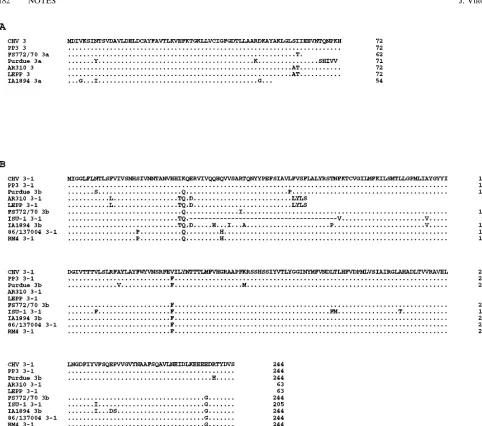
[image:3.612.61.294.70.183.2]Analysis of the ORF 3/3a and ORF 3-1/3b genes.The PCR products (primers 538 and 622) of the ORF 3 and ORF 3-1 genes of the PRCV isolates AR310 and LEPP and of CHV TGEV were the expected size, 1.2 kb. The PCR product from the PRCV isolate IA1894 migrated slightly faster, indicating that a small deletion of approximately 20 nucleotides was present. However, the PCR product from the PRCV isolate ISU-1 migrated much faster, indicating that approximately 290 nucleotides were deleted (data not shown). The alignment of the nucleotide sequence from the ORF 3/3a and ORF 3-1/3b genes of CHV TGEV and the PRCV isolates AR310, LEPP, IA1894, and ISU-1 is shown in Fig. 4. The nucleotide se-quences of the TGEV isolates PP3 (Miller) (32), FS772/70 (3), and Purdue (15) and of the PRCV isolates 86/137004 (18, 19) and RM4 (22) are included in Fig. 4 for comparison. Also, the predicted amino acid residues from the ORF 3/3a and ORF 3-1/3b genes are shown in Fig. 5A and B, respectively. Se-quencing of the PCR product from the ORF 3 and ORF 3-1 genes revealed that the PRCV isolates AR310, LEPP, IA1894, and ISU-1 all had a three-nucleotide deletion present in the noncoding region preceding the ORF 3/3a gene at position 42 identical to that found in another United States PRCV isolate, Ind/89 (34). The ORF 3 gene from the isolates PRCV AR310, PRCV LEPP, and CHV TGEV was preceded by a consensus CTAAAC leader RNA-binding site, and the predicted protein encoded from the ORF 3 gene of the PRCV isolates AR310 and LEPP was found to be 72 amino acids in length and differed from the ORF 3 protein of CHV TGEV by only 2 FIG. 2. Schematic diagram of the regions of TGEV and PRCV isolates
amplified by PCR for nucleotide sequence analysis in this study. The gray shaded area in the S gene represents the area that is deleted in PRCV isolates. (a) The 59half of the S gene of TGEV and PRCV isolates was amplified with the primers 021209 and 060704 under the conditions described in the text. (b) The ORF 3/3a and ORF 3-1/3b regions of TGEV and PRCV isolates were amplified with the primers 538 and 622 under the conditions described in the text.
on November 9, 2019 by guest
http://jvi.asm.org/
amino acids (Fig. 5A). Thus, the PRCV isolates AR310 and LEPP are the first PRCV isolates to have an intact gene 3.
The ORF 3-1 gene for PRCV AR310, PRCV LEPP, and CHV TGEV also was preceded by a consensus CTAAAC leader RNA-binding site. Analysis of the ORF 3-1 gene of PRCV AR310 and PRCV LEPP revealed that a one-nucle-otide deletion was present at position 581. This one-nucleone-nucle-otide deletion causes a frameshift resulting in early termination of the ORF 3-1 protein. The predicted ORF 3-1 protein for PRCV AR310 and LEPP is 63 amino acids in length, which is 181 amino acid residues shorter than the predicted ORF 3-1 protein for CHV TGEV and the other PRCV isolates (Fig. 5B).
Analysis of PRCV IA1894 revealed that there was a consen-sus CTAAAC leader RNA-binding site preceding the ORF 3a gene. Of note, the PRCV IA1894 ORF 3a gene contained a 23-nucleotide deletion at position 287 that would yield an early stop codon that would give rise to a truncated ORF 3a protein (Fig. 4). The truncated ORF 3a protein of PRCV IA1894 would consist of 54 amino acids compared with the 72 amino acids present for PRCV AR310, PRCV LEPP, and CHV TGEV ORF 3a protein (Fig. 5A). The consensus CTAAAC leader RNA-binding site preceding the ORF 3b gene of PRCV IA1894 was altered because of a five-nucleotide deletion that removed the last two nucleotides of the consensus sequence, thus making RNA 3-1 undetectable in PRCV IA1894-infected cells by Northern blot. However, the ORF 3b gene of PRCV IA1894 had an ATG start codon present, and the predicted ORF 3b protein was very similar to that of other TGEV and
PRCV isolates (Fig. 5B). Thus, the PRCV isolate IA1894 syn-thesizes an mRNA 3 similar to that of the Purdue, FS772/70, and D52 strains of TGEV in that the mRNA 3 is thought to code for both the ORF 3a and the ORF 3b proteins (2, 3, 15). The PRCV isolate ISU-1 was found to have a 168-nucleotide deletion present at position 53 that removed both the consen-sus CTAAAC leader RNA-binding site and the ATG start codon of the ORF 3 gene, thus rendering the ORF 3 gene a pseudogene. Further downstream at position 118, a five-nucle-otide deletion in the ORF 3 pseudogene of PRCV ISU-1 that is in the identical location of the five-nucleotide deletion in Ind/89 was found (32). Also, the 5-nucleotide deletion in the ORF 3 pseudogene of PRCV ISU-1 and Ind/89 starts at the same location as the 23-nucleotide deletion found in the ORF 3a gene of PRCV IA1894. The CTAAAC leader RNA-binding site preceding the ORF 3-1 gene of PRCV ISU-1 was present; however, the ORF 3-1 gene of PRCV ISU-1 had a 117-nucle-otide in-frame deletion beginning at position 329 that would be predicted to remove 39 amino acids from the 244 amino acids found in the intact ORF 3-1 protein of TGEV.
Studies in this report show that the PRCV isolates AR310, LEPP, IA1894, and ISU-1 are different from previously genet-ically characterized PRCV isolates. Until now, PRCV isolates have displayed two characteristics thought to typify PRCV, namely, a large in-frame deletion in the 59end of the S gene of 672 or 681 nucleotides in length and a nonfunctional ORF 3 gene (4, 13, 17, 19, 22, 34). The latter is due to either altered or deleted CTAAAC leader RNA-binding sites preceding the ORF 3 gene that result in the ORF 3 gene being nonfunctional FIG. 3. (A) Alignment of the nucleotide sequence from the 59end of the S gene of CHV TGEV and the PRCV isolates AR310, IA1894, ISU-1, Ind/89 (34), 86/137004 (4), and RM4 (22). The PRCV isolates AR310 and LEPP had identical S gene deletions of 621 nucleotides present. The PRCV isolate IA1894 had an S gene deletion of 678 nucleotides present. The PRCV isolates ISU-1 and Ind/89 both had identical deletions of 681 nucleotides present. The PRCV isolates 86/137004 and RM4 both had identical deletions of 672 nucleotides present. The start codon of the S gene is marked with113. The nucleotides composing the predicted signal peptide region of the TGEV S gene as determined by Rasschaert and Laude (23) are underlined. The nucleotide sequences of the TGEV isolate CHV and of the PRCV isolates AR310, LEPP, IA1894, and ISU-1 were determined in this study. (B) Alignment of the predicted amino acid residues from the 59end of the S gene of the TGEV isolate CHV and the PRCV isolates AR310, LEPP, IA1894, ISU-1, Ind/89 (34), 86/137004 (4), and RM4 (22). The boxed amino acid residues indicate the signal peptide region at the 59end of the S gene as determined by Rasschaert and Laude (23). Deleted amino acid residues are marked with a dash, and identical amino acid residues are marked with a dot. The PRCV isolates AR310 and LEPP had 207 amino acid residues deleted. The predicted amino acid residues were derived from the nucleotide
sequences of the TGEV isolate CHV and of the PRCV isolates AR310, LEPP, IA1894, and ISU-1 determined in this study.
on November 9, 2019 by guest
http://jvi.asm.org/
[image:4.612.67.539.71.327.2]on November 9, 2019 by guest
http://jvi.asm.org/
(19, 22, 30, 34). However, the ORF 3-1 gene of previously described PRCV isolates has been nearly identical to the ORF 3-1 of TGEV.
The PRCV isolates AR310 and LEPP differ from other PRCV isolates in four ways: (i) both have S gene deletions of
621 nucleotides, the smallest found thus far, (ii) both produce mRNAs 3 and 3-1, (iii) both have an intact ORF 3 gene, and (iv) both are predicted to yield a truncated ORF 3-1 protein due to a 1-nucleotide deletion in the ORF 3-1 gene. These characteristics are unique among PRCV isolates genetically FIG. 4. Comparison of the nucleotide sequences of the ORF 3/3a and ORF 3-1/3b regions of TGEV and PRCV isolates. Positions of the leader RNA-binding site upstream of genes are marked (}}}}}). The start codons are designated with open boxes and113. The stop codons are designated with shaded boxes. Positions having identical nucleotides are marked with dots, and positions of deleted nucleotides are marked with dashes. The nucleotide sequences of the TGEV isolate CHV and of the PRCV isolates AR310, LEPP, IA1894, and ISU-1 were determined in this study. The other nucleotide sequences presented, Miller PP3 TGEV (PP3) (32), FS772/70 TGEV (3), Purdue TGEV (15), 86/137004 PRCV (19), and RM4 PRCV (22), were previously reported.
on November 9, 2019 by guest
http://jvi.asm.org/
characterized thus far. The size of the S gene deletions for other PRCV isolates from Europe is 672 nucleotides (1, 17), and the size of the S gene deletion of other PRCV isolates from the United States is 681 nucleotides (8, 24). Even though the deletions in the 59end of the S gene of PRCV AR310 and PRCV LEPP are smaller than those of other PRCV isolates, the area deleted encompasses the S protein amino acid resi-dues of 92, 94, 218, and 219 that were predicted by Sanchez et al. (26) to be important in determining tissue tropism of PRCV and TGEV isolates.
Both the ORF 3 and the ORF 3-1 genes of the PRCV isolates AR310 and LEPP are preceded by a CTAAAC leader RNA-binding site that initiates synthesis of two separate sub-genomic mRNAs, mRNA 3 and mRNA 3-1. Also of
signifi-cance is that the PRCV isolates AR310 and LEPP are the first to have been found to have an intact and functional ORF 3 gene. The information from all other PRCV isolates previously sequenced has shown that there are deletions or alterations present that render the ORF 3 gene nonfunctional. Surpris-ingly, the ORF 3-1 gene-coding region of the PRCV isolates AR310 and LEPP contained a one-nucleotide deletion and is predicted to cause a frameshift in the ORF 3-1 gene resulting in a truncated ORF 3-1 protein of 63 amino acids. The ORF 3-1 gene of all other previously described PRCV isolates shows that gene 3-1 is preceded by a CTAAAC leader RNA-binding site and that the following ORF 3-1/3b was intact. Whether the truncated ORF 3-1 protein present in PRCV AR310 and LEPP would still be functional is not known, but it may be FIG. 5. (A) Alignment of the predicted amino acid residues from ORF 3/3a genes of TGEV and PRCV isolates. Identical amino acid residues are marked with dots. The TGEV isolates CHV and PP3 have an identical amino acid homology, and both are 72 amino acid residues long. The TGEV isolate FS772/70 ORF 3a protein is predicted to be 62 amino acid residues long. The Purdue TGEV isolate has an ORF 3a protein 71 amino acid residues in length, and the last 6 amino acid residues are changed from that of CHV TGEV. The PRCV isolates AR310 and LEPP have identical ORF 3 proteins that are 72 amino acid residues in length and differ from that of CHV TGEV by 2 amino acid residues. The PRCV isolate IA1894 has a truncated ORF 3a protein of 54 amino acid residues in length due to a 23-nucleotide deletion in the coding sequence of the ORF 3a gene. (B) Alignment of the predicted amino acid residues from the ORF 3-1/3b genes of TGEV and PRCV isolates. Identical amino acid residues are marked with dots. The TGEV isolates CHV, PP3 (32), Purdue (15), and FS772/70 (3) and the PRCV isolates IA1894, 86/137004 (19), and RM4 (22) all have nearly identical ORF 3-1/3b proteins of 244 amino acid residues in length. The PRCV isolates AR310 and LEPP both have truncated ORF 3-1 proteins of 63 amino acid residues in length due to a one-nucleotide deletion that results in a frameshift in the coding region of the ORF 3-1 gene. The PRCV isolate ISU-1 has a smaller ORF 3-1 protein of 205 amino acid residues due to a 117-nucleotide in-frame deletion in the coding region of the ORF 3-1 gene.
on November 9, 2019 by guest
http://jvi.asm.org/
[image:7.612.64.546.50.476.2]possible that this truncated ORF 3-1 protein still has the nec-essary domains to be functional. The presence of small dele-tions in putative nonstructural coronaviral proteins is not un-common, for example, porcine epidemic diarrhea virus ORF 3 (7), mouse hepatitis virus ORF 4 (31) and ORF 5 (36), bovine coronavirus 4.9- and 4.8-kDa ORFs (1), and human coronavi-rus 229E ORF 4 (14).
The PRCV isolate IA1894 is predicted to have the interest-ing feature of a truncated ORF 3a protein and intact ORF 3b protein made from a single mRNA 3. This would mean, with the exception of the 23-nucleotide deletion present in the ORF 3a gene, that the PRCV IA1894 mRNA 3 is like that of the Purdue, FS772/70, and D52 strains of TGEV in which the mRNA 3 is thought to code for both the ORF 3a and the ORF 3b proteins (2, 3, 15). Other than this unique feature, the PRCV isolate IA1894 is more in keeping with the previously described PRCV isolates in that its S gene deletion of 678 nucleotides is very similar in size to those of other PRCV isolates and that a 23-nucleotide deletion in the ORF 3a gene would result in a truncated ORF 3a protein.
The PRCV isolate ISU-1 also fits into the category of pre-viously described PRCV isolates, since PRCV ISU-1 had an S gene 681-nucleotide deletion and its ORF 3 gene altered to a pseudogene because of a 168-nucleotide deletion in ORF 3 gene that removed both the CTAAAC leader RNA-binding site and the ATG start site. However, the PRCV isolate ISU-1 had the unique feature of its ORF 3-1 gene having a 117-nucleotide in-frame deletion at amino acid residue 32 that would effectively remove 39 of a possible 244 amino acid res-idues of the ORF 3-1 protein. Whether this 117-nucleotide in-frame deletion in the ORF 3-1 gene of PRCV ISU-1 would still yield a functional protein is not known.
As of yet, no definitive function has been assigned to the ORF 3 and ORF 3-1 genes; however, these genes have been hypothesized to be important virulence determinants in TGEV. This has been based on genetic analysis of TGEV mutants that have regions of the ORF 3 and 3-1 genes deleted, exhibit a small-plaque phenotype in cell culture, and present a marked decrease in virulence in baby pigs (2, 33). Preliminary studies in our laboratory have shown that the PRCV isolates AR310 and LEPP are more virulent in a 5-week-old specific-pathogen-free pig model than the PRCV isolate IA1894 (11, 28). On the basis of our preliminary studies on the pathoge-nicity of PRCV isolates in pigs, combined with the molecular studies reported here, it is possible that the ORF 3 and ORF 3-1 genes are involved in the determination of PRCV viru-lence, similar to that reported for TGEV (33). A schematic diagram of the comparison of gene deletions and tissue tro-pism of TGEV and PRCV isolates is shown in Fig. 6.
From this study, we can confirm, as have others, that the ORF 3/3a gene of both TGEV and PRCV isolates is highly variable. In addition, the ORF 3-1/3b gene of at least the PRCV isolates appears to be variable also. The extent of the deletions present in the PRCV isolates AR310, LEPP, IA1894, and ISU-1 allows for some intriguing speculation as to the relationship of the S, ORF 3/3a, and ORF 3-1/3b genes in PRCV isolates. All previously analyzed PRCV isolates have been found to have a large deletion in the 59end of the S gene accompanied by an altered or deleted ORF 3/3a gene, while the ORF 3-1/3b gene has remained intact. In this study we have reported on additional PRCV isolates that do have S gene deletions as large as reported previously. However, the region of the ORF 3/3a and 3-1/3b genes of these new PRCV isolates was found to be more variable than previously reported.
It is believed that PRCV evolved from TGEV (17, 21, 34). PRCV AR310 was isolated originally from an intestinal tissue
homogenate from a piglet with TGE, and TGEV was isolated from other piglets in the same swine herd (29, 30). In contrast, PRCV LEPP was isolated from a nasal swab collected from a pig with pneumonia (29). Considering the differences in their origin, it is interesting to note the nucleotide sequence and biological similarities between the PRCV isolates AR310 and LEPP. Unlike other previously described PRCV isolates, both PRCV AR310 and LEPP retain a characteristic of TGEV in that they have an intact ORF 3 gene. Therefore, these isolates may represent intermediate genotypes in the evolution of PRCV from TGEV. Examination of early-passage virus from the original PRCV AR310 intestinal homogenates may indi-cate whether a mixture of TGEV-like or PRCV-like variants can exist in vivo, and such knowledge will further our under-standing on the origin of PRCV.
This study was in part supported by grants from the Iowa Livestock Health Advisory Council and Oxford Laboratories, Inc., a subsidiary of The Upjohn Co.
We thank Susan Carpenter for helpful discussions and Kelly Hicks, Steve Riley, and Marsha Morgan for excellent technical assistance.
REFERENCES
1. Abraham, S., T. E. Kienzle, W. E. Lapps, and D. A. Brian. 1990. Sequence and expression analysis of potential nonstructural proteins of 4.9, 4.8, 12.7, and 9.5 kDa encoded between the spike and membrane protein genes of the bovine coronavirus. Virology 177:488–495.
2. Britton, P., S. Kottier, C.-M. Chen, D. H. Pocock, H. Salmon, and J. M. Aynaud.1994. The use of PCR genome mapping for the characterisation of TGEV strains, p. 29–43. In H. Laude and J.-F. Vautherot (ed.), Coronavi-ruses: molecular biology and virus-host interactions. Plenum Press, New York. 3. Britton, P., C. Lopez Otin, J. M. Martin Alonso, and F. Parra. 1989. Se-quence of the coding regions from the 3.0 kb and 3.9 kb mRNA subgenomic species from a virulent isolate of transmissible gastroenteritis virus. Arch. Virol. 105:165–178.
4. Britton, P., K. L. Mawditt, and K. W. Page. 1991. The cloning and sequenc-ing of the virion protein genes from a British isolate of porcine respiratory coronavirus: comparison with transmissible gastroenteritis virus genes. Virus Res. 21:181–198.
[image:8.612.318.551.71.254.2]5. Britton, P., K. W. Page, D. J. Pulford, D. J. Garwes, K. Mawditt, F. Stewart, FIG. 6. Schematic diagram showing the comparison of gene deletions and tissue tropism of TGEV and PRCV isolates. Heavy black lines indicate that no deletions are present. Thin black lines indicate that deletions are present. De-letions are indicated as nucleotides (nt) deleted (D). The nucleotide sequences of the TGEV isolate CHV and of the PRCV isolates AR310, LEPP, IA1894, and ISU-1 were determined in this study. The nucleotide sequences of the other PRCV isolates, Ind/89 (34), 86/137004 (19), and RM4 (22), were previously determined. Unknown for the small-plaque TGEV isolate indicates that the small-plaque TGEV does not replicate in the villus enterocytes but rather rep-licates in cells found in the lamina propria and that the extent of small-plaque TGEV replication in respiratory tissue is not known (33).
on November 9, 2019 by guest
http://jvi.asm.org/
F. Parra, C. L. Otin, J. M. Alomso, and R. S. Carmenes.1990. Genomic organization of a virulent isolate of porcine transmissible gastroenteritis virus, p. 357–364. In D. Cavanagh and T. D. K. Brown (ed.), Coronaviruses and their diseases. Plenum Press, New York.
6. Cavanagh, D., D. A. Brian, L. Enjuanes, K. V. Holmes, M. M. C. Lai, H. Laude, S. G. Siddell, W. Spaan, F. Taguchi, and P. J. Talbot.1990. Recom-mendations of the coronavirus study group for the nomenclature of the structural proteins, mRNAs, and genes of coronaviruses. Virology 176:306– 307.
7. Duarte, M., K. Tobler, A. Bridgen, D. Rasschaert, M. Ackermann, and H. Laude.1994. Sequence analysis of the porcine epidemic diarrhea virus ge-nome between the nucleocapsid and spike protein genes reveals a polymor-phic ORF. Virology 198:466–476.
8. Garwes, D. J., F. Stewart, and P. Britton. 1989. The polypeptide of Mr 14,000 of porcine transmissible gastroenteritis virus: gene assignment and intracel-lular location. J. Gen. Virol. 70:2495–2499.
9. Godet, M., R. L’Haridon, J. F. Vautherot, and H. Laude. 1992. TGEV coronavirus ORF4 encodes a membrane protein that is incorporated into virions. Virology 188:666–675.
10. Halbur, P. G., P. S. Paul, E. M. Vaughn, and J. J. Andrews. 1993. Experi-mental reproduction of pneumonia in gnotobiotic pigs with porcine respira-tory coronavirus isolate AR310. J. Vet. Diagn. Invest. 5:184–188. 11. Halbur, P. G., E. M. Vaughn, and P. S. Paul. 1994. Unpublished data. 12. Hill, H., J. Biwer, R. Wood, and R. Wesley. 1990. Porcine respiratory
coro-navirus isolated from two U.S. swine herds, p. 333–335. In T. A. Neuzil (ed.), Proceedings of the American Association of Swine Practitioners. American Association of Swine Practitioners, Des Moines, Iowa.
13. Jackwood, D. J., I. Bae, R. J. Jackwood, and L. J. Saif. 1994. Transmissible gastroenteritis virus and porcine respiratory coronavirus: molecular charac-terization of the S gene using cDNA probes and nucleotide sequence anal-ysis, p. 43–48. In H. Laude and J.-F. Vautherot (ed.), Coronaviruses: mo-lecular biology and virus-host interactions. Plenum Press, New York. 14. Jouvenne, P., S. Mounir, J. N. Stewart, C. D. Richardson, and P. J. Talbot.
1992. Sequence analysis of human coronavirus 229E mRNAs 4 and 5: evi-dence for polymorphism and homology with myelin basic protein. Virus Res. 22:125–141.
15. Kapke, P. A., F. Y. T. Tung, and D. A. Brian. 1988. Nucleotide sequence between the peplomer and matrix protein genes of the porcine transmissible gastroenteritis coronavirus identifies three large open reading frames. Virus Genes 2:293–294.
16. La Bonnardiere, C., and H. Laude. 1983. Interferon induction in rotavirus and coronavirus infections: a review of recent results. Ann. Rech. Vet. 14:507–511.
17. Laude, H., K. Van Reeth, and M. Pensaert. 1993. Porcine respiratory coro-navirus: molecular features and virus-host interactions. Vet. Res. 24:125–150. 18. Page, K. W., P. Britton, and M. E. G. Boursnell. 1990. Sequence analysis of
the leader RNA of two porcine coronaviruses: transmissible gastroenteritis virus and porcine respiratory coronavirus. Virus Genes 4:289–301. 19. Page, K. W., K. L. Mawditt, and P. Britton. 1991. Sequence comparison of
the 59end of mRNA 3 from transmissible gastroenteritis virus and porcine
respiratory coronavirus. J. Gen. Virol. 72:579–587.
20. Parker, S. E., T. M. Gallagher, and M. J. Buchmeier. 1989. Sequence anal-ysis reveals extensive polymorphism and evidence of deletions within the E2 glycoprotein gene of several strains of murine hepatitis virus. Virology 173: 664–673.
21. Pensaert, M., P. Callebaut, and J. Vergote. 1986. Isolation of a porcine respiratory, non-enteric coronavirus related to transmissible gastroenteritis. Vet. Q. 8:257–261.
22. Rasschaert, D., M. Duarte, and H. Laude. 1990. Porcine respiratory coro-navirus differs from transmissible gastroenteritis virus by a few genomic deletions. J. Gen. Virol. 71:2599–2607.
23. Rasschaert, D., and H. Laude. 1987. The predicted primary structure of the peplomer protein E2 of the porcine coronavirus transmissible gastroenteritis virus. J. Gen. Virol. 68:1883–1890.
24. Saif, L. J., and E. H. Bohl. 1986. Transmissible gastroenteritis, p. 255–274. In A. D. Leman, R. D. Glock, W. L. Mengeling, R. H. C. Penny, E. Scholl, and B. Straw (ed.), Diseases of swine, 6th ed. Iowa State University Press, Ames. 25. Sambrook, J., E. F. Fritsch, and T. Maniatis. 1989. Molecular cloning: a laboratory manual, 2nd ed. Cold Spring Harbor Laboratory, Cold Spring Harbor, N.Y.
26. Sanchez, C. M., F. Gebauer, C. Sune, A. Mendez, J. Dopazo, and L. En-juanes.1992. Genetic evolution and tropism of transmissible gastroenteritis coronavirus. Virology 190:92–105.
27. Spaan, W., D. Cavanagh, and M. C. Horzinek. 1988. Coronaviruses: struc-ture and genome expression. J. Gen. Virol. 69:2939–2952.
28. Vaughn, E. M., P. G. Halbur, and P. S. Paul. 1994. Unpublished data. 29. Vaughn, E. M., P. G. Halbur, and P. S. Paul. 1994. Three new isolates of
porcine respiratory coronavirus with various pathogenicities and spike (S) gene deletions. J. Clin. Microbiol. 32:1809–1812.
30. Vaughn, E. M., and P. S. Paul. 1993. Antigenic and biological diversity among transmissible gastroenteritis virus isolates of swine. Vet. Microbiol. 36:333–347.
31. Weiss, S. R., P. W. Zoltick, and J. L. Liebowitz. 1993. The ns4 gene of mouse hepatitis (MHV), strain A 59 contains two ORFs and thus differs from ns4 of the JHM and S strains. Arch. Virol. 129:301–309.
32. Wesley, R. D., A. K. Cheung, D. D. Michael, and R. D. Woods. 1989. Nucle-otide sequence of coronavirus TGEV genomic RNA: evidence for 3 mRNA species between the peplomer and matrix protein genes. Virus Res. 13:87– 100.
33. Wesley, R. D., R. D. Woods, and A. K. Cheung. 1990. Genetic basis for the pathogenesis of transmissible gastroenteritis virus. J. Virol. 64:4761–4766. 34. Wesley, R. D., R. D. Woods, and A. K. Cheung. 1991. Genetic analysis of
porcine respiratory coronavirus, an attenuated variant of transmissible gas-troenteritis virus. J. Virol. 65:3369–3373.
35. Wesley, R. D., R. D. Woods, H. T. Hill, and J. D. Biwer. 1990. Evidence for a porcine respiratory coronavirus, antigenically similar to transmissible gas-troenteritis virus, in the United States. J. Vet. Diagn. Invest. 2:312–317. 36. Yokomori, K., and M. M. C. Lai. 1991. Mouse hepatitis virus S RNA
se-quence reveals that nonstructural proteins ns4 and ns5 are not essential for murine coronavirus replication. J. Virol. 65:5605–5608.