0022-538X/95/$04.0010
Copyrightq1995, American Society for Microbiology
Relationship between TATA-Binding Protein and Herpes Simplex
Virus Type 1 ICP4 DNA-Binding Sites in Complex
Formation and Repression of Transcription
RUHUL KUDDUS, BAOHUA GU,ANDNEAL A. DELUCA*
Department of Molecular Genetics and Biochemistry, University of Pittsburgh School of Medicine, Pittsburgh, Pennsylvania 15261
Received 24 March 1995/Accepted 31 May 1995
The herpes simplex virus (HSV) regulatory protein, infected-cell polypeptide 4 (ICP4), represses the tran-scription of promoters that have binding sites for ICP4 located near the trantran-scription start site. It has also been shown that ICP4 binds such promoter DNA cooperatively with the TATA-binding protein (TBP) and TFIIB to form a tripartite protein-DNA complex (C. Smith, P. Bates, R. Rivera-Gonzales, B. Gu, and N. A. DeLuca, J. Virol. 67:4676–4687, 1993). In this study, we analyzed the effects of position and orientation of the ICP4-binding site relative to the TATA box in the ICP4 promoter on transcriptional repression by ICP4 and on the ability of ICP4 to form tripartite complexes with TBP and TFIIB. The results of this parallel study provide a strong correlation between tripartite complex formation and repression. Both tripartite-complex formation and transcriptional repression were efficient when the ICP4-binding site was downstream of the TATA box, within a short distance and in proper orientation. In addition, both tripartite-complex formation and repression were partially sensitive to the stereoaxial positioning of the ICP4-binding site relative to the TATA box. As a preliminary characterization of the tripartite complex, circular permutation analysis was performed to assess the distortion of the proximal promoter region in the tripartite complex. As previously reported, both TBP and ICP4 independently could bend DNA and the relative magnitude by which each of these proteins bent DNA in the tripartite complex was preserved. The results of this study suggest that the formation of tripartite complexes on a promoter is part of the mechanism of repression and that simple blocking as a sole result of ICP4 binding is not sufficient for full repression.
Transcription of eukaryotic viral genes by RNA polymerase II is often modulated by virus-encoded transcription factors. Therefore, regulation of the cellular transcription machinery by viral gene products has served as a helpful paradigm for understanding eukaryotic transcription. Viral transcriptional regulators, like their cellular counterparts, can interact with both general transcription factors, such as the TATA-binding protein (TBP), TBP-associated factors, and TFIIB (4, 40, 59, 61), or gene-specific transcription factors, such as RB, p53, and oct-1 (7, 13, 18, 20, 54). Several cellular factors, such as RB (33, 52) and YYI (50, 57), and viral transcription factors, such as simian virus 40 large T antigen (5, 46), are known to both positively and negatively regulate transcription. Herpes sim-plex virus (HSV) encodes a 175-kDa phosphoprotein (6), re-ferred to as infected-cell polypeptide 4 (ICP4), which can pos-itively or negatively regulate RNA polymerase II-dependent transcription of many HSV genes. Genetic and biochemical analyses revealed that ICP4, like many cellular transcriptional regulators (reviewed in reference 45), contains discrete do-mains responsible for DNA binding, nuclear localization, and transcriptional regulation (9, 49, 56) and that the domain re-quirements for activation are greater than those for repression (56).
ICP4 interacts with both DNA and transcription factors in the transcription initiation complex to exert its regulatory ef-fects. ICP4 is a sequence-specific DNA-binding protein (15, 37), which binds a fairly degenerate consensus sequence (27,
44). The most recent consensus derived from a statistical anal-ysis of different potential binding sites is RTCGTCNNYNY SG, where R is purine, Y is pyrimidine, S is C or G, and N is any base (11). In vitro and in vivo studies collectively suggest that the binding of ICP4 to specifically located sites is not required for activation and that nonspecific DNA contacts may suffice (21, 58). However, specifically located ICP4-binding sites are required for repression (22, 43, 53). The HSV genes that ICP4 negatively regulates include those for the ICP4 gene itself (8, 48), the promoter for the latency-associated transcript (1), and the promoter for L/ST (3, 64). All these promoters contain strong ICP4-binding sites located near the transcrip-tion initiatranscrip-tion site (2, 3, 16, 37, 47, 64). Mutatranscrip-tion of this binding site also causes alteration in the temporal expression of the promoter, shifting its kinetics of expression to an earlier time postinfection and altering its sensitivity to inhibitors of DNA synthesis (35, 51). Biochemical studies with purified ICP4, TBP, and TFIIB showed that these proteins cooperatively in-teract on the ICP4 promoter containing a TATA box and a functional ICP4-binding site (59). It was hypothesized that the formation of the TBP-TFIIB-ICP4-DNA complex is involved in the mechanisms of ICP4 function, including repression.
The requirement for an ICP4-binding site near the transcrip-tion start site for repression and the observatranscrip-tion that ICP4 forms tripartite complexes with TBP and TFIIB on a repressed promoter led us to investigate whether ICP4 and the basal initiation complex require specific alignment for the observed regulation from such promoters and whether the extent of tripartite complex formation correlates with repression. To address this hypothesis, we altered the stereoaxial positioning, spacing, and orientation of the ICP4-binding site relative to the TATA box in the ICP4 promoter. The promoter constructs
* Corresponding author. Mailing address: E1257 Biomedical Sci-ence Tower, Department of Molecular Genetics and Biochemistry, University of Pittsburgh School of Medicine, Pittsburgh, PA 15261. Phone: (412) 648-9947. Fax: (421) 624-1401.
5568
on November 9, 2019 by guest
http://jvi.asm.org/
were used as probes for DNA-binding studies with purified ICP4, TBP, and TFIIB and as templates for in vitro transcrip-tion studies. The results demonstrate that each of the three parameters spacing, phasing, and orientation affect both tri-partite-complex formation and repression of transcription in vitro, to different degrees. Moreover, the degree of cooperative DNA binding by ICP4, TBP, and TFIIB correlated with the extent of repression, supporting a role for both DNA-protein and protein-protein interactions in ICP4-mediated repression.
MATERIALS AND METHODS
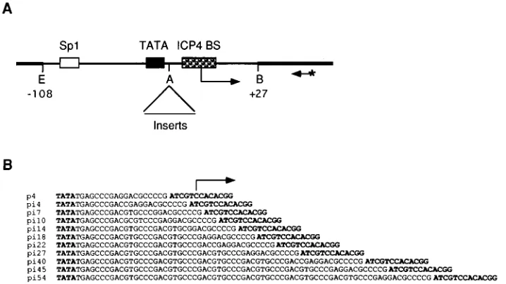
DNA templates.The plasmid p4, which contains a fragment of the ICP4 promoter from2108 to127 relative to the transcription start site was used to construct all the ICP4 promoter templates. The ICP4 promoter fragment in p4 contains a strong ICP4-binding site centered at12, a TATA box centered at
225, and an Sp1-binding site centered at280. This fragment is sufficient for Sp1-responsive transcription of ICP4 promoter from the correct initiation site (22, 29). Insertion mutants of the ICP4 promoter were generated by digestion of p4 by AvaI (Fig. 1A) followed by insertion of synthetic DNA fragments of various lengths. Oligonucleotides 59CCGACGTGC39and 59TCGGGCACG39
were phosphorylated with T4 polynucleotide kinase. Upon hybridization, the double-stranded oligonucleotide provides AvaI-compatible ends and can mul-timerize. After ligation of the double-stranded oligonucleotides with DNA con-taining AvaI-compatible ends, the AvaI site in the left end (of the top strand) is not preserved. Excess molar concentration of the double-stranded oligonucleo-tides were ligated with AvaI-digested p4 to generate pi18, pi27, pi36, pi45, and pi54, which contain two, three, four, five, and six copies of the double-stranded oligonucleotides, respectively. Plasmids pi7 and pi14 were fortuitous recombina-tion products, generated presumably from plasmids with longer inserts. Plasmids pi4, pi22, and pi40 are generated by digesting p4, pi18, and pi36, respectively, with AvaI, filling in the ends with Klenow enzyme in the presence of deoxyribo-nucleotide triphosphates, and then self-ligating with T4 DNA ligase. Plasmid pi10 was generated by inserting a single copy of double-stranded oligonucleotide generated upon hybridization of 59CCGACGCGTC39and 59TCGGGACGCG39
in the AvaI site of p4. The sequences of the promoter insertion mutants are shown in Fig. 1B. Plasmid p4R is identical to p4 except that the ICP4-binding site of ICP4 promoter (59ATCGTCCACAGG39) was reversed by replacing DNA sequence from the AvaI site to the BamHI site by the hybridization product of two 45-mer oligonucleotides.
Plasmid pBEND4 was constructed for use in the circular permutation analysis by inserting an 81-bp NaeI-SalI fragment from plasmid p4 into the XbaI and SalI sites of plasmid pBEND2 (31). The NaeI-SalI fragment of p4 contains the TATA box and ICP4-binding site of the ICP4 promoter. The XbaI end of pBEND2 was filled in with Klenow enzyme prior to SalI digestion. All the plasmid constructs
were sequenced to confirm the identity of the constructs by the dideoxy-chain termination method.
Proteins.HeLa cell nuclear extract was prepared by the method of Dignam (12). Recombinant human TBP (rTBP) was purified from extract prepared from Escherichia coli BL21 carrying plasmid pETHIID (30) as described previously (26). A fast protein liquid chromatography (FPLC) Mono S fraction of TBP was used throughout this study. Recombinant human TFIIB (rTFIIB) was purified from extract derived from E. coli carrying plasmid pHIIB (23) as modified by Smith et al. (59). An FPLC Superose 12 column fraction was used for this study. ICP4 was purified from HSV-1 (strain KOS)-infected Vero cell nuclear extract as previously described (27, 55).
In vitro transcription and primer extension.Supercoiled DNA template (26.6
mg/ml) was incubated for 80 min at 308C with nuclear extract (50mg of protein) in a buffer containing 40 mM N-2-hydroxyethylpiperazine-N9-2-ethanesulfonic acid (HEPES)–KOH (pH 7.9), 60 mM KCl, 12% glycerol, 8.3 mM MgCl2, 0.6 mM each ATP, CTP, GTP, and UTP, 0.3 mM dithiothreitol, and 12 U of RNasin (Promega). Where indicated, 200 ng of ICP4 was added to the transcription mixture. The order of addition was (i) nuclear extract, (ii) ICP4, and (iii) DNA template suspended in reaction buffer. The reaction was stopped by the addition of stop buffer (0.25 M sodium acetate [pH 5.2], 15 mM EDTA), and the tran-scribed RNA was purified by two phenol extractions followed by ethanol pre-cipitation. The purified transcripts were subjected to primer extension as previ-ously described (25). The240 sequencing primer for the pUC plasmids (New England Biolabs, Beverly, Mass.) was used for primer extension. Primers were labeled by phosphorylation with T4 polynucleotide kinase in the presence of [g-32
P]ATP. Primer extension products were resolved on 5% polyacrylamide–7.0 M urea sequencing gels. Following electrophoresis, the gels were fixed in 10% methanol–10% acetic acid solution and then dried. The dried gels were used for autoradiography and were also scanned for quantitative analyses with a radio-analytic imaging system (AMBIS Inc., San Diego, Calif.).
Electrophoretic mobility shift assays.The EcoRI-BamHI fragments of p4, p4R, and pi4 through pi54 were used as probes for gel shift analysis. For pBEND4, the plasmid was digested with MluI, NheI, SpeI, DraI, SmaI, or SspI and the fragments containing the direct repeat sequences of pBEND2 and the ICP4 promoter insert were used as the probes. In all cases, the DNA fragments were dephosphorylated with calf intestine alkaline phosphatase and then phos-phorylated with T4 polynucleotide kinase in the presence of [g-32P]ATP. The labeled DNA fragments were isolated from 5% polyacrylamide gels and were further purified with Elutip-D minicolumns (Schleicher & Schuell, Keene, N.H.) and Sephadex G-50 spin columns.
[image:2.612.128.487.71.273.2]DNA-binding reactions and the subsequent gel shift analysis were performed as previously described (59), except that the KCl concentration of the binding buffer was 80 mM and the DNA-protein complexes were resolved with 3.5 to 4.0% native polyacrylamide gels containing 0.53TBE buffer (45 mM Tris base, 45 mM borate, 1 mM EDTA [pH 8.0]). The amounts of proteins used in the binding reaction were 50 ng of TBP, 100 ng of rTFIIB, and 50 ng of ICP4. The FIG. 1. Wild-type and mutant ICP4 promoter constructs. (A) Schematic diagram of plasmid p4. The thicker and thinner lines represent plasmid and viral DNA, respectively. The Sp1-binding site, TATA box, and ICP4-binding site are indicated by boxes. The transcription start site and the direction of transcription are shown by the arrow. The site of oligonucleotide insertion was the AvaI site. The location of the240 primer used for primer extension is also shown. The primer was labeled with32P at the 59end is indicated by an asterisk. The numbers (2108 and127) are the nucleotide positions relative to the transcription start site. Abbreviations of the relevant restriction sites: A, AvaI; B, BamHI; E, EcoRI. (B) DNA sequences of the different promoter constructs from the TATA box to the ICP4-binding site. The TATA box and the ICP4-binding site are shown in boldface letters. The transcription start site is indicated by an arrow.
on November 9, 2019 by guest
http://jvi.asm.org/
gels were dried and exposed to Kodak X-Omat AR films and also scanned for quantitative analyses with the AMBIS radioanalytic imaging system.
Circular permutation analysis.The group of DNA probes generated from plasmid pBEND4 were incubated with ICP4, or TBP and TFIIB, or all three proteins, and the complexes were electrophoretically resolved, as described above. The mobilities of the bound and free probes were measured from the autoradiograms of the dried gels. The relative mobilities of the complexes (mo-bility of bound/free probes) were calculated and were plotted against the frac-tional distance. Fracfrac-tional distance is the length in base pairs from the center of DNA-binding site to the 59end of the top strand of the probe, divided by the length of the probe. The resulting data points were fitted by linear least-squares analysis to the equation of a parabola, y5ax21bx1c, and the bend angle was determined for the TBP–TFIIB-DNA and ICP4-DNA complexes from the first-and second-degree parameters [a52c(11cosu) and2b52c(11cosu)] of the equation, as previously described (17, 38). The angle of deflection of DNA from linearity (a) was calculated from the relationshipa 51808 2 u.
RESULTS
The focus of this study was to explore the relationship be-tween the cooperative association of ICP4, TBP, and TFIIB on promoters containing both an ICP4-binding site and a TATA box and repression by ICP4. The formation of this complex was previously observed and was hypothesized to be involved in ICP4 regulatory functions, including repression (59). As a test of this hypothesis, we varied the position and orientation of the two binding sites relative to each other and conducted a quan-titative comparison of the ability of ICP4 to participate in tripartite complexes and its ability to repress transcription.
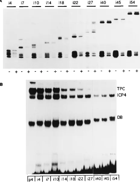
Effect of relative TATA box and ICP4-binding-site position on tripartite complex formation and repression.Plasmids pi4, pi7, pi10, pi14, pi18, pi22, pi27, pi40, pi45, and pi54, which contain the ICP4 promoter sequence from2108 to127 rela-tive to the transcription start site (Fig. 1A) and 4 to 54 bp of DNA inserted between the TATA box and the ICP4-binding site (Fig. 1B), were used as templates for in vitro transcription in the presence and absence of ICP4. The inserted sequences (Fig. 1B) do not have any significant match to an ICP4-binding site according to the predictive model proposed by DiDonato et al. (11). Plasmid p4 was included in each transcription re-action mixture in equimolar amounts to the test template to serve as an internal control for minor sample losses incurred in the isolation of the transcribed RNA and the extended primer and for slight variations in template concentration. The tran-scripts were visualized as primer extension products resolved in sequencing gels. The primer extension products generated from each set of reactions are shown in Fig. 2A. Transcription was initiated in vitro from the wild-type ICP4 promoter from the authentic initiation site. The ICP4-binding site in the ICP4 promoter overlaps with the transcription start site. Therefore, insertion of linker DNA increased the distance between the TATA box and the ICP4-binding site, moving the original start site further downstream. As expected, transcription from all the insertion mutant templates started about 25 bp down-stream from the TATA box. Thus, the length of the extension products increased by the length of the inserted sequences. Transcription from the p4 template was higher than from the remaining insertion templates, probably because of the alter-ation in the natural start site. However, repeated experiments demonstrated that transcription from all the insertion tem-plates was nearly the same in the absence of ICP4. In the presence of ICP4, transcription from the different templates was affected differently. Repression by ICP4 was greatest on the natural p4 template. This template was repressed 5- to 10-fold. The absolute level of repression seen for individual insertion templates varied approximately twofold from exper-iment to experexper-iment, as did the absolute level of repression of the p4 internal control. The global twofold variation was seen from experiment to experiment and may reflect slight
[image:3.612.320.552.74.374.2]differ-ences in the relative concentrations of transcription factors and ICP4. Small variation within an experiment was evident from repeat experiments. This was controlled for by normalization to the internal control. Figure 2A shows that ICP4 noticeably repressed transcription from templates pi4 through pi22. How-ever, repression was not seen with templates pi27 through pi54. To observe the effect of the insertion on the ability of ICP4 to form tripartite complexes, labeled DNA probes generated from the ICP4 promoter (plasmid p4) and the insertion mu-tants (plasmids pi4 through pi54) were incubated with ICP4, TBP, and TFIIB and the resulting protein-DNA complexes were resolved by gel shift analysis. Control gel shift experi-ments showed that ICP4 alone or TBP-TFIIB specifically formed stable protein-DNA complexes with each of the DNA probes and that ICP4 bound equally well to all the probes in the absence of ICP4 (data not shown). In the simultaneous presence of all three proteins, three different complexes, namely, the TBP-TFIIB-DNA, the ICP4-DNA complex, and the ICP4-TBP-TFIIB-DNA complex, were formed with the DNA probes (Fig. 2B). The tripartite complex was predomi-nant on the p4 probe but was also readily detectable with templates pi4 through pi22 (Fig. 2B). The formation of this FIG. 2. The effect of phasing and spacing of the TATA box and the ICP4-binding site on repression and tripartite-complex formation. (A) Effect on re-pression. Equimolar amounts of the indicated insertion mutant and the p4 template (as the internal control) were used in in vitro transcription reactions. The bottom bands represent transcription initiated from p4 template. Tran-scripts initiated from the insertion mutants have lower mobility because of the inserted sequences of variable lengths. The presence or absence of ICP4 in the reaction mixtures is indicated by1and2, respectively. (B) Effect on tripartite-complex formation. Labeled DNA probes generated from p4 and pi4 through pi54 were incubated with TBP, TFIIB, and ICP4 for 40 min at 308C. The reaction mixtures were resolved on 3.5% polyacrylamide gels. The protein-DNA bands corresponding to the TBP-TFIIB-DNA complex, the ICP4-DNA complex, and the tripartite complex are indicated by DB, ICP4 and TPC, respectively.
on November 9, 2019 by guest
http://jvi.asm.org/
complex was greatly reduced with the rest of the templates (pi27 through pi54); however, trace amounts of complex could be detected upon prolonged exposure of the film. The appar-ent reduced binding of ICP4 in probes that form tripartite complexes poorly may result from the lack to cooperativity between ICP4 and TBP binding. This would also reduce the amount of ICP4 complex migrating at the position of the ICP4-only complex, since a proportion of the tripartite complex would be expected to dissociate in the process of electrophore-sis. Also, consistent with previous findings (59), DNase I foot-printing of isolated tripartite complexes formed on the p4 and pi7 templates revealed specific protection of both the TATA box and the ICP4-binding site (data not shown).
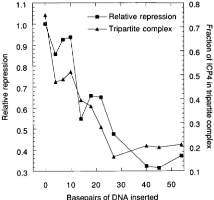
To determine whether the degree of repression from the different promoter constructs was related to the degree of tripartite-complex formation, the data derived from the gel shift and in vitro transcription experiments were quantified and compared graphically. The degree of ICP4-mediated sion from each template was normalized relative to the repres-sion of the wild-type promoter. The data from duplicate ex-periments were averaged and plotted against the number of base pairs of DNA inserted between the TATA box and the ICP4-binding site (Fig. 3). The fraction of total ICP4-bound probe present in the tripartite complex was also determined for each of the templates. Again, the data from duplicate experi-ments were averaged and also plotted against the number of base pairs of DNA inserted between the TATA box and the ICP4-binding site (Fig. 3). It is evident from Fig. 3 that both tripartite-complex formation and ICP4-mediated transcrip-tional repression dropped in a similar fashion as the distance between the TATA box and the ICP4-binding site was in-creased. Second, in templates with TATA boxes and binding sites separated by less than 40 bp of DNA, both
ICP4-mediated repression and, to a lesser extent, tripartite-complex formation were noticeably affected by the stereoaxial position-ing of the two bindposition-ing sites. Thus, tripartite-complex formation and ICP4-mediated repression were reduced more for the tem-plate pi4 than for pi10. This trend was evident but less pro-nounced for templates pi14 through pi27. Since the TATA box and the ICP4-binding site are separated by about 2.5 helical turns of DNA in the ICP4 promoter (10), the two binding sites are separated by about 3, 4, and 5 helical turns in the templates pi4, pi14, and pi27, respectively, and by approximately 3.5 and 4.5 helical turns in the templates pi10 and pi22, respectively. When the TATA box and the ICP4-binding site was separated by longer distances (50 bp or longer), tripartite-complex for-mation and transcription repression by ICP4 were not seen. The data in Fig. 3 demonstrate that both repression and tri-partite-complex formation are concomitantly reduced when the ICP4-binding site and the TATA box are moved further apart, strongly suggesting that ICP4-mediated repression is related to the degree of tripartite-complex formation on the ICP4 promoter. To a lesser degree, the data suggest that a separation of an odd number of half-helical turns of DNA is preferred for both repression and tripartite-complex forma-tion.
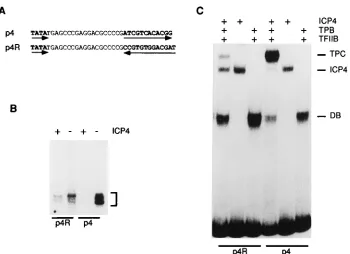
To investigate whether the orientation of the ICP4-binding site in the ICP4 promoter is functionally important in tripar-tite-complex formation and repression, we constructed a mu-tant promoter with the ICP4-binding site reversed in orienta-tion relative to the TATA box (Fig. 4A). Both promoter constructs were used as templates for in vitro transcription. The transcripts generated from each template in the absence and in the presence of ICP4 are shown in Fig. 4B. In the absence of ICP4, transcription from p4R was less than that from p4 (about fourfold), probably because of the alteration of the native start site region. In the presence of ICP4, transcrip-tion of the less active p4R template was reduced approximately two-fold relative to that seen in the absence of ICP4, while transcription of the more active p4 template was barely detect-able and was reduced by greater than 10-fold. Therefore, the magnitude of the repression was at least fivefold greater with the p4 template than with the p4R template.
To investigate the ability of ICP4 to form tripartite com-plexes on these promoter constructs, labeled DNA fragments encompassing the ICP4-binding site, TATA box, and the tran-scription start site from p4 and p4R were incubated with ICP4 and/or TBP and TFIIB and the resulting complexes were re-solved by electrophoresis in native polyacrylamide gels (Fig. 4C). ICP4 formed complexes with both the probes with nearly identical avidity. TBP also recognized the TATA boxes of both promoters and formed stable DNA-protein complexes in the presence of TFIIB. Tripartite-complex formation was clearly prevalent with the probe p4, which contains the ICP4-binding site in its natural orientation. However, tripartite-complex for-mation was significantly reduced with the p4R probe relative to the p4 probe. The fraction of ICP4-bound DNA in the tripar-tite complexes formed on the p4 and p4R probes was 91 and 39%, respectively. Data from two independent experiments indicated that tripartite-complex formation was approximately threefold greater with the ICP4-binding site in the natural orientation. Again, the data provide a correlation between the ability of ICP4 to form tripartite complexes and its ability to repress transcription.
[image:4.612.71.284.72.272.2]Distortion of the promoter in the tripartite complexes.Both ICP4 (14) and TBP (24, 32, 34, 38) induce bends upon binding to DNA. Whether the bends induced by ICP4 and TBP are retained in the tripartite complexes was investigated, since distortion of the proximal promoter may be involved in some FIG. 3. Graphical representation of the phasing and spacing effects on
re-pression and tripartite-complex formation. The data for this representation were derived from two independent experiments for repression and tripartite-complex formation, including that in Fig. 2. For repression, the ratio of the number of counts obtained in the presence of ICP4 to the number obtained in the absence of ICP4 for each insertion template was determined. This ratio was normalized to the ratio obtained for the internal control (p4 template) and is represented as the relative repression for the different promoter constructs. For complex for-mation, the fraction of DNA-bound ICP4 in the tripartite complex was calculated by dividing the number of counts in the area corresponding to the tripartite-complex band by the total number of counts present in the areas corresponding to the tripartite complex and the ICP4-DNA complex.
on November 9, 2019 by guest
http://jvi.asm.org/
aspect of ICP4-mediated repression. DNA probes with circu-larly permutated TATA boxes and ICP4-binding sites (Fig. 5A) were incubated with TBP and TFIIB and/or ICP4 and then resolved in native polyacrylamide gels. As expected, the mo-bility of DB and the ICP4-DNA complexes varied depending on the positions of TATA box and ICP4-binding site, respec-tively (Fig. 5B). The mobility of the tripartite complex also varied depending on the locations of the DNA-binding sites; however, the pattern produced in the gel shifts with the six different probes was not symmetrical (Fig. 5B). This is readily seen by comparing the pattern of the gel shifts produced by ICP4 and the tripartite complex in the same experiment (Fig. 5B, bottom).
Binding-site-dependent mobility variation of protein-DNA complexes is indicative of DNA bending (62). The relative mobility of the complexes versus fractional distance was plot-ted (Fig. 6) and analyzed as described in Materials and Meth-ods. The analysis of the curve in Fig. 6A revealed that the DNA bend induced in the DB complex was approximately 87658. This is consistent with previous observations with TBP alone and indicates that the TBP-induced DNA bend is retained in the DB complex. Similar analysis of the curve in Fig. 6B yielded a bend angle of 62628for the DNA in the ICP4-DNA complexes. Therefore, the obtained values for the human TBP-TFIIB and HSV-1 ICP4 are comparable to those previously reported for yeast and Arabidopsis TBP (32, 34, 38) and HSV-1 ICP4 (14), respectively. The relative mobility of the tripartite complexes plotted against the fractional distance of the ICP4 binding sites, TATA boxes, or points midway between the two sites is shown in Fig. 6C. It is clear that none of these treat-ments of the data resulted in a parabolic relationship, from which a bend angle can be correctly determined. This can be
reconciled by considering that the DNA in tripartite complex is bent by two different proteins, each to a different degree, at two different positions. If both proteins bent DNA to the same degree or if only one of the proteins bent DNA in the tripartite complex, the circular permutation analysis would result in a symmetrical pattern. In addition, the data imply that TBP is still bending DNA to a greater extent in the tripartite complex than in ICP4. The mobility of a protein-DNA complex is more strongly retarded when the bend occurs in the middle of the fragment. When the TATA box is more centrally located rel-ative to the ICP4-binding site, the mobilities of the probes are retarded the most (Fig. 5B, probes M, N, and S). When the ICP4-binding site is more centrally located relative to the TATA box, the mobilities of the probes are retarded the least (Fig. 5B, probes A and P). Therefore, the lack of a parabolic relationship suggests that both ICP4 and TBP are bending DNA in the complex and the asymmetrical nature of the ob-served pattern of probe mobilities suggests that TBP is still bending the DNA in the tripartite complex more than is ICP4.
DISCUSSION
Biochemical and genetic studies have demonstrated that ICP4-binding sites located near the start of transcription are required for repression by ICP4 (22, 43, 53). In the case of the ICP4 promoter, there is an ICP4-binding site approximately 23 bp downstream of the TATA box. It has also been shown that ICP4 will form a multiprotein complex (tripartite complex) on this promoter in the presence of TBP and TFIIB (59). The assembly of this complex is cooperative since its formation is favored over the formation of the TBP-TFIIB-DNA or ICP4-DNA complexes. This report provides a quantitative correla-FIG. 4. Effect of the orientation of the ICP4-binding site on repression and tripartite-complex formation. (A) DNA sequences between the TATA box and the ICP4-binding site of templates p4 and p4R. The TATA box and the ICP4-binding site are in boldface letters. The orientations of the TATA box and the ICP4-binding site are shown by the arrows. (B) The p4 and p4R templates were included in in vitro transcription reaction mixtures in the presence (1) and absence (2) of ICP4. The bands shown in the brackets indicate the primer extension products from transcripts initiated in the presence and in the absence of ICP4. (C) Effect of ICP4-binding-site orientation on tripartite-complex formation. Labeled DNA probes generated from p4 and p4R were incubated with ICP4 and/or TBP and TFIIB as indicated and then resolved in a 3.5% nondenaturing polyacrylamide gel. The protein-DNA bands corresponding to the DB complex, the ICP4-DNA complex, and the tripartite complex are indicated by DB, ICP4, and TPC, respectively.
on November 9, 2019 by guest
http://jvi.asm.org/
[image:5.612.134.484.66.320.2]tion between the ability of ICP4 to form this complex and its ability to repress transcription of its own promoter. These studies also address the requirement for the proper orientation and phasing of the ICP4-binding site relative to the TATA box for both repression and complex formation. The approach taken to measure repression by ICP4 was the use of a recon-stituted in vitro repression assay. We chose this approach over inserting the promoter constructs into the virus because it has previously been shown that repression by ICP4 can alter the kinetics or maximal time of expression during viral infection (35, 51) and because the in vitro system is more amenable to analysis of multiple mutant constructs.
The findings of the functional studies demonstrated the fol-lowing: (i) increasing the spacing between the ICP4-binding site and the TATA box results in a decrease in the ability of ICP4 to repress transcription and a proportional decrease in the ability of ICP4 to participate in tripartite complexes; (ii) there is a small degree of helical preference for both repression and complex formation when the distance between the ICP4-binding site and the TATA box is relatively small; and (iii) reversing the orientation of the ICP4 binding site results in a decrease in the ability of ICP4 to repress transcription and form tripartite complexes. These observations provide a strong
mechanistic connection between the ability of ICP4 to repress transcription and the participation of ICP4 in complexes with TBP and TFIIB.
[image:6.612.60.298.68.371.2]The observation that the axial phasing of the ICP4-binding site and the TATA box is not as important as absolute distance may reflect some degree of flexibility inherent in the system. Precedent for this consideration is provided from studies ex-amining the activation of the adenovirus E1B promoter by Sp1 (63). In that study, it was found that the spacing between the TATA box and the Sp1-binding site was more important than phasing for activation. However, some degree of optimal phas-ing was observed when the distance between the Sp1 site and FIG. 5. DNA bound to the TBP-TFIIB, ICP4, and tripartite complexes is
[image:6.612.340.524.74.476.2]distorted. (A) The set of DNA probes generated from plasmid pBEND4 for the circular permutation analysis. The locations of the TATA box and the ICP4-binding site (ICP4 BS) are shown in each of the probes by a square and a rectangle, respectively. The restriction endonuclease sites used to generate the probes are also indicated (A, SmaI; D, DraI; M, MluI; N, NheI; P, SspI; S, SpeI). (B) Circular permutation analysis of the DB, ICP4-DNA, and tripartite com-plexes. The probes shown above were individually used for gel shift analysis following incubation with TBP and TFIIB (top), ICP4 (middle), and all three proteins (bottom). Shown are the relevant complexes seen in the gel shift. The letters above the lanes represent the restriction endonuclease used to generate the probes used in the indicated gel track.
FIG. 6. Curves of relative mobility versus fractional distance. (A) For the DB complex, the relative mobility was plotted as a function of the fractional distance of the TATA box. The data points were fitted by linear least-squares analysis to the equation y51.43x22
1.17x10.68 (r50.99). A DNA bend angle of 876 58for the DB complex was calculated from the first- and second-order terms of the equation from two independent experiments, as described in Materials and Methods. (B) For the ICP4-DNA complex, the relative mobility was plotted as function of the fractional distance of the ICP4-binding site. The data points were fitted by linear least-squares analysis to the equation y50.31x22
0.33x10.30, (r50.99). The calculated bend angle for ICP4 was 62628. (C) For the tripartite complex, the relative mobility was plotted as a function of the fractional distance of the ICP4-binding site, the TATA box, and a point between the ICP4-binding site (ICP4 BS) and the TATA box.
on November 9, 2019 by guest
http://jvi.asm.org/
the TATA box was short. This situation is similar to what we observed for repression by ICP4. How suboptimal spacing is partially compensated for remains to be determined. ICP4 is a very large and elongated dimer molecule, and it would not be surprising if it were multivalent or if regions of the molecule were flexible. McGeoch et al. (41) noted that the regions of ICP4, which are conserved between HSV and varicella-zoster virus, are sometimes separated by ‘‘spacer regions’’ predicted to form a random-coil structure. We have noted that some of these regions greatly contribute to the Stokes radius of the molecule (unpublished observations). Perhaps these regions are also flexible to some extent. This hypothesis is supported by the observation that the effect of phasing was most clearly seen within one helical turn of the natural spacing between the two sites. In this situation, the ability to compensate for suboptimal phasing would be more limited by spatial constraints.
The finding that reversal of the ICP4-binding site relative to the TATA box also simultaneously decreased both repression and tripartite-complex formation again provides a mechanistic connection between the two phenomena. That some repres-sion and tripartite-complex formation was seen with the re-versed site may reflect the above considerations or the exis-tence of a low-affinity ICP4-binding site due to the degree of degeneracy in the ICP4-binding site (11). However, the data clearly show that one orientation is greatly preferred for re-pression, suggesting that ICP4 predominantly represses tran-scription in a direction-dependent manner and not by just simply blocking transcription. The directional requirement for the ICP4-binding site in repression is also supported by previ-ous findings that three promoters, which are repressed by ICP4, i.e., ICP4 itself (8, 48), latency-associated transcript (1), and L/ST (3, 64), all possess ICP4-binding sites oriented in the preferred orientation for tripartite complex formation.
The data in this study all demonstrate a correlation between the extent of tripartite-complex formation and repression. From this and the cooperative nature of complex formation, we infer that there are protein-protein contacts between ICP4 and the DB complex on DNA. It is possible that the distortion created by the binding of ICP4 to DNA is transmitted down the helix to the TATA box such that TBP binds with higher affinity. The binding of ICP4 to DNA has previously been shown to affect the susceptibility of a nearby TATA box to chemical modification (10). We have shown that DNA binding by ICP4 is necessary but not sufficient to form tripartite com-plexes (21a), and therefore we favor the notion that a region of ICP4 is necessary to contact the DB complex, leading to co-operative binding. Moreover, it has been previously demon-strated that the spacing between the TATA box and the ICP4-binding site in the ICP4 promoter is 2.5 helical turns (10). When the ICP4-binding site was moved farther away, we ob-served that the optimal spacing for complex formation and repression was an odd number of half-helical turns. Since TBP (32, 34, 39, 60) and ICP4 (11, 27) bind DNA in the minor and major grooves, respectively, a separation of an odd number of half-helical turns would align the binding sites on the same side of the helix, providing the best opportunity for protein-protein contacts between the two DNA-binding proteins.
ICP4 may negatively affect transcription by multiple mech-anisms. Indeed, small negative effects that do not approach the effects seen with ICP4-binding sites located near cap sites have been seen with upstream ICP4-binding sites (43). Perhaps an upstream ICP4-binding site interferes weakly with the commu-nication between upstream activators and the transcription complex. Our hypothesis for how ICP4 strongly represses tran-scription through the cap site mechanism is that it initially engages the cellular transcription machinery in structures
sim-ilar to the tripartite complexes we have described. While this implies a more complicated process than simply blocking via DNA binding, it does not rule out the possibility that overpro-duction of ICP4 leads to some repression by blocking. We have also noted that both ICP4 and TBP (TFIID in the cell) bend DNA, and these distortions are apparently conserved in the tripartite complexes. The distortion of the start site/TATA box region may lead to repression by not allowing other proteins such as activators to function. We have previously shown that ICP4 does not repress basal transcription but, rather, generally inhibits the function of activators (21a, 22). It is possible that the conformation of this complex does not allow activators to function by prohibiting the formation of higher-order tran-scription complexes. The cellular proteins NC1, NC2, and Dr 1 sequester TBP on the promoter in an unproductive complex by inhibiting the further formation of the basal transcription com-plex (28, 42). ICP4 would work similarly to inhibit activated transcription. The participation of ICP4 in the transcription complex may limit the access of coactivators to the complex, which are necessary for activator function. One such coactiva-tor, p15 or PC4 (19, 36), has amino acid sequence similarity to a region of ICP4 which is important for the formation of tripartite complexes (59). p15 also forms tripartite complexes with TBP and TFIIA (36). It is possible that the formation of the ICP4-TBP-TFIIB complex precludes the association of p15. Alternatively, the participation of ICP4 in tripartite com-plexes may prohibit activators from functionally interacting with TBP-associated factors. These possibilities are currently being tested.
ACKNOWLEDGMENTS
This work was supported by NIH grant AI27431.
We thank Michael Carrozza for valuable discussions and comments on the manuscript.
REFERENCES
1. Batchelor, A. H., and P. O’Hare. 1990. Regulation and cell-type-specific activity of a promoter located upstream of the latency-associated transcript of herpes simplex virus type 1. J. Virol. 64:3269–3279.
2. Batchelor, A. H., K. W. Wilcox, and P. O’Hare. 1994. Binding and repression of the latency-associated promoter of herpes simplex virus by the immediate early 175K protein. J. Gen. Virol. 75:753–767.
3. Bohenzky, R. A., A. G. Papavassiliou, I. H. Gelman, and S. Silverstein. 1993. Identification of a promoter mapping within the reiterated sequences that flank the herpes simplex virus type 1 ULregion. J. Virol. 67:632–642. 4. Boyer, T. G., and A. J. Berk. 1993. Functional interaction of adenovirus E1A
with holo-TFIID. Genes Dev. 7:1810–1823.
5. Brady, J., J. B. Bolen, M. Radonovich, N. Salzman, and G. Khoury. 1984. Stimulation of simian virus 40 late gene expression by simian virus 40 tumor antigen. Proc. Natl. Acad. Sci. USA 81:2020–2024.
6. Courtney, R. J., and M. Benyesh-Melnick. 1974. Isolation and characteriza-tion of a large molecular weight polypeptide of herpes simplex virus type 1. Virology 62:539–551.
7. DeCaprio, J. A., J. D. Ludlow, J. Figge, J. Y. Shew, C. H. Huang, W. H. Lee,
E. Marsilio, E. Paucha, and D. M. Livingstone.1988. SV40 large tumor antigen forms a specific complex with the product of the retinoblastoma susceptibility gene. Cell 54:275–283.
8. DeLuca, N. A., and P. A. Schaffer. 1985. Activation of immediate-early, early, and late promoters by temperature-sensitive and wild-type forms of herpes simplex virus type 1 protein ICP4. Mol. Cell. Biol. 5:1997–2008.
9. DeLuca, N. A., and P. A. Schaffer. 1988. Physical and functional domains of the herpes simplex virus transcriptional regulatory protein ICP4. J. Virol.
62:732–743.
10. DiDonato, J. A., and M. T. Muller. 1989. DNA binding and gene regulation by herpes simplex virus type 1 protein ICP4 and involvement of the TATA element. J. Virol. 63:3737–3747.
11. DiDonato, J. A., J. R. Spitzner, and M. T. Muller. 1991. A predictive model for DNA recognition by the herpes simplex virus protein ICP4. J. Mol. Biol.
219:451–470.
12. Dignam, J. D., R. M. Lebovitz, and R. G. Roeder. 1983. Accurate transcrip-tion by RNA polymerase II in soluble extract from isolated mammalian nuclei. Nucleic Acids Res. 11:1475–1489.
13. Dyson, N., P. M. Howley, K. Mu¨nger, and E. Harlow.1989. The human
on November 9, 2019 by guest
http://jvi.asm.org/
papiliomavirus-16 E7 oncoprotein is able to bind the retinoblastoma gene product. Science 243:934–937.
14. Everett, R. D., J. DiDonato, M. Elliot, and M. Muller. 1992. Herpes simplex virus type 1 polypeptide ICP4 bends DNA. Nucleic Acids Res. 20:1229–1233. 15. Faber, S. W., and K. W. Wilcox. 1986. Association of the herpes simplex virus regulatory protein ICP4 with specific nucleotide sequences in DNA. Nucleic Acids Res. 14:6067–6083.
16. Faber, S. W., and K. W. Wilcox. 1988. Association of the herpes simplex virus regulatory protein ICP4 with sequences spanning the ICP4 gene transcrip-tion initiatranscrip-tion site. Nucleic Acids Res. 16:555–570.
17. Ferrari, S., V. R. Harley, A. Pontiggia, P. N. Goodfellow, R. Lovell-Badge,
and M. E. Bianchi.1992. SRY, like HMG 1, recognizes sharp angles in DNA. EMBO J. 11:4497–4506.
18. Finley, C. A., W. H. Philip, and A. J. Levine. 1989. The p53 proto-oncogene can act as a suppressor of transformation. Cell 57:1083–1093.
19. Ge, H., and R. G. Roeder. 1994. Purification, cloning, and characterization of a human cofactor, PC4, that mediates transcriptional activation of class II genes. Cell 78:513–523.
20. Gerster, T., and R. G. Roeder. 1988. A herpes virus trans-activating protein interacts with transcription factor OTF-1 and other cellular proteins. Proc. Natl. Acad. Sci. USA 85:6347–6351.
21. Gu, B., and N. A. DeLuca. 1994. Requirements for activation of the herpes simplex virus glycoprotein C promoter in vitro by the viral regulatory protein, ICP4. J. Virol. 68:7953–7965.
21a.Gu, B., R. Kuddus, and N. A. DeLuca. 1995. Repression of activator-medi-ated transcription by herpes simplex virus type 1 ICP4 via a mechanism involving interactions with the basal transcription factors TATA-binding protein and TFIIB. Mol. Cell. Biol. 15:3618–3626.
22. Gu, B., R. Rivera-Gonzalez, C. A. Smith, and N. A. DeLuca. 1993. Herpes simplex virus infected cell polypeptide 4 preferentially represses Sp1-acti-vated over basal transcription from its own promoter. Proc. Natl. Acad. Sci. USA 90:9528–9532.
23. Ha, I., W. S. Lane, and D. Reinberg. 1991. Cloning of a human gene encoding the general transcription initiation factor IIB. Nature (London) 352:689–695. 24. Horikoshi, M., C. Bertuccioli, R. Takada, J. Wang, T. Yamamoto, and R. G.
Roeder.1992. Transcription factor IID induces DNA bending upon binding to the TATA element. Proc. Natl. Acad. Sci. USA 89:1060–1064. 25. Imbalzano, A. N., D. M. Coen, and N. A. DeLuca. 1991. Herpes simplex virus
transactivator ICP4 operationally substitutes for the cellular transcription factor Sp1 for efficient expression of the viral thymidine kinase gene. J. Virol.
65:565–574.
26. Imbalzano, A. N., and N. A. DeLuca. 1992. Substitution of a TATA box from a herpes simplex virus late gene in the viral thymidine kinase promoter alters ICP4 inducibility but not temporal expression. J. Virol. 66:5453–5463. 27. Imbalzano, A. N., A. A. Shepard, and N. A. DeLuca. 1990. Functional
rele-vance of specific interactions between herpes simplex virus type 1 ICP4 and sequences from the promoter-regulatory domain of the viral thymidine ki-nase gene. J. Virol. 64:2620–2631.
28. Inostroza, J. A., F. H. Mermelstein, I. Ha, W. S. Lane, and D. Reinberg. 1992. Dr1, a TATA-binding protein-associated phosphoprotein and inhibitor of class II gene transcription. Cell 70:477–489.
29. Jones, K. A., and R. Tjian. 1985. Sp1 binds to promoter sequences and activates herpes simplex virus immediate early gene transcription in vitro. Nature (London) 317:179–182.
30. Kao, C. C., P. M. Liebermann, M. C. Schmidt, Q. Zhou, R. Pei, and A. J.
Berk. 1990. Cloning of a transcriptionally active human TATA binding factor. Science 248:1646–1650.
31. Kim, J., C. Zwieb, C. Wu, and S. Adhya. 1989. Bending of DNA by gene regulatory proteins: construction and use of a DNA bending vector. Gene
85:15–23.
32. Kim, J. L., D. B. Nikolov, and S. K. Burley. 1993. Co-crystal structure of TBP recognizing the minor groove of a TATA element. Nature (London) 365: 520–527.
33. Kim, S.-J., H.-D. Lee, P. D. Robbins, K. Busam, M. B. Sporn, and A. B.
Roberts.1991. Regulation of transforming growth factorb1 gene expression by the retinoblastoma-susceptibility gene. Proc. Natl. Acad. Sci. USA 88: 3052–3056.
34. Kim, Y., J. H. Geiger, S. Hahn, and P. B. Sigler. 1993. Crystal structure of a yeast TBP/TATA box complex. Nature (London) 365:512–520.
35. Koop, K., J. Duncan, and J. R. Smiley. 1993. Binding sites for the herpes simplex virus immediate-early protein ICP4 impose an increased depen-dence on viral DNA replication on simple model promoters located in the viral genome. J. Virol. 67:7254–7263.
36. Kretzschmar, M., K. Kaiser, F. Lottspeich, and M. Meisterernst. 1994. A novel mediator of class II gene transcription with homology to viral imme-diate early transcriptional regulators. Cell 78:525–534.
37. Kristie, T. M., and B. Roizman. 1986. The binding site of the major regu-latory proteina4 specifically associated with promoter-regulatory domains of a genes of herpes simplex virus type 1. Proc. Natl. Acad. Sci. USA 83:4700–4704. 38. Kuddus, R., and M. C. Schmidt. 1993. Effect of the non-conserved N-terminus on the DNA binding activity of the yeast TATA binding protein. Nucleic Acids Res. 21:1789–1796.
39. Lee, D. K., M. Horikoshi, and R. G. Roeder. 1991. Interaction of TFIID in the minor groove of the TATA element. Cell 67:1241–1250.
40. Lin, Y., I. Ha, E. Maldonado, D. Reinberg, and M. R. Green. 1991. Binding of general transcription factor to an acidic activation region. Nature (Lon-don) 353:569–571.
41. McGeoch, D. J., A. Dolan, S. Donald, and D. H. K. Brauer. 1986. Complete DNA sequence of the short repeat region in the genome of herpes simplex virus type 1. Nucleic Acids Res. 14:1727–1744.
42. Meisterernst, M., and R. G. Roeder. 1991. Family of proteins that interact with TFIID and regulate promoter activity. Cell 67:557–567.
43. Michael, N., and B. Roizman. 1993. Repression of the herpes simplex virus 1a4 gene by its gene product occurs within the context of the viral genome and is associated with all three identified cognate sites. Proc. Natl. Acad. Sci. USA 90:2286–2290.
44. Michael, N., D. Spector, P. Mavromara-Nazos, T. M. Kristie, and B.
Roiz-man.1988. The DNA-binding properties of the major regulatory proteina4 of herpes simplex virus. Science 239:1531–1534.
45. Mitchell, P. J., and R. Tjian. 1989. Transcriptional regulation in mammalian cells by sequence-specific DNA binding proteins. Science 245:371–378. 46. Mitchell, P. J., C. Wang, and R. Tjian. 1987. A positive and negative
regu-lation of transcription in vitro: enhancer-binding protein AP-2 is inhibited by SV40 T antigen. Cell 50:847–861.
47. Muller, M. T. 1987. Binding of the herpes simplex virus type 1 gene product ICP4 to its own transcription start site. J. Virol. 61:858–865.
48. O’Hare, P., and G. S. Hayward. 1985. Three trans-acting regulatory proteins of herpes simplex virus modulate immediate-early gene expression in a pathway involving positive and negative feedback regulation. J. Virol. 56: 723–733.
49. Paterson, T., and R. D. Everett. 1988. The regions of the herpes simplex virus type 1 immediate early protein Vmw175 required for site specific DNA binding closely correspond to those involved in transcriptional regulation. Nucleic Acids Res. 16:11005–11025.
50. Riggs, K. J., K. T. Merrell, G. Wilson, and K. Calame. 1991. Common factor 1 is a transcriptional activator which binds in the c-myc promoter, the skel-etala-actin promoter, and the immunoglobulin heavy-chain enhancer. Mol. Cell Biol. 11:1765–1769.
51. Rivera-Gonzalez, R., A. N. Imbalzano, B. Gu, and N. A. DeLuca. 1994. The role of ICP4 repressor activity in temporal expression of the IE-3 and latency associated transcript promoters during HSV-1 infection. Virology 202:550–564. 52. Robbins, P. D., J. M. Horowitz, and R. C. Mulligan. 1990. Negative regula-tion of human c-fos expression by retinoblastoma gene product. Nature (London) 346:668–671.
53. Roberts, M. S., A. Boundy, P. O’Hare, M. C. Pizzorno, D. M. Ciufo, and G. S.
Hayward.1988. Direct correlation between a negative autoregulatory re-sponse element at the cap site of the herpes simplex virus type 1 IE175 (a4) promoter and a specific binding site for the IE175 (ICP4) protein. J. Virol.
62:4307–4320.
54. Sarnow, P., Y. S. Ho, J. Williams, and A. J. Levine. 1982. Adenovirus E1B-58Kd tumor antigen and SV40 large tumor antigen are physically as-sociated with the same 50Kd cellular protein in transformed cells. Cell
28:387–394.
55. Shepard, A. A., and N. A. DeLuca. 1991. Activities of heterodimers com-posed of DNA binding and transactivation deficient subunits of the herpes simplex virus regulatory protein ICP4. J. Virol. 65:299–307.
56. Shepard, A. A., A. N. Imbalzano, and N. A. DeLuca. 1989. Separation of primary structural components conferring autoregulation, transactivation, and DNA-binding properties to the herpes simplex virus transcriptional regulatory protein ICP4. J. Virol. 63:3714–3728.
57. Shi, Y., E. Seto, L.-S. Chang, and T. Shenk. 1991. Transcriptional repression by YY1, a human GL1-Kru¨ppel related protein, and relief of repression by adenovirus E1A protein. Cell 67:377–388.
58. Smiley, J. R., D. C. Johnson, L. I. Pizer, and R. D. Everett. 1992. The ICP4 binding sites in the herpes simplex virus type 1 glycoprotein (gD) promoter are not essential for efficient gD transcription during virus infection. J. Virol.
66:623–631.
59. Smith, C. A., P. Bates, R. Rivera-Gonzalez, B. Gu, and N. A. DeLuca. 1993. ICP4, the major transcriptional regulatory protein of herpes simplex virus type 1, forms a tripartite complex with TATA-binding protein and TFIIB. J. Virol. 67:4676–4687.
60. Starr, D. B., and D. K. Hawley. 1991. TFIID binds in the minor groove of the TATA box. Cell 67:1231–1240.
61. Stringer, K. F., C. J. Ingles, and J. Greenblatt. 1990. Direct and selective binding of an acidic transcriptional activation domain to the TATA-box factor TFIID. Nature (London) 345:783–786.
62. Wu, H. M., and D. M. Crothers. 1984. The locus of sequence-directed and protein-induced DNA bending. Nature (London) 308:509–513.
63. Wu, L., and A. J. Berk. 1988. Constraints on spacing between transcription factor binding sites in a simple adenovirus promoter. Genes Dev. 2:403–411. 64. Yeh, L., and P. A. Schaffer. 1993. A novel class of transcripts expressed with late kinetics in the absence of ICP4 spans the junction between the long and short segments of the herpes simplex virus type 1 genome. J. Virol. 67:7373– 7382.