CONDYLAR ANGLE VALUE OF THREE DIFFERENT
CONCEPTS THROUGH COMPUTERISED DIGITAL
CEPHALOMETRIC TRACING
A Dissertation Submitted to the
Tamil Nadu Dr. M.G.R. Medical University
In partial fulfillment of the requirement for the degree of
MASTER OF DENTAL SURGERY
(BRANCH I)
(PROSTHODONTICS AND CROWN & BRIDGE)
This is to certify that the dissertation titled “A COMPARATIVE STUDY OF HORIZONTAL CONDYLAR ANGLE VALUE OF THREE DIFFERENT CONCEPTS THROUGH COMPUTERISED DIGITAL CEPHALOMETRIC TRACING ” is a bonafide record of work carried out by Dr. Pallavi Vashisht, during the period of 2010-2013. This dissertation is submitted in partial fulfillment, for the
degree of Master of Dental Surgery awarded by Tamil Nadu Dr. MGR Medical University, Chennai in the branch of Prosthodontics and Crown and Bridge (Branch-I).
It has not been submitted partially or fully for the award of any other degree or
diploma.
Guided by
Dr. C Thulasingam, MDS
Professor and Head of Department Department of Prosthodontics
Tamil Nadu Government Dental College and Hospital
Chennai
Head of the institution
Dr. K.S.G.A Nasser. MDS Principal
Tamil Nadu Government Dental College and Hospital
I, Dr. PALLAVI VASHISHT, do hereby declare that the dissertation titled “A COMPARATIVE STUDY OF HORIZONTAL CONDYLAR ANGLE VALUE OF THREE DIFFERENT CONCEPTS THROUGH COMPUTERISED DIGITAL CEPHALOMETRIC TRACING” was done in the Department of Prosthodontics, Tamil Nadu Government Dental College & Hospital, Chennai - 600 003. I have utilized the facilities provided in the Government Dental College for this study in partial fulfillment of the requirements for the degree of Master of Dental Surgery in the specialty of Prosthodontics and Crown & Bridge (Branch I) during the course period 2010-2013 under the conceptualization and guidance of my dissertation Guide Dr. C. THULASINGAM, M.D.S.
I declare that no part of the dissertation will be utilized for gaining financial assistance for research or other promotions without obtaining prior permission from the Tamil Nadu Government Dental College & Hospital.
I also declare, that no part of this work will be published either in the print or electronic media except with those who have been actively involved in this dissertation work, and I firmly affirm that the right to preserve or publish this work rests solely with the permission of the Principal, Tamil Nadu Government Dental College & Hospital, Chennai 600 003, but with the vested right that I shall be cited as the author(s).
Signature of the PG Student Signature of the HOD
This agreement herein after the “Agreement” is entered into on this day 21/12/2012 between the Tamil Nadu Government Dental College and Hospital represented by its PRINCIPAL having address at Tamil Nadu Government Dental College and Hospital, Chennai - 600 003, (hereafter referred to as, ’the college)
And
Dr. C. THULASINGAM MDS, aged 57 years working as Professor and Head in the Department of Prosthodontics at the college, having residence ad dress at No. 10/35, VENKIER STREET, MINT.P.O, ChennaI-600079 (herein after referred to as the Principal investigator’)
And
Dr. PALLAVI VASHISHT, aged 26 years currently studying as Post Graduate Student in the Department of Prosthodontics, Tamil Nadu Government Dental College and Hospital, Chennai-03 (herein after referred to as the ‘PG Student and co- investigator’).
Whereas the PG student as part of her curriculum undertakes to research on “A COMPARATIVE STUDY OF HORIZONTAL CONDYLAR ANGLE VALUE OF THREE DIFFERENT CONCEPTS THROUGH COMPUTERISED DIGITAL CEPHALOMETRIC TRACING” for which purpose the PG Student/ Principal Investigator shall act as principal investigator and the college shall provide the requisite infrastructure based on availability and also provide facility to the PG student as to the extent possible as a Co-investigator
shall become the vested right of the college, including in particular all the copyright in the literature including the study, research and all other related papers.
2. To the extent that the college has legal right to do go, shall grant to licence or assign the copyright so vested with it for medical and/or commercial usage of interested persons/entities subject to a reasonable terms/conditions including royalty as deemed by the college.
3. The royalty so received by the college shall be shared equally by all the three parties.
4. The PG student and Principal Investigator shall under no circumst ances deal with the copyright, Confidential information and know – how - generated during the course of research/study in any manner whatsoever, while shall sole west with the college.
5. The PG student and Principal Investigator undertake not to divulge (or) cause to be divulged any of the confidential information or, know -how to anyone in any manner whatsoever and for any purpose without the express written consent of the college.
6. All expenses pertaining to the research shall be decided upon by the Principal Investigator/Co-investigator or borne sole by the PG student.(co-investigator)
7. The college shall provide all infrastructure and access facilities within and in other institutes to the extent possible. This includes patient interactions, introductory letters, recommendation letters and such other acts required in this regard.
9. It is agreed that as regards other aspects not covered under this agreement, but which pertain to the research undertaken by the PG student, under guidance from the Principal Investigator, the decision of the college shall be binding and final.
10.If any dispute arises as to the matters related or connected to this agreement herein, it shall be referred to arbitration in accordance with the provisions of the Arbitration and Conciliation Act, 1996.
In witness whereof the parties herein above mentioned have on this the day month and year herein above mentioned set their hands to this agreement in the presence of the following two witnesses.
College represented by its Principal PG Student
Witnesses Student Guide
1.
I am extremely grateful to my esteemed guide Dr. C. THULASINGAM, M.D.S, Professor and Head, Department of Prosthodontics, Tamil Nadu Govt. Dental College
and hospital, for his filial attitude, valuable guidance, encouragement, lending me his
precious time and never ending patience without which this study would not have been
possible. I also thank him for the valuable guidance he has given me throughout my
post-graduation.
I consider it my utmost privilege to express my sincere and heartfelt gratitude to
Dr. K.S.G.A. NASSER, MDS., Professor, Dept. of Prosthodontics, Principal, Tamil Nadu Government Dental College and Hospital, for his able guidance and kind help, and
permitting me to use the facilities in the institution.
My sincere thanks and gratitude to Dr. C. SABARIGIRINATHAN, MDS Professor and Dr. A.MEENAKSHI, MDS., Professor, Department of Prosthodontics, Tamil Nadu Government Dental College and Hospital for their support and guidance
throughout this study.
I am thankful to Dr. P.RUPKUMAR,MDS., Dr. T. JEYANTHI KUMARI,
MDS., Dr. G.SRIRAMPRABHU,MDS., Dr. S. VINAYAGAM, MDS., Dr. G .GOMATHI, MDS., Dr. K.RAMKUMAR, MDS., Dr.M.KANMANI, MDS Dr.V.HARISHNATH, MDS., Assistant Professors, Department of Prosthodontics for helping me at different stages of this study.
Narrow border of language could never express my gratitude to the BDS
undergraduate students of this college who volunteered and co-operated with me for this
the various test results.
I am extremely thankful to DR.AMIT BHAGAT and DR. SANDEEP. B. PATIL for their constant help for completing my study.
I am highly indebted to my PARENTS, my sister RAJNI VASHISHT for their constant motivation, encouragement and support throughout my life.
S.NO TITLE PAGE
1. INTRODUCTION 1-3
2. AIM AND OBJECTIVES 4
3. REVIEW OF LITERATURE 5-23
4. MATERIALS AND METHODS 23-35
5. RESULTS 36-42
6. DISCUSSION 43-51
7. SUMMARY AND CONCLUSION 52-54
8. BIBLIOGRAPHY 55-59
S.NO TITLE 1. Armamentarium for examination
2. Armamentarium for impression making 3. Bio-art professional face bow assembly
4. Armamentarium for marking on face before lateral cephalogram 5. Upper and lower alginate impressions
6. Bio-art face bow transfer (front view)
7. Bio-art face bow transfer (lateral view) 8. Maxillary cast mounted to articulator 9. Maxillary cast mounted to articulator 10. Centric jaw relation registration 11. Condylar guidance set at 30 degrees 12. Mounting of mandibular cast
13. Articulated upper and lower casts in Arcon articulator
14. Protrusive edge to edge interocclusal record 15. Programming of articulator
16. Condylar guidance obtained after programming
17.
A point 43 mm marked above from incisal edge of lateral incisor on
patient skin
18. 23 gauge orthodontic wire placement 19. Lateral cephalogram obtained
21.
cephalogram by using posterior slope of articular eminence and
contacting incisal edges(anterior and posterior reference) to draw
condylar path
22.
Values of horizontal condylar angle with three different planes on lateral
cephalogram by using posterior slope of articular eminence (posterior
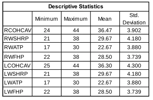
S.NO TITLE PAGE 1. The descriptive statistics of the variables with Minimum,
Maximum, Mean and Standard deviation of clinically obtained
horizontal condylar angle value with three different radiographically
obtained values
36
2. The correlation between the comparing paired groups of Clinically obtained horizontal condylar angle value with all the three
radiographic values respectively
36
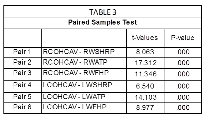
3. The paired t-test comparison between the Clinically obtained horizontal condylar angle value in right and left side with all the
three radiographically obtained values respectively
37
4. The descriptive statistics of the variables with Minimum, Maximum, Mean and Standard deviation of clinically obtained
horizontal condylar angle value with three different radiographically
obtained values
38
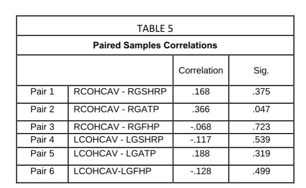
5. The correlation between the comparing paired groups of clinically obtained horizontal condylar angle value with all the three
radiographic values respectively
38
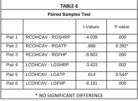
6. The paired t-test comparison between the Clinically obtained horizontal condylar angle value in right and left side with the three
radiographic values respectively
S.NO TITLE
1. Comparison between radiographic value and right side clinical horizontal condylar angle value when posterior slope of articular eminence and
contacting incisal edges used to trace condylar path
2. Comparison between radiographic value and left side clinical horizontal condylar angle value when posterior slope of articular eminence and
contacting incisal edges used to trace condylar path
3. Comparison between radiographic value and right side clinical horizontal condylar angle value when posterior slope of articular eminence used to
trace condylar path
4. Comparison between radiographic value and left side clinical horizontal condylar angle value when posterior slope of articular eminence used to
BACKGROUND:
Condylar path is a path travelled by the mandibular condyle in the temporomandibular joint during various mandibular movements.
The condyle will descend along the slope of articular eminence when it will move from centric relation to perform excursive movements. The angle at which the condyle moves from a horizontal reference plane is called Horizontal condylar angle. This condylar path inclination must be determined in relation to a horizontal plane of reference.
AIM:
To evaluate the Horizontal Condylar angle value using Computerised Digital Cephalometric Tracing.
OBJECTIVES:
1) To assess the Horizontal Condylar angle established by FENN, SHILLINGBURG and FRANKFORT horizontal plane using Computerised Digital Cephalometric tracing.
2) To compare these values obtained, with that of clinically established Horizontal Condylar angle
METHODS:
This clinical study was performed on 30 dentate individuals and horizontal condylar angle was measured clinically from these subjects by using Semi-Adjustable Arcon type articulator and these values were compared with the three values obtained by lateral cephalogram to find out which value comes closest to clinically established horizontal condylar angle value.
RESULTS:
When both anterior and reference posterior reference points were used to trace the condylar path, it was observed that there was significant difference between the radiographic horizontal condylar angle values and clinically established horizontal condylar angle value.
When only posterior reference point was used to trace condylar path, there was no significant difference between the radiographic value of Ala-tragus line (camper’s plane) and clinically established horizontal condylar angle value.
INTERPRETATION AND CONCLUSION:
1
It is a well-accepted matter in the field of dentistry that three vital factors
which influence the mandibular movements are namely, Incisal guidance, Condylar
guidance and overall neuromuscular control. According to GPT-81, Condylar
Guidance is defined as a mandibular guidance generated by the condyle and articular
disc transversing the contour of the glenoid fossae. It depends on the steepness of
articular eminence. It is the first factor of occlusion. It is obtained from the patient and
cannot be changed.
Factors that show major impact on the occlusal morphology of posterior teeth
are the protrusive condylar path inclination and mandibular lateral translation. The
inclination of the condylar path during protrusive movement can vary from steep to
shallow in different patients. Studies by Zamacona et al2, Lundeen and Wirth3,
Woelfel et al4, Hobo and Mochizuki5, Preti et al6, and dos Santos et al7 found
variations in condylar guidance angles ranging from 5 to 55◦, average angle being
30.4 degrees.
If the protrusive inclination is steep, the cusp height may be longer, with more
pronounced anterior guidance. However, if the inclination is shallow, the cusp height
must be shorter, with less pronounced anterior guidance. Therefore correct recording
of condylar guidance and its accurate transfer to semi-adjustable articulator
determines the success of restorative procedure.
Condylar inclination affects the angulation of the cusps of the teeth in both
protrusive and lateral excursive movements8. A steep condylar inclination allows
steeper inclines on the cusps of the teeth, while a less steep inclination demands a
flatter occlusal surface with shallower cuspal inclination. If the articulator condylar
path is set at a steeper angle than that which exists in the patient, the resulting
restoration will have cusps that have overly steep inclines. A positive error exists and
2
lateral excursion at the time of mastication. Condylar path is a path traveled by the
mandibular condyle in the temporomandibular joint during various mandibular
movements.
The condyle will descend along the slope of articular eminence when it will
move from centric relation to perform excursive movements. The angle at which the
condyle moves from a horizontal reference plane is called Horizontal condylar angle.
There are three classes of records for transferring maxillomandibular relations
from patient to articulator.(1) the method based on Christensen phenomenon in which
the interocclusal records are made and then horizontal condylar angle is calculated in
the articulator, commonly advocated in dentate individuals (2) A graphic method in
which the condylar path is recorded on a flag with tracing paper by means of a
facebow (pantographic tracing) and correct adaptation of recording is facilitated by an
intraoral bearing device, and the articulator is programmed accordingly.
The angle of the condylar path is obtained by measuring the tangent to the
functional portion of the tracing. Pantographic tracing method although quite
accurate, is quite cumbersome to perform in clinical situations and not feasible in all
the patients. Both orthopantogram and lateral cephalogram can be used to measure
condylar angle radiographically, as the condyle transverses the slope of articular
eminence during protrusion and radiograph made at edge to edge position of anterior
teeth.
This condylar path inclination must be determined in relation to a horizontal
plane of reference, but the values obtained cannot be directly compared to the
inclinations measured in relation to Frankfurt plane or Axis-orbital plane. In the
literature, there is no uniform consensus regarding the horizontal reference plane
3
H.R.B Fenn9 recommends occlusal plane parallel to Ala-tragus line (Campers
line) as horizontal reference plane whereas Shillingburg10 recommends a plane
formed by point 43mm above incisal edge of maxillary lateral incisor to superior
margin of external auditory meatus to measure condylar angle.
The purpose of this study is to evaluate the horizontal condylar angle using
Computerised Digital Cephalometric tracing and to assess the effectiveness of three
different horizontal reference planes described by Fenn, Shillingburg and Frankfort
AIM
AND
4 Aim of the study:
To evaluate the Horizontal Condylar angle value using Computerised Digital
Cephalometric Tracing.
Objectives of the study:
1) To assess the Horizontal Condylar angle established by FENN using
Computerised Digital Cephalometric tracing.
2) To assess the Horizontal Condylar angle established by SHILLINGBURG
using Computerised Digital Cephalometric tracing.
3) To assess the Horizontal Condylar angle established by FRANKFORT
horizontal plane using Computerised Digital Cephalometric tracing.
4) To compare these values obtained, with that of clinically established
REVIEW
5
Boos R H11 (1951) conducted a study to measure condylar angle by Roentgenograph. A metal form was devised which would fit onto an occlusion rim or
onto natural teeth which may be present. Two steel rods, l/16 inch in diameter, are
attached on either side of the metal form. The rods are arranged so that they may be
placed at the side & of the face below the temporo mandibular joint. They are
U-shaped with straight end segments. They are attached to the metal form so that they
may be set at the Fox plane. The Fox plane extends from the tragus of the ear to the
ala of the nose and provides an anatomic landmark on the face. The rod on the side of
the head which is being x-rayed is placed in position on the Fox plane, and the
opposite side is dropped down.
X ray was obtained, the developed film should show the steel rod as an
opaque line, and the temporomandibular joint with the angle of the articular tubercle
of the fossa. The measurement of the angle of the condylar path is made in degrees on
the developed x-ray film. (Boos R H: condylar path by roentgenograph. J. Prosthet Dent
1951;1:387-92)
Cohen R12 (1956) conducted a study to find out the effect on the condylar guidance when the shape of the anterior guidance is altered and when the vertical
dimension is increased. Gnathograph and Gnathoscope was used to record and
duplicate the paths of movement of the condyle. This experiment concluded that (1)
within the range of opening of the vertical dimension used, there is no change in the
paths of condylar movement regardless of the vertical dimension or the shape of the
anterior guidance for the mandible (2) within the range of opening of the vertical
6
the mandible. (Cohen R: The relationship of anterior guidance to condylar guidance in mandibular
movement, J.Pros.Dent.1956;6:758-67)
Issacson D13 (1958) conducted a study to record Bennett movement with a clinical instrument Gnathograph in 26 patients.. The stylus points attached to
Gnathograph made tracing of various mandibular movements on the glass slides.
Study demonstrated average Bennett angle ranges in age from 19 to 60 years was
12.33 degrees and range of bennett path angulation was zero degree to 35 degrees.
(Issacson D: A clinical study of the Bennett movement J.Pros.Dent.1958;8:641-49)
Issacson D14 (1959) designed a study to measure the angle and radius of the condyle path. He used orbital plane as horizontal reference plane. complete
movements of mandible in sagittal plane were traced on glass slides by gnathograph
styli and mounted on gnathoscope, and was adjusted to follow path of tracing. He
concluded that the condyle path angle ranged from a minimum of 22 to a maximum of
53 degrees. The age and sex of patient had little bearing on the size of condyle path
angle, which was determined as approx. 35 degrees. (Issacson D: A clinical study of condyle
path, J. Pros. Dent.1959; 9:927-35).
Posselt U and Skytting B15 (1960) conducted a study to ( 1) to assess any variations of sagittal condyle path inclination as obtained by graphic recording and (2)
to compare them with the results obtained from the intraoral wax record method. He
concluded that the error of the graphic method was found to be about twice that of the
intraoral wax method. It is probable that the error is mainly caused by the difficulty in
drawing a tangent to the curved condylar path rather than to the recording itself.
(Possel and Skytting B: Registration of the condyle path inclination: variation using the Gysi technique,
7
Olsson A and Posselt U16 (1961) advocated condylar path inclination measured in relation to one horizontal reference plane cannot be compared directly to
inclinations measured in relation to Frankfort plane or its near equivalent, the Axis
orbital plane. He compared nasion sella line, Frankfort plane, campers line and
occlusal line. Average angles between these planes were calculated on lateral
cephalogram and ranked according to magnitude of variation. (Olsson A and Posselt U.:
Relarionship of various skull reference lines, J. Pros.Den 1961;11:1045-4)
Posselt U and Nevstedt P17 (1961) carried an investigation to determine the frequency of condyle path inclinations, with the error of measurement found with the
dentatus articulator as a background in 101 patients. Protrusive bite registration was
obtained and dentatus articulator was programmed to obtain condylar inclination and
it varied between 0 and 60 degrees. The greatest frequency was around 40-50 degrees
as related to Frankfort plane. (Posselt U and Nevstedt P: Registration of the condyle path
inclination by intraoral wax records-its practical value,J.Pros.Den 1961;11:43-47)
Ismail YH and Bowman JF18 (1968) conducted a study to compare the occlusion plane established prosthetically with the one that existed before extraction
of the teeth in each subject. Lateral Cephalograms were taken for 20 subjects with
their teeth in centric occlusion. Following extraction of teeth, denture construction
was started using a standardized technique. The occlusal plane was tentatively
determined. The height of the maxillary occlusion rim in the anterior region was
placed 1-3mm below the resting upper lip and parallel to the Ala-tragus line
posteriorly.
He told camper’s line is a anthropologic measurement projected to the living
head as a line passing from the ala of the nose to the centre of the tragus of the
8
fulfill esthetic requirements and posteriorly middle third of retromolar pad was used
as reference point for orienting the occlusal surface of second molars. After the
complete dentures were placed, another set of lateral cephalogram were made. (Ismail
YH and Bowman JF: Position of occlusal plane in the natural and artificial teeth. J Pros.Dent
1968;20:407-11)
Pipko D19 (1969) conducted a study on 10 subjects to check the validity of the concept of curvilinear condylar path movement. The condylar path curvature was
recorded by a radiographic method, by the Ney technique, by pantographic tracings
and by the electronic methods of instrumentation. He concluded that condylar path
movement recordings are more curvilinear than rectilinear in nature, and positive
correlation exists among the methods used for determining the specific condylar
radius path curvature. (Pipko D: Evaluation of validity of condylar path curvature. J. Pros.Den
1969;21:626-38)
Lee R20 (1969) conducted a study to obtain a scientifically accurate and practical method of duplicating jaw movements. A new method and apparatus was
designed to record right lateral, left lateral and protrusive movements. All jaw
movements were recorded directly from the patient in the form of engravings in solid
plastic. The paths of these points are represented by groves in the plastic blocks at the
tips of their respective recording styli. These engravings were related to hinge axis.
(Lee R: Jaw movements engraved in solid plastic for articulator controls,Part I. Recording apparatus
J.Pros.Dent 1969;22:209-24)
Corbett et al21 (1971) designed a study to determine the relationship between the form of articular eminence and their corresponding condylar paths. A lateral
cephalogram showing the relation of the condylar head to articular eminence at
9
the movement of the condyles in mandibular protrusions. He concluded that
gnathologically recorded protrusive condylar paths and radiographically derived
protrusive condylar paths were found to be same and in protrusion, the condylar head
of mandible follows closely the anatomical form of the articular eminence. (Corbett et
al: The relation of the condylar path to the articular eminence in mandibular protrusion, J.Pros.
Den1971;41:286-92)
Rothstein R22 (1972) described prerequisites of interocclusal protrusive record and method to adjust the condylar elements on the articulator using this protrusive
interocclusal records. (Rothstein R: Condylar guidance settings on articulator from protrusive
records. J.Pros.Den 1972;28:334-36)
Ingervall B23 (1972) studied the movements of the mandibular condyles in the sagittal plane and the inclinations of the condyle path with a roentgencephalometric
method in children aged 7 and 10 years and in adults.. In both children and adults, the
condyles moved,on the average, forwards and downwards from intercuspal position to
postural position. The inferior movement was positively correlated with the
inclination of the condyle path recorded between intercuspal position and 5mm
protruded position. The inferior movement of the condyle from intercuspal position to
protruded position and to maximal opening increased as did the inclination of the
condyle path with age.
No correlation was found between the inclination of the condyle path and the
inclination of the incisal path in children or in adults. No correlation was found
between the inclination of the condyle path and the number of tooth contacts on the
working and non working sides. (Ingervall B: range of sagittal movement of the mandibular
10
Spartley MH 24(1980) in his article demonstrated that it runs from the center of the Ala to the center of the tragus. (Spartley MH: A simplified technique for determining the
occlusal plane in full denture construction. J Oral Rehab 1980;7:31-33)
Preti G,Scotti R, Bruscagin C and Carossa S6(1982) conducted a research to study the statistical investigation of the angular values of the condylar sagittal
pathway obtained with the Graphic record method and to verify its repeatability.
Gerber’s graphic registration instrument was used to obtain protrusive graphic
registration and a tangent is drawn to each tracing to express the angular value of
condylar sagittal pathway in 309 subjects. He concluded that the data when was
subjected to statistical analysis gave median angular value of the sagittal pathway
equals to 33 degrees and showed a greater median value dispersion. A low error index
was obtained in tracing the tangent to the CSP tracing with Gerber registration
method. (Preti G,Scotti R, Bruscagin C and Carossa S: A clinical study of graphic registration of the
condyle path inclination. J.Pros.Dent 1982;48:461-66)
Fattore et al25 (1984) conducted a study to determine the clinical accuracy of waxes, zinc oxide-eugenol, and polyether dental materials for recording interarch
relationships. Interocclusal records of 31 patients were placed on an arcon articulator
with an arbitrary face-bow to measure the magnitude and direction of distortion. He
concluded that 1) Polyether interocclusal recording medium without a carrier was the
most accurate. 2) Polyether and zinc oxide-eugenol pastes with carriers were the next
most accurate recording mediums, but they required a disciplined technique. 3)
Recording waxes were consistently unreliable. 4) Distortion occurred more frequently
in a vertical direction, followed by an anteroposterior direction. (Fattore et al: clinical
11
El-Gheriani AS and Winstanley RB26 (1989) carried out study to determine the accuracy of different methods of measuring condylar inclinations from graphical
recordings of condylar paths. Thirty subjects made protrusive mandibular movements
while condylar inclination was recorded on a graph paper card. This method proved to
be too variable. The spline curve fitting technique was shown to be accurate, but its
use clinically may prove complex. The mathematical method was more practical and
overcame variability of the tangent method. (El-Gheriani AS and Winstanley RB: Graphic
tracings of condylar paths and measurements of condylar angles. J. Pros. Dent1989;61:77-87)
Muller J, Gotz G, Horz W and Kraft E27 (1990) conducted a study to determine the three-dimensional errors in mounting casts affected by the interocclusal
recording materials. Eight materials and/or combinations of two different materials
were selected for this study (1) impression plaster, (2) Palavit G self-curing resin, (3)
Palavit G resin combined with Temp-Bond zinc oxide-eugenol paste, (4) Beauty pink
(X-hard) wax, (5) Beauty pink wax combined with Temp Bond material, (6)
impression compound, (7) impression compound combined with Temp Bond
material, and (8) polyether. The results indicated that all the materials induced
asymmetric deviations of the condyles after each storage period. Impression plaster
was the most accurate and dimensionally stable material; polyether was the second
most accurate material, but it must be used within 6 hours. (Muller J,Gotz G,Horz W and
Kraft E: Study of the accuracy of different recording materials. J Prosthet Dent 1990;83:41-6.)
Muller J,Gotz G,Horz W and Kraft E28 (1990) conducted a study to analyze the accuracy of transferring jaw relations with recording materials and the derived
casts. A specific measuring system was designed to determine three-dimensional
deviations of the condyles of an articulator. Four interocclusal recording materials
12
compound, wax and zinc oxide and eugenol. He concluded that the greatest
three-dimensional deviations were evident in plaster recordings. Impression compound was
the most accurate of the materials tested, but deviations to 300 pm need review.
(Muller J,Gotz G,Horz W and Kraft E: An experimental study on the influence of the derived casts on
the accuracy of different recording materials. Part 1: Plaster, impression compound, and wax. (J
Prosthet Dent1990;63:263-9.1)
Zamacona J, Otaduy Eand Aranda E2(1992) studied 55 patients making three graphic registrations of the protrusive condylar movement on each side. Three
examiners independently used tangential method to measure the angulation relative to
the Camper’s plane. Range of inclination of condylar path varied from 10 to 62
degrees on the left side, with a mean of 35.75 degrees, while on right side, it was from
23 to 55 degrees,with a mean of 36.6 degrees. He recommended tangential method of
measuring inclination on graphic records. (Zamacona J,Otaduy Eand Aranda E: Syudy of the
sagittal condylar path in edentulous patients. J.Pros.Dent1992;68:314-17)
Ogawa T, Koyano K and Suetsugu T29 (1997) conducted a study to reveal the influence of the incisal and condylar guidance on mandibular protrusive
movement. The protrusive movements were measured on 54 young adults using a
three-dimensional mandibular movements analyzing system. The inclinations of the
sagittal path on the incisor, canine, first molar, second molar and condylar points were
calculated, and multiple regression analysis was performed to evaluate the influence
of the incisal and condylar path on the path of each tooth quantitatively.
The influence of the incisal path on any tooth path was consistently greater
than that of the condylar path. The condylar path had a greater influence on the paths
13
(Ogawa T, Koyano K and Suetsugu T: The influence of anterior guidance and condylar guidance on
mandibular protrusive movement Journal of oral rehabilitation 1997;24;303-09)
Rudolph, Sinclair, and Coggins30 (1998) Computerized cephalometric analysis currently requires manual identification of landmark locations. This process
is time-consuming and limited in accuracy. The purpose of this study was to develop
and test a novel method for automatic computer identification of cephalometric
landmarks. Spatial spectroscopy (SS) is a computerized method that identifies image
structure on the basis of a convolution of the image with a set of filters followed by a
decision method using statistical pattern recognition techniques. By this method,
characteristic features are used to recognize anatomic structures.
This study compared manual identification on a computer monitor and the SS
automatic method for landmark identification on minimum resolution images. Fifteen
landmarks were selected on a set of 14 test images. The results showed no statistical
difference in mean landmark identification errors between manual identification on
the computer display and automatic identification using SS. We conclude that SS
shows potential for the automatic detection of landmarks, which is an important step
in the development of a completely automatic cephalometric analysis. (Rudolph, Sinclair,
and Coggins: Automatic computerized radiographic identification of cephalometric landmarks. (Am J
Orthod Dentofacial Ortho1998;113:173-9)
Chen, Chen, Chang, Chen31 (2000) conducted a study to assess landmark identification on digital images in comparison with those obtained from original
radiographs. Ten cephalometric radiographs were selected randomly. Seven
orthodontic residents identified 19 cephalometric landmarks on both the original
14
cephalometric analysis, the differences of landmark location between original
cephalometric radiographs and their digital counterparts were statistically significant.
The reliability of landmark identification in digital images was comparable to
that in original radiographs except for the points Po, Ar, PNS, and UM. These
landmarks with significant lower reliability in digital images should be scrutinized
more carefully when we take potential advantages of the use of digital cephalometry.
(CHEN, CHEN, CHANG, CHEN: Comparison of Landmark Identification in Traditional Versus
Computer-Aided Digital Cephalometry. Angle Orthodontist 2000;50:387-92)
Perillo et al32 (2000) Identification of craniofacial landmarks, particularly condylar anatomy, on the lateral cephalometric radiograph is erratic. They conducted
a study to to evaluate the identification of condylion and other cephalometric
landmarks commonly used, or thought to be easily identifiable. A lateral
cephalograph was taken on each of 34 adult subjects. Five examiners, three
orthodontists, a dental radiologist and a second-year orthodontic resident rated the
condyle, along with sella (S), nasion (Na), point A (A), infradentale (I), pogonion
(Pog) and menton (Me) as identifiable, non-identifiable and interpreted. The left
condyle, subject to less magnification than the right condyle because it is closer to the
film, was more identifiable than the right condyle, which had the highest rating as
non-identifiable. (Perrilo et al: Effect of landmark identification on cephalometric measurements:
guidelines for cephalometric analyses. Clin Orthod Res 3, 2000:29–36)
Dos Santos J, Nelson S and Nowlin T7 (2003) conducted a study to compare the condylar inclinations angles found by the use of the wax protrusive record in a
Hanau articulator with those found by use of the Whip-Mix Protrusive tracing
quick-set recorder in ten patients. He concluded that measurement of the extraoral tracing of
15
intraoral wax protrusive method. (Dos Santos J,Nelson S and Nowlin T: Comparison of condylar
guidance setting obtained from a wax record versus an extraoral tracing: A pilot study. J Pros Dent
2003;89:54-9)
Kucukkeles N, Ozkan H, Demirkaya A and Cilingirturk A33 (2003) conducted a study to compare measurements between mechanical and computerized
axiographs in recording the rotational and translation movements of the mandible in
31 subjects. A single operator obtained 3 separate axiographic tracings of right and
left condylar paths for each subject, using repeated opening, closing, protrusive and
retrusive movements. Data were collected for both the mechanical and computerized
axiographs. He used axio-orbital plane as horizontal reference plane. He concluded
that data from the manual and the computerized axiographs are compatible with each
other. (Kucukkeles N, Ozkan H, Demirkaya A and Cilingirturk A: compatibility of mechanical and
computerized axiographs:A pilot study.J.Pros.Dent 2005;94:190-4)
Chen, Chen, Yao, Chang34(2004) conducted a study to explore the effects of differences in landmark identification on the values of cephalometric measurements
on digitized cephalograms in comparison with those obtained from original
radiographs. Ten cephalometric radiographs were randomly selected from orthodontic
patients’ records. Seven orthodontic residents identified 19 cephalometric landmarks
on the original radiographs and digitized images. Twenty-seven cephalometric
measurements were computed with a customized computer aided program. To assess
the concordance between cephalometric measurements derived from landmarks
identified on the original radiographs and those from digitized counterparts, the values
of 27 cephalometric measurements were compared to quantify the absolute value of
16
He concluded that the measurement differences between the original
cephalograms and the digitized images are statistically significant but clinically
acceptable. The interobserver errors for cephalometric measurements on our digitized
cephalometric images are generally comparable with those on the original
radiographs. The results of our study substantiated the benefits of digital
cephalometry in terms of the reliability of cephalometric analysis. (CHEN, CHEN, YAO,
CHANG: The Effects of Differences in Landmark Identification on the Cephalometric Measurements
in Traditional Versus Digitized Cephalometry. Angle Orthod 2004;74:155–161)
Matsumura H, Tsukiyama Y and koyano K35 (2006) investigated the sagittal condylar path during protrusive and lateral excursions by analysing the
actually measured jaw movement data and re-evaluated the setting of the sagittal
condylar path inclination in consideration of Fischer’s angle. Protrusive and lateral
excursions of 10 healthy subjects were measured using a three-dimensional
mandibular movement analysing system. Condylar path inclinations at the hinge-axis
point and the corresponding external point laterally extending from the condyle were
evaluated in the sagittal plane. Fischer’s angle was defined as the difference between
the sagittal condylar inclinations during protrusive and lateral excursions on the
non-working side, by keeping the corresponding horizontal distance from the intercuspal
position (ICP) equivalent at the incisal point.
Analysis was performed at three different magnitudes of excursions, where
the incisal point was located at 1, 3 and 5 mm away from the ICP. He concluded that
The sagittal condylar path inclination was significantly different at the 1-, 3- and
5-mm eccentric mandibular positions from ICP, but not different between the reference
point in the centre of the condyle and the corresponding external point laterally
17
different magnitudes of excursions, but not different between the two condylar
reference points. (Matsumura H, Tsukiyama Y and koyano K: Analysis of sagittal condylar path
inclination in consideration of Fischer’s angle. Journal of Oral Rehabilitation 2006 33; 514–519)
Gilboa et al36 (2008) conducted a study to determine the correlation between the anatomic shape of the articular eminence and the corresponding panoramic image
in dry skulls. Two metal wires were adapted and fixed to the inner and outer surfaces
of the articular eminences in 25 human skulls. The inner (thicker) wire was fixed to
the middle of the most concave aspect of the articular eminence in an
anterior-posterior direction. The outer (thinner) wire was attached to the inferior aspect of the
zygomatic arch adjacent to the articular eminence. Panoramic radiographic images
were recorded. Impressions were made of the condylar fossae in 25 human dry skulls.
Tracings of the incline of the articular eminence on the panoramic radiographs and the
impression sections were compared. He concluded that the panoramic radiographic
image of the sagittal inclination of the articular eminence consistently replicated the
eminence inclinations in the 25 human skulls evaluated. (Gilboa I,Cardash HS,Kaffe
I,Gross MD.Condylar guidance:correlation between articular morphology and panoramic radiographic
images in dry human skulls. J.Pros.Dent 2008;99:477-82)
Roden-Johnson, English, and Gallerano37 (2008) conducted a study (1) to investigate the variations of landmark identification between film and digital
cephalometric tracings, (2) to compare the ability of Quick Ceph 2000(Quick Ceph
Systems, Inc, San Diego, Calif) to measure the linear and angular measurements with
the hand-traced method, and (3) to compare Quick Ceph 2000 superimpositions to the
hand-traced method of superimpositions that are currently accepted by the American
Board of Orthodontics. They concluded that there was no difference in the
18
Ceph 2000 and there was no difference in the regional superimpositions of the
mandible, the maxilla, and the cranial base, manually vs digitally with Quick Ceph
2000. (Roden-Johnson, English, and Gallerano: Comparison of hand-traced and computerized
cephalograms: Landmark identification, measurement, and superimposition accuracy. (Am J Orthod
Dentofacial Orthop 2008;133:556-64)
Yu, Nahm, and Baek38 (2008) conducted a study to compare the reliability of landmark identification with hard-copied film images vs monitor-displayed images
from digital lateral cephalograms in 50 orthodontic patients. Identification and
digitization of the cephalometric landmarks were performed 3 times at 2-week
intervals by 2 observers. The 2 methods of landmark identification were the
hard-copied film-based method (HFM) and the monitor-displayed method (MDM) and
concluded that there were no statistically significant differences in landmark
identification between the 2 methods. (Yu, Nahm, and Baek: Reliability of landmark
identification on monitor-displayed lateral cephalometric images. Am J Orthod Dentofacial Orthop
2008;133:790)
Zoghby A,Re J and Perez C39 (2009) conducted a study to find a correlation between the mean Functional Incisal path of the maxillary anterior teeth and the
Functional Condylar Path. The tracing of multiple cuts of silicone of maxillary
anterior block which were analyzed by an odontometry software program and a
mechanical axiography to register the protrusive path bilaterally was performed for 50
dental students. He concluded that the functional incisal path is superior by 9.52
degree as compared to the functional condylar path. (Zoghby A,Re J and Perez C. Functional
harmony between the sagittal condylar path inclination and the anterior guidance inclination.
19
Huja, Grubaugh, Rummel, Fields, Beck40 (2009) conducted a study to determine the ability to produce comparable superimpositions using hand tracing and
digital methods and to determine if a difference existed between the best-fit cranial
base superimposition and S-N superimpositions using the digital method. Sixty-four
initial (T1) and final (T2) cephalometric film radiographs were obtained. Cranial base
and regional superimpositions were completed independently for each pair of
radiographs by either hand tracing and digital methods. They concluded that there are
no differences between cranial base and regional superimpositions produced by
Dolphin Imaging version 10 and those completed by hand when using the described
methods and provides support for transition from hand to digital superimposition
methods. (Huja, Grubaugh, Rummel, Fields, Beck: Comparison of Hand-Traced and Computer-Based
Cephalometric Superimpositions. Angle Orthod. 2009;79:428–435)
AL quran et al41 (2011) conducted a study to determining the most reliable Ala-tragus line as a guide for the orientation of the occlusal plane in complete denture
patients by use of cephalometric landmarks on dentate volunteers. Analysis was made
for prosthodontically related craniofacial reference lines and angles of lateral
cephalometric radiographs taken for 47 dentate adults. Variables were determined and
data were analyzed using SPSS. He concluded that the superior border of the tragus
with the inferior border of the Ala of the nose was most accurate in orienting the
occlusal plane. (AL quran et al: The Position of the Occlusal Plane in Natural and Artificial
Dentitions as Related to Other Craniofacial Planes. J. Prosthodon 2011:19;601-05)
Goyal MK and Goyal S42 (2011) conducted a study to compare evaluation of sagittal condylar values of arcon and non-arcon articulators with cephalometric
20
values between arcon and non-arcon articulators using same protrusive record in
twenty subjects.
He concluded that the mean difference in the sagittal condylar guidance values
obtained from non arcon and arcon articulators show a low level of reproducibility,
and no significant difference found in mean sagittal condylar values obtained from
Arcon articulator and cephalometric tracings indicates replication of sagittal condylar
guidance value from image of articular eminence. (Goyal MK and Goyal S. A comparative
study to evaluate the discrepancy in condylar guidance values between two commercially available
arcon and non-arcon articulators: a clinical study IJDR,22;2011)
Hue O43 (2011) conducted a study to determine the condylar form, incline, and movement characteristics during protrusive movement in fully edentulous
complete denture wearers. The study included 60 complete denture wearers (aged 58
to 74 years), who received a new set of complete dentures for this study. The patients
did not present signs of muscular or articular pain. Protrusive movements were
recorded by a SAM electronic axiography system.
He concluded that during protrusive movement in completely edentulous
patients, the condylar path patterns were different than conventionally described
patterns. In particular, the sinusoidal form was frequently found, and the incline of the
condylar slope was low. These factors need to be taken into account during the final
occlusal selective grinding for new sets of complete dentures. (Hue O : Condylar Paths
during Protrusion in Edentulous Patients Analysis with Electronic Axiography. J. Prosthodon 2011;
20:294-98)
Tannamala P V, Pulagam M, Pottem S and Swapna B44 (2012) conducted a study to compare the sagittal condylar angles set in the Hanau articulator by use of a
21
panoramic radiographic image in ten patients. A panoramic radiographic image of
each patient was made with the Frankfurt horizontal plane parallels to the floor of the
mouth. He concluded that radiographic values were on average 4 degree greater than
the values obtained by protrusive interocclusal record method and the protrusive
condylar guidance angles obtained by panoramic radiograph may be used in
programming semi-adjustable articulators. (Tannamala P V, Pulagam M, Pottem S and
Swapna B: Condylar guidance :correlation between protrusive interocclusal record and Panoramic
radiographic image: A pilot study JOP 2012 ;181-84)
22 References from Text books
Schillingburg H T, Hobo S, Whitsell L D, Jacobi R and Brackett S E10: Fundamentals of Fixed Prosthodontics, Third edition. Page 20
The inclination of condylar path during protrusion movement can vary from steep to
shallow in different patients. It forms an average angle of 30.4 degrees with the
horizontal reference plane (43mm above the maxillary central incisor edge).
Fenn H.R.B, Liddelow K.P and Gimson A.P9 :Clinical Dental Prosthetics Page 270
By condylar path is meant, the path taken by the head of the condyle when moving up
and down the articular eminence. The angle which this makes with the occlusal plane
is known as Condylar Angle.
Fenn H.R.B, Liddelow K.P and Gimson A.P9: Clinical Dental Prosthetics Page 243-44
When recording the occlusion in the patient’s mouth, the occlusal surface of the
record blocks are trimmed parallel to the occlusal plane, which is a horizontal plane
parallel to the Nasoauricular line which is line joining the lower border of the ala of
nose to the external auditory meatus.
Boucher CO45: Complete denture prosthodontics—state of the art. J Prosthet Dent 1975;34:372-383
23
Grant AA, Johnson W47: An Introduction to Removable Denture Prosthetics. Edinburgh, Churchill Livingstone, 1983
Neill DJ, Naim RI48: Complete Denture Prosthetics. Bristol, John Wright Sons, 1975
Among seven of the most famous prosthodontic textbooks, only Boucher’s provides a
definition., Two other textbooks recommend the concept without defining it, while
Basker et al, Grant and Johnson, and Neill and Naim provide only pictorial
representation, illustrating Camper’s line as extending to a point, not at the superior
border, but at the center of the tragus of the ear.
Winkler S49: Essentials of Complete Denture Prosthodontics, Second edition, page 140-41
After forming the occlusal rim with prescribed vertical heights, the plane of occlusion
is modified until it is parallel with a line projected from the Ala of nose to the superior
edge of the tragus of ear (camper’s line). When viewed from the front, the occlusal
plane should also be parallel to the interpupillary line. The relationship of the
interpupillary line, Camper’s line and the occlusal plane is also shown in a pictorial
MATERIALS
AND
24 STUDY DESIGN:
This clinical study was performed in dentate individuals to compare values of horizontal condylar angle in relation to three different horizontal planes described in
the literature. Horizontal condylar angle was measured clinically from dentulous
subjects by using Semi-Adjustable Arcon type articulator and these values were
compared with the three values obtained by lateral cephalogram to find out which
value comes closest to clinically established horizontal condylar angle value. This
study was performed from June 2012 to November 2012 in the Department of
Prosthodontics, Tamil Nadu Government Dental College and Hospital, Chennai.
ETHICAL COMMITTEE APPROVAL:
The study was conducted with the approval from the institutional ethical
committee.
The following materials and equipments were used to conduct the study.
S.No NAME
(commercial name)
FORM OF THE MATERIAL
MANUFACTURER DETAILS
1. Vignette chromatic alginate impression material Irreversible hydrocolloid impression material Dentsply, India
2. Jabbar trays Stock tray Jabbar & co,India
3. Kalstone Type III Dental stone
Kalabhai, India
4. White gold Type II Dental plaster
Asian chemicals, India
5. Professional face bow
Bio art, Brazil
6. Samit super Tracing Sticks
Green Stick Compound
25
7. A7 Plus Articulator Semi Adjustable Arcon Articulator
Bio Art , Brazil
8. Konark ever bright dental stainless steel wire
Stainless steel orthodontic wires, 23 gauge
Khokhar, india
9. Cellotape Premier
10. Multimark 1513 Black marker pen Faber-castell
11. Camlin scale Scale 15cms Camlin limited, Mumbai
12. Kodak Digital Panoramic and Cephalogram system 13. DICOM viewer
2.1.7
Medsynaptics Pvt. Ltd. , Pune, India
ARMAMENTARIUM FOR CLINICAL EXAMINATION:
1. Kidney Tray
2. Mouth mirror
3. Periodontal probe
4. Cheek retractor
5. Disposable gloves and mask
ARMAMENTARIUM FOR OBTAINING IMPRESSIONS:
1. Alginate (Vignette chromatic)
2. Maxillary and Mandibular stock trays
3. Type III Dental stone (Kalstone)
4. Rubber bowl and Spatula
26
ARMAMENTARIUM FOR FACE BOW TRANSFER:
1. Bio-Art Professional face bow
2. Green stick Impression Compound
3. Rubber bowl
ARMAMENTARIUM FOR FACE BOW MOUNTING:
1. Bio-Art Arcon Semi-adjustable Articulator
2. Accessories for mounting
3. Type II Dental Plaster
4. Rubber bowl and spatula
5. Cotton
ARMAMENTARIUM FOR JAW RELATION REGISTRATION:
1. Green Stick Impression Compound
2. Rubber bowl
ARMAMENTARIUM FOR OBTAINING LATERAL CDEPHALOGRAM:
1. Protrusive registered bite
2. Two straight 21 gauge orthodontic wires
3. Cellotape
4. Ruler
5. Permanent Marker Pen
ARMAMENTARIUM FOR COMPUTERISED DIGITAL CEPHALOMETRIC TRACING:
27
2. Digital Lateral Cephalogram
3. DICOM Software
METHODOLOGY
1. SUBJECT SELECTION
2. PREPARATION OF STUDY MODELS
3. FACE BOW TRANSFER
4. MOUNTING OF MAXILLARY CAST ON SEMIADJUSTABLE
ARTICULATOR
5. CENTRIC JAW RELATION REGISTRATION
6. MOUNTING OF MANDIBULAR CAST ON SEMIADJUSTABLE
ARTICULATOR
7. OBTAINING PROTRUSIVE AND RIGHT LATERAL AND LEFT
LATERAL BITES
8. PROGRAMMING THE SEMI ADJUSTABLE ARTICULATOR TO
OBTAIN RIGHT AND LEFT SIDE CONDYLAR ANGLE VALUE
9. OBTAINING LATERAL CEPHALOGRAM OF PATIENT
10.COMPUTERISED DIGITAL CEPHALOMETRIC TRACING TO OBTAIN
VALUES RADIOGRAPHICALLY
SUBJECT SELECTION
Study participants were selected from the undergraduate dental students
of Tamil Nadu Government Dental College and Hospital with the age group of
20-27 years of age and both male and females subjects were included in the study. All
the patients were informed about the purpose and methods of the study and signed the
28
The inclusion criteria for entry in the trial were:
(a) Class I molar and canine relation
(b) Average horizontal and vertical overlap
(c) Minimal spacing or crowding of anterior teeth
(d) No prosthesis with minimum or no occlusal restorations
(e) No history of orthodontic treatment
The exclusion criteria for entry in trial were:
a) Class II or III relation
b) Any missing teeth
c) Traumatic occlusion of anterior teeth
d) TMJ clicking,crepitation,tenderness
e) Parafunctional habits
SAMPLE SIZE
Study was designed with sample size of 30.
PREPARATION OF STUDY MODELS
Maxillary and mandibular arch impressions were recorded with irreversible
hydrocolloid and casts poured with type III dental stone and bases were formed with
type II plaster.
FACE BOW REGISTRATION
Bio-art Professional face bow was used in this study. Three points are made on
the fork, one frontal point, in the exact center of the fork, and two points at the back,
29
fork handle is aligned with the midline of the maxilla and placed on the upper arch, it
was held up firmly in place until the registration material hardens. Individual was
made to sit in reclined position in the chair to reduce the induction of tensions on the
fork set and face-bow. Individual was asked to keep the fork in the same position by
supporting the thumbs against the maxilla. The face bow was taken to the patient and
the bite fork assembly was introduced into the fork handle, assuring the wing nut is
upside down. The face-bow earpieces were carefully inserted into the patient’s
external auditory meatus.
The nasion relator was placed on the face-bow cross bar and centered on
patient nose. The fork fixator assembly was pushed forward, sliding it on the fork
handle until it is as close as possible to the lips, without touching them. First the
double articulated nut of the fork was tightened followed by the horizontal slide bar
nut. Patient was asked to remove his thumbs from the bite fork and it was checked if
the fork and facebow are stable and immobilized. The wing nut of the nosepiece was
loosened to remove the face bow and nasion relator assembly removed from the face
bow. Then, the central wing nut of the facebow was loosened, cross bar was held and
patient was told to open mouth slowly, removing the whole set carefully.
MOUNTING OF MAXILLARY CAST ON SEMIADJUSTABLE ARTICULATOR
As study was planned to be carried out on Bio-Art Arcon Semi-adjustable
articulator (Model A7 plus), to facilitate the mounting of the casts, condylar guidance
angle was adjusted at 30 degrees and the Bennett angle at 0 degree as recommended
by the manufacturers. The upper part of the central lock was pushed back until the
30
feature of this model is the Jig Transfer Assembly for mounting the casts on the
articulator. This assembly is placed in the lower member of the articulator through the
jig assembly lower base thus eliminating the need for the face bow frame while
transferring the patient’s registration onto the articulator and providing quick
mounting of the cast.
Incisal guide table was removed from the lower member of the articulator and
lower base transfer was inserted, assuring that the guide pin is properly touching the
end of the slot. The Jig Transfer Assembly was removed from the face bow and
connected to the jig transfer lower base and wing nut was fastened. The upper
member support was inserted on the jig transfer connection rod, supporting the upper
member and assuring the parallelism between the upper and lower members of the
articulator. Upper cast was placed on the fork registration and the upper member of
the articulator was lifted and plaster placed on the upper mounting plate and some on
the top of upper cast. Upper member was closed and articulation of upper cast was
completed.
CENTRIC JAW RELATION REGISTRATION
Green stick compound was used to record the centric jaw relation, the
compound was taken and placed in a bowl of warm water. It was formed into a shape
of U shaped arch and placed on mandibular teeth of patient. Then the patient’s
mandible was manipulated in centric relation by bimanual manipulation method.
Material was left in patient’s mouth for adequate time for proper cooling. Occlusal
31
MOUNTING OF MANDIBULAR CAST ON SEMI-ADJUSTABLE ARTICULATOR
The incisal guide in the upper member of the articulator is placed with its
rounded tip pointing downward so that the upper and lower members are kept parallel
i.e. when the incisal pin is on zero marking. Articulator is turned upside down and
lower cast affirmed upon the interocclusal register that was placed in the mounted
mandibular cast. A small amount of plaster was placed on the lower part of
mandibular cast and a small amount on the mounting plate of the lower member of
articulator to fill in the gap between them. After the plaster hardens, articulator is
turned back to its correct position and finishing touches given to plaster.
OBTAINING PROTRUSIVE AND RIGHT AND LEFT LATERAL JAW RELATIONS REGISTRATIONS
Green stick compound was taken and placed in a bowl of warm water. It was
formed into a shape of U and placed on mandibular teeth of patient. Patient was asked
to bring his/her incisors edge to edge relation and the protrusive bite was registered. It
was allowed to cool, removed and examined.
Then again green stick impression compound was molded in U shape and placed
on mandibular teeth and patient was asked to move towards right lateral side. Material
was allowed to cool down and then inspected. It was repeated in a similar way for left
lateral position. Thus three jaw relations were obtained.
1. Protrusive
2. Left lateral
32
PROGRAMMING THE SEMI ADJUSTABLE ARTICULATOR TO OBTAIN RIGHT AND LEFT SIDE CONDYLAR ANGLE VALUE
Before mounting of the casts, condylar guidance was adjusted to 30 degree
and Bennett angle at zero degree as recommended by manufacturers in instruction
manual. For programming of the articulator, upper member was required to be
separated completely from the lower member. Before proceeding with it, condylar
guidance screw was loosened to allow free movement of condylar guidance assembly
and angle was brought down to zero degree. Then Stablilizing elastic band was
removed from stabilizing elastic band pin on both right and left sides. Central lock of
upper frame was released to let free condylar element on both sides. Now upper
member was lifted from the lower member and kept aside.
Protrusive bite was placed on the mandibular teeth and checked for proper
adaptation on teeth. Now the upper member of articulator was placed gently over this
protrusive record so that the maxillary teeth fit into the indentations on the protrusive
record. It was noticed at this stage that the condylar element should not be touching
the superior wall of the condylar guidance assembly after placing protrusive record.
At this stage, the condylar guide assembly was rotated gently towards the increasing
angulation till the condylar element touches the superior wall of the condylar guide
assembly. The point where contact would be established is recorded as the condylar
guidance value (Horizontal condylar angle value) obtained for that patient on that