NATIONAL INSTITUTE OF SIDDHA
CHENNAI
– 600 047
THE TAMIL NADU DR. M.G.R. MEDICAL UNIVERSITY, CHENNAI - 600 032
A STUDY ON
CEGANAVAATHAM
(DISSERTATION SUBJECT)
For the partial fulfillment of the requirements to the Degree of
DOCTOR OF MEDICINE (SIDDHA)
BRANCH III - SIRAPPU MARUTHUVAM
CERTIFICATE
This is to Certified that I have gone through the dissertation submitted by
Dr.M.PRATHIBA a student of final M.D.(S) Branch-III, Department of Sirappu
Maruthuvam, National Institute of Siddha, Tambaram Sanatorium, Chennai-47 and the
dissertation work “A Study on Ceganavaatham” has been carried out by individual
only. The dissertation does not represent or reproduce the dissertation submitted and
approved earlier.
Place: Tambaram Sanatorium, Chennai-47
Date:
Associate Professor and HOD,
Department of Sirappu Maruthuvam,
National Institute of Siddha,
ACKNOWLEDGEMENT
The author is extremely grateful to the Almighty for the successful completion of
this dissertation work.
The author expresses her sincere thanks to the Vice- Chancellor, The Tamilnadu
Dr.M.G.R Medical University, Chennai - 32.
The author extends her sincere thanks to Dr.V.Arunachalam M.D.(S), Director,
National Institute of Siddha, Tambaram Sanatorium, Chennai-47, for granting permission
to undertake a study in this dissertation topic and also for providing all the basic facilities
in order to carry out this work.
The author is very great pleasure to Dr.G.Thiagarajan M.D.,(S), Associate
Professor and Head of the Department of Sirappu Maruthuvam, National Institute of
Siddha, Tambaram Sanatorium, Chennai-47, for his encouragement, suggestions and
valuable guidance in this dissertation work.
The author is very grateful to Dr.R.S.Ramaswamy M.D.,(S), Associate Professor,
Department of Sirappu Maruthuvam, National Institute of Siddha, Tambaram Sanatorium,
Chennai-47, for his suggestions, valuable guidance and support in this dissertation work.
The author is grateful to Dr.T.R.Siddque Ali M.D.,(S), Lecturer, Department of
Sirappu Maruthuvam, National Institute of Siddha, Tambaram Sanatorium, Chennai-47,
for his guidance and support in this dissertation work.
The author owes special thanks to Dr.K.V.Chandrasekaran M.B.B.S., D.Ortho,
M.S.Ortho, Head of the Orthopaedics Department, Dr.P.Thirunavukarasu M.B.B.S.,
D.Ortho, D.Phys.Med&D.N.B(PM&R), Dr.M.Sureshbabu M.B.B.S., M.S.Ortho,Tutor,
Dr.R.Sundarapandian M.B.B.S., D.Ortho, Tutor of Department of Orthopaedics,
The author expresses her sincere thanks to Dr.D.Porselvi M.B.B.S., D.M.R.D,
Department of Radiology, Chengalpattu Medical College hospital, Chengalpattu.
The author expresses her thanks to Mr.P.Jayabal M.Sc., Assistant Professor
(Statistics), National Institute of Siddha, Tambaram Sanatorium, Chennai -47.
The author expresses thanks to Dr.S.Venkatraman Ph.D, Director, C.L.Baid
Metha College of pharmacy, Thoraippakkam, Chennai - 96.
The author expresses her thanks to Mr.Mathan, Mettex Laboratories of India,
Guindy, Chennai-32.
The author expresses her thanks to Dr.B.Ravindran S.M.P., D.Y.N., Agaram
hospital, Mathuranthakam.
The author thanks her colleagues and other staff members who helped her in this
dissertation work.
The author expresses her hearty thanks also to her friends Dr.K.Natarajan
M.D.,(S), II year, Dr.S.Lalitha M.D.,(S), II year National Institute of Siddha, Tambaram
Sanatorium, Chennai -47.
CONTENTS
PAGE NO
1.
Introduction
2.
Aim and objectives
3.
Review of literature
a.
Siddha aspects
b.
Modern aspects
4.
Treatment
5.
Protocol
6.
Results and observations
7.
Discussion
8.
Summary
9.
Conclusion
10.
Annexures
I.
Drug review
II.
Preclinical studies
III.
Proforma
11.Bibliography
1
3
4
21
38
42
47
73
77
78
INTRODUCTION
The Siddha System of Medicine is prevalent in South India, Sri Lanka, Malaysia,
and Singapore, where the Dravidian civilization was documented. This system owes its
origin to the Dravidian culture which is of the Pre – vedic Period.
Siddha means perfection, Siddhar is the one who has attained immortality. The
Siddhars were the ancient Tamils who are in their quest for knowledge for longevity,
developed two ways by which man can achieve mastery over nature. One is the yogic way
and the other is through medicines. The persons who dedicated themselves to this task were
great yogis known as Siddhars. Hence, the system of medicine propounded by them came out
to be known as Siddha System of Medicine.
Siddha System is well founded on the basic principles of nature and its elements and
it offers a careful and thorough study of the human and animal systems. Siddha science
considers nature and human as essentially one. One who knows the secerecy of nature and its
five elements, knows well the secerecy of human and nature is the foremost physician.
According to Siddha Medical Science, the universe consists of five elements
(Panchaboothas) namely earth, water, fire, air and ether (Aakayam) which correspond to the
five senses of the human body.
The three vital forces namely Vali (Vaatham), Azhal (Pittham), Ayyam (Kabam)
called as Uyir Thaathukkal are activated by the functions of Punchaboothas. According to
the Punchabootha theory all the substances in the universe are created by the actions or
reactions of the Punchabootha only.
includes yoga which prolongs the life span and it also includes Vaithiya Muppu which
enhances the efficacy of drugs manifold. Even though no modern equipments were available
in olden days for research, Siddhars could understand the secret doctrines of the five
elements, and could change a base metal into gold. Siddhars’ alchemy is called as
“Vaathamuppu”.
Thokkanam is a special kind of treatment in Siddha System and it is of 9 types –
Asaiththal, Izhuthal, Azhuthuthal, Pidithal, Murukkal, Kaikattal, Mallathuthal, Irukkal and
Thattal.
Traumatological aspects have also been dealt in Siddha System of Medicine in the
name of “Varmam”.
Siddha Science, though very ancient, is applicable even to the present modern age.
In Siddha System the diseases are classified into Vaatham, Pittham and Kaba diseases
based on the mukkutrams. Vaatha diseases are 80, Pittha diseases are 40 and Kaba diseases
are 20 in numbers.
AIM AND OBJECTIVES
Ceganavaatham is one of the Vaatha diseases with signs and symptoms comparable to
cervical spondylosisis. It is a painful and a distressing one involving nape, upper back and
upper limbs. It affects the people in their active period of life and causes embarassment both
physically and mentally. The clinical study of Ceganavaatham was done in 22 cases admitted
and treated in inpatient ward and 38 cases in outpatient Department of Sirappu Maruthuvam
at National Institute of Siddha, Chennai - 47.
i) The author has attempted to study in this clinical trial the action of Sarvaangavaatha
Chooranam as internal medicine [Reference: Kannusamy Vaithya Chinthaamani] and Vaatha
Noii Thylum as external medicine [Reference: Aathmaratchaamirtham Ennum Vaithya
Saarasangirakam]
ii) The author has explained the clinical course of Ceganavaatham and its various
aspects such as aetiology, signs, symptoms, pathology and complications on the basis of both
Siddha and Modern Science.
iii) The author has attempted to do a complete study of this disease under the following
topics:
Mukkutra vaerupaadugal – Imbalance or abnormalities of the three thodams.
Udal Thaathukkal – Seven physical constituents
Poriyal arithal
– Examination by sense organs
Ennvagai thervugal
– Eight types of examination
iv) To observe the incidence of the disease in relation to age, sex, occupation, food and
other habits and paruvakaalam (season).
v) To evaluate the drugs by Qualitative, Pharmacological and Toxicological analyses.
vi) To use modern parameters to confirm the diagnosis and prognosis of the disease.
CEGANAVAATHAM
DEFINITION:
Ceganavaatham is one of the varieties of Vaatha diseases. It is a condition involving
the neck which is identical to the cervical spine, presenting with the symptoms of pain in the
nape, radiating pain in the upper limbs, feeling of heaviness of the body, mental depression,
giddiness, burning sensation of the eyes and constipation.
-Yugi Vaidhya Chinthaamani- 800
AETIOLOGY:
The common aetiological factors for all types of Vaatha diseases including
“Ceganavaatham” have been decribed generally in Yugi Vaidhya Chinthaamani- 800 and
Agasthiyar Gunavaagadam.
1. In Yugi Vaidhya Chinthaamani, the following causes have been given:
"en p rp p
y inj im Nm
en i i m
!"m
en k i$i%ip
d"i i '(l $ eyl
en e%i r Ri+ l
,k i$y eik!+ ".
- /l 244, k m 23. " $ i p
N eN n ey il
4 $ e!5$%i 4/k il
46i$ R( iR d/l
i $ y L i i%8! n
i 9+i ,i irn e N/l
µ $ µe m µ(k i e4+
µ%8 m k m p uN/".
1.
Consumption of bitter, astringent and pungent food items excessively.
2.
Eating food which cooked the previous day
3.
Drinking polluted water
4.
Changing sleep rhythm
5.
Excessive starvation
6.
Lifting heavy objects
7.
Excessive lust
8.
Sexual indulgence
9.
Walking long distance
10.
Living in chill environment
11.
Excessive consumption of tubers, fruits, curd, etc.
2. In Agasthiyar Gunavaagadam:
sel ey inνm e! 4y L
el il +9k!k NN
el il 4y 4rn
i l ai+i/ i$8 6s
si$/ aini E6 4'
i$i E6 i9 i
aiil i/ p m
ap Ei$k !N"k y i i m
µir ,r k $ m
nFL6 µNk e " i i
aip $i 4$mG8 N/y
aΝ!/ 4y !m$s.
saΝ!/ iin i i m
ap I in e9k m
!il i$m 8 m inm
!"e uN/p".
1.
Tiredness
2.
Brain diseases
3.
Renal disorders
4.
Convulsions
5.
Sexually transmitted diseases
6.
Diseases of the vertebral column and Spinal cord
7.
Menorrhagia
8.
Intake of improperly prepared medicines of mercury and lead will cause
Vaatha disease.
Kanmavinai is also indicated in the aetiology of Vaatha including Ceganavaatham
The aetiological factors are as follows:
Jen m +
N yk nin k K
i ni p
il µ/k i ,k
i in i9d
!%+ $+n ed/ll l,l
4i ,e+ l µil
4l e N % µil 4il ".
-/l 56, a i r n N/m 300, k m-23.
"en + eN!m
i i ir Kk e yF(
in en $njey
e$i r L i$$ 5/im
n N eil $njey
i !9 + rk!m
n 4i+ eyl
iR +i +".
" $n i +r
a i $i r d n , r
!9e%i + r L
e 6' ey 8 !i rk!
Q /+nil m +
uRik!m in uN".
- /l 253, O i i i+i 800, k m -78.
1.
Cutting trees, peeling of tree bark, cutting tender leaves
2.
Breaking legs of animals
3.
Abusing the elderly people and priests.
4.
Exploitation of charitable properties.
5.
Ingratitude to mother, father and gurus
6.
Irrespectful attitude towards God
7.
Refusing food for destitutes and refugees
8.
Involvement in murder, theft, uttering lies and lustful activities.
PATHOPHYSIOLOGY
Changes in lifestyle, occupation, food and habits leads to development of this disease
by causing derangement of micro elements in the body (Panchaboothangal). Improper food
habits alter the elemental composition directly while the other activities cause derangement
of these elements indirectly. When elemental composition is altered Uyir Thaathukkal or the
three humours which are made up of these elements naturally get deranged. This
simultaneously leads to derangement of seven udal thaathukal, which produces symptoms of
Ceganavaatham.
Here –
Vali + Aahaayam
– Vaatham
Earth + water
– Kabam
Fire
– Pittham
So Vaatham, Pittham and Kabam are deranged and the Udal Thaathukkal get
deranged. These changes give rise to clinical features of Ceganavaatham
Uyir Thaathukkal
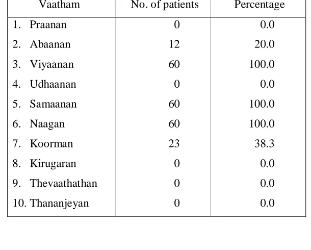
Vaatham – Commonly affected Vaatham are Viyaanan, Abaanan, Samaanan, Naagan and
Koorman.
Derangement of Viyaanan (Vaayu + Earth) leads to pain in the cervical and dorsal
spine, pain along the upper limbs, pain like scorpion sting and heaviness of the body.
Derangement of Abaanan (Vaayu+ fire) leads to constipation.
Involvement of Samaanan (Vaayu+ Aahayam) leads to imbalance of functions of
other Vaayus.
Involvement of Naagan leads to sluggishness and mental depression
Involvement of Koorman leads to burning sensation in eyes, diminished vision.
Pittham – commonly affected Pittham is Saathaga Pittham
Involvement of Saathaga Pittham - produces the features like mental depression and
difficulty in performing regular duties because of the pain in the neck and upper limbs
Kabam – Avalambagam, Tharpagam, and Santhigam are affected.
Involvement of Avalambagam leads to imbalance of functions of other Vaayus.
Derangement of Tharpagam produces burning sensation in eyes
Derangement of Santhigam produces pain and stiffness in joints.
Udal Thaathukkal :
Panchaboothas forming the basic constituents of these Thaathukkal get deranged.
Saaram - water + water
Oon - Earth + water
Kozhuppu - Earth + Air
Enbu - Earth + Earth
Narambu - Earth + Vaayu
Thol - Earth + Fire
Gnanenthiriyam
Panchaboothas forming the basic constituents of these Gnanenthiriyams are deranged.
Commonly affected Gnanenthiriyams are Mei, Kan.
Mei – Fire + Air,
Kan – Fire + Fire
Kanmenthiriyam:
Panchaboothas forming the basic constituents of these Kanmenthiriyams are
deranged.Commonly affected Kanmenthiriyams (organs of action) are Kai, Eruvaai.
Kai - Earth + Vaayu,
Eruvaai - Earth + Earth
CLINICAL FEATURES:
The signs and symptoms of Ceganavaatham are described in the following verses.
•
In Yugi Vaithya Chinthaamani and Pararaasasekaram
" K Gin , %$k! m
e " $i$N i e4+
K $,$el8 i 9k!m
irk! 8 NΝ k !m
RK i$N NΝ e$iN N/m
RRy +ν i( ik Νm
K e d" R k!m
e ii/ ,rk + "
" N/r i n 8 Gin ,%$k! m
iN/8 $i$N i e4+ i9k!m
N" iirk !m i i!6'N/!m
N/r !%i6 i inν ".
- $$ $m.
1.
Pain in the neck
2.
Radiating pain in the shoulders and upper limb
3.
Heaviness of the body.
4.
Mental depression
5.
Giddiness
6.
Burning Sensation of the eyes
7.
Constipation
8.
Pain like scorpion sting
9.
Tingling sensation and numbness of the upper limbs.
•
In Chikitcha Rathna Dheebam Part II (Kannusamy Vaithya Chinthaamani)
-Page 61
The clinical features have been described as,
1.
Swelling below the neck and above the groin
2.
Swelling in both lower limbs
3.
Mental depression
4.
Burning Sensation of the eyes
5.
Retention of urine
DIAGNOSIS:
Diagnosis of Ceganavaatham in Siddha is based on,
•
Ennvagai Thervu (eight types of examination)
and also on other factors like -
•
Uyir Thaathukkal
•
Udal Thaathukkal
•
Gnanenthiriyam
•
Kanmenthiriyam
Ennvagai Thervu (Eight types of examination):
"4"p $im 4 4im e%i i%i
m Ei$i 9$Fm".
"eyk!i 4iei i%i 4i9m k!i".
- $ r (4y 4/l m1, k m 253)
The eight types of examination are:
1.
Naadi (Pulse reading)
2.
Sparism (Tactile sensation)
3.
Naa (Tongue)
4.
Niram (Colour)
5.
Mozhi (Speech or Voice)
6.
Vizhi (Eye)
General definition for each type
Features in Ceganavaatham
1.
Naadi:
Naadi means a vital force responsible for birth - Agathiyar
This vital force is divided into three humours -Vaatham,
Pittham and Kabam. It can be assessed in 10 sites. The commonest site is
radial artery.
In Ceganavaatham the naadi felt are,
Vaatha Pittham
Pittha Vaatham
Pittha Kabam
KabaVaatham
-"e il inj r
---
l ip kk nm.
- "iil il i!"i#$ %$ #$,
'($ )i ey)m in,
- siµe-k ed/p i!"i(il ."
- s0d/(i!"iν$n 'ikils
- y!l m1, km 171, 174,175
2. Sparism:
By Sparism the temprature of the body, smoothness or
roughness, dryness, hard patches, abnormal growth, sweating, swelling,
tenderness and nourishment can be felt.
In Ceganavaatham patients:
General body temperature – slight warmth
Tenderness present in neck & upper extremities
3.
Naa:
Colour, Coating, dryness, movement, deviation, sensory changes,
ulcer, conditions of the tooth and gums are noted.
In Ceganavaatham patients:
4. Niram :
Colour of skin, mucous membrane, hair and nail are examined.
In Ceganavaatham
General colour – mixed colour (thonthaniram due
to mixed thodams)
5. Mozhi :
Disturbances in voice, hoarseness of voice, etc are assessed.
In Ceganavaatham
No change or disturbance of voice are found
6.Vizhi:
Testing for – acuity of vision, colour – redness, pallor, whiteness,
any burning sensation, excessive lacrimation.
In Ceganavaatham
Burning sensation of eyes is present. In aged
patients acuity of vision is diminished.
7. Malam:
The waste and excretory products of body are called as malam. The
feces should be semi-solid without hardness & looseness.
Nature, quantity, colour, odour, froth, presence of blood and mucus
are noted.
In Ceganavaatham
8) Moothiram :
The urine is examined by two methods.
•
Neerkuri
•
Neikkuri
Neerkuri:
Urine is collected after taking a well balanced diet (Appetite corrected, seasonally
correlated), which do not alter the three thodams. It should be examined within 3-3/4 nazhigai.(90
minutes)
" rkie m enjen
i µ ."
- r rki eyki $l (i' (')*+ ,(km, .km 334)
In Neerkuri the Niram (Colour), Manam (Odour), Nurai (froth), Eadai (specific gravity)
and Enjal (quantity) is noted. Apart from these, the frequency of urination, presence of abnormal
constituents such as sugar, Protein etc., and sediments are also noted.
Neikkuri :
The collected urine is kept in a glass bowl and is placed under direct sunlight. A drop of
gingelly oil is added and nature of Neikkuri is noted. If the drop of oil lengthens like a snake it
indicates Vaatham, if it spreads like a ring it indicates Pittham, if it appears like a pearl it indicates
Kabham.
"ae3 N5 3q )ms - )y)l .)m 1, .km 279
s<=i .)R.in aq .i'ms - )y)l .)m 1, .km 279
sµ'e)' iRin e)=ien . "- )y)l .)m 1, .km 280
UYIR THAATHUKKAL
VAATHAM
Vaatham
Physiological Function
Features in Ceganavaatham
1) Praanan
Inspiration and Expiration responsible
for sneezing, coughing and belching
Not affected
2) Abaanan
Acts with downward movement
Affected
constipation present
3) Viyaanan
Helps in various movements in our
Body and also responsible for sensation
Affected.
Restricted neck movements, radiating
pain in shoulder and arm, tingling
sensation, numbness.
4) Uthaanan
Regulates the higher functions of brain,
responsible for physiological reactions
like Hiccough and Vomiting.
Not affected
5) Samaanan
Regulates all other Vaayus
Affected
Due to derangement of other Vaayus
6) Naagan
Responsible for intelligence, helps in
opening and closing of eyes
Affected.
Sluggishness and depressive mood in
aged patients
7) Koorman
Responsible for lacrimation , helps in
visualization of all things of world
Affected.
Burning sensation in eyes, in aged
patients acuity of vision is diminished
8) Kirugaran
Produces sensation from tongue and
nostrils, hunger, sneeze and cough
Not Affected
9) Thevathathan Responsible for laziness, rotation of
eye balls
Not Affected
PITTHAM
1) Anar Pittham
Digests all the ingested particles
Not affected
2) Ranjaka Pittham
Increase the blood & gives color to the blood
Not affected
3) Saathaka Pittham
Execution of work prompted by the mind
Affected.
Neck pain and Restricted
movement
4) Aalosaka Pittham
Responsible for visual perception of things
Affected in aged
5) Praasaka Pittham
Gives color to skin.
Not affected
KABAM
1) Avalambagam
Controls the other 4 types of Kabam
Affected
Santhigam affected
2) Kilethagam
Moistens the food
Not affected
3) Bothagam
Helps to know the taste.
Not affected
4) Dharpagam
Gives cooling effect to the eyes
Affected
Burning sensation of eyes
present.
5) Santhigam
Gives lubrication to the joints
Affected
GNAANENTHIRIYAM
1) Mei
Feels all types of sensations
Affected.
Numbness present in both upper limbs
2) Vaai
Taste
Not affected
KANMENTHIRIYAM
1)Kai
Works done by the hands
Affected
Radiating pain, tingling sensation, numbness.
2)Kaal
Walking
Not affected
3)Vaai
Speaking
Not affected
4)Eruvaai
Defaecation
Affected; constipation present.
5)Karuvaai
Reproduction
Not affected
UDAL THAATHUKKAL
1)Saaram
Strengthen the body and mind
Affected.
weakness, mental depression
2)Senneer
Preserves brightness, boldness, power
and knowledge to the mankind.
Not affected
3)Oon
Gives structure and shape to the body,
responsible for body movements.
Early Stage- Not Affected
Late Stage - Affected
4)Kozhuppu
Lubricates the joints
Affected
Stiffness present
5)Enbu
Gives shape and stability to the body.
Products soft internal organs of the body.
Affected
Degenerative changes present
6)Moolai
Present in the bones and gives strength
Not affected
7)Sukkilam&
Suronitham
Meant for reproduction
Not affected
NOI KANIPPU VIVAADHAM (DIFFERENTIAL DIAGNOSIS)
Some other types of Vaatha diseases mimicking Ceganavaatham are mentioned. Careful
and clear history taking and examination will reveal the diagnosis.
They are,
1.
Pei Vaatham
2.
Kumba Vaatham
3.
Kanda kiraga Vaatham
4.
Sirakkamba Vaatham
5.
Paanikkamba Vaatham
1. PeiVaatham (y m)
"eRim e m ei enj i k
Ri ei em Ri
ky e" #$m em %i uRi ' iire)
u)iyp i+k# e' ,m
k)iy y" k m
)i)i$eN ' y )" ).".
- $l 276, 6i )i i")'i - 800, km 86
The clinical features are,
1. Pain and swelling in neck, abdomen, upper and lower limbs
2. Weakness of hand muscles, difficulty in holding things in the hand
3. Vomiting
4. Giddiness
5. Swelling all over the body
2. Kumba Vaatham (m m)
"i, )L% )in % m
,i" e) i #N$m i, n.e ." )νm
iip< ei# Νm
i, +pnj i> )n.iR
>?Ri ik%z ,i µN$y
ai, +ki ,?n Νm a, m )" ).".
- $l 264, 6i )i i")'i - 800, km 82
The clinical features are,
1. Burning pain in shoulder and upper limbs
2. Burning sensation in the cheek and eyes
3. Twitching over the scalp
4. Spasmodic pain in the lower abdomen
3. Kanda Kiraga Vaatham (N i m)
" p Ri e iiiil iNi elm e !Ri
Νky% & 'p l µ )%& i
µ*i iri i ,Nm n*p .ed
i N i*in N' ".
- l 303, 5i *i i6i - 800, km 95
The clinical features are:
1. Pain in the throat, chest and occipital region and all over the body
2. Breathing through mouth
3. Backache
4. Sweating on face and pain in ribs
5. Loss of appetite
4. Sirakkamba Vaatham (ikm m)
"my uirN miR 'ki*
el kip 'k m m; iN i,; <
iNm k m imy in ;ik Νm
e)%&; edii* i m imy* ;ik p' µNm
ikm en ep ".
- l 300, 5i *i i6i - 800, km 94
The clinical features are
1. Stiffness of neck
2. Deafness
3. Difficulty in using lower and upper limbs
4. Confused thinking / impaired memory
5. Difficulty in breathing
6. Yawning, excessive sleeping
5. Paanikkamba Vaatham ( ikm m)
"rky y, y eyi i>iR ii ?Ν R> rky @* k R>
k; iN ii. µNm Ark kil 6r%i R>
ui e; µr Νm rky yid a* m
6ikm *in ; ".
- l 266, 5i *i i6i - 800, km 83
The clinical features are,
1. Gaseous accumulation and anorexia
2. Tingling sensation and numbness of upper limbs
3. Tremor of upper limbs
4. Sleeplessness and
5. Dryness all over the body
6. Kazhuthu Vaatham (Koonikiraga Vaatham)
m (ii m)
“E* i.p iF*
iid u<R Νm e!i* m' F* iR m
ak ed iE* iF* ipHm
i eym E* m E*m 6;L 6i nil
y air NF.s
- y .*m, km 89
The clinical features are
1. Stiffness and restriction in movements of the neck
2. Boring pain in neck
3. Thickening of nerves in neck
THE ANATOMY
The Vertebral Column:-
The vertebral column which lodges and protects the spinal cord, its meninges, and
the continuation of the central nervous system lies in the dorsum of the body. It forms a
pillar which contains 33 segments and lengths about 70cm in an average male and 60cm in
a female. It supports the body weight and transmits it to the ground through the lower
limbs.
The segments can be divided into cervical, thoracic, lumbar, sacral and coccygeal
segments. The cervical segment has seven vertebral bones, thoracic twelve, lumbar five,
sacral five and coccygeal four. All are separate bones except the sacrum and coccyx.
The Curvatures of the Spine:-
There are four curvatures in the vertebral column. They are two primary and two
secondary curvatures.
The primary curvatures are the thoracic and the sacral. They are convex
posteriorly. The secondary curvatures are the cervical and lumbar. They are anteriorly
convex. The cervical curvature becomes prominent when the child is able to hold its head
up and fit upright. The lumbar curvature appears by 12-18 months after the child starts
walking. A slight lateral curvature is seen in the upper thoracic region. It is curved to the
right in right handed persons and vice versa.
The General features of the Vertebrae:-
The vertebrae can be divided into vertebral body and a dorsal vertebral arch. The
vertebral arch has 2 pedicles, 7 processes and 2 laminae. Pedicles are thick bars projecting
backward from the body. The laminae are vertical plate like structures, fuses together to
form spinous process. The spinous processes projects downwards and are the lever for the
muscles. The articular processes are four in number, bearing the articular facets and
articulate with the adjacent vertebrae. Transverse processes project laterally from the
The Cervical Vertebrae:-
The cervical segment of vertebral column contains 7 vertebrae. The cervical
spine is divided into anterior & posterior columns. The component parts of anterior
columns are
• Anterior longitudinal ligament (ALL)
• Annulus fibrosus (ANN)
• Unco vertebral joint
Posterior column consist of
• Nerve root (NR)
• Facet
• Superior ligament
• Posterior longitudinal ligament
There are 8 pairs of cervical Nerves. Each nerve root contains sensory, motor,
sympathetic fibres that innervate the upper extremities. The first, second and the seventh
Cervical Vertebrae are atypical and the third to sixth are typical. They are smaller and
delicate than the thoracic and lumbar vertebrae. All the cervical vertebrae have a foramen
in the transverse process known as foramen transversarium. This is identical to the
cervical vertebrae.
Typical Cervical Vertebrae:-
1. Body:
It is small and oval. It’s superior surface is concave transversely with
upward projecting lips on each side and its inferior surface is saddle shaped,
convex from side to side and concave from before backwards.
2. Vertebral Foramen :-
It is larger than the body and triangular.
3. Vertebral Arch :-
i. Pedicles :
These are short and directed outwards and backwards from the middle of
postero lateral parts of the body and they form the postero medial wall of the
ii. Laminae :
These are long and narrow, being thinner above than below.
iii. Articular Facets :
The superior and inferior articular processes form the articular pillars which
project laterally at the junction of the pedicle and the laminae. The superior
articular facets are flat and directed backwards and upwards. The inferior
articular facets are also flat but directed forwards and downwards.
iv. Transverse Processes :-
Each transverse process is short and pierced by foramen transversarium. Each
process has an anterior and posterior root which ends in tubercles joined by the
costotransverse bar. The anterior tubercle of the sixth cervical vertebra is large
and is called carotid tubercle.
v. The Spine :-
It is short and bifid.
Foramen Transversarium:-
It transmits the vertebral artery, vertebral veins and sympathetic plexus.
The Atypical Cervical Vertebrae:-
1. Atlas :-
It is the first cervical vertebrae which lodges the skull. It has no body and spine. It
has anterior and posterior arch, right and left lateral masses and transverse processes. The
anterior arch bears an anterior tubercle in the anterior aspect. Its posterior aspect bears an
oval facet which articulates with dens. The posterior surface of the posterior
arch has a median posterior tubercle. The two lateral masses bear an elongated superior
articular facet for atlanto-occipital joint and an inferior articular facet for atlanto axial
joint.
The transverse process of atlas is long and thick. It is pierced by the foramen
2. The Axis :-
The Axis has a peg like projection in its upper part of the body known as the dens
(or) odontoid process. It has a circular facet anteriorly articulating with atlas. There are
two articular facets on either side of the dens on the upper surface of the body. The
laminae are thick. The spine is large and bifid. The transverse process is small and
possesses a tubercle in its tip.
3. The Seventh Cervical Vertebrae :
It is also known as “Vertebral Prominent”. The transverse process does not posses
anterior tubercle. The foramen transversarium is small (or) absent. It transmits accessory
vertebral vein only. The spine is long.
Palpable parts of Cervical Vertebrae:-
i. The spine of C2 is in the nape of the neck 5 cm, below the external occipital
protruberance.
ii. The spine of C7 where the collar bone crosses the posterior medium line of the neck.
iii. The transverse process of C1 through the anterior border of sternocleidomastoid,
immediately below the tip of the mastoid process.
Inter- Vertebral Discs:-
They are fibro cartilagenous discs interposed between the adjacent surfaces of the
vertebral bodies. They are thicker in lumbar region than in thoracic. Their peripheral parts
are supplied by the adjacent blood vessels but the central parts are avascular.
They receive their nutrients by diffusion from spongy bone of adjacent
vertebrae.The central portion of disc is known as Nucleus Pulposus and the peripheral
zone is know as Annulus Fibrosus. The central portion is made up of gelatinous mucoid
material and it is composed of around 80-90% water. . On aging it is converted into fibro
cartilagenous material and its water binding capacity is reduced. Annulus Fibrosus (outer
ligamentous ring) which hydraulically seals the nucleus and this annulus fibrosus contains
collagen bundle in periphery & fibro cartilaginous tissue in the inner part.
The annulus has overlapping radial bands, not unlike the plies of a radial tire.
The thickness of the discs varies daily. In the morning it is thick due to absorption of
Intervertebral Discs – Physiology
1) As a spacer:
Proper spacing of intervertebral disc allows the intervertebral foramen to
maintain its height, which allows the segmental nerve roots to exit spinal level without
compression.
2) As a shock absorber: 3) As a motion unit:
The elasticity of the disc allows motion coupling. So that the spinal segment
can flex, rotate, and bend to the side all at the same time during a particular activity.
4) As a hydraulic cylinder:
The annulus interacts with the nucleus. As the nucleus is pressurized the
annular fibres serve a containment function to prevent the nucleus from bulging or
herniating. The gelatinous nuclear material directs the forces of axial loading outward &
the hoops of annular fibres help to distribute that force without injury.
Joints of the Vertebral Column:-
The vertebrae from the 2nd cervical to 1st sacral are articulated to one another by a
series of cartilagenous joints between vertebral bodies and a series of synovial joints
between the vertebral arches. The vertebral bodies are united by anterior posterior
longitudinal ligaments and by centevertebral disc of fibrocartilage.
1. Atlanto Occipital Joint :-
It is a synovial condyloid variety.
Articular ends:-
Superiorly - Occipital condyles.
Inferiorly - Superior articular facet of the atlas.
Adjacent structures - Ligaments, capsule, anterior & posterior occipital
membranes.
Blood supply - Vertebral artery
Ligaments:-
1. Capsular ligament
2. The Anterior Atlanto-occipitial membrane.
3. The posterior Atlanto-occipital membrane.
Movements:-
Flexion, extension, and slight lateral flexion are possible.
2. Atlanto Axial Joints :
Comprise of
a. A pair of lateral atlanto-axial joints.
b. Median atlanto-axial joint.
a. Lateral atlanto-axial joints
Synovial joint - Plane variety
Articular ends - Inferior facets of atlas and the superior facets of axis.
Ligaments - Ant. longitudinal ligament and ligamentum flavum
b. Median atlanto-axial joint.
Synovial joint - Pivot variety
Articular ends - Between the dens of axis, antrier arch of atlas
Ligaments - Transverse ligament
Movement - Rotatory movements around a vertical axis
Ligaments between axis and the occipital bone:-
1. Membrana tectoria
2. Cruciate ligament
3. Apical ligament of dens
4. Linear ligament
The Unco Vertebral (Luschka’s) Joints:-
Luschka’s joints are not true synovial joints; which develop as a result of
degenerative changes in the edges of the disc in early adult.
Luschka’s joints are important, because
i. They are the commonest site of osteophyte formation. The osteophytes may
ii. Vertebral artery lies lateral to the joints intruding on the canal can cause
distortion of the artery and leads to Vertibro Basilar Insufficiency in
athero-sclerotic vessels.
Movements of the Vertebral Column:-
The greater thickness of the discs in the cervical and lumbar regions as compared
with the thoracic region is associated with the greater individual range of movements
occurring in thoracic regions.
Flexion (or) forward bending, extension (or) backward bending, lateral flexion and
rotation are possible in vertebral column. Numerous muscles are attatched directly on the
vertebrae.
Movements of the Head and Neck:-
Movements Muscles Nerve Supply
Sternocleidomastoid Accessory ventral rami of cervical
spinal nerves C2.C3,C4
Longus Coli Cervical ventral rami C2 – C6.
Longus capitis Cervical ventral rami C1 –C3
Flexion
Rectus capitis Anterior C1 ventral ramus
Splenius cervicis and capitis Dorsal cervical nerve
Erector spinae Dorsal rami
Rectus capitis posterior major and
minor
Dorsal rami C1
Obliques capitis Superior, C1 – Dorsal ramus
Extension
Trapezius Accessory
Sternocleido mastoid Accessory, ventral rami of cervical
spinal nerves C2,C3,C4
Scalene Cervical ventral rami C3 – C8
Longus Coli Cervical ventral rami C3 – C8
Levator scapulae Cervical ventral rami C3, C4, C5
Rectus capitis C1 – ventral ramus
Splenius Cervical dorsal ramus
Lateral flexion
and rotation
Longismus obliques capitis superior
and inferior.
Structures passing through:
a. Foramen transversarium
Vertebral artery, Vertebral vein, Plexus of sympathetic nerve
b. Intervertebral foramen
Spinal nerves form dorsal medulla
Blood supply of Vertebral Column:-
The vertebrae and longitudinal muscles attached to them are supplied by segmental
arteries. The arteries give multiple small branches to the vertebral bodies. The extensor
muscles of neck are supplied by the occipital, deep cervical and transverse cervical
arteries.
Venous Drinage:-
The Internal vertebral venous plexus lies within the vertebral canal, but outside the
spinal dura. It received tributaries from
i. The vertebrae through the basilo vertebral veins.
ii. The meninges and the spinal cord.
The internal vertebral venous plexus is drained by the intervertebral veins, which
pass out through the intervertebral foramen. Here they are joined by the tributaries from
the external vertebral and sacral veins. The internal venous plexus communicates with the
CERVICAL SPONDYLOSIS
Nomenclature
Cervical - Neck region
Spondylosis - Vertebral ankylosis
Synonyms and related keywords:
Cervical Degenerative Joint Disease, Cervical Degenerative Disk Disease, Cervical
Osteoarthritis, Cervical Spondylotic Myelopathy, Disk Degeneration, Degenerative
Cervical Disease, Osteophytic Bars, Cervical Radiculopathy.
Definition:-
Cervical spondylosis is a disorder characterised by increasing degeneration of the
intervertebral disc, with subsequent changes in the bones and soft tissues. Spondylosis is
usually asymptomatic. Symptoms are usually manifested by encroachment on local neural
elements such as cervical nerve roots, spinal cord, vertebral artery (or) sympathetic nerves.
The symptoms and signs appear to be related to the cause and time course of compression
as well as the structures being compressed.
Epidemiology:
Age: Cervical spondylosis is present in 5 – 10% over the age of 20 to 30 years, 20-25% by the age of 50 years and 70-85% by the age of 65 years.
Sex: Men are affected more than women.
Location:
• C5-C6 levels are commonly involved due to maximal movements occurring at this cervical spine.
AETIOLOGY
I. Degenerative Causes
They are primary & secondary
• Primary - Senility, genetic factors, metabolic factors and manual labour
• Secondary - osteoarthritis, rheumatoid arthritis, metastatic carcinoma or lymphomas of the spine and TB spine.
II. Injury
• Automobile accidents with “Whiplash” injury, athletic injury,
• Sudden jerks on the arms during fall down,
• Previous injury with fracture or disc prolapse
III. Occupational causes
The physical discomfort, which arises through an occupation is occupational
stress. The physical strain, intensity of work and duration of working hours all constitutes
the occupational strain
IV. Hereditary factors
• Congenital narrowing of the cervical spinal canal (myelopathy is often seen when canal’s sagital diameter is 12 mm or loss)
• Segmental defects – Hemi vertebra, fused vertebra.
V. Aquired narrowing of cervical spinal canal due to
• Osteophytes
• Ossified posterior longitudinal ligament (OPLL)
• Facet joint hypertrophy (results foraminal stenosis & compression of foot of radicular artery)
• Hypertrophied ligamentum flavum (Compress the cord during extension).
VI. Outgrowths of bone that some times occur with aging.
VII. Inter vertebral disc protrusions are commonest in the cervical region which is due
to degeneration of the intervertebral disc and if it involves several discs with
osteoarthrosis liable to interfere with blood supply of the cord and thus leads to further
PATHOLOGY
In cervical spondylosis, involvement of following structures has to be considered:
• Intervertebral disc
• Uncovertebral joints
• Apophyseal joints
• The foramina(intervertebral) and
• The transverse foramina.
Intervertebral disc
All the three parts of disc cartilage plate, nucleus and annulus are involved.
Cartilage plate:
First the cartilage plate thins out and cracks. Fissuring and erosion is
common. The whole plate is replaced by fibrous tissue.
Nucleus:
Nucleus becomes fibrous with degeneration. The process of dehydration
occurs and concludes with reduction in water binding mucoprotein.
Annulus:
Annulus undergoes some changes as in nucleus. Focal necrosis and
calcification is common. They form hard ridge within the cervical canal.
Osteophytes are formed as a result of instability producing stress on the
periosteum.
Uncovertebral joints
The uncovertebral joints are most affected as C5-C6 and C6-C7 levels. Progressive
decrease in disc height the uncinate process approximates against the vertebral body
undergoes erosion and formation of osteophytes.
Apophyseal joints
They may remain unaffected for long time. When they subjected to heavy weight
pathological changes like erosion, degeneration, lipping, and osteophyte formation occurs.
The foramina (intervertebral and transverse)
Foramina are narrowed by fibrosis and posterior longitudinal ligament thickening
Vertebral Artery
Spondylotic changes in the foramina transversarium are not uncommon. They can
cause buckling or tortuosity of the vertebral arteries which is commonly found in older age
group. The artery may also be affected by uncovertebral and apophyseal osteophytes.
Vertebral artery compression is most common at C4/5 and C5/6 levels.
PATHOGENESIS
1. Cervical spondylosis is very common and histological evidence of degenerative
changes is present in virtually, even present over the age of 70.
2. The disc degeneration the primary event which is a progressive decrease in the
degree of hydration. Glycoproteins diminish in size and number and their ability to
retain water diminishes. This results in loss of disc height, disc fibrosis and annular
weakening. Adjacent vertebral bodies approximate each other and uneven abnormal
movement in the affected areas probably results in oseteophyte formation. These
occur at all the joints, namely the disc, zygoapophyseal joints and the neurocentral
joints of luschka. Though osteophyte formation may be the body’s attempt to
stabilize the joints their growth.
3. Osteophytes may form posteriorly with osteoarthritis of the apophyseal joints and
also anteriorly in relation to degenerative changes and narrowing of the
intervertebral disc with sclerosis of the bony end plates. The osteophytes may cause
symptoms by encroaching on the spinal nerve foramina or in narrowing of the spinal
canal and cord compression or in the cervical regions on the vertebral artery
foramen. In the cervical region intermittent pain and discomfort may be followed
eventually by stiffness and limitation of movements.
4. The predisposing factors which may accelerate of these changes viz.
• Occupation requiring repetitive motion and chronic flexion of the
• Previous injury with fracture or disc prolapse.
• Segmentation defects like hemivertebrae or fused vertebrae.
5. Factors responsible for Myelopathy in cervical spondylosis:
• Uncovertebral osteophytes cause anterior compression of cord
• Bony ridges on the posterior vertebral bodies cause central compression on the cord.
• Zygapophyseal osteophytes causing posterior compression
• In curving of the ligamentum flavum causing posterior compression on the cord
• Development of narrow cervical canal
• Dynamic effect of narrowing of the cervical canal
• Calcification of the posterior longitudinal ligament
• Teethering of the roots to the osteophytes
• ArachNoiditis, postoperative scar
• Interference of blood supply of cord.
CLINICAL FEATURES
Sympotms and Signs:
Symptoms and signs of cervical spondylosis can be acute, subacute or chronic
occasionally acute exacerbation of chronic symptoms can occur.
Symptoms:
Symptoms can be described as
1) Neck pain
2) Radiculopathy
3) Headache
4) Myelopathy
5) VBI [Vertebro Basilar Insufficiency]
6) Autonomic symptoms
1) Neck pain:
Pain is present in nape of the neck and its nature is aching or boring quality, which
radiates to the shoulder blades top of the shoulder, upper arm and hands or back of the
head. Clinically it is very difficult to decide, which disc is responsible to pain. Patients feel
2) Radiculopathy:
Results from nerve root compression and consists of shooting pain (pins and
needles) radiating along the dermatomal distribution of particular nerve root usually into
forearm or fingers. There is also frequently referred pain and tenderness along the
scapula’s medial border.
3) Headache:
Headache is a common symptom; its pathogenesis is not fully understood. It is
more a pain than a headache usually located in occipit on bothsides. It spreads to the
temple or eyes. It is described as a tight band round the head.
4) Myelopathy:
Myelopathy can be classified in varies ways and depends on the involvement of the
lateral or medial cord or vascular involvement. The signs may be a mixure of upper motor
neuron signs in the lower limbs and lower motor neuron signs in the upper limbs.
Generally myelopathy may be predicated by central disc herniation, but is more
commonly the result of spondylytic change superimposed on a congenitally narrow
cannal. Motor weakness is rare. If they occurs there may be marked wasting of the
muscles.
5) VBI [Vertebro Basilar Insufficiency]:
VBI usually requires a combination of arterosclerosis & osteophytes intrusion into
the foramen transversorium. The symptoms of this insufficiency are typically a brief attack
of giddiness without loss of consciousness and generally brought by head movements.
6) Autonomic symptoms:
Vertigo, flushing, tinnitus & visual blurring are the autonomic symptoms produced
by cervical disc diseases. These may be mediated by sympathetic distribution to the
sinuvertebral nerves from stellate ganglion.
Signs: 1. Motor
• Atrophy of the hand musculature (Intrinsic muscle atrophy)
• Muscle weakness
2. Sensory
Loss of vibratory sense or proprioception in the extremites especially in the feet,
3. Reflexes
• Hyper reflexia
• Ankle clonus 4. Babinski’s sign
5. Lhermitte’s sign positive (Electric shock like sensations down to the center of the
back following flexion of the neck)
Summary of the site of lesion
Cervical spondylosis can produce cord compression (upper motor neuron signs) or root
compressions (lower motor neuron signs)
C5 Motor Raised elbows (axillary n.)
Reflex Biceps (musculocutaneous n.)
Sensory Upper, lateral arm, near/over deltoid (axillary n.)
Pain Upper, lateral arm, never below elbow
C6 Motor Elbow supination (radial n.) / pronation (median n.)
Reflex Brachioradialis (radial n.)
Sensory Lateral forearm (musculocutaneous n.)
Pain Lateral forearm, possibly into thumb
C7 Motor Elbow extension (radial n.)
Reflex Triceps (radial n.)
Sensory Over triceps, mid-forearm, and middle finger
Pain Deep pain in triceps, front, and back of forearm & into middle finger
C8 Motor Thumb index pinch (ant.interosseus n. off median n. at the elbow )
Sensory Medial forearm (antebrachial cutaneous n.)
Pain Medial forearm, into the 2 medial fingers
T1 Motor Finger abduction (ulnar n.)
Sensory Medial arm (brachial cutaneous n.)
DIAGNOSIS
Mainly based on X – ray
1) X- ray cervical spine
• Anteroposterior (AP) view
• Lateral view
• Right oblique
• Left oblique
• AP odontoid view
Plain x-rays can demonstrate loss of disc space height, anterior and posterior
end-plate osteophytes, fusion or instability. A lateral view will also show the
antero-posterior diameter of the spinal canal; and if this is less than 14 mm then cord
compression is a real possibility.
2) CT scan
A CT scan of your spine uses X- ray technology, but produces a more
detailed image than X- ray can.
3) Myelogram
This test involves generating images using X-rays or CT scans after dye is
injected into the spinal canal. The dye makes areas of your spine more visible.
4) Electromyography(EMG) and Nerve conduction study
Needle EMG, nerve conduction studies and evoked potentials may help to
differentiate spondylotic neurological problems from motor neurone disease,
multiple sclerosis, peripheral nerve compression and so on.
5) MRI (Magnetic Resonance Imaging)
MRI uses a magnetic field and radio waves and can produce detailed,
cross-sectional images of your spine. These tests may help your doctor determine
DIFFERENTIAL DIAGNOSIS
1. Motor neuron disease
2. Multiple sclerosis
3. Syringomyelia
4. Spinal cord tumors
5. Tropical spastic paresis from HTLV – 1 infection
6. Amyotrophic lateral sclerosis
7. Carcinomatous infiltration or radiotheraphy
8. Peripheral nerve lesions (distal ulnar or median nerve)
9. Reffered pain
• Cardiac ischaemia
• Sub-diaphragmatic lesions
• Gall bladder lesions
COMPLICATIONS
1. Cord compression- Quadriplegia, spastic gait, affecting the bladder.
2. Nerve root compression – Neurological injury, Brachialgia.
TREATMENT OF CEGANAVAATHAM
Treatment – Directed towards relief of untoward symptoms, leaving cure of the disease to
natural forces.
y+ yµ)l + a )'ikm
y+ ypD el. - )ikL
uRn aE#m i'iE#m ,µm
Rn )iD el. - )ikL
As per Thiruvalluvar, after diagnosis and finding the etiology, a physician prescribes
the line of treatment based on patient’s condition, condition of the disease and climatic
condition.
For Ceganavaatham patients, the following line of treatment is given,
Purgation
Internal Medicine
External Medicine
Patthiyam
Thokkanam
Exercise
Kanmaneekkam
Purgation:
"i.)l )m )m" - y$l m 1, km 248
"ai")im )m a$ ,)i.il" - i) D >km km 97
Purgation is used to normalize the vitiated Vaatham and eliminate other toxic products
of digestion, metabolism and catabolism.
Purgative medicine: Sitthaathi ennai – 15 ml with Sombu kudineer at early morning on first
day only.
Internal medicine:
External medicine:
Vaatha Noii Thylum – 30 to 50 ml
Patthiyam:
Dietary regimen (or) regulation of diet.
The sort of diet to be observed either simple or rigorous depends on various factors such
as patient’s strength and nature, nature of the disease, quality of medicine, time, climate etc.
The greatness and usefulness of observing diet,
")i)N$m N+)Rp N
)i)N$m N+)L - )i)
id$r i'iL i)ikm id+$,
id$R km i."
- )n eN eyL 600(T.V. mimiLE, Vol V km 139)
Patthiyam supports the treatment and produce successful result.
Substances that should not be consumed are:
"<Ei r inj>i RPik m)m
oEi rp il i D %m - iEie?i
rpi.ip< injiR minj> njd+)D
p <'r Ν)".
- )r) 'i")'i (y$l m 1, km 22)
si,'m <EieN ' µ),
ee ' e?i - i,iDi
TzpN$ Dm eN)i eLi)ii
)%zpi e)i %".
- )n eN 601 (T.V. mimiLE, VOL V km 141)
Salt, tamarind, mustard, gingelly - any of them should be prohibited as warranted by
the medicine taken. Further flesh, fish ash, pumpkin, tobacco, horse gram, and lustful
In Siddha System of Medicine “Sirappu Maruthuvam” deals with cure and also
prevention of diseases especially with special treatment techniques such as Yogam,
Thokkanam, Varmam, in addition to internal medicine and external medicine. The author has
explained here the methods of Thokkanam and and also some exercise for treating
Ceganavaatham patients
Thokkanam:
Thokkanam is one of the oldest and simplest forms Siddha treatment. It improves
muscle tone, stimulates blood circulation and helps elimination of waste products throughout
the body. At its best thokkanam has the potential to restore the individual physically, mentally
and spiritually.
e)k'm - e)k + a'm
e)k - )l
a'm - a'" ey)l
Thokkanam - A process consisting in striking with fist and then pressing the body or
its part of a person suffering from some ailment. On account of this treatment the body grows
strong, the skin gets luster and the person gets sound sleep.
e)k')i .,i)" )l X'id
ik #kinj %νm - eyk)i
<d+km <'rDi i)ikm
d$ a,D,m r. (%m - )
- edN< Yl(ir a m, km 30)
r).i e)k')in el p. - )
i")µm )m i'i) i'ipD ep.
l,. i$r eni E, - i'i
l,i eyi.iR )ip" )i E,
)d$,ikl i+)l µl )" -
d$,) ,i)l , )l "
a"),iv en .)i")i.m - i)il
i) .ED el,p + .m
The nine techniques mentioned in the verse above are the thokkanam procedures
described in our Siddha system. In our inpatient ward and outpatient department, the author
treated the patients of Ceganavaatham with the following thokkanam methods.
1. Azhutthal
2. Piditthal
Exercise:
Simple movements of the head ,neck and upperlimbs were prescribed to patients of
Ceganavaatham.
Expiation:
(Kanmaneekkam
)Siddha literature says, poorvakanmam is one of the reasons for diseases among mankind.
It should be expiated.
",i, ")n" )%enl
nL )p< $, )l
e)Ei. i'ed$l EL ed$l
e)y), il d$)%m
eEi). ,rk '%)l
ene)n )el, i$" m
?i.y ")ip+ ey
)i )i)p i ey".
- $l 57, aS)ir n N$m- 300, km 23
To expiate misdeeds of kanma, planting trees, establishing gardens, laying roads and
pathways, digging wells, pools, lakes, construction of temples, donating ornaments to poor
PROTOCOL
AN OPEN TRIAL OF SARVAANGAVAATHA CHOORANAM AND
VAATHA NOII THYLUM FOR THE TREATMENT OF CEGANAVAATHAM
(CERVICAL SPONDYLOSIS)
BY
Dr. M. PRATHIBA
1. BACK GROUND
Cervical spondylosis is defined as arthrosis of the posterior intervertebral joints in
the cervical vertebrae. It is common in the middle aged and in the elderly particularly in
those whose occupation involves a posture of prolonged neck flexion.
NEED FOR THIS STUDY
The Modern System of Medicine gives temporary relief for Cervical spondylosis.
Commonly used methods are NSAIDS, traction, cervical collar. According to the literature
of Aathmaratchamirtham and Kannusamy Vaithya Chinthamani, Sarvaangavaatha
Chooranam and Vaatha Noii Thylum are the most efficient drugs for Ceganavaatham
2. AIMS
a)
Primary aim:
To find out the efficacy of Sarvaangavaatha chooranam and Vaatha Noii
Thylum for Ceganavaatham
b)
Secondary aim
: To evaluate the side effects of the trial drug, if any.3. POPULATION AND SAMPLE
POPULATION
A) About the disease:-
In Siddha literature the clinical features of Ceganavaatham according to Yugi
B) According to modern aspect, clinical features of cervical spondylosis:-
1. The patients complain of pain in the neck with or without radiating down to arm.
2. There will be diffuse tenderness in the cervical spine with limitation of all
movements.
3. The neck is held rigidly and neck movements may exacerbate pain.
4. Paraesthesia and sensory loss may be found in the affected segment.
5. Headache in the occipital region at morning.
6. Burning and tingling sensation in the upper limbs.
7. Giddiness (or) drop attacks precipitated by neck movements.
SAMPLE
Ceganavaatham patients attending Ayothidoss Pandithar Hospital of National
Institute of Siddha, Tambaram Sanatorium, Chennai - 47.
4. SAMPLE SIZE
It is proposed to study a sample of 60 patients.
a) INCLUSION CRITERIA
Definition of the disease:
Cervical spondylosis is defined as a disorder characterised by increasing
degeneration of the intervertebral disc, with subsequent changes in the bones and soft tissues
assessed by using clinical, physical and radiological methods (Reduction of the
1. Ceganavaatham patient
2. Age between 25 years and 60 years
3. Willing to be admitted as In-patient in our ward for minimum 25 days and continue
the remaining treatment in OPD or Willing to attend OPD once in 8 days for 48 days
4. Willing to undergo radiological investigation before and after treatment
5. Willing to give blood specimen before and after treatment
b) EXCLUSION CRITERIA
A patient is not eligible for admission to the trial if any of the following is applicable.
1. Any history of trauma, hypertension, cardiac disease
2. Use of intravenous (or) oral narcotic drugs
3. Pregnancy
4. Lactation
5. Patients with any other serious illnesses
c) WITHDRAWAL CRETERIA
1. Development of any adverse drug reaction (ADR)
2. Occurrence of any other serious illness
d) TRIAL DRUG & DURATION
1. PURGATIVE Sitthathi ennai 15 ml with Sombu kudineer at early
morning on first day only.
2. INTERNAL DRUG Sarvaangavaatha Chooranam
1½ gm pack twice a day with hot water.
3. EXTERNAL DRUG Vaatha Noii thylum
30-50 ml for local application twice a day.