A COMPARATIVE STUDY OF CONTINUOUS POSTERIOR
LUMBAR PLEXUS BLOCK AND CONTINUOUS EPIDURAL BLOCK
A COMPARATIVE STUDY OF CONTINUOUS POSTERIOR
LUMBAR PLEXUS BLOCK AND CONTINUOUS EPIDURAL BLOCK
IN TOTAL HIP REPLACEMENT SURGERY
- A prospective randomized single blind controlled clinical trial
DISEERATION SUBMITTED IN PARTIAL FULFILMENT OF THE REQIREMENT
OF THE Dr. M G R MEDICAL UNIVERSITY , CHENNAI, FOR THE DEGREE OF
CERTIFICATE
This is to certify that the dissertation entitled
“
“
A COMPARATIVE STUDY OF
CONTINUOUS POSTERIOR LUMBAR PLEXUS BLOCK AND CONTINUOUS
EPIDURAL BLOCK IN TOTAL HIP REPLACEMENT SURGERY”
is the bonafide
work by
Dr. R. Ravi Raj
, in the partial fulfillment of the requirement for the M.D.
Degree (Anaesthesiology –Branch-X) of the Tamil Nadu Dr. M.G.R Medical
University, Chennai, to be held in March 2007.
Guide:
_____________________ ____________________
Dr. Sarah Ninan, MD Dr. Manickam Ponniah, MD
Professor, Prof. & Head,
Department of Anaesthesiology, Department of Anaesthesiology,
Christian Medical College, Christian Medical College,
Vellore 632004, Vellore 632004,
Tamil Nadu, India. Tamil Nadu, India.
TABLE OF CONTENTS
PAGE NO.
INTRODUCTION
…
5
AIMS & OBJECTIVES
…
7
REVIEW OF LITERATURE
…
8
MATERIALS & METHODS
…
41
RESULTS
…
48
DISCUSSION
…
65
CONCLUSIONS
…
71
BIBLIOGRAPHY
…
72
APPENDIX:
I.
Master Chart
II.
Proforma
III.
Consent form
INTRODUCTION
Total hip replacement (THR) is the most common joint replacement procedure that aims to relieve joint pain, increase mobility and improve the quality of life of the patients with,
chronic degenerative disease of hip joint and patients with proximal femoral fracture.
Anaesthesia and post operative analgesia for THR is a challenge, as the age of the
patients presenting for THR varies, and 60% of the procedures are performed on patients
above 65 years of age1. Most of these elderly patients have significant comorbid conditions
like hypertension, ischemic heart disease, renal dysfunction, diabetes mellitus and obstructive
pulmonary disease, all of which can adversely affect the patient management in
peri-operative period. It is therefore important to choose an effective analgesic regimen with
minimal side-effects to allow timely mobility, optimal functional recovery and decrease
postoperative morbidity and mortality.2
The major intraoperative anaesthetic concerns during THR are blood loss,
cardiovascular instability and hypoxemia. Blood loss is related to intraoperative tissue trauma,
especially the reaming of medullary bone, and increases if an osteotomy of greater trochanter
is performed to improve surgical exposure or with revision procedures. Blood loss is variable,
averaging 0.5L to 1.5L intraoperatively and 300-500ml in the postoperative period3.
Intraoperative blood loss is significantly reduced with regional anaesthesia than general
anaesthesia alone.4 Combination of general anaesthesia with regional techniques has
revealed significantly lower blood loss than regional or general anaesthesia alone.
Pain after total-hip replacement (THR), although variable and of unclear duration, is
severe in half of the patients at rest and is often exacerbated by movement. Postoperative
Analgesia (PCA), epidural analgesia, and lumbar plexus block. Intravenous PCA is inefficient
in controlling pain during mobilization.5 The benefit of postoperative analgesia with epidural
block is offset by adverse effects such as nausea, vomiting, pruritus, urinary retention and
respiratory depression.6
Now-a-days, general anaesthesia with continuous lumbar epidural block is the
standard anaesthetic technique. Because of the efficiency and relative safety of continuous
neuraxial nerve blocks, the lower extremity received little attention during the early
development of continuous nerve blockade7. Lumbar plexus block (LBP) is an effective but
underused regional technique that was described nearly three decades ago. Since the
introduction of peripheral nerve stimulators, ultrasound guided nerve localization and
continuous peripheral nerve block technique, a growing interest in the field of regional
anaesthesia has been established with more deep and unconventional nerve blocks being
AIM
Is Continuous lumbar plexus blockade, superior to continuous lumbar epidural blockade in
REVIEW OF LITERATURE
Total hip replacement surgery:
The first total hip replacement surgery was performed in 1960. It has attained many
modifications according to the latest technology in the following years. The surgery has the
following steps.
• Dislocation and removal of the femoral head.
• Reaming of the acetabulum and insertion of a prosthetic plastic or ceramic acetabular
cup.
• Reaming of the femur with insertion of a femoral component (metal or ceramic femoral
head, and metal stem) into the femoral shaft (with or without cement). Metals used
include stainless steel, alloys of cobalt and chrome, and titanium. Bone cement (with or
without antibiotics) may be used to anchor the prosthesis into the bone. Joint
replacements implanted without cement are designed to fit and lock into the bone
directly.
Types of total hip replacement are:
1. Traditional cemented total hip replacement.
2. Uncemented total hip replacement.
3. Revision hip replacement.
4. Articular surface replacement.
5. Computer navigated hip replacement.
Special consideration related to the surgery:
• Bone cement implantation syndrome - Mixing polymerized methylmethacrylate
(PMMA) powder with liquid MMA monomer causes exothermic reaction, which leads to
cement hardening and expansion against prosthetic components. The resultant
intramedullary hypertension can cause embolization of fat, bone marrow, cement, and
air into the femoral venous channels.8 The residual monomer can also cause
vasodilatation and a decrease in systemic vascular resistance, thought to be the cause
for the transient hypotension often seen with cement insertion. The release of tissue
thromboplastin may trigger platelet aggregation, microthrombus formation in the lungs,
and cardiovascular instability as a result of circulation of vasoactive substances. This
can manifest clinically as hypoxia due to increased pulmonary shunt, hypotension,
arrhythmias, pulmonary hypertension and decreased cardiac output. These
complications are avoided by using uncemented prosthesis.
• Perioperative haemorrhage- Intra operative blood loss depends on type of surgery,
duration and type of anaesthesia. The blood loss continues in to the post operative
period with the mean total loss of 1510 ml and the 470ml of concealed blood.9
• Thromboembolism- Venous thromboembolism is a significant cause of morbidity and
mortality following hip-replacement surgery. The incidence has come down by
continuous regional anaesthetic techniques, intermittent leg-compression devices and
low-dose anticoagulant prophylaxis.10
History of lumbar plexus block:
In 1973 Winnie11 first described the anterior lumbar plexus block or 3 in 1 block. Based
on paraesthesia technique in 1974 Winnie described the posterior approach of lumbar plexus
compartment block.” Their hypothesis was that branches of the lumbar plexus and some of
the sacral plexus lay close to each other at the level of L4 vertebrae, which they called the
“psoas compartment,” and could be reached by single injection. They used a
loss-of-resistance technique to judge needle placement within the psoas compartment. In 1989
Parkinson et al13 compared the L3 approach with that used by Winnie and Chayen at L4-L5.
They modified both approaches by using a peripheral nerve stimulator.
Winnie claimed that with his approach, he could achieve blockade of both the lumbar
and sacral plexuses. But Parkinson proved that none of the approaches resulted in complete
sacral plexus blockade. In 1995, a human cadaver study failed to confirm the existence of a
femoral nerve fascial sheath capable of conveying a liquid from below the inguinal ligament to
the lumbar plexus14. This study also suggested that the obturator nerve would not be reliably
Location of Lumbar plexus within Psoas muscle
The first continuous nerve block was described in 1946 by Paul Ansbro who
performed repeated supraclavicular injection of brachial plexus to prolong the duration of
anaesthesia15. More than 30 yrs latter Selander et al published a study on axillary catheter.
Because of extensive use of continuous neuraxial techniques for lower extremity anaesthesia
Psoas major muscle 1/3
2/3
Femoral nerve
L 4
and analgesia, it was not until 1978 that Brands and Callanan reported on the first lumbar
plexus placement of an epidural catheter.16
Anatomy: Location
The exact location of the lumbar plexus remains controversial. Winnie11 and Wedel17
locate the lumbar plexus between the psoas and quadratus lumborum muscles. Recent
anatomical studies on the plexus place by cadavaric dissection and computed tomography
locate the nerve branches within the psoas musclebetween its anterior two third and posterior
one third.18, 19 At the level of L5-S1 the nerve roots of L3, L4, L5 and even L2 run downwards
like a compact bundle before it branches.
Origin and branch:
The lumbar plexus arises from the first four lumbar ventral rami that join within the substance of the psoas major. The first lumbar nerve, frequently supplemented by a twig from
the last thoracic, splits into an upper and lower branch; the upper and larger branch divides
ilioinguinal nerves; the lower and smaller branch unites with a branch of the second lumbar to
form the genitofemoral nerve. The remainder of the second nerve, and the third and fourth
nerves, divide into ventral and dorsal divisions. The ventral division of the second unites with
Quadrtus Lumborum M
Psoas Major M
Inguinal Ligament Illiohypogastric N
Illioinguinal N
Genito Femoral N
Lateral cutaneous N
Femoral N
Lumbar Plexus Anatomy
the ventral divisions of the third and fourth nerves to form the obturator nerve. The dorsal
divisions of the second and third nerves divide into two branches, a smaller branch from each
uniting to form the lateral femoral cutaneous nerve, and a larger branch from each joining with
the dorsal division of the fourth nerve to form the femoral nerve. The accessory obturator,
when it exists, is formed by the union of two small branches given off from the third and fourth
nerves.20
The femoral nerve is the largest terminal branch of the lumbar plexus. It descends
through the psoas major muscle and emerges from the postero-lateral border at the junction
of the upper two third and lower third of the muscle sandwiched between the psoas muscle
and iliacus muscle deep to the iliacus fascia and medial to the lateral cutaneous nerve.It
supplies branches to the iliacus muscle and innervates the pectineus muscle. It enters the
thigh after passing underneath the inguinal ligament and posterior to the femoral artery where
it divides into anterior and posterior divisions.It provides sensory innervation of the anterior
aspect of the thigh and the medial lower leg and motor innervation of the quadriceps muscle.
The obturator nerve descends through the fibers of the psoas muscle and emerges
from its medial border at the level of L5- S1. It then passes through the obturator foramen to
Anatomy of lumbar plexus in relation to other structures Cadaver dissection
posterior branches. It supplies the obturator externus and the adductor muscles of the thigh
and sends sensory fibers to the hip and knee joints. Sensory innervation of the skin is quite
variable. Isolated block of the obturator nerve resulted in no cutaneous distribution in 57%, an
area of hypoesthesia in the superior part of popliteal fossa in 23%, and sensory deficit in the
muscle weakness making it a more reliable sign to evaluate obturator nerve block.
The lateral cutaneous nerve of the thigh (lateral femoral cutaneous nerve) arises
from L2 to L3 in the lateral part of the psoas muscle and crosses its lateral border at the level
of L4. It crosses the iliacus obliquely, running toward the anterior superior iliac spines. It then
passes under the inguinal ligament approximately 1 cm medial to the anterior superior iliac
spines. It provides sensory innervation of the skin over the anterior and lateral aspects of the
thigh as far inferiorly as the knee.
The iliohypogastric nerve perforates the transverse abdominis muscle just above the
iliac crest and divides into anterior and lateral cutaneous branches. The anterior cutaneous
branch innervates the skin over the anterior aspect of the abdomen above the pubis. The
lateral cutaneous branch supplies the skin over the posterolateral aspect of the gluteal region.
The ilioinguinal nerve also emerges at the lateral border of the psoas major muscle
just caudal to the iliohypogastric nerve. It supplies the skin over the superomedial aspect of
the thigh and the genital region.
The genitofemoral nerve divides into the genital and femoral branches superior to the
inguinal ligament. The genital branch supplies the cremaster and skin of the scrotum in men,
and in women it accompanies the round ligament of the uterus and ends by supplying the skin
of the mons pubis and labium majus.The femoral branch enters the femoral sheath lateral to
the femoral artery. It supplies the skin over the upper part of the femoral triangle.21, 22
The branches commonly blocked by posterior lumbar plexus block are femoral nerve
100%, obturator 93%, and lateral femoral cutaneous 91%.22
Depth of the lumbar plexus
Traversing from posterior to anterior at the level of L4-L5, the following structures
quadratus lumborum, and the psoas muscle. The estimated distance from the skin to the
lumbar plexus is 8.35 cm in men (range 6.1–10.1 cm) and 7.1 cm in women (range 5.7– 9.3
cm). The depth of the lumbar plexus correlated with gender and body mass index.
Importantly, the distance from the transverse process to the lumbar plexus was extremely
consistent at a distance of less than 2 cm and it is independent of body mass index or
gender.23 Thus, contact with the transverse process provides a consistent landmark to avoid
excessive needle penetration during psoas compartment block. The distance between the
internal border of the psoas muscle and the median sagittal plane is 2.73 ± 0.63 cm.
medial approach or a medially directed needle may result in bilateral epidural/spinal
anaesthesia.
Technique:
Surface landmarks
The surface landmarks of the posterior approach to lumbar plexus block are the
spinous process of L4 and the posterior superior iliac spine. Identification of L4 spine and its
transverse process are the most important anatomical considerations. The line connecting the
A B
A – Winnie’s approach
highest points of iliac crests (intercristal line) usually crosses the L4 spinous process or L4/5
interspace. The transverse process of L5 is the shortest and L3 is the longest one.
Approach:
A number of posterior approaches to the lumbar plexus have been described including:
1. Winnie et al 11(L4-L5 approach): a needle is inserted perpendicular to skin surface at the
intersection of the intercristal line with the line drawn parallel to the spine through the PSIS
(posterior superior iliac spine). This places the needle tip between the transverse processes
of L4 and L5. If contact with the transverse process (TP) of L5 is encountered, the needle is
slightly withdrawn and redirected caudally. As the transverse process of L5 is the shortest of
2. Chayen et al 12 approach: After identifying the spinous process of L4 as above, a line is
drawn 3 cm caudal and 5 cm lateral. This entry point equates to the tip of L5 transverse
process. The needle is inserted perpendicular to the skin until it encounters the transverse
process of L5, where the course of the needle is changed to a slightly cephalic inclination.
3. Modified Winnie’s approach. To avoid the more lateral approach by Winnie the needle is inserted with slight medial inclination of 15 degree at the intersection of the intercristal line
with the line drawn parallel to the spine through the PSIS. This places the needle tip between
the transverse processes of L4 and L5. If contact with the transverse process of L5 is
encountered, the needle is slightly withdrawn and redirected caudally.
4. Parkinson et al13 L3 approach: This technique, involves placing the tip of the needle in the
psoas muscle at the level of L3. The entry point is 3 to 4 cm lateral to the SP of L3, and the
needle is directed slightly cephalad to contact the transverse process of L3. After contacting
A
B
5 Cms
3 Cms
A – Chayen’s approach B – Pandin’s approach
the transverse process, the needle is redirected caudad and advanced 1.5 cm deeper to
reach the lumbar plexus. Usually the lower poles of the kidneys reach the level of L3, and
during deep inspiration they may descend to reach the level of L3-L4. Aida et al. reported 2
cases of renal subcapsular hematoma caused by lumbar plexus blockade at L3.
5. Pandin et al 23modified psoas compartment approach. The puncture site was located 3cm
below the intercrest line and 3 cm lateral to the interspinous line 1 to 2 cm medial to original
Chayen et al12 approach. This modification was done to increase the obturator nerve block
(92%) and to optimize catheter insertion.
2/3 1/3
6. Capdevila` s 24 approach. The needle was inserted at the junction of the lateral third and
medial two thirds of a line between the spinous process of L4 and a line parallel to the spinal
column passing through the posterior superior iliac spine. By CT he has confirmed the
distance from the transverse process to the lumbar plexus was consistent at a distance of
less than 2 cm. The importance of locating the transverse process was well documented in
his study.
Locating the lumbar plexus:
Identifying the transverse process of L4 at a depth of approximately 5 to 6 cm
provides a safeguard to the technique 24 and the lumbar plexus should be no more than 2 cm
from this bony landmark.
Parasthaesia technique:
Winnie11 used parasthaesia to locate the plexus, but this technique is not
recommended now due to postoperative neurological sequelae.
Loss of resistance technique:
Chayen et al 12 used a loss-of-resistance technique with a 22g spinal needle and
20-mL syringe containing air to locate the psoas compartment. This is similar to other
paravertebral blocks. Himat et al 25 used 17g epidural needle with 5ml glass syringe
containing saline to elicit loss of resistance. As the needle advances through muscle, light
tapping on the plunger elicits resistance. When the needle tip enters the psoas compartment,
loss of resistance was elicited.
A major advantage of using a peripheral nerve stimulator is having an objective
endpoint. The motor response of the quadriceps muscle is required as an endpoint for a
successful block of the lumbar plexus. Stimulation of the lumbar roots could result in
contraction of the adductor muscles or the quadriceps muscles (patellar dance). Sciatic nerve
stimulation occurs when the needle is too caudal and results from stimulation of L4 or L5
roots. By the use of nerve stimulators the success rate of plexus blocks increased
significantly.
Ultrasound technique:
Unlike other plexus block ultrasound is not much useful in lumbar plexus block,
because of the depth of LP. Kirchmair et al26 showed ultrasound is a useful tool in increasing
the efficacy of block by visualizing the solution spread and avoid complications such as renal
Use of radiography:
Though use of radiography to see the spread of solution and catheter tip placement is not needed as a routine27, it is a valuable tool in situations like difficulty in locating the bony
landmark and misplacement of catheter.
Physical spread of solution in LPB:
The initial controversies regarding the spread of drug in lumbar plexus block are cleared with the recent advancement in radiological imaging like contrast MRI and CT.
According to the recent study by Stephen mannion28, the most common pattern of injectate
spread seen on MRI with modified Winnie and capdevilas16 approaches to lumbar plexus
block was a spread in the fascial plane within the body of the psoas muscle around lumbar
branches (L2-3 and L4). The radiography with contrast shows a bundle shape spread which
extends from L2 to L3.19
Continuous catheter technique:
The use of perineural catheter can extend the analgesic effect of block to 48-72hrs.
This has significantly reduced the opioid consumption, earlier ambulation and improved
patient satisfaction. The advantage of posterior lumbar plexus block over other continuous
approaches to the lumbar plexus is the decreased likelihood of catheter dislodgement
because of the large muscle mass that must be traversed to reach the lumbar plexus.22
Placement of lumbar plexus catheters can be performed using a stimulating needle. Once
proper placement of the needle tip is confirmed by contraction of the quadriceps muscle, the
Types of continuous plexus needles
Types of needle tip
as dextrose 5% solution (D5W) through the introducer will help expand the psoas
compartment and maintain conductivity and hence preserve current density. The catheter is
then advanced 3 to 5 cm beyond the needle tip, maintaining at the same time stimulation of
quadriceps muscle.
Stimulating needles for continuous plexus block:
The block needle for continuous plexus block is available in two types. The first type
has a stimulating needle with a sheath, after stimulating the particular plexus the needle is
removed and the catheter is threaded through the sheath. In the second type, the catheter is
threaded directly through the stimulating needle.
The needle tip is either short or long beveled, Sprotte tip and Tuohy tip. The theoretical
advantage of tuohy tip needle is ease in threading the catheter in a particular direction. The
sets with the "catheter through the needle" particularly with the stimulating Tuohy needle is
recommend by some authors.29
Lumbar plexus block is not used as a sole anaesthetic technique which is often
supplemented with a neuraxial or general anesthetic intraoperatively, and the need for
sustained postoperative analgesia is achieved with long-acting amides administered either as
single injections or continuous infusions. The use of adjuvants such as clonidine, opioids, and
ketorolac is common during lower-extremity peripheral techniques, their efficacy in improving
the quality or duration of blockade has not been consistently shown. 0.75% ropivacaine and
0.5% bupivacaine are equally effective for lumbar plexus block. About continuous lumbar
plexus block adequate data’s regarding the concentration of drugs are not available. Anker et
al 30 study on femoral 3 in 1 continuous block shows that the quality of the analgesia
of0.125% bupivacaine infusion is similar to 0.25% bupivacaine infusion with less motor
duration of lidocaine from 186 minutes to 264 minutes. Odoom et al 31 study shows that
significant reduction in plasma bupivacaine concentrations following lumbar plexus block with
addition of epinephrine. The decision to add epinephrine (and the dose of epinephrine) is
based on the concerns related to cardiac or neural ischemia versus the ability to discern an
intravascular injection.
The recommended dose for lumbar plexus block is 30-40ml or 0.4ml/kg of local anaesthetic.
The incidence of epidural spread is directly proportional to the volume of the bolus drug. The
bolus drug should be given in small increments after a test dose. For continuous infusions
8-10ml/h rate is recommended.
Complications:Proximal spread (neuraxial block)
Epidural spread is the most common complication. The incidence of epidural spread varies
greatly in the literature from less than 1% to 16%.13, 23, 32 The epidural spread is due to the
local anaesthetic traveling proximally into paravertebral space rather than the needle being
placed directly in to the epidural space. The more medial approach has the highest (Chayens
approach12) incidence of epidural spread. Epidural spread of the local anaesthetic is not a
severe complication, but an expected event with minor side effects. If this occurs, the catheter
should not be removed, but left in place to be used as an epidural catheter.
1. Total spinal anaesthesia is a feared complication of posterior lumbar plexus block.
Two case reports of total spinal anaesthesia are available in the literature.33, 34 The
probable mechanism for intrathecal spread is due to the placement of the needle in the
dural sleeve of a nerve root. Spinal nerve roots are surrounded by dural sleeves that
follow the nerve root some centimeters outside the intervertebral foramina before
becoming continuous with epineurium. This complication can be avoided by a test
Hemorrhagic complications:
1. Psoas haematoma is a compartment syndrome that can occur due to puncture of
blood vessels with in the space. One case report of lumbar plexopathy with
neurological deficit is reported with a failed lumbar plexus block and received LMWH
within 4.5h after the block35
2. Retroperitoneal haematoma is a dangerous complication because the symptoms
appear late on 3rd – 4th post operative day. It usually present with low hemoglobin level
with or without neurological deficit and ecchymosis in the back. Most of the cases
associated with post operative anticoagulation and multiple attempts.36 The
combination of its deep location and inability to apply pressure after an inadvertent
puncture of deeply situated blood vessels supplying the local muscles and other
structures may make this block less suitable in the setting of anticoagulation as
compared with other more superficial lower extremity nerve blocks.
3. Renal subcapsular hematoma. The lower pole of kidney especially on right side is at
the level of L3. Needle directed above L3 transverse process can hit the renal
parenchyma and produce subcapsular hematoma.In patients with nephroptosis and
thin muscles, renal injury during the block is considered to occur more easily. In the
reported two cases, the symptoms were microscopic hematuria and low back pain.37
The American society of Regional Anesthesiologists recommended in a consensus
statement that continuous perineural block catheters be regarded as similar to neuraxial
catheters in the presence of anticoagulation therapy38
Local anaesthetic systemic toxicity:
An adequately performed test dose helped to detect 7 out of 13 intravascular injections in one
study. The best way to prevent toxicity is still a negative test dose and slow fractionated
injection. The persistence of a myotonic response with neurostimulation after the injection of 1
ml of normal saline or local anaesthetic can be due to intravascular injection. Deep sedation
can mask initial symptoms of systemic local anaesthetics absorption. Cardiac arrests with
intravascular injection of bupivacaine and ropivacaine are reported during lumbar plexus
block.39, 40
Neurologic complications:
Neurotoxicity of a local anesthetic is caused by its neurotoxic potency, its concentration,
and the duration of its contact with the nerve. A case report of femoral nerve injury due to the
direct needle trauma to the nerve root that recovered fully after 6 months.41 Factors
influencing transient or permanent nerve injury after peripheral nerve blocks include direct
needle or catheter trauma and local ischemia from internal compression (intraneural injection
of local anesthetic) or extraneural compression (volume of local anesthetic or hematoma).42
Misplacement of catheter:
Catheter tips have been located in the epidural space, abdominal cavity, retroperitoneal
cavity, subarachnoid space, L4-L5 intervertebral disc and in paravertebral space.24, 27,33,43,44 The
optimal distance of catheter advancement is controversial. De Biasi et al27 reported
advancement of 2 cm, Pandin et al23. 3 cm, and Capdevila et al165 cm to 8 cm. The chance of
misplacement is high with the catheter length more than 5cm inside space. Test dose after
catheter placement instead of after needle placement will avoid complications. In case of
doubtful placement radiography with contrast should be done to find the catheter tip location.
advantage of stimulating over nonstimulating catheters is still in debate.45
Infection:
So far only few studies have come on continuous perineural catheter infection. The
incidence is highest up to 57% with femoral catheters. Bacterial species found include
staphylococcus epidermidis, gram negative bacilli, and staphylococcus aureus7. Risk factors
for local inflammation are patients in ICU, males, catheter duration longer than 48 h, absence
of prophylactic antibiotics, diabetes, and femoral nerve blockade. Catheter should be
removed and appropriate antibiotics should be prescribed when signs of infection are present.
Contraindications:
Similar to the neuraxial techniques of regional anaesthesia lumbar plexus block is
contraindicated in the following situations
1. Patient refusal
2. Infection in the area of needle puncture 3. Anatomical abnormality of the spine 4. Coagulation abnormalities
5. Neurological deficit in the lower limbs 6. Renal enlargement like hydronephrosis
7. Morbidly obese BMI>30 kg / m2
Epidural block: History:
Epidural anaesthesia is a central neuraxial block technique with many applications.
The epidural space was first described by Corning in 1901, and Fidel Pages first used
most commonly used for epidural anaesthesia.
Anatomy:
The epidural space surrounds the dural sac and is bounded by the posterior
longitudinal ligament anteriorly, the ligamenta flava and the periosteum of the laminae
posteriorly, and the pedicles of the spinal column and the intervertebral foramina containing
their neural elements laterally. The space communicates freely with the paravertebral space
through the intervertebral foramina. Superiorly, the space is anatomically closed at the
foramen magnum where the spinal dura attaches with the endosteal dura of the cranium. The
epidural space contains loose areolar connective tissue, semiliquid fat, lymphatics, arteries,
an extensive plexus of veins, and the spinal nerve roots as they exit the dural sac and pass
through the intervertebral foramina.
Techniques and approach to identify the epidural space:
Loss of resistance technique with saline or air is commonly used to identify the space.
The epidural space is located by a midline or paramedian approach.
Mechanism of epidural blockade:
Local anaesthetic acts on the nerve roots in the epidural space and produces
reversible blockade by preventing the passage of sodium ions through nerve membrane.
Another mechanism for neural blockade assumes that local anaesthetic passes through the
Factors Affecting Epidural Anaesthesia:
The major factors affecting the epidural anaesthesia are the site of injection, dosage of
local anaesthetic, age and the minor factors are height, weight and posture.
Physiological Effects of Epidural Blockade:
Vasodilatation of resistance and capacitance vessels occurs, causing relative
hypovolaemia and tachycardia, with a resultant drop in blood pressure. This is exacerbated
by blockade of the sympathetic nerve supply to the adrenal glands, preventing the release of
catecholamines.
Complications and Side Effects:
Hypotension, urinary retention, pruritus, pressure sore and transient neurological
injuries are the minor complications and the major complications are total spinal, epidural
haematoma, epidural abscess severe respiratory depression and arachnoiditis.
Contraindications:
Absolute contraindications
• Patient refusal
• Coagulopathy. Clotting abnormalities may lead to the development of a large
haematoma leading to spinal cord compression.
• Therapeutic anticoagulation.
• Skin infection at injection site. Leads to serious complications such as meningitis or
epidural abscess.
• Raised intracranial pressure. Accidental dural puncture in a patient with raised ICP
• Hypovolaemia. The sympathetic blockade produced by epidurals, in combination with
uncorrected hypovolaemia, may cause profound circulatory collapse.
Relative contraindications
• Pre-existing neurological disorders, such as multiple sclerosis, may be a
contraindication.
• Fixed cardiac output states. Probably relative rather than absolute. This includes aortic
stenosis, hypertrophic obstructive cardiomyopathy (HOCM), mitral stenosis and
complete heart block. Patients with these cardiovascular abnormalities are unable to
increase their cardiac output in response to the peripheral vasodilatation caused by
epidural blockade.
• Anatomical abnormalities of vertebral column may make the placement of an epidural
technically impossible.
• Prophylactic low dose heparin
Epidural for total hip replacement:
The incidence of deep venous thrombosis and of pulmonary embolism after total hip replacement surgery was lower in patients given continuous lumbar epidural anaesthesia than
in others with general anaesthesia alone. The thromboprophylactic effect of continuous
lumbar epidural anaesthesia is explained by its beneficial influence on all factors of the triad
proposed by Virchow, viz. blood flow, factors within the blood itself and increases the blood
flow. Other characteristics include an inhibitory action on platelet aggregation and stabilizing
effect on leukocytes and endothelial cells--effects exerted by the local anaesthetics. It also
reduce the intraoperative and post operative blood loss and earlier mobilization.46, 47, 48
Though there are many studies 49, 50 on single shot posterior lumbar plexus blockade
lumbar plexus block.
Jacques et al 51 compared continuous lumbar plexus with PCA morphine for
postoperative pain management after open reduction and internal fixation of acetabular in 26
patients. The lumbar plexus group demonstrated a lower requirement of opioid and earlier
effective unassisted ambulation.
Nava et al 52 compared continuous lumbar plexus block with continuous lumbar
epidural block in combination with spinal anaesthesia for total knee arthroplasty. Patient
controlled infusion of local anaesthetic was used for both groups. Mean visual analog pain
score was always lower than 3 at rest and lower than 5 during movements in lumbar plexus
group. No differences between groups were noted in complications like vomiting, pruritus,
motor block and catheter related problems.
Capdevila et al 24 performed continuous lumbar plexus block in total 80 cases and
computed tomographic measurements in 35 patients. By injecting 10ml of contrast through
the lumbar plexus catheter they have located the catheter within the psoas major muscle in
74%, it was located in the area between the psoas and quadratus lumborum muscles in 22%
and three catheters were in improper location. At 1h, sensory blockade of femoral nerve,
obturator nerve and lateral cutaneous nerve was successful in 95%, 90%, and 85%
respectively. At 24h, these same rates were 88%, 88% and 83% respectively. The branches
of lumbar plexus could not be localized on the line passing through the posterior superior iliac
spine. The depth of lumbar plexus was more in men and this was positively correlated with
the weight of the patient.
Turker et al 53 compared continuous lumbar plexus and continuous lumbar epidural for
partial hip replacement surgery in 30 patients. The epidural group required significantly more
attempts and longer duration than the psoas block. Epidural Group also showed significantly
similar regarding pain scores and patient satisfaction, but epidural group had higher incidence
of motor blockade and significantly more complications.
Singelyn et al 54 compared the effects of intravenous patient controlled analgesia
(PCA), continuous epidural analgesia and continuous femoral sheath block for total hip
arthroplasty in total 45 patients. The quality of pain relief, postoperative rehabilitation, and
duration of hospital stay were comparable in all the groups. When compared with the two
other techniques, continuous femoral nerve block was associated with a lower incidence of
side effects.
Brain et al 55 recently conducted a prospective feasibility study to make total hip.
arthroplasty as an overnight procedure using an ambulatory continuous psoas compartment
nerve block. . Patients were discharged home when they met specific, prospectively defined
criteria, as early as post operative day 3 for the first phase and post operative day 1 for the
second phase. Of the patients in the first phase (n = 7) who remained hospitalized for at least
3 postoperative nights, 5 met discharge criteria on post operative day 1 and the remainder on
post operative day 2. Of the patients in phase 2 (n = 5), all but 1 met discharge criteria on
post operative day 1 and 3 were discharged directly home on post operative day 1.
Postoperative pain was well controlled, opioid requirements and sleep disturbances were
MATERIAL AND METHODS
The aim of the study was to assess if continuous lumbar plexus blockade was superior
to continuous lumbar epidural blockade in total hip replacement surgery and compare the
hemodynamic stability, pain relief and incidence of complications between the two groups.
After obtaining institutional ethics committee approval and written informed consent, a
single blinded randomized controlled trial was conducted on 60 patients scheduled for elective
total hip replacement under general anaesthesia.
The sample size was calculated as 30 in each group based on the previous study53 and
a pilot study
Adults belonging to ASA grade I, II, III, body mass index less than 30 were included in
the study. Those with ankylosing spondylitis, moderate to severe COPD, ASA grade IV, body
mass index more than 30, were excluded from the study.
Patients were randomly allocated to receive either general anaesthesia combined with
a continuous lumbar plexus block (n=30 patients) or general anaesthesia combined with
continuous lumbar epidural block.
All patients were premedicated with diazepam 10mg and metaclopramide 10mg
orally an hour before surgery.
In group-L (Lumbar plexus), the patient was positioned laterally with the operative side
Capdevila`s Landmark
spinous process was marked, and the posterior superior iliac spine (PSIS) was identified. A
line passing through the PSIS was drawn parallel to a line joining the spinous process. The
point of needle insertion was at the junction of the lateral third and medial two-thirds of a line
between the spinous process and the PSIS and 1cm cephalad to the intercristal line. The
needle was inserted perpendicular to all planes. Proper placement of the needle tip in the psoas compartment was confirmed by contraction of the quadriceps femoris muscle with the
use of a nerve stimulator, delivering 0.2 – 0.5mA impulses at 2Hz linked to a 18g, 100mm
stimulating needle.
5ml of normal saline was injected to expand the space and a 20g catheter was threaded
through the needle. 5-7 cm of catheter was placed inside the psoas compartment. After
aspiration to ensure absence of blood or cerebro spinal fluid, 3ml of 2% lignocaine with
adrenaline 1:200,000 was injected as a test dose. The patient was observed for any untoward
complication like neuraxial spread and intravascular injection. While maintaining a verbal
contact, 20ml of 0.5% bupivacaine was injected in incremental doses. The onset of block was
assessed by loss of sensation to cold in the region of the femoral, lateral cutaneous nerve
and obturator nerve distribution. This was followed by a standard general anaesthetic
technique comprising of sodium thiopental 4 – 6mg / kg; 2µg / kg fentanyl and maintained
with 0.5 – 1% isoflurane. Tracheal intubation was facilitated with 0.8 – 1mg / kg of vecuronium
Stimulating needle in position
The second bolus of 0.25% bupivacaine (20ml) was injected through the catheter after
2 hours. An infusion of 0.125% bupivacaine with fentanyl 1µg / ml was started at the rate of 7
– 10ml / hour for next 48 hrs.
In group-E (Epidural), the patient was placed laterally or in the sitting position. An 18g
Tuohy neelde was used to locate the L3-L4 lumbar epidural space by loss of resistance
technique. A 20g catheter was threaded through the needleand 4cm of catheter was passed
into the epidural space. A test dose containing 2% lignicaine 3ml with adrenaline 1:200000
was injected through the epidural catheter.After ensuring there was no intravascular injection
or Intrathecal spread 10 ml of 0.5 % bupivacaine was injected into the epidural space. The
sensory level of epidural block was assessed by loss of sensation to cold. This was followed
by a standard general anesthetic technique described in the group L. If the duration
procedure was more than 2 hrs, a supplemental bolus dose of 0.25% bupivacaine was titrated
based on the hemodynamic parameters.
During the operation mean arterial pressure, heart rate, Spo2 and ETCO2 were
recorded at 3 minutes interval. Intraoperative blood loss was assessed by measuring blood in
the suction container and estimation of blood loss in the surgical pads. All of them were
transfused with one unit of blood. Crystalloid, colloid and further blood transfusion were
guided by visible and the measured blood loss and by hemodynamic data.
The aim was to maintain the mean arterial pressure (MAP) between 70-100mm Hg.
Each MAP drop below 30% of the baseline was treated with a 6mg of ephedrine IV bolus.
Each MAP raise above 30% of the baseline was treated by administering 1µg/kg fentanyl IV
bolus. At the end of surgery patient was extubated and transferred to post anaesthesia care
unit (PACU).
Patients remain in the PACU and monitored until they met the recovery criteria like
In group E epidural infusion containing 0.1% bupivacaine with fentanyl 1µg /ml was
started in the PACU at the rate of 5-8ml when the vital signs were stable.
Hemoglobin estimation was done by the portable Hemocue apparatus. If the
patient’s hemoglobin was below 10.5, they were transfused with one unit blood. The sensory
assessment of blockade was done and any undesirable effects were noted. Vital sign
assessment was carried out by nurses, who were blinded to the type of block performed.
In the ward analgesic requirement were evaluated by the nurses using the visual
analog scale (VAS).The patient with VAS score more than 3 received the rescue analgesia of
intramuscular injection of Pethidine 1mg/kg. Pain score, blood pressure and heart rate were
monitored by nurses 4th hourly for 48 hrs. Catheter related problems and any complications
were monitored by the acute pain service (APS) team. Both the nurse and APS team were
blinded to the type of block performed.
Data are presented as mean (standard deviation), median (range). Data was analyzed
with the help of SPSS software using Mann and Whitney U test of Chi- square test as
RESULTS
A total of 72 patients were assessed for eligibility. Among them, 12 patients were
excluded from the study because 11 of them did not satisfy the inclusion criteria and one
patient refused to participate in the study. 60 patients were enrolled and randomized to two
groups of 30 each.
In group-L all 30 were included in the analysis. Two patients were excluded from the
post operative analysis as, one had pulmonary edema due to airway obstruction and
inadequate reversal of neuromuscular blockade, and another patient’s post operative data
was found missing.
In group-E, two cases were excluded from the analysis due to failure of technique and
six patients were excluded from the post operative period analysis due to discontinuation of
Table 1: Demographic data
Allocation
Assessed for eligibility (n= 72)
Excluded (n=12 ) Not meeting inclusion criteria (n= 11)
Refused to participate (n= 1)
Analyzed (n=28 )
Totally Excluded from analysis (n= 2) Reason- failed procedure
Partially excluded from analysis (n=6) Reason- persistent hypotension
Lost to follow-up (n= 0 )
Discontinued intervention (n= 6 )
Reason- persistent
hypotension
Group – E
Allocated to intervention (n = 30 )
Received allocated intervention (n =28 )
Did not receive allocated intervention (n = 2 )
Reason - failed block
Lost to follow-up (n= 0)
Discontinued intervention (n= 1) Reason- had pulmonary edema in PACU
(Post Anaesthesia care unit)
Group – L
Allocated to intervention (n = 30 )
Received allocated intervention (n = 30 )
Did not receive allocated intervention (n = 0 )
Analyzed (n=30 ) Partially excluded from analysis (n= 1)
Reason- one had pulmonary edema in PACU
Analysis
Follow-Up
Enrollment
There were no significant difference in age, sex, weight, height and body mass index
distribution between the two groups.
Group E (n=30) Group L (n=30)
Age (Yrs.) Mean Range
45.83 ± 13.06 24-73
40.20 ± 14.00 20-74 Sex: Male Female 20 10 23 7 Body Weight (Kg)
Mean Range
59.80 ± 1.70 36-85
57.40 ± 2.16 37-87
Height (cm) Mean Range
163.57 ± 1.08 150-175
165.13 ± 1.31 150-176 BMI ( kg/m2 )
Mean Range
22.16 ± 0.57 15-30.5
20.88 ± 0.56 15.4-28.1 ASA Risk I /II /III
(In no.)
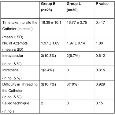
Table 2: Procedure Variables
Group E (n=28)
Group L (n=30)
P value
Time taken to site the Catheter (in mins.)
(mean ± SD)
18.38 ± 10.1 16.77 ± 3.75 0.417
No. of Attempts (mean ± SD)
1.97 ± 1.09 1.97 ± 0.14 1.00
Intravascular
(in no. & %)
3(10.3%) 2(6.7%) 0.612
Intrathecal
(in no. & %)
1(3.4%) 0 0.315
Difficulty in Threading the Catheter
(in no. & %)
3(10.7%) 3(10%) 0.929
Failed technique
(in no.)
2 0 0.15
The time taken to site the catheter, number of attempts, intravascular placement of needle or
catheter and difficulty in threading the catheter were comparable in both the groups. In the
epidural group, two cases were excluded because of, one had a dural puncture and the other
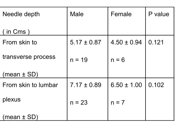
Table 3. Depth of lumbar plexus
Needle depth
( in Cms )
Male Female P value
From skin to
transverse process
(mean ± SD)
5.17 ± 0.87
n = 19
4.50 ± 0.94
n = 6
0.121
From skin to lumbar
plexus
(mean ± SD)
7.17 ± 0.89
n = 23
6.50 ± 1.00
n = 7
0.102
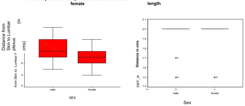
In group-L, the mean depth of lumbar plexus was 7.17cm in male and 6.5 cm in female.
The distance between the transverse process to lumbar plexus was 2cm for both male and
female.
In 80%, the lumbar plexus was located by caudal or cephalad angulation of the needle
after striking the transverse process of L-4 or L-5 and in 20% of the patients, the plexus was
Table 4: Sensory distribution in lumbar plexus block
Branches n % of blockade
Femoral nerve 30 100%
Lateral cutaneous nerve 28 93.3%
Obturator nerve 27 90%
The nerves blocked by lumbar plexus were femoral 100%, lateral cutaneous nerve
93% and obturator nerve 90 %.
SEX female male fr o m S ki n t o L u m b a r P le xu s 10 9 8 7 6 5 4 D is ta nc e fro m S ki n t o L u m b a r p le xu s (i n cm s)
Mean depth of lumbar plexus between male and
female Difference between male and female in the length
between transverse process and lumbar
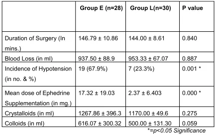
Table 5: Intra operative variables
Group E (n=28) Group L(n=30) P value
Duration of Surgery (In mins.)
146.79 ± 10.86 144.00 ± 8.61 0.840
Blood Loss (in ml) 937.50 ± 88.9 953.33 ± 67.07 0.887 Incidence of Hypotension
(in no. & %)
19 (67.9%) 7 (23.3%) 0.001 *
Mean dose of Ephedrine Supplementation (in mg.)
17.32 ± 19.03 2.37 ± 6.403 0.000 *
Crystalloids (in ml) 1267.86 ± 396.3 1170.00 ± 49.6 0.275 Colloids (in ml) 616.07 ± 300.32 500.00 ± 131.30 0.059
*=p<0.05 Significance
All data are expressed as mean ± SD (except Incidence of Hypotension)
There was no significant difference between two groups regarding the duration of the surgery
and blood loss. The mean dose of ephedrine requirement was significantly (P < 0.000) high in
group-E. The requirement of crystalloids and colloids were less in lumbar plexus but
statistically not significant.
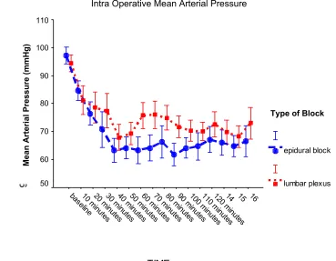
Time (in mins) Group E (n=28) Group L (n=30) MAP MAP P value Group E (n=28) Group L (n=30)
Heart Rate Heart Rate
Base Line
97.18 ± 7.87 94.43 ± 8.26 0.201 80.00 ± 7.32 81.90 ± 6.85
0 84.68 ± 8.98 81.2 ± 13.94 0.267 88.82 ± 15.87 82.30 ± 16.21
10 76.46 ± 10.78 78.57 ± 14.75 0.541 92.68 ± 17.37 83.30 ± 13.83
20 70.57 ± 60.57 77.40 ± 16.46 0.131 92.25 ± 19.36 88.47 ± 15.98
30 63.36 ± 11.17 67.93 ± 12.66 0.151 90.29 ± 21.61 84.63 ± 16.10
40 64.18 ± 9.76 69.33 ± 10.9 0.064 86.43 ± 19.93 82.00 ± 15.89
50 63.36 ± 12.24 75.73 ± 12.46 0.000 * 82.57 ± 16.07 79.80 ± 15.21
60 64.18 ± 11.85 75.03 ±12.98 0.001 * 82.86 ± 15.75 79.20 ± 14.43
70 66.36 ± 15.08 74.93 ± 11.62 0.018 * 84.39 ± 16.62 80.53 ± 15.78
80 61.75 ± 10.29 71.70 ± 10.67 0.001 * 80.68 ± 15.46 78.63 ± 14.59
90 64.11 ± 9.44 70.30 ± 10.02 0.019 * 79.86 ± 15.23 78.47 ± 14.41
100 64.71 ± 12.60 70.17 ± 8.84 0.060 81.21 ± 14.26 78.43 ± 14.73
110 67.15 ± 11.71 72.66 ± 11.73 0.085 81.26 ± 13.77 78.76 ± 15.66
120 66.08 ± 10.99 69.87 ± 9.98 0.219 79.16 ± 13.35 77.17 ± 14.24
130 66.78 ± 8.74 68.26 ± 8.91 0.188 81.22 ± 13.10 77.35 ± 14.07
140 66.48 ± 12.06 73.00 ± 12.32 0.091 82.10 ± 14.12 76.10 ± 14.53
150 66.74 ± 13.25 73.84 ± 13.30 0.108 80.37 ± 15.33 78.00 ± 14.41
* p<0.05 Significance Note: MAP – Mean Arterial Pressure.
The incidence of hypotension was significantly high in group-E (P<0.001) at 50 to 90 minutes
Table 7: Post Anaesthesia Care Unit Variables
Parameters Group E (n=25) Group L (n=29) P value
TIME 16 15 14 12 0 m inu tes 11 0 m inu tes 10 0 m inu tes 90 m
inu tes 80 m
inu tes 70 m
inu tes 60 m
inu tes 50 m
inu tes 40 m
inu tes 30 m
inu tes 20 m
inu tes 10 m
inu tes base line (b efo re sur
9 5 % C I b a se li n e b lo o d p re ss u re 110 100 90 80 70 60 50 TYPEOFBL epidural block lumbar plexus Mean Arteri al P ressu re (mmHg )
Type of Block
Duration of Stay in mins. (mean ± SD)
119.60 ± 50.37 111.21 ± 35.19 0.476
Incidence of Hypotension
(in no. & %)
6 (22.2%) 1 (3.6%) 0.036 *
In post anaesthesia care unit, the duration of stay was comparable in both the groups.
The incidence of hypotension was significantly more in group-E (P<0.036), whereas only one
Table 8: Mean arterial pressure and heart rate in Post Anaesthesia Care Unit (mean ± SD)
Time (in mins.)
Group E (n=25)
Group L (n=29)
MAP MAP P value
Group E (n=25)
Group L (n=29)
Heart Rate Heart Rate
0 83.36 ± 18.19 92.76 ± 12.69 0.030 * 91.52 ± 17.74 89.45 ± 15.29
30 84.12 ± 15.99 93.34 ± 11.94 0.019 * 87.12 ± 18.17 85.62 ± 13.12
60 85.61 ± 16.37 89.66 ± 9.65 0.272 86.17 ± 17.64 84.24 ±12.50
90 80.94 ± 10.94 89.00 ± 8.99 0.012 * 90.61 ± 18.07 82.58 ±12.46
120 80.40 ± 11.59 90.27 ± 7.86 0.023 * 90.87 ± 17.54 79.45 ± 11.85
150 81.57 ± 9.58 83.00 ± 9.40 0.792 86.43 ± 17.25 83.83 ± 13.55
*=p<0.05 Significance
Note: MAP – Mean Arterial Pressure.
Compare to group L, group E had statistically significant drop in the mean arterial pressure
TIME 8 7 150 minutes 120 minutes 90 minutes 30 minutes bp at 0
baseline 9 5 % C I b a se li n e b lo o d p re ss u re 140 120 100 80 60 40 20 TYPEOFBL epidural block lumbar plexus
0 30 60 90 120 150 Time in mins
Mean Arterial Pressure in PACU
Type of block
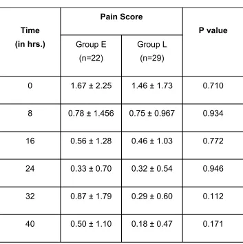
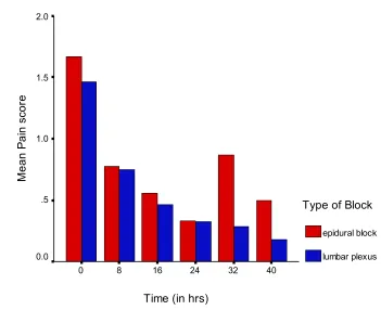
Table 9: Pain Score in Postoperative Ward (in mean ± SD)
Time (in hrs.)
Pain Score
Group E (n=22)
Group L (n=29)
P value
0 1.67 ± 2.25 1.46 ± 1.73 0.710
8 0.78 ± 1.456 0.75 ± 0.967 0.934
16 0.56 ± 1.28 0.46 ± 1.03 0.772
24 0.33 ± 0.70 0.32 ± 0.54 0.946
32 0.87 ± 1.79 0.29 ± 0.60 0.112
40 0.50 ± 1.10 0.18 ± 0.47 0.171
The pain score was similar in both the groups.
Table 10: Postoperative Complications Time (in hrs)
40 32
24 16
8 0
M
e
a
n
P
a
in
s
co
re
2.0
1.5
1.0
.5
0.0
Type of Block
epidural block
Parameters Group E
(n=28)
Group L
(n=29)
P value
Hypotension 7 (25.0%) 1 (3.4%) 0.019 *
Discontinuation of infusion due to hypotension
6 (21.4%) 0 0.009 *
Urinary dysfunction 14 (50%) 7 (24.1%) 0.043 *
Nausea-Vomiting 7 (25.0%) 8 (27.6%) 0.825
Pruritus 6 (21.4%) 1 (3.4%) 0.039 *
Subarachnoid migration of catheter 1 0 0.305
Dislocation of catheter 5 (17.9%) 2 (6.7%) 0.191
Mean dose of pethidine supplement (in mg)
17.59 ± 44.83 8.62 ± 19.22 0.329
Number of complete unilateral epidurals
7 (25%)
Number of epidural spread 1 (3.4%)
* p<0.05 Significance
In the post operative ward, the incidence of hypotension (P<0.019), discontinuation of
infusion (P<0.009), pruritus (P<0.039) and urinary dysfunctions (P<0.043) were significantly
high in group-E. In group-L incidence of urinary retention, dislocation of catheter and post
operative pethidine requirements were comparatively less. A patient with subarachnoid
migration of catheter developed blister in the gluteal region which was confirmed by
biochemical analysis of cerebro spinal fluid for sugar and protein content.
25% of group-E had unilateral spread to the operated side and were totally pain free.
10 % of group-E had lateralization on non-operated side and required more opioid for pain
The epidural pain relief was rated excellent by 27% of the patients, good by 63% and
DISCUSSION
Total hip replacement has been a major advancement in the treatment of chronic
arthritis of hip and provides pain relief and increase mobility. Total hip replacement involves
the prosthesis replacement of femoral and acetabular component of hip joint. There is
significant operative trauma to both soft tissue and bone marrow with resultant haemorrhage.1
Spinal, epidural and various techniques of general anaesthesia have been used
successfully for total hip replacement. The major intraoperative anaesthetic concerns are
blood loss, cardiovascular instability and hypoxaemia. The blood loss is variable, averaging
0.5L – 1.5L intraoperatively and 300 – 500ml in the post operative period.3
Anaesthetic technique is an important factor affecting blood loss during total hip
replacement. Intraoperative blood losses are significantly reduced with spinal and epidural
techniques when compared with general anaesthesia alone.4, 59
Patients undergoing total hip replacement experience substantial and sustained post
operative pain. Inadequate analgesia may limit early mobilization, impede the physical
therapy and delay the discharge.60 Postoperative pain relief is achieved by varieties of
techniques such as IV PCA with morphine, epidural and continuous perineural blockade.5, 54,58
Each had its advantages and disadvantages. IV PCA with morphine is inefficient for pain relief
during movement and ineffective in preventing the reflex spasm of quadriceps muscle, which
is frequent after hip surgery. The benefit in consistency of pain control that epidural analgesia
provides is offset by high incidence of catheter related problems and side effects such as
urinary retention and hypotension.6 Continuous lumbar plexus block has emerged as an
alternative analgesic approach with continuous, efficient, and consistent pain relief and as
provided a similar quality of analgesia like epidural, but with fewer side effects.53, 55, 57
Lumbar plexus block is technically easy to perform in patients with ankylosing
spondylitis and elderly age group with calcified spinal ligaments. It can be performed in the
lateral position and does not require hip flexion.
The X-ray of the pelvis with the lumbosacral spine served as a guide in localizing the
L-4 spine and the transverse process. In our study, by using this simple landmark assessment
100% success was achieved in lumbar plexus block without any complication. So far various
techniques like parasthaesia by Winnie11, loss of resistence by Chayen12 and the use of nerve
stimulator by many others. And also, lots of modification in the landmark has been performed
to reduce the failure rate and the complications of the block.
We have used Capdevila’s 24 approach to locate the lumbar plexus and the sensory
block of femoral nerve, lateral cutaneous nerve and obturator nerve was successful with
100%, 93% and 90% respectively in this study compared with that of Capdevila’s 24 study with
95%, 85% and 90% respectively.
Similar to Capdevila’s 24 study the difference between the depth of the transverse
process and the lumbar plexus was 2cms in both male and female patients.
Rapid and painless recovery is the main advantage in combining general anaesthesia
with epidural technique. But this combination causes more severe hypotension than is
observed, when either method is used alone. The reason for this, large drop in pressure is
because of the negative inotropic effect of general anaesthesia which is augmented by
peripheral vasodilatation effect of epidural anaesthesia. Healthy and young individuals
tolerate these effects well, where as in elderly patients with intravascular volume deficit, the
combination of general anaesthesia and epidural may lead to hemodynamic instability.
In contrast to epidural block, the lumbar plexus block is reported to cause limited
the mean arterial blood pressure at 50-90 minutes after the start of general anaesthesia
revealed significant drop in arterial blood pressure in the patients who were given a general
anaesthesia with epidural block than those who were given general anaesthesia with lumbar
plexus block. These decrease in blood pressure occurred in spite of preoperative volume
loading with IV crystalloids 10ml / kg. In the epidural group 67%of patients had significant
hypotension compared to 23% in lumbar plexus block. This is statistically significant and this
finding correlated well with the previous study 53.The ephedrine requirement in the epidural
group, to treat the hypotension was significantly high with a mean dose of 17mg whereas it
was 2mg in lumbar plexus group.
In the epidural group, due to the persistent hypotension 6 patients had discontinuation
of infusion in the ward and were provided with other modes of pain relief. None of the patients
in lumbar plexus group had such a complication.
Urinary retention in the postoperative period is common in the elderly age group done
under epidural anaesthesia. William et al 61 reported urethral catheterization 67% and
Singelys et al 54 reported 40% of similar incidence in the epidural group. In our study urinary
dysfunctions like urinary retention and urinary incontinence were observed in 50% of the
epidural group. Incidence of pruritus was significantly high in epidural group.
The reported incidence of epidural spread varies from 1-16%.13, 23, 32 In our study, only
one patient had epidural spread to L-4 dermatome in the contralateral side. The body mass
index of that patient was only 18. A bolus of 20ml bupivacaine must have been a larger
volume for her weight. The same patient underwent similar procedure a week later and had
lumbar plexus block with a bolus of 15ml of 0.5% bupivacaine without epidural spread. When
continuous nerve block technique is compared with single shot technique the incidence of
epidural spread is less because lesser volumes are used. The entry point of the needle close