ONE YEAR STUDY OF “TRANSVERSE AND
VERTICAL DIAMETER OF THE ORBIT AS A
PARAMETER IN SEX
DETERMINATION OF HUMAN SKULL”
(JUNE 2005 – MAY 2006)”
DISSERTATION SUBITTED FOR M.D. Degree (FORENSIC MEDICINE)
MARCH 2007
Dept. of Forensic Medicine
Madurai Medical College,
Madurai – 625 018.
CERTIFICATE
This is to certify that this dissertation entitled A ONE YEAR STUDY OF “TRANSVERSE AND VERTICAL DIAMETER OF THE ORBIT AS A PARAMETER IN SEX DETERMINATION OF
HUMAN SKULL June 2005 - May 2006)” has been prepared by Dr. T.JEYASINGH under my overall supervision, in partial fulfilment of the regulations for the award of Degree of DOCTOR OF MEDICINE in FORENSIC MEDICINE of Tamil Nadu Dr.M.G.R. Medical University, Chennai.
I certify regarding the authenticity of the work done to prepare this dissertation.
Date :
ACKNOWLEDGEMENT
I wish to record my profound sense of gratitude towards Prof.Dr. K.Meiyazhagan M.D., my Professor, Co-guide and Head of the Department and who is also a fountain head of inspiration during my post graduation course, and during this thesis work.
I am greatly indebted to THE DEAN for encouraging and securing me all the required facilities during the work.
I express my sincere thanks to all the Teaching Staffs and Department Staffs for their timely help and guidance.
I extend my thanks to my post-graduate colleagues for their support and encouragement throughout this thesis.
With great pleasure I extend my gratitude to all my friends who assisted me in analysis and write up of this work.
Finally, I am indebted to all the deceased souls, which formed a substance of this study without which this study would never have been possible.
Date :
CONTENTS
S.NO INDEX PAGE NO.
1.
Introduction
1
2.
Review
of
Literature
10
3.
Anatomy
of
Orbit
16
4.
Materials
&
Methods
26
5.
Observations
Discussion 40
6.
Summary
&
Conclusions
58
INTRODUCTION
The present study is determination of sex by taking measurements of transverse diameter and vertical diameter of the orbit of the skull.
There are a number of factors, which inveighs against high degree of accuracy in the sexing of unknown material. Intrinsic variability and absence of any real standards and the evident age of the bone poses problems. Here is the problem of subjectivity Vs. objectivity of description Vs. measurements, of experience Vs. statistical standardization.
Most of the older studies of sex differences in the skeleton (skull and pelvis mainly centered on morphological traits in a descriptive manner. The newer studies focus on morphometry in a largely in a quantitative and statistical sense.
The accuracy of these are variable. Stewart (48, 51) feels that for the entire skeleton, or for the adult pelvis, or for one adult hip bone, he can identify sex correctly in 90-95% of cases; for adult skull alone about 80% for adult major long bones alone ‘size alone determines the answer’.
The foregoing discussion refers to adult material. As a general rule definitive sexual traits in the skeleton are not manifest until after the full achievement of the secondary sex traits that appear during puberty. The dividing line between immaturity and maturity is some where around 15-18 years.
remains. Equally traditionally, the sexing of skulls has been based on an anatomical basis (Osteological), so that descriptive features (traits) have ruled rather than dimensions (size, proportions).
In sexing a skull the initial impression is the deciding factor, a large skull is generally a male and a small skull female. The cranial capacity in the female averages 200 cc less than the male, though in the female the index of cerebral value is relatively higher. The female skull is usually rounder than the male i.e the cranial index is two or more points greater. The cranio facial proportions are about the same, though in the female the skeleton may be relatively more gracile. The general impression may be verified by observation of mandible, nasal aperture, orbits, cheek bones, supra orbital ridges glabella, forhead contour, mastoid processes, supra orbital ridges, occipital region, palate and teeth and base of the skull. In so far as several of these criteria are age phenomenon, appearing or becoming pronounced at puberty, and many are affected by the changes of senility, the description of sex differences must be limited to the ages approximately 20-55. These characters in their entirety give a pretty good idea of sex in adults.
In 1939-40, Hug put sex differences in cranial dimensions on a fairly sound basis. A scrutiny of that data will reveal considerable size overlap, as between male and female. Obviously no single dimension is diagnostic, but if all dimensions are large, in a male direction, then it is pretty safe to diagnose the skull as male.
infra temporal fossa, length of the mastoid process). For these he calculated the mean and standard deviation for each sex which gave a male range, a female range and a neutral zone. This gave an accuracy of sex of skull about 85%.
In recent years elaborate and detailed statistical studies, involving discriminate analysis have been made of sex differences in skull and long bones. Harihra (‘59) reported on such a study on Japanese skull.
The entire pelvis structure i.e its constituent bones and its total configuration, has long been regarded as a critical factor in the sexing of the human skeleton. It takes on added value by virtue of the fact that it apparently differs from other skeletal sexual criteria in that sex differences are present from foetal life and onward.
One of the most obvious sex differences, in the long bones is that typically male bones are longer and more massive than typically female bones. Schultz (37) observed that the various human races differ but very little in the degree with which the male surpass the females in the length of the long bones.
Krogman outlined the essential descriptive morphology of the skull in the three main human stocks. Caucosoid, Negroid and mongoloid. There really are no pure races and hence no pure Negro skulls or pure Mediterranean skulls and so forth. There are only skulls which to a greater or lesser degree present a combination of traits that suggests stock or race category.
In 1955, Ray reported possible racial differences in the configuration of the superior orbital fissure. It is difficult to evaluate just how a single skull is classified as white or negro or mongoloid. Certain measurements and proportions may be useful. For example the height and breadth index of the orbit the Negro orbit tends to be lower and wider, the mongoloid higher and more rounded, the white in between.
The orbital measurements of George Buchan – with those of other skulls from Person’s study shows:
Average male of London of 17th Century of Poor shows : Breadth of R. Orbit : 42.3 Breadth of L. Orbit : 42.4
Height of R. Orbit : 34.3 Height of L. Orbit : 34.3 Skull of Buchanan shows :
Breadth of R. Orbit : 45.8 Breadth of L. Orbit : 44.8
Height of R. Orbit : 35.9 Height of L. Orbit : 36.8 Skull of Sir Thomas Brown Shows :
Height of R. Orbit : 36.3 Height of L. Orbit : 36.7 Skull of Jeremy Benthom shows :
Breadth of R. Orbit : 45.2 Breadth of L. Orbit : 46.0
Height of R. Orbit : 36.0 Height of L. Orbit : 36.0
The changes in the orbit during the period of growth depend partly on the development of cranium and skeleton of face, between which the orbit is placed, and also on the growth of the neighbouring air sinuses.
The orbital margin is sharp and well ossified at birth. At seven years, except at its upper parts, it is less sharp and as the supero-medial and infero lateral angles are better marked than the others the orbital openings tends to be triangular.
The form of the orbit on coronal section behind the orbital margin is that of a quadrilateral with rounded corners. In the new born it has the form of an ellipse higher on lateral than the medial side.
From Height Width Index
Foetus Oval 14mm 18mm 77.7 New Born (6 Months) Round 27mm 27mm 100
Child (7 Years) Quadrilat 28mm 33mm 84.8
Adult Quadrilat 35mm 39mm 89.7
In long headed (Dolichoephalic) skulls the orbits tend to look more laterally than in the short headed (brachycephalic).
There is a great difference between the measurements given by different authorities.
Height of orbital opening 35mm Width of orbital opening 40mm
Upto puberty there is little difference between the orbits and, in fact, the skulls of males and females.
After this the male skull takes on its secondary sexual characters seen especially in the formation of lower jaw and in the forehead region.
The female remains more infantile in form. The orbits tend to be rounder and the upper margin sharper than in the male. The female orbit is more elongated and relatively larger than the male (Markel).
diminishes as a result of loss of teeth and subsequent absorption of the alveolor margins.
Witton Marion Krogman says in his book the orbits are higher, more rounded and relatively larger, compared to upper facial skeleton in the female. The orbital margins are sharper, less rounded than in the male. The male show orbits squared, lower, relatively smaller with rounded margins.
Harrison stresses that the determination of the sex of the skull cannot be made from one characteristic alone but must be assesses from the examination of a number of features taken together. The best characteristics are probably those of the supra orbital ridges, the mastoid processes, the dimensions of the palate, the outline of the orbit and characteristics of the mandible. It is only after the age of fourteen to sixteen years that the male characteristics begin to develop, upto that age it is often exceedingly difficult, if not impossible, to sex the skull.
Dr. S.C.Basu says the size of orbital openings, is large relating to the face as a whole. The male orbits are square, lower, relatively smaller and rounded margins. The female orbits are rounded, higher relatively larger and sharp margins.
Francis E. Camps say that features of the skull that are characteristically female include the tendency for the orbital openings to be more nearly circular. With a sharp upper edge and for their size to be larger relative to the face as a whole.
Dr.K.S.Narayana Reddy states that the male orbit is square, lower, relatively smaller, rounded margins and the female orbits rounded, higher, relatively larger and sharp margins.
Dr.C.K.Parikh states that male orbital opening is comparatively smaller and that of female orbital opening comparatively bigger.
Dr.D.J.Gee states that the orbits of male are square and orbital ridges are prominent and in the female the orbits are rounded and orbital ridges are not marked.
Sir Sydney Smith states that upper ciliary ridges are less pronounced thus causing the upper orbital margin to be thinner and sharper than in the male.
In the present study an attempt has been made to arrive at the determination of sex by taking the measurements of transverse diameter and vertical diameter of the orbital openings. The orbital index is the length of the orbit as compared with its breadth (Gonzales).
Length of Orbit
--- x 100 Breadth of Orbit
There is an encouragement of results thus obtained. The graphic representation also suggested that there is a scope of further study in this topic. The Z values and P values obtained also suggested that there is certain marked difference in the vertical diameters of the orbit in males and females. The study is confined to adults, taking the age group above sixteen years onwards.
The age group below is not taken into account for the fear of questionable developmental growth of orbits. But the literature suggests that there is minimal changes occur after a particular age i.e above twelve years. In children both sexes show the same diameter for vertical or transverse.
The orbital index obtained in these studies have pointed our that there is a clear male pattern and a clear female pattern.
REVIEW OF LITERATURE
There are a number of factors which inveigh against a high degree of accuracy in the sexing of unknown material. Most of the older studies of sex differences in the skeleton (skull and plevis mainly) centres on morphological traits in a descriptive manner. The newer studies Focus on morphometry in a largely quantitative and statistical sense.
In a series of 100 Adult American Negro skeletons (Complete) sexed by inspection, Stewart scored 94%; but in the same series, using skull plus mandible, he scored only 77%.
Traditionally the skull is the single most studied bone in physical anthropology. Archaeologists have concentrated on excavating and preserving skulls. Equally traditional, the sexing of skulls has been based on an anatomical basis so that descriptive features have ruled, rather than dimensions. In sexing a skull the initial impression often is the deciding factor.
In 1939-40, Hug put sex differences in cranial dimensions on German crania on a fairly sound basis. In 1950, Keen attempted to set a battery of cranial traits and dimensions for adult skulls of the cape coloured population of S. Africa, which will sex skulls with 85% of accuracy.
Reynolds, ’45, made roentgenometic studies of the bony pelvic girdle in early infancy from birth to one year age of American white people. For the pre-puberal periods, age two to nine years, in ’47 he studied on American white people with X-ray films.
In ’59 Genoves studied sex differences in the adult pelvis.
Schuttz (’37) observed that the various human races differ but very little in the degree with which the males surpass the females in the length of the long bones. The male: female ratio for long bone length around 100:90.
The sexing of unknown skeletal material depends, of course, upon relative completeness of skeleton. Percentage of accuracy for adult material, is about as follows: (Krogman).
Skull alone = 90% Skull plus pelvis = 98%
Skull plus long bone = 90-95%
Krogman, ’55 says that there really are no pure races. There are only skulls which to a greater or lesser degree, present a combination of traits that suggest stock or race category.
Krogman writes that certain measurements are useful to evaluate a race; for example the height / breadth index of the orbit: the Negro orbit tends to be lower and wider; the Mongoloid higher, more rounded and the whites in between.
Dwight concluded that the breast bone is no trustworthy guide either to the sex or age.
Pearson ’26, has studied in detail the skull of Buchanan. There in that he compares the average London Poor 17th century males, skull of Sir Thomas Browne, skull as Jeremy Bentham. He describes the breadth and height of each orbit and also gives the orbital index of each orbit.
Diameter in mm Vertical ( R) Vertical (L) Orbital Index Transverse ( R) Transverse
(L) ( R) (L)
Average
London Poor 17th Century Male
42.3 42.4 34.3 34.3 81.0 80.9
Skull of George
Buchanan
45.8 44.8 35.9 36.8 78.4 82.1
Skull of Sir Thomas
Brown
43.7 42.8 36.3 36.7 79.6 78.3
Skull of Jeremy
Bentham
45.2 46.0 36.0 36.0 79.6 78.3
great differences between the measurements given by different authorities. He writes that the width of the orbital opening 40mm and height of orbital opening 35mm.
Wincler’s table gives a resume of the changes in the orbital opening.
Form Height Width Index
Foetus Oval 14mm 18mm 77.7 New Born (6 Months) Round 27mm 27mm 100
Child (7 Years) Quadrilat 28mm 33mm 84.8
Adult Quadrilat 35mm 39mm 89.7
Conard Bereus in his book states that the orbital vertical diameter is 35mm and the horizontal diameter is 40mm and there are of course variations in different individuals.
Ernest Gardner in his book (’67) describes “that each orbit is shaped roughly like a pear or a four sided pyramid, with its apex posteriorly and its aditus or base anteriorly. The base is about 35mm high and 40mm wide.
Dr.S.N. Sahana in his book (82) states the vertical diameter of orbital base is 41.3mm and horizontal diameter is 41.3mm.
Markel mentions that-the female orbit is more elongated and relatively larger than the male.
Harrison stresses that the determination of the sex of the skull cannot to be made from one characteristic alone but most be assessed form the examination of a number of features taken together; one of the best characteristic is the out line of the orbit.
Dr.S.C. Basu says that the size of orbital opening is large relating to the face as a whole; male orbits are square and have rounded margins and female orbits are rounded and have sharp margins.
Francis E. Camps say that features of the skull that are characteristically female include the tendency for the orbital openings to be more nearly circular. With a sharp upper edge and for their size to be larger relative to the face as a whole.
N.J.Modi states the female orbits have sharp margins and rounded and comparatively higher and longer.
Dr.K.S.Narayana Reddy states that the male orbit is square, lower, relatively smaller, rounded margins and the female orbits rounded, higher, relatively larger and sharp margins.
Wilton Marian Krogman says that the orbital margins are sharper, less rounded than in the male for females; the male show orbits squared, lower relatively smaller with rounded margins.
Dr.D.J.Gee states that the orbits of male are square and orbital ridges are prominent and in the female the orbits are rounded and orbital ridges are not marked.
Sir Sydney Smith states that upper ciliary ridges are less pronounced thus causing the upper orbital margin to be thinner and sharper than in the male.
Ganzole points out that the orbital index is the length of the orbit as compared with its breadth.
The relation between the height and breadth of the orbit is given by the orbital index, which varies in different races of mankind.
Height of Orbit
The orbital index (of Borca) = Height of Orbit --- x 100 Width of Orbit
Taking the orbital index as the standard, three classes of orbit are recognized.
1) Megaseme (Large) : The orbital index 89 or over. This type is characteristic of the yellow races except the Equimanx. The orbital opening is round.
2) Mesoseme (Intermediate): The orbital index is between 89-83. This type is found in the white races. (European 87, English 88.4 according to Flower)
ANATOMY OF ORBIT
The Bony Orbit
The orbital cavities are placed on either side of the mid vertical line of the skull between the cranium and the skeleton of the face. Thus situated, they encroach about equally on these two regions. (Winckler)
Above each orbit is the anterior cranial fossa, medially are the nasal cavity and air sinuses, below is the maxillary sinuses, while laterally from behind forwards are the middle cranial and temporal fossa.
The orbit it essentially intended as a socket for the eye balls and also contains the muscles, vessels and nerves, which are essential for its proper functioning. More over, it serves to transmit certain vessels and nerves destined to supply the areas of the face around the orbital aperture.
Seven bones take part in the formation of the orbit, namely: the maxilla and palatine, the frontal, the sphenoid, and zygomatic bone, the ethmoid and lacrimal bone.
As stated above, the comparison with a quadri-lateral pyramid is a rough one only, for since the floor (which is the shortest orbital wall) does not reach the apex, the cavity is tri-angular on section in this region.
Also, since the orbit is developed around the eye, and is bulged out by the lacrimal gland, it has a tendency towards being spheroid in form and it’s widest part is not at the orbital margin but about 1.5 cm behind this. Moreover, the results in the fact, that its four walls are for the most part separated from each other by ill defined rounded borders, so that Whitnall compares the shape of the orbit to a pear whose stalk is the optic canal. It is important to note that the medical walls of the orbit are almost parallel where as the lateral walls on angle of 90 degree with each other. The direction of each orbit is given by its axis which runs from behind forwards laterally and slightly downwards.
The orbital margin has the form of a quadri lateral with rounded corners. The orbital Margin has the form of a spiral; the inferior orbital margin is continuous with the anterior lacrimal crest, while the superior is continued down into the posterior lacrimal crest. The lacrimal fossa thus lies in the orbital margin. (Poirier).
Each side measure some 40mm, but usually the width is greater than the height: the relation between the two is given by orbital index, which varies in the different races of mankind. The opening is directed forward and slightly laterally, and is tilted so that the upper and lower margins slope gently downwards from the medical to the lateral side. The orbital margin is made up of three bones: the frontal, the zygomatic and maxilla.
two thirds; and rounded in the medial third. At the junction of two portions, some 25mm, from the midline and situated at the highest part of the arch is the supra orbital notch, whose lateral border is usually sharper than the medial. Not infrequently it is converted into a foramen by its ossification of the ligament which closes it below. The posterior opening than is 3 to 6 mm from the orbital margin. It transmits the supra orbital nerve and vessels. Notch and foramen are palpable in the living.
Some times medial to this second notch and foramen is found (Arnold’s). This transmits the medial branches of the supra orbital nerves vessels where these have divided inside the orbit.
Supra grooves leading from these notches or foramen are sometimes seen.
A groove may also present some 10mm; medical to the supra orbital notch for the supra trochlear nerve and artery.
A supra ciliary canan (Ward) is found in about half of cases. It is a small opening near the supra orbital notch and transmits a nutrient artery and a branch of the supra orbital nerve to the frontal air sinuses.
The lateral orbital margin, being the most exposed to injury, is the strongest portion of the orbital outlet. It is formed by the zygomatic process of the frontal and by the zygomatic bone. It looked at from the side, it appears to be concave forwards and not to reach as far forwards as the medical margin.
The inferior orbital margin is raised slightly above the floor of the orbit. It is formed by the zygomatic bone and the maxilla, usually in equal portions.
The zygomatic portion forms a long thin supr (the maxillary or marginal processes) which lies on the maxilla.
The suture between the two, which is not infrequently by a tubercle, can be felt lying usually about half way along the margin, just above the infra orbital foramen.
Sometimes, however, the zygomatic (malar), may reach the anterior lacrimal crest thus excluding the maxilla or may take only a very small part it self in the formation of this margin.
The medial margin is formed by the anterior lacrimal crest on the frontal process of the maxilla and the posterior lacrimal crest on the lacrimal bone. These crests overlap; the medial margin is thus not a continuous ridge, but runs up from the anterior lacrimal crest across the frontal process of maxilla to the superior margin.
Certain points of importance in the neighbourhood of the orbital margin:
The frontal eminences are rounded elevations on the vertical plate of teh frontal bone 2” above the orbit; they are more prominent in the female and even more so in infants.
the infra orbital forament lies 4-5mm, below the tubercle on the lower orbital margin which marks the suture between the zygomatic bone and the maxilla. It is usually oval. The foramen may be double-indeed, upto five have been described.
The supra orbital notch, the infra orbital foramen are on the same vertical line which passes between the two bicuspid teeth.
SURFACE ANATOMY
The upper orbital margins forms a well marked prominence, more so in the lateral shape portion than in the medial more rounded part. Its form can be made out easily by touch. It should be noted carefully that the eye brow corresponds in position only in part to the upper orbital margin.
The head of the eye brow lies for the most part under medial part of the margin, to palpate which that finger must press forwards. The body lies along the margin, while the tail runs well above the lateral part of the margin, which can be felt and usually can be seen below it.
The zygomatic process of the frontal bone often forms a marked prominence under the skin.
The supra orbital notch can be felt at the junction of the lateral 2/3 with the medial third and not infrequently supra orbital nerve can be rolled under the fingers.
The lateral margin of the orbit is only visible down to the level of the lateral can thus but can easily felt in its whole extent.
The lower orbital margin, as opposed to the upper, forms no prominence, since the skin of the lower lid passes without sudden change of plane into that of cheek. Just beyond it, especially in the old, lie he naso-jugal and maral furrows.
The lateral tubercle can be felt in the shape anterior lacrimal, crest, as can the tubercle at the middle of the lower margin which marks the suture between the zygomatic bone and maxilla.
The lateral orbital tubercle (Whitenall’s) felt just within the lateral margin at its middle by passing the finger into the orbit and rubbing it up and down against the margin.
The infra orbital foramen or rather its cresentric sharp upper margin can be made out, not infrequently, 4-5 mm, below the tubercle on the lower orbital margin which marks the zygomatic-maxillary suture.
The zygomatic tubercle (malar) can be felt below and behind the zygomatic process of the frontal bone and bone and between the two a “V” shaped interval, at the bottom of which is the fronto-zygomatic suture.
The anterior lacrimal crest is easily defined. Behind it the finger passes into lacrimal fossa and behind this again the posterior lacrimal crest can be felt.
The temporal crest can be felt arching backwards from the zygomatic process of the frontal bone.
The nasal bone, sitting on the frontal process on maxilla, can be seen and palpated down to its lower end, where it joins the mobile cartilage of the nose.
Walls of the Orbit
forwards as well. It is markedly concave anteriorly and more or less flat posteriorly. The anterior concavity is greatest about 1.5 cm, from the orbital and corresponds to the equator of the globe.
The roof of the orbit is separated from the medial wall by fine sutures between the frontal bone above and the ethomid, lacrimal and frontal process of the maxilla below. The roof is separated from the lateral wall posteriorly by the superior orbital fissure anteriorly by the slight ridge that marks the fronto-zygomatic suture.
The medial wall of the orbit is the only wall which is not obviously triangular. It is roughly oblong either quite flat or slightly convert towards the orbital cavity. It runs parallel with sagittal plane and consists from before backwards:
1. The frontal process of maxilla. 2. The lacrimal bone.
3. The orbital plate of ethmoid.
4. A small part of the sphenoid, with some times sphenoidal concha.
the maxilla, take varying parts in the formation of the fossa, and so the position of the vertical suture between them varies also.
The anterior lacrimal crests on the frontal process of the maxilla is ill defined above but well marked below, where it becomes continuous with the lower orbital margin and here often presents a lacrimal tubercle.
The floor of the orbit is roughly triangular, corresponding to the shape of the roof. It is not quite horizontal, but slopes slightly downwards from the medial to lateral side. The lowest part of the floor of the orbit is found in a concavity some 3mm, deep at the lateral and anterior part. The floor (47.6mm, long) the shortest of orbital boundaries is formed by three bones.
1. The orbital plate of maxilla.
2. The orbital surface of the sygomatic. 3. The orbital process of the palatine bone.
Of these the maxillary takes by far the largest portion. The zygomatic forms the anterior lateral part, while the palatine bone occupies the small area behind the maxilla. The sutures between the three bones forming the floor of the orbit are almost invisible.
The floor of the orbit is separated from the medial wall only by a fine suture, the lateral wall is separated from it posteriorly by the inferior orbital fissure, while anteriority it is continuous with it.
It is slightly convex posteriorly, flat at its centre, while anteriorly the orbital surface of the zygomatic bone 1cm behind the orbital margin as concave.
The lateral wall of the orbit is formed by two bones.
a. Posteriorly by the orbital surface of the greater wing of sphenoid. b. Anteriorly by the orbital surface of the zygomatic bone.
The sphenoidal portion is sharply separated from the roof and floor by the superior and inferior orbital fissures, respectively.
METERIAL AND METHODS
In the present study from known sexes, the measurements of transverse and vertical diameter of orbital openings are taken in millimeters.
The instrument used is a sliding calipers.
The dead bodies belong to the anatomy, and forensic medicine kept in mortuary. Not much destruction is made to the bodies while taking the measurements.
Orbital height : It measures the straight distance between the upper and lower
margins of the orbital cavity, taken at right angle to the orbital breadth.
Orbital breadth : It measures the straight distance between maxillo frontal (mf)
and ecto conchion (ecto, Ecto conchion (ect) must be marked before the measurement. The diameter should out the orbital cavity in almost to equal halves.
Care has been taken while selecting the specimens that there are no deformaties or diseases of the orbits present.
The age of the individuals are also noted.
MEASUREMENTS
MALE
Sl. No. Age in Years
Diameter in mm
Right Eye Orbit Left Eye Orbit
Transverse Vertical Transverse Vertical
(1) (2) (3) (4) (5) (6)
1. 55 32 28 32 28 2. 55 33 25 33 25 3. 60 36 25 36 25 4. 60 38 27 38 27 5. 65 33 25 33 25 6. 60 33 25 33 25 7. 18 32 28 32 28 8. 20 33 26 33 25 9. 48 33 25 33 25
10. 51 32 27 32 27
11. 30 33 25 33 25
12. 58 33 25 33 25
13. 45 33 25 33 25
14. 63 32 28 32 28
15. 30 33 25 33 25
16. 35 33 25 33 25
17. 60 39 30 39 30
18. 65 39 30 38 30
19. 30 33 25 33 25
(1) (2) (3) (4) (5) (6)
21. 70 38 30 38 30
22. 70 38 29 38 29
23. 25 33 25 33 25
24. 28 32 26 32 26
25. 30 33 25 33 25
26. 35 33 26 33 26
27. 25 33 27 33 27
28. 65 33 28 33 28
29. 18 32 27 32 27
30. 30 32 25 32 25
31. 30 32 25 32 25
32. 35 33 27 33 27
33. 60 34 28 34 28
34. 29 33 28 33 28
35. 30 32 24 32 24
36. 30 33 28 33 28
37. 50 32 25 32 25
38. 60 33 27 33 27
39. 40 33 27 33 27
40. 60 34 28 34 28
41. 70 33 27 33 27
42. 28 32 25 32 25
43. 60 32 25 32 25
44. 55 33 25 33 25
(1) (2) (3) (4) (5) (6)
46. 26 33 25 33 25
47. 20 33 25 33 25
48. 40 33 25 33 25
49. 50 33 26 33 26
50. 28 34 26 34 26
51. 40 33 25 33 25
52. 50 33 27 33 27
53. 23 33 27 33 27
54. 35 33 28 33 28
55. 75 34 28 34 28
56. 20 33 28 33 28
57. 35 33 28 33 28
58. 40 32 28 32 28
59. 45 32 28 32 28
60. 50 33 27 33 27
61. 47 32 27 32 27
62. 23 32 25 32 25
63. 50 32 26 32 26
64. 27 32 26 32 26
65. 42 32 28 32 28
66. 45 32 28 32 28
67. 32 32 26 32 26
68. 40 33 26 32 26
69. 45 33 26 33 26
(1) (2) (3) (4) (5) (6)
71. 50 33 27 33 27
72. 21 33 27 33 27
73. 21 33 28 33 28
74. 35 32 27 32 27
75. 70 40 31 40 31
76. 22 33 28 33 28
77. 23 32 25 32 25
78. 40 33 26 33 26
79. 60 33 26 33 26
80. 50 33 26 33 26
81. 40 33 25 33 25
82. 60 33 26 33 26
83. 43 33 25 33 25
84. 25 33 27 33 27
85. 35 32 25 32 25
86. 40 33 26 33 26
87. 22 32 26 32 26
88. 50 34 27 34 24
89. 40 33 26 33 26
90. 36 33 26 33 26
91. 30 33 25 33 25
92. 50 33 26 33 26
93. 40 33 27 33 27
94. 60 34 26 34 26
95. 25 32 25 32 25
MEASUREMENTS
FEMALE
Sl. No. Age in Years
Diameter in mm
Right Eye Orbit Left Eye Orbit
Transverse Vertical Transverse Vertical
(1) (2) (3) (4) (5) (6)
1. 50 33 25 33 25 2. 24 33 30 33 30 3. 19 33 31 33 31 4. 30 34 30 34 30 5. 42 34 30 34 30 6. 22 34 31 34 31 7. 55 33 30 33 30 8. 45 34 31 34 31 9. 65 34 30 34 30
10. 22 34 30 34 30
11. 68 33 29 33 29
12. 25 34 30 34 30
13. 18 33 30 33 30
14. 28 34 30 34 30
15. 80 33 30 33 30
16. 25 34 30 34 30
17. 28 33 30 33 30
18. 28 34 31 34 31
(1) (2) (3) (4) (5) (6)
20. 21 34 30 34 30
21. 25 33 30 33 30
22. 26 33 33 33 30
23. 20 32 30 32 30
24. 23 34 28 34 28
25. 65 33 30 33 30
26. 22 34 31 34 31
27. 28 34 31 34 31
28. 30 33 30 33 30
29. 35 33 30 33 30
30. 40 34 31 34 31
31. 30 34 30 34 30
32. 20 33 30 33 30
33. 25 35 31 35 31
34. 35 35 30 35 30
35. 30 34 29 34 29
36. 45 34 30 34 30
37. 35 35 30 35 30
38. 25 34 31 34 31
39. 24 35 31 35 31
40. 18 34 30 34 30
41. 19 34 30 34 30
42. 60 35 31 35 31
(1) (2) (3) (4) (5) (6)
44. 25 35 30 35 30
45. 22 34 30 34 30
46. 25 35 30 35 31
47. 18 34 30 34 30
48. 40 34 25 34 25
49. 25 34 31 34 31
50. 22 34 31 34 31
51. 35 34 31 34 31
52. 30 35 31 35 31
53. 30 34 30 34 30
54. 45 34 30 34 30
55. 40 35 31 35 31
56. 50 34 30 34 30
57. 25 34 30 34 30
58. 50 33 30 33 30
59. 25 33 30 33 30
60. 35 33 29 33 29
61. 35 33 30 33 30
Table I
Age wise Distribution of Cases
Age Sex Total Male Female
11-20 5 8 13
21-30 29 29 58
31-40 20 11 31
41-50 17 8 25 51-60 16 2 18 61-70 8 3 11
71-80 1 1 2
Total 96 62 158
Table II
Transverse Diameter of the Orbit Sex wise Distribution
Diameter in mm
Male Female Total Rt Nos. Lt Nos. Rt Nos. Lt. Nos.
30-32 28 28 1 1 58
33-35 61 61 61 61 244
36-38 4 5 - - 9
39-41 3 2 - - 5
Average Diameter in mm
Rt Lt Average
Male 33.15 33.14 33.15
Female 33.83 33.83 33.83
Table II A
Transverse Orbital Diameter (Average of Right & Left)
Diameter in mm
Male Female Total
30-32 28 1 29 33-35 61 61 122
36-38 4 - 4
39-41 3 - 3
Total 96 62 158
Z = 2.8024 Significant
Table III A
Vertical Diameter of the Orbit
Diameter in mm
Male Female Total
22-24 2 - 2
25-27 69 2 71
28-30 24 41 65
39-41 3 - 3
Total 96 62 158
Table IV
Sex wise variation in Vertical and Transverse Diameter of Orbit
Mean
Diameter (mm) S.E.D. Z Remarks
Transverse
Diameter
Male
Female
33.15
33.83
0.2141 2.8024 Significant
Vertical
Diameter
Male
Female
26.45
30.06
0.2149 16.75 Significant
Table V
Age wise Distribution of Orbital Diameter in mm
Age Group Vertical Transverse
Male Female Male Female
Table VI
Distribution of Orbital Index – Sex Wise
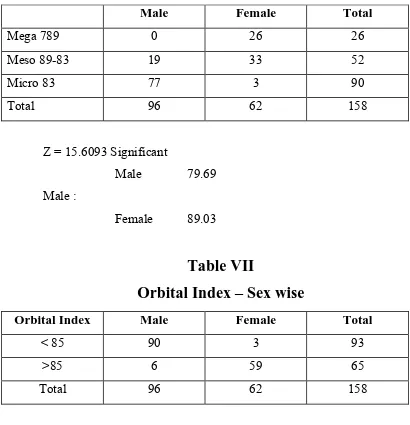
Male Female Total
Mega 789 0 26 26
Meso 89-83 19 33 52
Micro 83 77 3 90
Total 96 62 158
Z = 15.6093 Significant
Male 79.69 Male :
[image:43.612.101.511.145.570.2]Female 89.03
Table VII
Orbital Index – Sex wise
Orbital Index Male Female Total
< 85 90 3 93
>85 6 59 65 Total 96 62 158
DISCUSSION
A study of transverse diameter and vertical diameter of male and female orbital openings from the available bodies in the Institute of Anatomy and Mortuary of Madurai Medical College, Madurai was undertaken by me to complete the present work.
The study was made between the periods of June 2005 and May 2006. The particulars of the cases were available from the respective Anatomy and Forensic Medicine Departments of Madurai Medical College Madurai.
The total number of orbits studied is in female 62 x 2 = 124 and in male 96 x 2 = 192. Care has been taken to exclude any pathological component is present in the examined orbit.
The examined people belong mainly to the district of Madurai, Ramnad adn Virudunagar of Tamil Nadu and particularly neighbourhood of Madurai.
Most of the deceased are moderately nourished and moderately built. There is no gross evidence of any endocrinal or nutritional disorders or diseases. The age of the persons vary from sixteen to eighty years.
The straight distance between the upper and lower margins of the orbital cavity, taken at right angle to the orbital breadth is considered as orbital height.
The relation between the width or breadth to the height is given by orbital index.
Height of Orbit
The orbital index = --- x 100 Width of Orbit
The measurements taken are sex wise separated. In each sex, age wise they are grouped. The right orbit and left orbit measurements were compared. Orbital index for each sex has been noted and conclusions were made. Necessary graphic charts were also prepared.
The following are the tables :
TABLE TITLE
I Age wise Distribution of Cases.
II Transverse diameter of the orbit – Sex wise Distribution. IIA Transverse orbital diameter (Average of Right and Left) III Vertical diameter of orbits – Sex wise.
IIIA Vertical diameter of orbits – Sex wise.
V Sex wise variation in vertical and transverse Diameter of the orbit.
V Age wise distribution of orbital diameter.
VA Sex wise and age wise Distribution of orbital diameter. VB Sex wise and age wise distribution or orbital diameter. VI Distribution of orbital index – Sex wise.
The table 1 shows the study material age wise. A good number of orbits studied i.e. between the age of second to seventh decade.
The table II shows the Transverse diameter of the Orbit in Millimeters. The average mean transverse diameter in males is about 33.145mm.
The average mean transverse diameter in females is about 33.83mm.
There is not much difference in right orbital and left orbital measurements.
The Table IIA Shows the transverse Orbital diameter (average of right and left). Mean diameter for males is about 33.15mm and that of females is about 33.83mm.
The Z value is 2.8024. The Z test suggests that the statistical value of the present one is significant.
The Table III vertical diameter of orbits sex wise shows in millimeters. The average mean diameter for males is about 26.45mm and the average mean diameter for females is about 30.60mm. There is not much difference in right orbit and left orbit of the same individual in either sex.
The ‘Z’ value is 16.75. It is highly significant.
The Table IV shows the sex wise variation in vertical and transverse diameter of orbit. It shows Mean Diameter, Standard error of differences (SED) and Z test value. This table at a glance shows that the obtained values are statistically significant.
The Table V shows age wise distribution of orbital diameter.
The Table V-A shows sex wise and age wise orbital vertical diameter. The P value have been recorded. The values obtained upto the age group 51 to 60 are significant. Above the age of 60 are not so significant. This may be due to the quantum of study material is less is this age group in the present work.
The Table V-B is sex wise and age wise transverse orbital diameter with ‘P’ values. Here upto the age group of 41-50 appeared significant.
The Table VI shows the orbital index sex wise. The mean in males is 79.69 and in female is 89.03. The Standard error of difference is 0.5990. The ‘Z’ test value is 15.6093. The ‘Z’ test value is statistically highly significant.
The table VII shows orbital index sex wise. The chi square test λ2 value is 119.34. The P value is <0.01.
The above values suggest that it is significant qualitatively in a statistical study.
the sex of the given object. In the other way, it can be said that there can be determination of sex with the available measurements of transverse diameter and vertical diameter of the orbital opening. It means in an object of unknown sex, the transverse and vertical dimensions of the orbit furnishes a near clue of the sex of the object. Sexing of a given skull can be make near to certainity by taking the measurements transverse and vertical diameter. The statistical data obtained by the present work certainly points out that much reliability can be put on the orbital opening dimensions.
As Krogman puts that in sexing a skull, the initial impression often is the deciding factor, in my opinion the diameters of transverse and vertical of orbital margins or openings gives an initial impression of sexing a given skull. To say so, the present observations show that committing an error in determining the sex of a given skull will be less than one in hundred which givens much creditability to the present work. It is also highly significant to note that the orbital index show a clear marginal variation for each sex.
The high orbital index of female and relatively low orbital index of a male also points out that the measurements of transverse and vertical diameter of orbital opening of a given skull is a must while determining the sex. In the other way to say the same, that the orbital index can be taken as one of the major criteria while determining the sex of a skull or an individual.
An attempt has been made to bring the present work on the orbital index into the work that on the orbital index (of Broca). Another attempt has been made to mark a dividing line for the orbital of a male and female.
might be the one of the reasons to write a number of text books of Forensic Medicine the male orbit is square or rectangular in shape.
In majority of the observations made in this study, it is pointed out that there is difference of more than half centimeter to he transverse diameter to that of vertical diameter of the orbit in the case of males. In the case of female orbits the difference of the transverse diameter and vertical diameter is less than half a centimeter in majority of cases.
This may be one of the reasons to state that male orbital margins or openings, are square and that of female orbital openings are round or nearly round. These terms have been used by many authors of the text books of Forensic Medicine.
Another feature is a very few cases show the difference of Transverse and vertical diameter of one side orbit with the other side orbit. It means right and left orbit shows the difference of transverse and vertical diameter is same except in a very few cases. (In female two cases and in males two cases were observed).
a) An average male of Poor, London, of 17th Century show: The breadth of right orbit : 42.3 The breadth of left orbit : 42.4 The height of Right orbit : 34.3 The height of left orbit : 34.3 The orbital Index, R : 81.0 The orbital Index, L : 80.9 b) The Skull of George Buchanan Show :
The breadth of right orbit : 45.8 The breadth of left orbit : 44.8 The height of right orbit : 35.9 The height of left orbit : 36.8 The orbital Index, R : 78.4 The orbital Index, L : 82.1 c) The Skull of Sir Thomas Browne Show :
The breadth of right orbit : 43.7 The breadth of left orbit : 42.8 The height of right orbit : 36.3 The height of left orbit : 36.7 The orbital Index, R : 83.1 The orbital Index, L : 85.7 d) The Skull of Jeremy Bentham show :
The study of Person’s show that the orbital index in the present material varies between 78.2 to 85.74. In each case the right orbit and left orbit show different orbital index but not of great difference.
In my present work also it has been noticed that in the case of males the orbital index mean 79.69 and out of 96 bodies i.e. 192 orbits nearly 90 males i.e. 180 orbits fall below the orbital index of 85. This finding nearly coincides with that Pearson’s work.
Another feature noticed in study of Pearson’s work shown that the difference of transverse diameter to vertical diameter in males varies from 6.1 to 10.
In my present work also, it is noticed, that in males, that in the majority of cases show the difference of transverse diameter to vertical diameter from 5mm to 11mm.
But the present work differs with that of Pearson’s studies in that the measurements of transverse and vertical diameter of males is not the same. In my study the mean transverse diameter of male orbit is 33.15 and mean vertical diameter is 26.46. The range of transverses diameter of orbit in males varies from 32 to 40mm. Majority falling between 32 to 35mm.
The range of vertical diameter of orbit in males is between 24mm to 31mm; majority of them falling in the range of 25 to 28mm.
But the common feature is the indices of the orbit and the difference of transverse and vertical diameter measurements.
In Eugene Wolff’s Anatomy of the eye and Orbit and author mentions the measurements of orbital margin in:
Height of the orbital opening : 35mm. Width of the orbital opening : 40mm. It was not mentioned sex wise.
The author points out there is a great difference between the measurements given by different authorities.
Dr.S.N. Sahana, in his text book of “Human Anatomy Volume I” states that the orbital measurement at the base.
Vertical : 41.30 Horizontal : 41.30 It was not mentioned sex wise.
In their text book of “Anatomy – a Regional study of Human structure” the authors Ernest Gardner et all, wrote that ‘each orbit is shaped roughly like a pear or a four sided pyramid with it’s apex posteriorly and its aditus or base anteriorly. The base is about 35mm high and 40mm wide”. It was not mentioned sex wise.
They add that at birth, the base of the orbit is circular or elliptical; and relatively large and the two orbits are relatively nearer to each other. The full dimensions of the orbit are probably not attained until puberty.
In his book ‘The eye and its disorder Trevor-Roper, Patrik mentions that the orbital margin forms a rough square 4cm wide.
Winekler says that the adult orbital opening is quadrilateral in form and 35mm in height and 39mm in width and the orbital index is 89.7.
For a comparative study his table gives a resume of the changes in the orbital opening (Winckler).
Form Height Width Index
Foetus (8 months) Oval 14mm 18mm 77.7 New Born (6 months) Round 27mm 27mm 100.0 Child (7 years) Quadrilateral 28mm 33mm 84.8 Adult Quadrilateral 35mm 39mm 89.7
The Orbital Index of Broca is
Height of Orbit
--- x 100 Width of Orbit
Taking the orbital index as the standard there classes are recognised.
1) Megaseme (Large) : The orbital index is 89 or over. This type is characteristic of yellow races except the Eskimos. The orbital opening is round.
3) Microseme (Small) : Orbital index is 83 or less. This type is characteristic of black races. The orbital opening is rectangular. In the above mentioned classification, it is of racial one and do not specify sex variation. In my study for each sex the orbital index has been calculated.
In males 19 (Nineteen) out of 96 (Ninety Six) fall in the group of Mesoseme and 77 (Seventy Seven) out of 96 (Ninety Six) fall in the group of Microseme. The mean orbital index of male is about 79.90.
In the case of females 26 (Twenty Six) out of Sixty Two individuals fall in the category of Megaseme; 33 (Thirty Three) out of 62 individuals fall in the category of Mesoseme; and 3 (Three) out of 62 (Sixty Two) individuals fall in the category of Microseme.
My study on the orbital index, sex wise, reveals that: the males orbital index in Megaseme is NIL.
Majority of orbital index of males fall in the category of Microseme.
The female orbital index in majority lies in the groups of Megaseme or Mesoseme the female orbital index in Microseme is minimal.
An attempt has been made, taking the orbital index as criteria, to determine the sex.
85 (Eighty five) as the orbital Index.
It is noted that:
In case of females 3 (Three) fall below this range and 59 (Fifty Nine) out of 62 (Sixty Two) fall above this range.
In the other way to say the same that in majority of cases the orbital index in males is less than 85 and in females it is more than 85.
Mean of Orbital Index – Age Group Wise Male Orbits : 192 Female Orbits : 124
Mean Male Female
11-20 82.24 88.50 21-30 80.11 89.40 31-40 80.15 88.90 41-50 80.27 87.41 51-60 73.26 89.73 61-70 79.91 89.00 71-80 82.35 90.90 The Chi square test value and the ‘p’ value are significant in this study and also purposeful.
In the study of the sex determination of a skeleton Stewart (’48, ’51) feels that for the entire adult skeleton, or for the adult pelvis or for one adult hip bone, he can sex correctly in 90-95% of cases; for adult skull alone about 80% for adult major long bones alone” size alone determines” the answer.
skeletons sexed by inspection (skeleton complete) Stewart scored 94%; but in this same series, using skull plus mandible he scored only 77%.
Hug, in 1939 – 40, put sex differences in cranial dimensions on fairly sound basis on German Crania. A scrutiny of his data will reveal considerable size overlap, as between male and female. Obviously, no single dimension is diagnostic, but if all dimensions are large, in a male direction, then it is pretty safe to diagnose the skull as male.
Keen, in 1950, attempted to set up a battery of cranial traits and dimensions for adult skulls. (Juvenile and senile skulls excluded) which will sex skull with 85% accuracy. Keen’s work is on the skulls of the ‘cape coloured’ population of South Africa which he accepts as a homogeneous population. It is interesting to note that each of the anatomical feature shows a significant sex difference.
In recent years elaborate and detailed statistical studies, involving discriminate analysis, have been made of sex differences in skull and long bones. Harihara (’59) reports on such a studyon the Japanese skulls. He concluded that the method used here was not good for skulls.
In the determination of sexing of unknown skeletal material depends, of course, upon the relative completeness of the skeleton. Percentage of accuracy, for adult material is about as follows: (Krogman).
f) Long bones plus pelvis : 95% g) Long bones alone : 80%
Krogman narrates that standards of morphological and morphometric sex differences in the skeleton may differ with the population involved. This is especially true with the reference to dimensions and indices (average and range). As a general rule standards should be used with reference to the group from which they were drawn and upon which they are based; they are not ordinarily interchangeable. With statistics one can be sure; or at least more sure in an individual case.
Glanter John, in his book states that the sex of a subject can frequently be determined form the general characters of skull; it is necessary that the balance of all characters should be considered with case.
I. Gordon and R. Turner in their book say that even when all the necessary data for sexual differences in the skulls of a particular group are available, it is possible to arrive at a definite conclusion regarding sex only in 85% of cases.
Harrison states that it is only after the age of fourteen to sixteen year that the male characteristics begin to develop; upto that age it is often exceedingly difficult, if not impossible to sex the skull. He stresses that the determination of sex of the skull cannot be made from one characteristic alone. The best characteristics to note are probably those of
(a)The supra orbital ridges. (b)The mastoid processes. (c)The dimensions of the palate. (d)The outline of the orbit.
In toto, it is noticed that a stress has been given by many investigators and authors about the shape of the orbital opening or amargin or base in the determination of a race. But not significant work has been to describe shape rather than to note the dimensions or morphometry.
In the present study the following observations were made.
In Males : (the study of ninety six deceased person’s orbits or total 192 orbits (right 96 and left 96):-
a) The difference of vertical diameter and transverse diameter of orbits between 5 millimeters to 11 millimeters observed in majority of cases. i.e. 178 orbits.
b) The mean diameter of the vertical diameter of orbit on average is 26.45. c) The man diameter of the transverse diameter of orbits is 33.15.
d) The mean diameter of the vertical diameter of the orbit age wise. 11-20 26.8mm
21-30 26.1mm 31-40 26.4 mm 41-50 26.15mm 51-60 26.44mm 61-70 28.38mm 71-80 28.0mm
31-40 32.7 41-50 32.75 51.60 33.88 61-70 35.56 71-80 34.0
f) The mean orbital index : 79.69
g) The orbital index group wise :
Megaseme >89 NIL Mesoseme 89-83 19 Microseme <83 77 h) The orbital index : <85 90
>85 6 The orbital index is 62.5% cases only more than 85.
In Females : (The study of sixty two deceased person’s orbits or total one hundred and twenty four orbits.)
a) The difference of vertical diameter and transverse diameter of orbits is less than a half centimeter in fifty five cases or one hundred and ten orbits.
b) The mean diameter of the vertical diameter of the orbit is 30.06mm. c) The mean diameter of the transverse diameter of orbits 33.83. d) The mean diameter of the vertical diameter of the orbit age wise.
61-70 29.66 71-80 29.5
e) The mean diameter of the transverse diameter of orbits age wise. 11-20 33.37
21-30 32.72 31-40 33.91 41-50 33.88 51-60 34.00 61-70 33.33 71-80 33.00 f) The mean orbital index : 89.03 g) The orbital index group wise:
Megaseme >89 NIL Mesoseme 89-83 33 Microseme <83 3 h) The orbital index : <85 3
>85 59 The orbital index is 4.83% cases only is less than 85.
The Statistical Analysis Says :
A) The ‘Z’ value of transverse diameter of the orbital margin or base of the orbit is 2.8024 and is significant.
B) The ‘Z’ value of the vertical diameter of the orbital margin or base of the orbit is 16.75 and is significant.
C) The ‘P’ value for vertical diameter of orbital margin or base of he orbit are significant sex wise and age wise from 11-20 age group to 51-60 age group.
E) The ‘Z’ value of orbital index is 15.6093 and is significant. F) The orbital index sex wise show a chi square value
λ2 = 119.34 (d.f. = 1) P value <0.01 and is significant.
From the above observations made, it is significant that the determination of sex an be made by taking the measurements of transverse and vertical diameter of the base of the orbit or orbital opening or orbital margin.
The present work is confined to the examination of bony orbits of the cadavers. It is not of dry done of orbits. The study also limited to a particular sector of population and geography. And also there is smallness in the number of cases studied. The present work is taken up to pick up a good parameter for the determination of sex of unknown skull. The detailed study has suggested that a good amount of further work on this topic from different segments of the state will certainly give a broader and recognisable status for this parameter.
SUMMARY AND CONCLUSION
Introduction :
In view of certain amount of, uncertainity the sex differentiation of skull, it is thought to make out another parameter. As previous experience revealed certain amount of variants of orbital height between both sexes, it is thought that this parameter may be investigated. Hence this work.
Review of Literature :
The literature of this subject is reviewed. Broca studied orbital index for race differentiation but did not mention of the sex difference. Wincler reported on the estimation of age difference through orbital index but he too did not mention sex difference. Pearson studied the orbital index of males but did not observe on the females. This work aims to study the sex differentiation with the help of orbital index.
Material and Methods :
For this work, the orbital measurements of 96 male and 62 female adult Cadavers were taken.
Observation :
The average orbital breadth of males and females was found to be 33.15 and 33.83mm respectively. The average orbital height of males and females was found to be 26.45 and 30.06mm respectively. This difference of orbital height in statistically found to be significant (P < 0.01).
Conclusion :
and below is conclusively to be that of a male with-out any error. These conclusions have been found to be statistically highly significant.
Limitations :
The limitations of this work may be (a) a small sample – segregated geographical distribution and (b) little more probable error for dry bones.
Suggestions :
It is suggested that a bigger sample may be taken, the work may be continued at different geographical areas and on dry bones.
BIBLIOGRAPHY
1. Basu, S.C (1984) Handbook of Medical Jurisprudence and
Toxicology, Second Edition, Current Distributors, Sarani, Calcutta – 700 013 P.22
2. Boil, T.C. Grant E.A.U. Grant’s method of Anatomy – Seventh Basmajian J.V. Edition – Scientific book agency,
Calcutta-P.591.
3. Conard, Beraus (1950) Encyclopedia of the Eye, Diagnosis and Edward Siegal Treatment. J.B.Lippincott Company,
London. Page 158.
4. Francis E.Camps (1956) Practical Forensic Medicine,
Hutchinson’s Medical Publication Ltd., P.175.
5. Gardner, E. Donald J.G. “Anatomy-A regional Study of Human Ray (1967) O’ Rahilly, R. Structure” – Second Edition – W.B.
Saunders Company Page 794.
6. Gee, D.J. (1975) Lecture notes on Forensic Medicine
Second Edition-Black-Well Scientific Publications London-Page 185.
7. Glaister, John (1975) Medical Jurisprudence and Toxicology-
8. Gozales, T.A. Legal Medicine, Pathology and
Toxicology Century Crafts, INC New York – Page 39.
9. Gordon, I; Turner, R; Medical Jurisprudence-Third Edition- Price T.W. E&S Livingstone Ltd., London.
10. Harold, E. (1960) Clinical Anatomy-First Edition-
Blackwill, Scientific Publications, Oxford.
11. Hill, A.B. (1962) Principles of Medical Statistics.
12. Krogman, W.M. (1962) The Human Skeleton in Forensic
Medicine-Charles C. Thomas Illinois-USA. Pages 112-150; Pages 246-247.
13. Last, R.J. (1964) Eugene Woeff’s Anatomy of the Eye and Orbit-Fifth Edition H.K. Lexis & Co. Ltd., London Pages 1 to 19.
14. Mallik, C.C. (1969) A Hand book of Medical Jurisprudence-
Academic Publishers Panchanan Ghose Lane- Calcutta Page 23.
15. Modi, N.J. (1975) Modi’s Text Box of Medical
16. Narayanreddy K.S. (1983) The Essentials of Forensic Medicine and
Toxicology-Seventh Edition-Published by K.Sugua Devi, 16-11-1512/2, Salem Nagar Colony, Malakapet,
Hyderabad-500 036. Page 47.
17.Parikh C.H. (1970) A simplified Text Book of Medical
Jurisprudence and Toxicology, 1st Edition-Medical Publications-D/12, York House, Colaba House, Bombay-1, Page 32.
18. Patric. D (1974) The eye and its disorders, Third Edition- Black will Scientific Publications, London.
19. Polson, C.J. (1965) The essentials of Forensic Medicine –
Second Edition – Pergamon Press – Oxford – Page 53.
20. Shana, S.N Human Anatomy, volue 1, Third Edition
– New Central Book agency, 54-B-Patua Hoela Lane-Calcutta – 700 009, Page 82.
22. Smith, S, Forensic Medicine – Third Edition J & A Fiddles, F.S (1955) Churchill Ltd., 104, Glocester Place,
W.I.Page 68.