TOPOGRAPHIC ANALYSIS OF CHANGES IN
CORNEAL SHAPE AS A FUNCTION OF AGE
Dissertation submitted to
THE TAMILNADU Dr.M.G.R. MEDICAL UNIVERSITY
CHENNAI
with fulfillment of regulations for the award of degree
of
M.S. (OPHTHALMOLOGY)
Branch III
MADRAS MEDICAL COLLEGE
CHENNAI
CERTIFICATE
This is to certify Dr.Swarna Udayakumar M.S. Post graduate in Ophthalmology, Regional Institute of Ophthalmology, Government Ophthalmic Hospital attached to Madras Medical College, Chennai, carried out this dissertation titled "Topographic Analysis of changes in Corneal Shape as a function of Age" by herself under my guidance and direct supervision during May 2004 to March 2007.
This dissertation is submitted to the Tamilnadu Dr.M.G.R. Medical University, Chennai for the fulfillment of award of M.S Degree in Ophthalmology.
Prof . Dr.C.Vijayalakshmi M.S.D.O Prof. Dr.V.Velayutham M.S.D.O
Chief, Uvea Clinic Director and Superintendent
Regional Institute of Ophthalmology Regional Institute of Ophthalmology Govt. Ophthalmic Hospital Govt. Ophthalmic Hospital
Egmore, Chennai Egmore, Chennai
Prof.Dr.Kalavathy Ponniraivan B.Sc. M.D
ACKNOWLEDGEMENTS
I wish to express my sincere thanks to Prof. Dr.Kalavathy Ponniraivan B.Sc, MD, Dean, Madras Medical College, Chennai for permitting me to do this study at Regional Institute of Ophthalmology, Government Ophthalmic Hospital, Chennai.
It is with overhwhelming respect and profound gratitude, I thank Prof.Dr.V.Velayutham M.S., D.O., Dean and Superintendent, RIO GOH for assigning this topic, for his continuing help, encouragement and valuable guidance throughout my post graduate course in ophthalmology.
I am greatly indebted to my Chief, Prof.Dr.C. Vijayalakshmi M.S. D.O who with her constant help, patience and affection has constantly encouraged me in everyway during my post graduate course and conduct of this study.
With profound gratitude, I thank, Prof. Dr.K.Vasantha M.S., F.R.C.S (Chief Cornea Services GOH) MS for her support, thoughtful guidance and invaluable advice during the study.
I express my gratitude to the assistant professors in my unit.
To Dr.B.Chandrasekar M.S. for his effective guidance during the conduct of this study and also during the course.
To Dr.Nirmal Fredrick M.S.D.O for always being a source of constant help, guidance in all my endeavours.
To Dr. S.Kumaran M.S for his support and guidance through out my course in ophthalmology.
I would like to thank all my Professors, Assistant Professors, my Co post graduates who have played a salutary role.
CONTENTS
TITLE PAGE NO.
PART I
1 INTRODUCTION 1
2 REVIEW OF LITERATURE
a. Anatomy 2
b. Refractive Indices of the Eye 6
c. Astigmatism 8
d. Evaluation of Corneal Astigmatism 9 e. Aging Changes in Cornea 25
PART II
1 AIM 30
2 MATERIALS AND METHODS 31
3 OBSERVATION AND ANALYSIS 34
4 DISCUSSION 46
5 RESULTS 48
6 CONCLUSION 50
PART III
1 BIBLIOGRAPHY
2 PROFORMA
INTRODUCTION
Time is the fourth dimension, affecting all things living and nonliving. As time affects the structure of the body, the eye also undergoes certain inevitable consequences. With aging, the ability to maintain physiologic homeostasis under conditions of biologic stress declines. Further this decline is associated with an increase in vulnerability of individual and a decrease in viability potential. Although time and aging are on a continuum they begin at birth.
REVIEW OF LITERATURE
Aging Changes in Corneal dimension
There is a little change in the diametric proportion of cornea from birth till old age. A 2mm growth spurt occurs during the I year of life and after which corneal size remains static at 11.7 mm. Although diameter has been reported to decrease by 0.4 mm after the age of 40 yrs, this change is relatively insignificant and is primarily due to slight central extension of corneoscleral junction.
• Corneal thickness also does not change with aging in central, midperipheral or in the peripheral corneal areas.
• Corneal asphericity likewise progresses from relatively flat centrally at birth to steeper in adults.
• Astigmatism changes from principally with the rule in 92.8% of young patients to against the rule in 85.7% of patients aged 80 yrs .
This change occurs at the same time as eyelid structures are becoming more lax and a relative decease in pressure exerted on the globe has been implicated in age related changes in corneal toricity. Alterations in curvature of cornea can also be induced by non physiological but age related phenomenon such as pterygia.
Cornea as a Refractive Medium
Anatomy
The transparent cornea is the main structure responsible for refraction of light entering the eye. This clear transparent structure forms the anterior 1/6 th of eye ball.
The Cornea Proper
The Cornea has 5 concentric layers
1. The epithelium
4. Descemets membrane
5. Corneal endothelium
1. Epithelium :- has 3 major functions
Acts as a mechanical barrier to foreign material and micro organisms and via the Langerhans cells as an outpost for immune defense.
Creates a smooth, transparent optical surface by adsorption of tear film.
Maintains a barrier to diffusion of water, solutes and drugs.
Structure :-
¾ It is a nonkeratinised stratified squamous epithelium 5-7 layers thick (about 10% of cornealthickness)
¾ It has morphologically 3 layers
a. Single layer of columnar basal cells
b. 2 to 3 layers of wing cells.
c. 2 to 3 layers of superficial squamous cells.
the flattened squamous cells, which ultimately are sloughed into tear film. It takes about 1week for the entire corneal epithelium to be replaced.
Corneal epithelial cells are tightly adherent to each other and the underlying structures by means of specialized junctions. There are three types of junctions,
1 Occluding
2. Communicating 3. Anchoring
At the corneoscleral junction (limbus), the epithelium becomes thicker and may consist of 10 or more layers of cells.
Other than epithelial cells, neurons, melanocytes, Langerhans cells and occasional leucocytes are present within the epithelial layer. Langerhans cells are modified macrophages normally found in the peripheral corneal epithelium. They are thought to play a role in ocular hypersensitivity and other immunological phenomena by processing antigens and presenting them to lymphocytes.
epithelial cells but these cells lose their ability to regenerate this structure. So once damaged it cannot be regenerated.
3. Stroma - Comprises 90% of corneal thickness. It is composed almost entirely of an extracellular matrix with keratocytes dispersed throughout and Type I collagen fibrils running parallel with the surface. It is transparent, fibrous and compact. Keratocytes are the predominant cell type in the stroma. They are flat cells derived from neural crest. Major extracellular material in stroma is collagen, which is highly orderly arranged. Human cornea has 3 different types of collagen (I, V, VI)
4. Descemets membrane- It is the normal basement membrane secreted by the corneal endothelium. Its peripheral termination, visible by gonioscopy is known as schwalbe’s line It is composed prodominantly type IV collagen and glycoproteins including fibronectin.
Functions:
Forms a scaffold on which the endothelial cells spread themselves . Acts as a barrier to the penetration of leukocytes and blood vessels into stroma but allows passage of water and small molecules
limited capacity to divide and replace aging and injured endothelial cells. Instead the endothelium enlarges, reorganizes, migrates and maintains tight apposition to neighbouring cells. 1. Polymegathism describes heterogeneity in cell size.
2. Pleomorphism describes heterogeneity in cell shape. With decrease in cell count and increase in cell size and pleomorphism the ability to restore and maintain pump and barrier function is lost.
When cell density < 400-500 cells / sq.mm functional reservoir is minimal and corneal edema is likely to occur.
Functions:- Regulates the passage of aqueous into stroma and helps in maintaining deturgescence.
Refractive Indices of the Eye
Anterior segment structures Average Range Refractive index
Air Tear
Cornea
Corneal epithelium Anterior corneal stroma Posterior corneal stroma Aqueous Lens 1.00 1.336 1.376 1.401 1.380 1.373 1.336
The tear air interface is the most powerful refracting surface of the eye (80% of the eye’s optical power). The curvature of tear film surface reflects that of underlying cornea. The refractive power of cornea is determined by its index of refraction and by its radius of curvature.
The light then undergoes little further refraction till it reaches the lens because the refractive index of aqueous and vitreous are almost the same. At both surfaces of lens , it is refracted. While the refractive index of substance of lens is higher than that of aqueous and vitreous, the difference is not so marked as between that of cornea and air.
Anterior Corneal Curvature - A Prolate
Astigmatism
Astigmatism is that condition of eye where refraction varies in the different meridians and so a point focus of light cannot be formed upon the retina. Theoretically no eye is stigmatic Astigmatism may be an error of either curvature of cornea or of the lens or of centering.
Curvature astigmatism if any, of high degree has its seat most frequently in the cornea. This anomaly is usually congenital . Its occurrence in a small degree is almost universal. The most common error is about 0.25D with vertical curvature greater than horizontal. This is known as "With the rule astigmatism or physiological astigmatism". This is due to constant pressure of upper lid on the cornea.
As age advances, it disappears or even reverses itself into "Against the rule or inverse astigmatism" with vertical curvature less curved than the horizontal. Curvature astigmatism can also be due to lenticonus, subluxated lens etc.
Optical Condition in Astigmatism
The configuration of rays as it passes through a toric surface is called
Types of Astigmatism
A. Regular astigmatism: The 2 principal meridians are at right angles to each other. It can be either
• With the rule
• Against the rule
Simple myopic or hypermetropic astigmatism: where one the focal lines falls on the retina. The other may fall either in front as in simple myopic or behind the retina as in simple hypermetropic astigmatism.
Compound myopic or hypermetropic astigmatism: where neither of 2 lines fall on the retina but are placed either in front as is compound myopic or behind the retina as in compound hypermetropic astigmatism.
Mixed astigmatism: where one of focal lines falls in front of retina and the other focal line behind the retina.
B. Irregular astigmatism
Irregularities in the curvature of cornea is different in the various meridians and no geometrical pattern is adhered to.
Evaluation of Corneal Astigmatism
History
With the advent of widespread refractive correction at the beginning of 17th century, interest developed in the shape of cornea and the optical properties of eye. Early investigations of corneal topography were confined to gross estimates of corneal curvature.
In 1619, Scheiner made the first measurements of corneal shape. He held up a series of convex mirrors of different curvatures next to the eye until he found one which gave an image of same size as the image from the cornea.
In 1820's, Cuigent developed a keratoscope through which he observed the reflected image of illuminated target held in front of patients cornea.
In 1882 Placido, placed an observation hole in the centre of the target. His target was a disc bearing alternate black and white concentric rings and this pattern still forms the basis of many topographic systems today.
Quantification of corneal curvature became possible in 1854 with the development of Keratometer by Von helmholtz. The distance between 2 pairs of reflected points gave the spherocylindrical curvature of the central 3mm of cornea in 2 meridians.
With the development of microsurgical techniques for cataract extraction, corneal grafting and incisional refractive surgery, interest turned to the optical power provided by the cornea. Measurement of corneal curvature can be converted to dioptric power using the standard keratometric index.
Methods of Measurement
Falls into 2 main categories :
• based on principle of reflection
• based on principle of projection.
1. Reflectionbased
Majority of topography systems are based on this principle
Examples include Keratometer and Video keratoscope.
They measure slope of corneal surface and can use this information to calculate radius of curvature and power. It does not measure elevation.
2. Projection based
Newly developed systems make use of this principle.
Examples include Slit photography, Rasterstereography, Moires interference, Laster interferometery.
Computer assisted video keratoscopy
• Steps involved
1. Patient positioned correctly 2. Patient fixates target
3. Placido disc Illuminated
4. Mires reflected from corneal surface 5. Clinician focuses and aligns the mires 6. Clinician triggers image acquisition 7. CCD video camera records image 8. Framegrabber captures image 9. Digitisation of image
12. Data points located 13. Algorithm applied 14. Display of results
Measurements
Raw Image
Study of raw image captured by the camera in the topography can demonstrate focal irregularities which corresponds to surface pathology or tear film abnormalities.
Height
Slope and Curvature
They represent rate of change of height and are a much sensitive measure of variation in contour across the corneal surface.
Power
Refractive power can be calculated.
Two Dimensional Maps
1. Colours
Cool( blue, black,azure)- represents flatter surface with lesser dioptric power.
2. Scales : Label on the scale gives the type of measurement which is being displayed.
Height is expressed in mm/μm , slope(no units) ,curvature (mm)power (dioptre)
Absolute scale is one in which there is a fixed colour coding system; the same colours always represent the same curvatures or powers.
The allocation of colours is related to distribution of corneal powers in normal population.
Normalised / reactive scale uses a set number of colours which are automatically adjusted to fill the range of dioptric values for that single map.
Adjustable scale : This enables the operator to select the step interval and dioptric range of the contours.
Statistical Indices
Simulated Keratometry Reading (Sim K)
Provide information equivalent to that measured by keratometer. Calculated by determining the average power along each meridian in the central (within 3mm zone) or paracentral (rings 7-9) area. The major axis is that with the greatest power and the minor axis is 90o to it.
• Min K
It is the meridian with the lowest mean power. Cylinder is the difference between major and minor axes.
• Sphero Equivalent Power
It is the effective refractive power of the cornea within the 3mm pupillary zone, taking into account the Stiles - crawford effect. It is more reliable than keratometry for calculation of power of IOL.
• Asphericity
It is a measure of flattening or steepening of midperiphery. It is important for optical aberrations following refractive surgery.
• Surface Asymmetry Index
• Inferior-Superior Value - (I-SV) :
It is calculated from the refractive power difference between five inferior and five superior points 3mm from the centre at 30o intervals.
• Keratoconus Prediction Index: derived from
1. Sim K1
2. Sim K2
3. SAI
4. Differential sector index (DSI) 5. Opposite sector index (OSI) 6. Centre surround index (CSI) 7. Irregular astigmatism index (IAI) 8. Analysed area (AA)
• Surface regularity Index
It is a measure of local regularity of the corneal surface within the central 4.5mm diameter. This index correlates well with visual function.
TOPOGRAPHIC MODELING SYSTEM:(TMS)
• It is based on old principle of placido disc
• It incorporates many luminous concentric rings
On each ring 256 points are identified and sampled to provide data from which the radius of curvature is computed.
• These rings give corneal reflections at 180 μm internals
• It provides 7,000 data points in toto
• It gives an accuracy of 0.10 D
• 2 cones are used
• 25 ring cone is for standard use. It covers 8.5mm of cornea
• 31 ring cone projects rings farther peripherally and covers 11mm diameter of cornea. This is recommended for contact lens fitting.
4 types of map
1. Standard
2. Refractive
3. Instantaneous
• Standard
Displays refractive powers in parallel.
A spherical cornea without astigmatism is displayed in one colour in this map.
• Refractive
Indicates refractive power by regarding cornea as lens.
Good for optical analysis.
• Instantaneous
Shows local slope of cornea
Good indicator of keratoconus and used to identify the borderline between the portion cut out by PRK or laser and its surrounding area.
Bogan and co workers classified normal human corneas into 4 types
A. Round and oval pattern - very low astigmatism (Figure 1)
B. Symmetrical bow tie - symmetrical astigmatism (Figure 3)
C. Asymmetrical bow tie (Figure 4)
Figure 1
Astigmatism and topography
Topography displays difference in curvature of 2 principal corneal meridians as a bow tie pattern. In normal conditions, the bow tie is oriented along the steeper meridian, the colour of the bowtie is itself warmer than the surrounding corneal surface.
During measurement, the radius of curvature are referred to the videokeratoscopic axis that intersects the cornea at the normal vertex. This explains the bow tie pattern in astigmatic cornea, the bow tie demostrates that the 2 principal meridians have different powers. Intermediate meridians have intermediate power, this is greater closer the intermediate meridian is to the steeper principal meridian.
The normal vertex is in the centre of the bow tie. It behaves like the nodal point of a lens where there is no deviation of the light rays.
If the cornea were a regular spherocylindrical surface, astigmatism would be represented by a propeller profile. There are 2 reasons for bow tie aspect in which peripheral parts of the propeller tend to be reduced in dimensions.
• First - the flatter periphery of the cornea tends to reduce the dioptric power over those meridians next to the steeper principal meridians.
Size of the bowtie is not related to the amount of astigmatism.It indicates only the extent of astigmatic changes over the corneal surface.
It is the colour difference between the 2 principal meridians that reflects the amount of astigmatism, the greater the colour difference, greater the astigmatism.
• Symmetrical bowtie indicates a symmetrical cylinder.
• In asymmetrical bowtie the dioptric power is not symmetrically distributed along the two semimeridians.
Symmetry does not correspond to intersection of line of sight . Thus astigmatism may be topographically asymmetric but relatively symmetric if 2 parts of the bow tie overlying the pupil are symmetric.
Irregular astigmatism - Topographic pattern
Semi meridional - the dioptric power of a meridian is not equally distributed along the 2 semimeridians (eg. kerotoconus)
Oblique - 2 principal meridians are not perpendicular to each other. (eg after keratoplasty)
Age Associated Clinical Alterations in the Cornea
All the histological layers of the cornea undergo changes.
¾ Corneal Tear Film
An age associated increase in incidence of bacterial infections of the cornea has been suggested. There is an increase in the mucin component of tears from elderly individuals when compared to young subjects. This leads to adherence of several bacterial organisms and may explain the susceptibility to corneal infection in the elderly.
¾ Epithelium
Epithelium undergoes spontaneous alteration in the corneal limbal surface ascribed to senile changes called as fuchs dimples. These are shallow elliptical excavations measuring 1.5-2mm in diameter, with clearly defined edges, sloping borders and a faint opacity in the floor. They are due to focal thinning or a loss of surface epithelium, Bowman's membrane and anterior stroma. They are believed to be caused by local atherosclerosis or by dessications.
closure of the eyelids, a region of focal tear pooling during eye closure. Clinically, the line appears slight brown and is best seen under cobalt blue illumination. Histologically, the iron is concentrated in the deeper cellular areas of epithelium and is best seen with iron stains such as prussian blue or perl's stain. However, it is of no consequence to corneal health or vision.
¾ Corneal sensitivity
The sensory nerves to the cornea are derived from ophthalmic branch of trigeminal nerve. They enter the cornea at the limbus, travel centripetally near the Bowman's membrane and finally arborize with in the basal epithelial cells. Beginning at the age of 40, the threshold for sensitivity to corneal touch increases and continues to do so with advancing maturity. This can be attributed to change in corneal mechanoreceptors.
¾ Bowman's layer
Corneal arcus appears to be a prognostic factor for the development of CHD in men below 50 yrs and of elevated cholesterol and LDL in patients above 50 years. Arcus senilis results form the deposition of cholesterol, cholesterol esters and fat in the extra cellular spaces of peripheral corneal stroma; this phenomenon appears first in the more posterior layers and spares the peripheral 0.25mm. In advanced layers, it is also seen in the Bowman's layer, in descemets membrane and between the scleral lamella. The opacity first appears inferiorly in the cornea, then progresses circumferentially, separated from the corneal limbus by a clear zone, the lucid interval. The leakage of fatty products from the age affected perilimbal vasculature could be a cause.
Marginal atrophy is a corneal thinning in the periphery of the cornea that occurs in association with arcus senilis. A fragmentation of peripheral collagen results in a noninflammatory depression at the corneal limbus, beginning superiorly that can even progress circumferentially. The epithelium overlying the furrow is intact.
Posterior crocodile shagreen has a mosaic like clinical appearance similar to Anterior type. Cloudy regions appear in the central cornea as a result of alteration in collagen lamination in the posterior stromal layers.
Corneal Endothelium and descemets membrane
Descemets membrane generation of membrane material then continues throughout life with doubling in thickness every 40 yrs, it measures 2μm at age 10 years and 10μm at 80 years. The anterior 1/3 is produced in utero and is 3μm thick with a 100nm banding pattern.
The posterior portion produced exutero in nonbanded and homogenously granular. The membrane remains relatively uniform in thickness until age of 30 yrs after which nodular excrescences appear in the peripheral areas. These consitute "Hasal Henle bodies" – universally found in elderly. They represent focal over production of basement membrane material. It is postulated that focal alteration in endothelial cell homeostasis results in modified production or depsition of membrane material in non planar fashion.
AIM
To investigate the age related differences in corneal topography of a normal population.
MATERIALS AND METHODS
This documentation has been arrived at the end of study of 152 patients divided prospectively into three groups.
The entire evaluation was carried out at RIO- GOH, Egmore, Chennai. The study began in June 2005 to May 2006 covering a period of 12 months.
Similarity of the cases studied was maintained as far as possible. The strategy included selection of age factor and uncomplicated cases.
Inclusion Criteria
¾ Normal cornea
¾ No history of any ocular surgery
¾ No abnormalities on slitlamp examination
¾ Regular Keratometric mires
¾ No History of Contact lens wear.
¾ No History of any other ocular ailments other than refractive errors.
Before topography was done,
¾ Visual acuity
¾ Slitlamp examination
¾ Keratometry was done
The corneas were divided into 3 groups
a 21-40 years
b 41-60 years
c 61-80 years
Number of eyes in
¾ Group a - 86
¾ Group b - 77
¾ Group c - 80
making a total of 243 eyes of 152 patients.
The highest quality keratograph was selected and stored. Videokeratographic images were displayed with the absolute scale map (1.5D/contour interval). Colour coded topographic maps with the highest quality were then selected and analysed. All the mires with in the central 1.5mm radius (central 10 mires) needed to be complete to be included in the study. The astigmatism pattern was established by analysing the axis.
70o-110o with the rule 160o-20o against the rule
in between oblique
Statistical Analysis
OBSERVATION AND ANALYSIS
The results of the 3 groups were analysed.
Group a : 21-40 years
86 eyes of 50 patients were examined in this group 14 eyes had to be excluded because of history of trauma to the eye or irregular mires on examination etc.
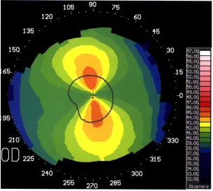
The topographic map of patients in this group showed the vertical bow-tie shaped steep area in central cornea in which the refractive power was between 43 and 44.5D (yellow) which thus indicates "with the rule" astigmatism. The peripheral cornea of these patients was relatively flat and refractive power was between 41.5 and 40.0D (green).
[image:39.612.128.502.563.632.2] The refractive power of the horizontal meridian (0-180o) were lower than those in vertical meridians (90-270o).
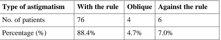
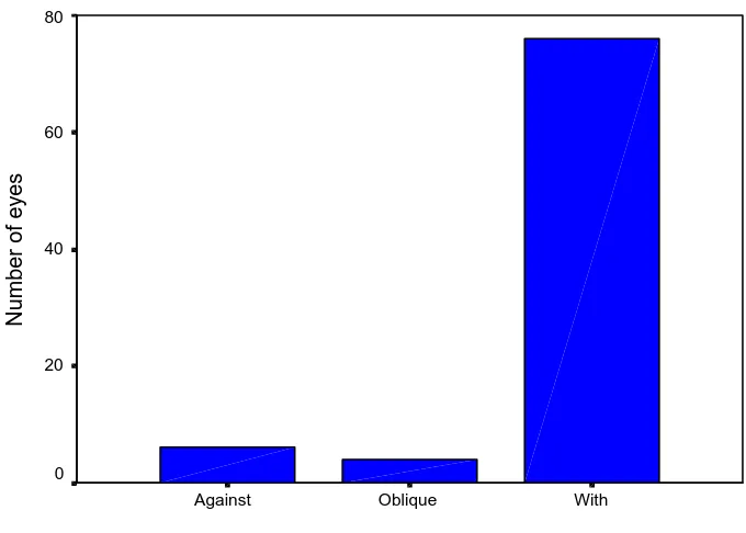
Table 1a
Type of astigmatism With the rule Oblique Against the rule
No. of patients 76 4 6 Percentage (%) 88.4% 4.7% 7.0%
Table 1b
Category Frequency Percentage P value
With the rule 76 88.4% Against the rule 6 7.0% Oblique 4 4.7%
< 0.001 (significant)
Reference
P value < 0.01 - significant at 1% level P value (0.011 to 0.05) - significant at 5% level P value (>0.05) - not significant at 5% level
By the chi square test, p value was found to be less than 0.001 and thereby it is statistically significant.
Types of Astigmatism
With Oblique
Against
Number of eyes
80
60
40
20
0
This coincides with the study conducted by Hayashi et al in 1995(14,15).
Table 1
Right Eye Left Eye
Mean Ks
Vertical Axis Mean Kf Horizontal Axis
Mean Ks
Vertical Axis
Mean Kf
Horizontal Axis
44.87 42.74 44.54 42.18
Group b: 41-60 years
77 eyes of 52 patients were examined in this group 13 eyes had to be excluded because of history of trauma to the eye or irregular mires on examination or history of ocular surgery.
The topographic map of patients in this group reveals the cornea on the whole becomes steeper and attains a virtually round configuration.
The refractive power of the horizontal meridian is almost nearing that of the vertical meridian.
Table 2a
Type of astigmatism With the rule Oblique Against the rule
No. of patients 31 20 26 Percentage (%) 40.25% 25.97% 33.76%
[image:44.612.121.504.377.455.2]On analysis of the data, of the 41-60 age group, it was found that 33.76% of eyes had against the rule astigmatism. 40.25% of eyes had with the rule astigmatism and 25.97% of eyes had oblique astigmatism.
Table 2b
Category Frequency Percentage P value
With the rule 31 40.25% Against the rule 26 33.76% Oblique 20 25.97%
Reference
P value < 0.01 - significant at 1% level
P value (0.011 to 0.05) - significant at 5% level
P value (>0.05) - not significant at 5% level
By the chi square test, p value was found to be 0.909 and thereby it is statistically not significant.
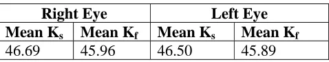
Table 2
Right Eye Left Eye Mean Ks Mean Kf Mean Ks Mean Kf
46.69 45.96 46.50 45.89
On analysis of Table 2 it is found that cornea as a whole becomes steeper.
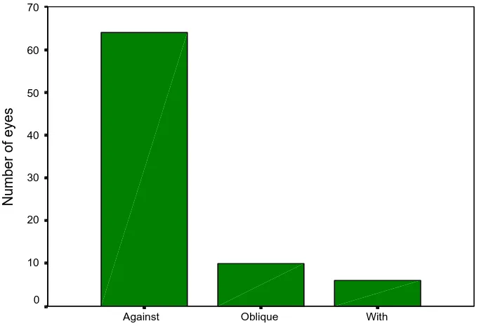
Group c : 61-80 years
80 eyes of 50 patients were examined in this group 20 eyes had to be excluded because of corneal abnormalities like degeneration, bullous keratopathy or because of pseudophakia or irregular mires.
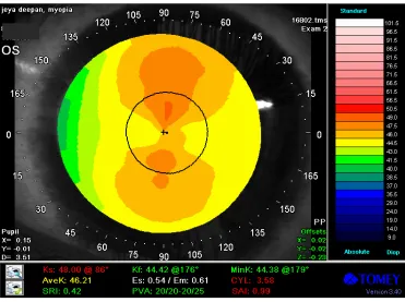
The topographic map of patients in this group showed the enlargement of central steep area to a horizontally oval shaped configuration, which suggests against the rule corneal astigmatism.
[image:47.612.194.433.124.169.2]On examining the refractive powers, it is found that the refractive powers in horizontal meridian was greater than those in vertical meridian.
Table 3a
Type of astigmatism With the rule Oblique Against the rule
No. of patients 6 10 64 Percentage (%) 7.5% 12.5% 80%
Table 3b
Category Frequency Percentage P value
With the rule 6 7.5% Against the rule 64 80% Oblique 10 12.5%
< 0.001 (significant)
Reference
P value < 0.01 - significant at 1% level P value (0.011 to 0.05) - significant at 5% level P value (>0.05) - not significant at 5% level
By the chi square test, p value was found to be less than 0.001 and thereby it is statistically significant.
This coincides with the study conducted by Hayashi et al in 1995(14,15).
Types of Astigmatism
With Oblique
Against
Number of eyes
70
60
50
40
30
20
10
Table 3
Right Eye Left Eye
Mean Ks
Horizontal Axis
Mean Kf
Vertical Axis
Mean Ks
Horizontal Axis
Mean Kf
Vertical Axis
46.45 44.82 46.62 45.08
DISCUSSION
Our study clearly shows that the normal cornea becomes steeper and shifts from with the rule astigmatism to against the rule astigmatism with age. The averaged map of the normal cornea at younger age showed vertical bow tie shaped steep area in central cornea which indicates with the rule astigmatism(14,15).
With aging, the cornea on the whole gradually becomes steeper and the central steep area extended horizontally. The subjects in and around 60's revealed a virtually round configuration(14,15) which implied that corneal astigmatism has been almost neutralised at this age.
The causes of aging alteration in the cornea still cannot be clearly explained. The against the rule astigmatism shift in older age predominantly depended on external influences.
1. decline in upper eyelid pressure due to dermatochalasis
2. reduction action of medial rectus muscle
which leads to flattening of the horizontal meridian of cornea in older individuals.
Internal Factors
1. Progressive thickening of stromal collagen bundles
2. Decrease in interfibrillar spacing
3. Thickening of descements membrane
4. Degeneration of endothelial cells
RESULTS
243 eyes of 152 patients were analysed in this study. 57 eyes had to be excluded because of either irregular mires, slitlamp abnormalities, corneal pathology, pterygium or because of pseudophakia.
The patients were categorized into 3 groups.
Group a - 21-40 years
b - 41-60 years
c - 61-80 years
Topographic examination was done and the depending on the axis of the steeper meridian they were classified as having with the rule if steeper axis was between 70o and 110o, against the rule if the steeper axis was between 160o-20o and oblique astigmatism if steeper axis was in between.
In our study, group a (21-40 years) had with the rule astigmatism predominantly (88.4%) when compared to oblique (4.7%) and against (7.0%) the rule.
This coincides with the study conducted by Hayashi et al in 2001.
astigmatism. There is a gradual increase in against the rule astigmatism in this age group.
In group c patients of age 61-80 years, 80% was against the rule, 12.5% had oblique, 7.5% had with the rule astigmatism.
On analysis of topographic pattern of each group it is found that the vertical bow the pattern in the younger group (group a) gradually changes to almost round configuration in the group b (41-60 yrs) and as age further advances, it is found that the horizontal meridian steepens further and becomes a horizontal bow tie in group c (61-80 yrs).
CONCLUSION
This study clearly demonstrates that the normal cornea becomes steeper and shifts from with the rule to against the rule astigmatism with age. The data anlaysis of change in refractive powers clarified that the against the rule shift occurs predominantly due to steepening of horizontal meridians.
BIBLIOGRAPHY
Text books and Journals
1. The eye in Aging – Jackobeic chapter 344, pp.4784 - 4831.
2. The cornea – Scientific foundations and clinical practice Smolin and thofts, fourth edition.
3. Grayson’s diseases of cornea – fourth edition.
4. Berger, Brown Peymen – Principles and Practice of Ophthalmology – Vol.1.
5. Parson’s Diseases of the eye - Nineteenth edition.
6. Clinical anatomy of the eye – Richard S. Snell -second edition.
7. Wolffe Anatomy of eye and orbit – Anatomy J. Bron, Eighth edition.
8. Adlers physiology of eye – Ninth edition.
9. Abrams- Duke elder’s practice of refraction – 10th edition.
10. Optics, refraction and contact lens– Basic and clinical science course – American Academy of Ophthalmology – 2004-2005.
12. Corneal topography – The clinical atlas-Lucio Buratto – 1996.
13. Corneal topography - Principles and applications, Corbett, Rosen O'Bratt.
14. Topographic Analysis of changes in corneal shape due to aging - Cornea 14(5): 527-532-1995- Ken Hayashi M.D., Hideyuki Hayashi. M.D. and Fumihiko Hayashi, M.D.
15. Gender and age related different in corneal topography- Tomoko Goto, M.D., Stephen D. Klyce, Ph.D, Cornea, vol.20, no.3, 2001, pp.270-76.
16. Measurement of corneal curvature in young and older normal subjects - JRS vol.15 July/Aug 1999, Parahan et al.
17. Aging and Cornea Faragher RGA, Mucholland, BJO 1997, 81:84-817.
18. Wilson SE Klyce. SD Advances in Analysis of corneal topography. Surv.oph. 1991 35:269-77.
19. Roberts C. Corneal Topography: a review of items and concepts JCRS 1996 22: 624-9.
20. Methods of analysis of corneal topography. Invest ophthal JCRS 1989 5: 368-71, Klyce SD. Wilson SC.
22. Zabel Rw, Tuft SJ, Fitzke FW, Marshall J. Corneal topograph: a new photokeratoscope eye 1989, 3:298-301.
PROFORMA
Sl.No. :
IP. No/OP/No. :
Name :
Age /Sex :
Presenting complaint :
Defective vision Re/Le-Duration.
H/o Presenting Illness :
Past History :
H/o Ocular surgery
H/o Wearing spectacles
H/o Contact lens wear
H/o Dm/TB/HT
Family History :
Local Examination :
1. Head Posture
2. Facial Symmetry
3. Visual axes
Slit lamp examination
RE LE
Lids
Conjunctiva
Cornea
Anterior chamber
Iris
Pupil
Lens
UCVA
Distance:
Refraction:
BCVA
IOP
Fundus
Keratometry
KEY TO MASTER CHART
M male
F female
RE Right eye
LE left eye
D Dioptre
cyl cylinder
WTR with the rule
ATR Against the rule
Kf flatter axis
MASTER CHART
Right Eye Left Eye
Serial
Number Name Sex Op No Ks Kf Cyl axis Ks Kf Cyl ax
1 amudha 28 f 39209 43.8 42.32 1.48
2 anuradha 38 f 41954 43.77 42.91 0.86 11
3 amudha 38 f 37109 44.91 44.44 0.47 166 44.9 44.29 0.61
4 amulu 60 f 41994 45.29 44.34 0.95
5 sara 57 f 65431 46.33 45.65 0.68 135 46.44 46.04 0.4
6 iyyanar 63 m 43218 42.84 42.28 0.56 111 43.51 42.76 0.75
7 bharati 26 f 46120 46.04 45.08 0.96 97 43.49 42.99 0.5
8 bharath 37 m 23348 45.68 44.37 1.32 96 45.02 44.02 1
9 christi priya 22 f 60411 45.59 44.1 1.5 80 44.2 43.09 1.01
10 umadevi 23 f 23411 44.69 43.34 1.35 94 44.47 43.43 1.05
11 dhanalakshmi 35 f 22610 44.29 43.26 1.03 98 42.63 41.63 1
12 edwin 40 m 40908 101 42.88 42.48 0.4
13 elumalai 39 m 44790 45.38 45.02 0.36 72 45.38 44.89 0.49
14 ezhil 35 m 58370 43.98 42.49 1.49 101 44.08 43.02 1.06
15 gajavalli 28 f 63086 44.56 43.73 0.84 174
16 ganesan 36 m 61042 45.6 45.03 1.57 96 46.57 44.39 2.18
17 gnanamani 63 f 63093 45.21 44.9 0.31 6 45.05 44.75 0.3
18 deivam 70 m 68342 46.41 46.03 0.38 6
19 gowri 24 f 37522 87 46.9 45.43 1.47
20 indumati 22 f 40987 87 43.5 43.2 0.3
21 rajamma 64 f 67544 47.04 46.75 0.29 169
22 indrani 36 f 31260 45.81 44.64 1.16 107 45.41 43.41 2
23 jaya 40 f 27118 46.02 43.72 1.3 89 45.86 45.29 0.57
24 jyadeepan 21 m 58168 45.88 42.88 3 98 46 42.42 3.5
25 kumar 32 m 67461 43.8 43.46 0.34 100
26 naidu 68 m 69276 47.74 46.79 1.08 6
27 kalimmal 68 f 63822 46.89 45.99 0.9 156
28 kanniama 68 f 47711 45.78 44.24 1.54 169
29 marriappan 70 m 67020 46.76 45.62 1.14 177
Right Eye Left Eye Serial
Number Name Sex Op No Ks Kf Cyl axis Ks Kf Cyl ax
33 kumaresan 39 m 50201 45.88 45.12 0.76 101 45.96 45.33 0.63
34 lakshmi 40 f 51231 43.99 42.98 1.01 101 43.69 42.4 1.29
35 lokanayaki 38 f 41620 43.85 43.03 0.83 98 43.98 43.09 0.89
36 mohan 45 m 42341 45.9 45.5 0.44 108
37 mani 39 m 50269 45.36 43.76 1.6 83
38 narasia 70 m 402068 46.64 46.21 0.43
39 marriama 68 f 41139 48.49 47.48 1.02
40 manniama 62 f 43682 46.87 45.92 0.95 76
41 leela 63 f 43312 47.3 45.75 1.55 1 48.08 46.89 1.19
42 muthulakshmi 52 f 23123 45.58 45.24 0.34 98 45.55 45.89 0.34
43 muthuselve 48 f 43211 45.43 43.67 0.76 106
44 shanti 38 f 44.49 43.04 1.45 83 44.85 43.71 0.74
45 paramman 72 m 43078 48.99 47.73 1.26 24 48.01 46.99 1.02
46 pughuzhali 52 f 43210 45.36 45.04 0.32 38 44.91 45.77 0.86
47 pushpa 50 f 41201 47.04 46.24 0.8 109 47.23 46.45 0.78
48 pyari 62 f 43121 46.2 45.72 0.49
49 sarala 58 f 31221 46.81 46.21 0.59 129 46.02 45.27 0.75
50 ramji 32 m 43105 42.75 42.32 0.44
51 natesan 79 m 23132 46.35 45.8 0.65 3 46.92 46.12 0.8
52 raganadhan 72 m 23121 46.4 45.77 0.63 169
53 paranjothi 70 m 35498 46.35 45.43 0.92 162
54 rayappan 68 m 24791 46.19 45.31 0.88 13
55 rani 62 f 32965 48.28 45.96 2.33 4 47.68 46.2 1.47
56 saraswati 64 f 23175 47.49 47.08 0.41 176 46.89 46.32 0.57
57 sharmila 38 f 24718 44.62 43.71 0.9 68
58 siraj 28 m 23178 44.5 43.88 0.62
59 suba 23 f 37612 39.39 35.35 1.04
60 victor 38 m 43781 44.54 44.08 0.46
61 sugantha 68 f 44285 46.41 45.78 0.63 1 46 44.89 1.1
62 shanti 62 f 47351 47.33 46.17 1.16
63 papa 68 f 43613 46.53 46.07 0.45 6 46.2 45.96 0.24
64 sasirekha 29 f 42165 42.47 41.45 0.92 100 45.33 43.1 1.23
Right Eye Left Eye Serial
Number Name Sex Op No Ks Kf Cyl axis Ks Kf Cyl ax
66 shalini 20 f 45109 42.64 41.91 0.73 80
67 shoba 28 f 32745 43.24 42.19 1.06 89 43.37 42.91 0.46
68 swaroopa 26 f 34181 41.04 40.03 1.01 96 42.6 41.6 1
69 vanitha 24 f 34165 42.85 42.19 0.66 97 42.99 42.12 0.86
70 venketraj 26 m 36712 44.02 43.12 0.9 94 44.75 43.6 1.15
71 umadevi 26 f 42185 43.81 42.23 1.58 97 44.79 43.04 1.76
72 swati 24 f 34813 44.18 42.67 1.51 100 44.04 42.61 1.43
73 shanti 36 f 43163 44.79 44.08 0.71 98 44.7 43.5 1.2
74 nivaskumar 22 m 43816 44.27 43.11 1.16 79 44.49 42.97 1.52
75 peter 26 m 43981 43.13 42.43 0.7 82
76 ponmalar 25 m 41291 45.42 44.95 0.47 78 44.17 43.58 0.5
77 prakasan 38 m 43819 42.97 42.62 0.36 96 43.62 43.1 0.52
78 selve 21 f 41291 40.21 39.33 0.88 87
79 rajalaksmi 23 f 45183 43.74 42.27 1.47 87 43.12 41.07 1.07
80 rajina 28 f 45184 43.12 42.45 0.67 96 42.92 43.42 0.5
81 rupa 36 f 43291 45.99 45.25 0.74 104 46.9 46.5 0.4
82 roshan 25 f 31672 44.22 43.49 0.73 94 44.11 43.18 0.14
83 devani 70 21376 47.93 46.6 1.33 89 45.37 44.77 0.61
84 stalin 61 m 23458 43.85 41.87 1.98 87
85 sami 62 f 23781 42.39 41.67 0.72 118
86 savithri 66 f 28134 45.64 45.12 0.51 149
87 panchali 70 f 23712 45.53 44.44 1.09 187 45.64 46.39 0.75
88 ramaniaamal 68 f 26184 47.68 46.2 1.47 169 47.15 45.68 1.47
89 tilagam 62 f 23109 45.98 45.37 0.61 160 45.77 44.77 1
90 tirupati 72 f 27315 46.19 45.49 0.7 173 46.02 45.52 0.5
91 mangalam 62 f 43017 46.9 45.43 1.47 175 45.7 45.14 0.56
92 dasaradhan 72 f 42754 45.05 44.75 0.3 6 46.57 45.72 0.75
93 mangamma 67 f 43162 46.79 45.77 1.02 189 46.02 45.22 0.8
94 jayalakmi 62 f 43982 46.12 45.62 0.5 3 46.39 45.8 0.59
95 pandurangan 64 m 31627 46.95 46.35 0.6 18 47.7 46.2 0.5
Right Eye Left Eye Serial
Number Name Sex Op No Ks Kf Cyl axis Ks Kf Cyl ax
99 thirivegadam 70 m 35207 47.31 46.92 0.39 172 47.48 46.98 0.5
100 manonmani 64 f 37591 46.62 4612 0.5 4 46.35 45.82 0.63
101 ganga ye 65 f 43612 46.15 45.4 0.75 150
102 purushothaman 65 m 31020 45.31 44.94 0.36 138
104 puroshathaman 65 m 34518 47.02 46.22 0.8 172 47.23 46.32 0.91
105 domadaran 65 m 37162 46.91 45.89 1.02 167 46.52 46.02 0.5
106 pankajammal 62 f 46.74 45.82 0.92 6 47.09 46.34
107 rasti 70 f 45317 46.92 46.12 0.8 173 48 46.8 1.2
108 sundarimmal 69 f 43178 47.52 47.02 0.5 8 46.87 46.12 0.75
109 rajan 60 m 393890 46.78 46.39 0.39
109 mouthu 65 m 40920 48.01 46.89 1.12 121 46.75 46.25 1.5
110 vasnthakumari 59 f 393871 46.22 45.35 0.87
111 perumal 52 m 394130 45.81 46.66 0.79
112 jaythnbe 54 f 395123 46.45 46.02 0.43
113 kathiresan 60 m 395632 46.31 46.91 0.6 101 45.78 45.21 0.57
114 rahim 56 m 39602 46.7 46.02 0.68 7 47.04 45.54 1.5
115 lakshmi 54 f 395860 46.3 45.75 0.55 180 46.02 44.98 1.03
116 savithri 52 f 39652 46.63 45.91 0.72 4
117 periyanayagam 60 m 396316 46.39 45.98 0.41 180 46.28 46.58 0.38
118 natesan 60 m 392979 47.09 46.34 0.75 87 47.09 46.08 1.02
119 joseph 56 m 392546 46.74 45.95 0.9 3 46.22 45.42 0.8
120 lailabee 52 m 394908 46.91 45.89 1.02 78 46.52 47.02 0.5
121 baby 60 m 395425 46.89 45.7 1.19 173 46.12 45.62 0.5
122 rajeshwari 59 m 395855 46.13 45.61 0.52 4 46.39 45.5 0.89
123 gnanamal 55 m 393006 47.28 46.48 0.8 41 47.32 46.34 0.98
124 thotiamma 52 m 393428 46.33 45.31 1.02 29 45.86 46.68 0.62
125 rajendran 60 m 394130 47.23 46.32 0.91 42 47.5 46.48 1.02
126 balammal 58 m 34915 46.29 45.89 0.4 138 45.75 45.25 0.5
127 fathima 59 m 395855 46.24 45.44 0.8 60
128 kalyani 44 m 365428 46.02 45.27 0.45 136
129 govindhamal 55 m 34339 47.45 46.99 0.46
130 kesavan 55 m 393888 46.72 45.84 0.88 140
Right Eye Left Eye Serial
Number Name Sex Op No Ks Kf Cyl axis Ks Kf Cyl ax
132 parvatham 54 f 395298 47.3 46.8 0.5
133 seetha 55 f 395631 47.91 46.72 0.38 44
134 jamuna 44 f 396311 47.8 46.9 0.9 144
135 sunderaaswaran 60 m 395866 45.89 45.16 0.73 76 46.73 45.71 1.02
136 suseela 56 f 396107 47.36 46.57 0.79 170 46.93 45.92 1.01
137 murthy 54 m 396586 47.2 46.41 0.79 14 47.53 46.81 0.72
138 munisekhar 48 m 396603 47.44 46.95 0.53 12 47.07 46.18 0.89
139 saroja 46 f 396615 47.72 46.91 0.81 80 46.9 46.12 0.78
140 raniamma 56 f 396304 46.2 45.42 0.78 4 46.42 45.16 0.36
141 adhimoolam 45 m 396294 47.19 46.63 0.56 110 46.89 46.08 0.82
142 saraswati 58 f 393621 46.55 46.11 0.44 101
143 bhuvaneshavari 42 f 394874 46.66 45.91 0.75 90
144 chellama 55 f 395147 47.5 45.89 0.61 96
145 thiruvasagam 50 m 395451
146 kathirseslvan 42 m 396103 46.12 45.3 0.82
147 narayanan 44 m 396108 46.19 45.69 0.5
148 perumal 52 m 394882 47.03 46 1.03
149 kannamal 48 f 395136 46.5 45.38 1.12
150 gnanamal 57 f 395847 47.21 46.12 1.09
151 ananadan 47 m 395875 47.5 46.56 0.94
LIST OF SURGERIES PERFORMED
Sl.No. Name Age / Sex
OP / IP No.
Diagnosis Surgery Performed
1 Mrs.
Muniamma
65 / F 41447 RE - Aphakia LE - MC
LE - ECCE with PCIOL 2 Mrs.
Jayalakshmi
62/F 38233 RE - MC LE - IMC
LE - ECCE with PCIOL 3 Mrs.
Logammal
80/F 382591 BE - MC LE - ECCE with PCIOL 4 Mr. Vadivel 65 /
M
382805 RE - MC LE - IMC
LE - ECCE with PCIOL 5 Mrs.
Subbammal
60/F 383102 RE - IMC LE - MC
LE - ECCE with PCIOL 6 Mr. James 63 /
M
383595 BE - PCC LE - ECCE with PCIOL 7 Mr. Hameed 53/
M
391020 BE - IMC RE - ECCE with PCIOL 8 Mr. Muthu 71/M 384442 RE - MC
LE - IMC
RE - ECCE with PCIOL 9 Mr. Pethaya 40 /
M
394862 RE -MC LE - IMC
RE - ECCE with PCIOL 10 Mrs.
Thengammal
55 / F 384012 RE - IMC LE - MC
LE - ECCE with PCIOL 11 Mrs.
Dhanabakiyam
70 / F 385999 RE - Aphakia LE - Nuclear Cataract
LE - ECCE with PCIOL 12 Mr. Engayya 58/
M
16840 RE - MC LE - IMC
RE - ECCE with PCIOL 13 Mrs.
Muniammal
63 / F 32624 BE - IMC RE - ECCE with PCIOL 14 Mr. Chitubabu 60 /
M
389924 RE - Aphakia LE - MC
LE - ECCE with PCIOL 15 Mrs.
Maragadham
50 / F 390453 RE - Pseudophakia
LE - MC
Sl.No. Name Age / Sex
OP / IP No.
Diagnosis Surgery Performed
16 Mrs. Meena 60 / F 39028 BE - IMC LE - ECCE with PCIOL 17 Mr.
Chinnadurai
65/ M
390736 RE-
Pseudophakia LE - MC
LE - ECCE with PCIOL 18 Mrs. Salmabee 80 / F 390986 RE - IMC
LE - Aphakia
LE - ECCE with PCIOL 19 Mr. Manickam 65 /
M
390947 RE-
Pseudophakia LE - MC
LE - ECCE with PCIOL 20 Mrs. Meera 75 / F 398469 BE - Nuclear
Cataract
RE - SICS with PCIOL 21 Mrs.
Meenakshi
65/ F 390462 RE - MC LE - IMC
RE - SICS with PCIOL 22 Mr. Hari 56 /
M
395787 RE - HMC LE - MC
RE - SICS with PCIOL 23 Mr. Rajendran 63 /
M
394565 BE - IMC RE - SICS with PCIOL 24 Mr. Ravindran 70 /
M
395324 BE - HMC RE - SICS with PCIOL 25 Mr. Ashok 25 /
M
32651 RE - Chalazion RE - I & C 26 Mrs. Rajammal 40 / F 406219 BE - Nasal
pterygium
RE - pterygium
excision 27. Mrs.
Muniammal
60 / F 40251 LE - Chronic dacryocystitis
LE - DCT 28 Mrs. Rajammal 68/ F 45385 RE - Chronic
dacryocystitis
RE - DCT 29 Mrs. Lakshmi 62 / F 49261 LE - Chronic
dacryocystitis
LE - DCR 30 Mr. Samuvel 35 /
M
357231 RE - Scleral tear with iris prolapse
Sl.No. Name Age / Sex
OP / IP No.
Diagnosis Surgery Performed
done BE - Both Eye
RE - Right Eye LE - Left Eye
IMC - Immature Cataract MC - Mature Cataract HMC - Hyper Mature Cataract
ECCE - Extra Capsular Cataract Extraction PCIOL- Posterior Chamber Intra Ocular Lens SICS - Small Incision Cataract Surgery DCT - Dacryocystectomy