0022-538X/96/$04.0010
Copyrightq1996, American Society for Microbiology
Identification and Characterization of a Small Modular Domain
in the Herpes Simplex Virus Host Shutoff Protein
Sufficient for Interaction with VP16
J. SCHMELTER,
1J. KNEZ,
1J. R. SMILEY,
2AND
J. P. CAPONE
1*
Department of Biochemistry
1and Molecular Virology and Immunology Programme,
2Department of Pathology,
McMaster University, Hamilton, Ontario L8N 3Z5, Canada
Received 18 September 1995/Accepted 19 December 1995
The herpes simplex virus transactivator VP16 and the virion host shutoff protein vhs are viral structural
components that direct the activation of immediate-early gene expression and the arrest of host protein
synthesis, respectively, during an infection. Recent studies show that VP16 and vhs physically interact with
each other in vitro and in infected cells, suggesting that their respective regulatory functions are coupled. In
this report, we used the yeast two-hybrid system and affinity chromatography with purified VP16 fusion
proteins to precisely map a region in vhs that directs interaction with VP16. Deletion analysis of vhs
demon-strated that a 21-amino-acid-long domain spanning residues 310 to 330 (PAAGGTEMRVSWTEILTQQIA) was
sufficient for directing complex formation with VP16 in vivo and in vitro when fused to a heterologous protein.
Site-directed mutagenesis of this region identified tryptophan 321 as a crucial determinant for interaction with
VP16 in vitro and in vivo and additional residues that are important for stable complex formation in vitro.
These findings indicate that vhs residues 310 to 330 constitute an independent and modular binding interface
that is recognized by VP16.
Herpes simplex virus (HSV) affords a useful model to
inves-tigate the mechanisms involved in the coordinated regulation
of gene expression at both the transcriptional and
posttran-scriptional levels. The HSV type 1 (HSV-1) genome encodes
more than 70 polypeptides that can be divided into at least
three classes based on their kinetics of appearance during a
lytic infection: immediate-early (IE, or
a
), early (E, or
b
), and
late (L, or
g
) (reviewed in references 43 and 53). The viral
genes belonging to the different temporal classes are expressed
in a tightly regulated cascade fashion during the course of an
infection, mediated in part through combinatorial interactions
among both viral and host regulatory factors.
At least two important viral regulatory proteins exist as
preformed structural components of the herpes simplex virion
and are delivered into the host cell by the infecting viral
par-ticle. The most prominent and best characterized of these
virion-associated regulatory factors is VP16 (also called
Vmw65 and
a
TIF), an abundant 490-amino-acid-long
phos-phoprotein that is present in the tegument of the virus (3).
VP16 potently stimulates transcription of the viral IE genes by
recognizing cis-regulatory consensus TAATGARAT (where R
is a purine) target sites present in the promoters of the IE
genes (reviewed in references 33, 44, and 52). VP16 does not
bind to these sites directly but instead orchestrates the
coop-erative assembly of a multicomponent DNA-binding complex
that includes at least two cellular factors, the octamer-binding
protein Oct-1, which binds directly to TAATGARAT
ele-ments, and HCF (also called VCAF-1 and C1), which binds
directly to VP16 and mediates its stable association with
DNA-bound Oct-1 (18, 21, 23–25, 34, 40, 50, 56, 57). The spatial
arrangement of the assembled complex positions the modular
carboxyl-terminal acidic activation domain of VP16 for
func-tional interaction with downstream targets, which include
members of the basal transcription machinery (29, 51).
Transcriptional activation of IE genes by VP16 greatly
in-creases the likelihood of productive infection; however, the
activation function of VP16 is not absolutely essential for virus
growth, since viruses that bear derivatives of VP16
compro-mised in protein-DNA complex assembly (1) or missing the
acidic activation domain (28) remain viable in tissue culture
when infections are carried out at high multiplicities. VP16
does however appear to play an essential structural role, since
deletion of the VP16 open reading frame is lethal (54). The
precise role of VP16 in virus morphogenesis remains to be
established.
A second important regulatory event induced by one or
more components of the infecting viral particle is the rapid
cessation of host protein synthesis and degradation of host
mRNAs soon after virus penetration and uncoating (12, 15, 16,
45). A large body of evidence indicates that this is mediated by
the virion host shutoff protein vhs, a 489-amino-acid-long
phosphoprotein which, like VP16, is a located in the viral
tegument (13, 14, 27, 31, 35, 36, 41, 42, 47, 49). Vhs is less
abundant than VP16 and is not essential for viral growth (42,
49). However, vhs null mutants display altered patterns of viral
protein synthesis during infection, and virus yield is reduced
approximately 10-fold, suggesting that vhs plays an important
role in the lytic cycle (41, 49). vhs is sufficient for inhibiting
protein synthesis and triggering mRNA turnover in the
ab-sence of other viral factors; however, its mechanism of action
remains to be elucidated (22, 38). Recent in vitro studies
sug-gest that vhs may promote RNA cleavage of 5
9
-capped mRNA
substrates (10).
Viral mRNAs are also targets for vhs-mediated turnover;
thus, activity from newly synthesized vhs must be
downregu-lated during a lytic infection in order to sustain viral protein
synthesis at late times (14–16, 26, 35, 36). Evidence suggests
that vhs activity is downregulated by one or more newly
syn-thesized viral proteins (13, 14, 16). Recently, we have
demon-* Corresponding author. Mailing address: Department ofBiochem-istry, McMaster University, 1200 Main St. West, Hamilton, Ontario L8N 3Z5, Canada. Phone: (905) 525-9140, ext. 22774. Fax: (905) 522-9033. Electronic mail address: [email protected].
2124
on November 9, 2019 by guest
http://jvi.asm.org/
The mechanism(s) by which VP16 serves to dampen vhs
function is not known. The simplest model is that binding of
VP16 directly inactivates vhs. Alternatively, or in addition,
VP16 may translocate vhs into the nucleus and/or into the viral
assembly pathway. Identification of the determinants in the
respective proteins that direct protein-protein interaction will
contribute to understanding the functional significance of
VP16-vhs complex formation in viral replication. In this study, we
examined the sequence requirements in vhs that direct
inter-action with VP16. Using the two-hybrid system in
Saccharo-myces cerevisiae (17) and in vitro solid-phase capture assays
with immobilized VP16, we demonstrate that a
21-amino-acid-long domain encompassing vhs residues 310 to 330 is sufficient
for interaction with VP16.
MATERIALS AND METHODS
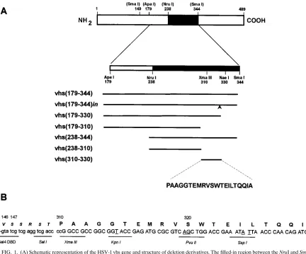
Plasmids. (i) Plasmids for two-hybrid analysis.DBD-vhs(179–344) is a yeast expression plasmid which expresses the GAL4 DNA-binding domain (DBD; amino acid residues 1 to 147) fused to the vhs ApaI-SmaI fragment (residues 179 to 344) and is suitable for two-hybrid analysis in S. cerevisiae; it has been de-scribed before (48). DBD-vhs(179–344)in is identical to DBD-vhs(179–344) ex-cept that it contains an in-frame XhoI linker (59-CCCCTCGAGGGG) inserted between the codons for amino acid residues 330 and 331. It was constructed by substituting a fragment from a plasmid bearing a vhs gene that contained this linker insertion (pUC/CMV-vhsN331 [22]) for the corresponding fragment be-tween the NruI and SacI sites in DBD-vhs(179–344). To generate DBD-vhs(179– 330), the NruI-EcoRV fragment from the in vitro expression vector pSPAS, which expresses vhs(179–344) from an SP6 promoter (48), was replaced with the NruI-XhoI fragment from pUC/CMV-vhsN331 (after first filling in the XhoI overhang). This plasmid, designated pSPAX, was digested with NcoI (which provides the initiator methionine codon) and BglII (present downstream in the polylinker) and cloned in place of the NcoI-BglII fragment in DBD-vhs(179–344) to generate DBD-vhs(179–330). DBD-vhs(238–344) was constructed by cloning the NruI-SacI fragment from pSPAS into the corresponding sites of the low-copy parental GAL4 DBD expression vector pPC97(5). Similarly, DBD-vhs(238–310) was constructed by cloning the NruI-XmaIII fragment of vhs into the SmaI and NotI sites of pPC97. DBD-vhs(310–330) was constructed by cloning the XmaIII-BglII fragment from pSPAX into the SmaI and XmaIII-BglII sites in the pPC97 polylinker after first filling in the XmaIII end. DBD-vhsMBD contains the syn-thetic oligonucleotide 59-GGCCGCCGGCGGTACCGAGATGCGCGTCAGC TGGACCGAAATATTAACCCAACAGATCGCCTA and its complement, 59 -GATCTAGGCGATCTGTTGGGTTAATATTTCGGTCCAGCTGACGCGC ATCTCGGTACCGCCGGC. This cassette encodes vhs residues 310 to 330, incorporates a TAG termination codon after codon 330, and contains XmaIII-and BglII-compatible ends.
GAD-VP16424, GAD-VP16404, GAD-VP16379, GAD-VP16369, GAD-VP16335, and GAD-VP16D141–178are yeast two-hybrid low-copy plasmids that express carboxyl-terminally truncated derivatives of VP16 (the number refers to the carboxyl-terminal deletion endpoint) or an internal deletion of VP16 residues 141 to 178 (derived from GAD-VP16424) as fusions to the GAL4 acidic activation domain (GAD) and have been described previously (39, 48).
(ii) Plasmids for in vitro expression.pSPAS and pSPAX, the in vitro tran-scription-translation plasmids for vhs(179–344) and vhs(179–330), respectively, are described above. pSP(vhs) expresses full-length vhs, and pSP-vhsDSma ex-presses vhs that bears an internal deletion of amino acids 149 to 344 (48). The in vitro expression vector for pSP-vhs(310–331) was constructed by cloning the XhoI-SacI fragment from DBD-vhs(179–344) into the SalI and SacI sites of the in vitro expression plasmid pSPUTK (11). This plasmid expresses residues 310 to
and then incubated in 2 ml of buffer Z (60 mM Na2HPO4, 40 mM NaH2PO4, 10 mM KCl, 1 mM MgSO4, 50 mMb-mercaptoethanol, pH 7.0) supplemented with X-Gal (5-bromo-4-chloro-3-indolyl-b-D-galactopyranoside; 1 mg/ml). Filters were incubated at 378C and observed for development of blue color. Strong two-hybrid interactions were detected within 30 min. Filters were incubated overnight in order to detect weak activity.
Oligonucleotide-directed cassette mutagenesis.The synthetic oligonucleotide encoding residues 310 to 330 of vhs was designed to incorporate unique XmaIII, KpnI, and PvuII restriction sites (see Fig. 1) to facilitate cassette mutagenesis of this region. Short, double-stranded oligonucleotide cassettes designed to convert selected amino acids to alanine residues and containing appropriate ends for cloning into the above restriction sites were synthesized, phosphorylated at their 59ends with T4 polynucleotide kinase, and purified on 4% NuSieve agarose gels. Mutant oligonucleotide cassettes were used to replace the corresponding wild-type regions in the yeast and in vitro vhsMBD expression plasmids following digestion of these plasmids with the appropriate restriction enzymes and dephos-phorylation with calf intestinal alkaline phosphatase. The point mutants gener-ated by this procedure are listed in Fig. 3. A deletion derivative lacking codons for amino acids 321 to 330 was also constructed. The accuracy of mutagenesis in all cases was verified by dideoxy DNA sequence analysis.
In vitro transcription and translation.In vitro transcription with SP6 poly-merase and translation in rabbit reticulocyte lysates were carried out with a commercially available coupled system (Promega) according to the manufactur-er’s protocol.
Purification of PA-VP16 and MBP-VP16 fusion proteins.The purification of protein A-VP16 (PA-VP16) and maltose-binding protein-VP16 (MBP-VP16) fusion proteins from induced bacterial cultures has been described before (39, 55, 57). Both MBP-VP16 and PA-VP16 contain residues 4 to 411 of VP16 and are thus missing the carboxyl-terminal acidic activation domain. Unfused protein A and maltose-binding protein were purified for use as controls in the binding assays.
Solid-phase capture assay. Purified PA-VP16 was covalently coupled to CNBr-activated Sepharose (Pharmacia) at 2 mg of protein per ml of settled beads as before (57). Beads were stored at 48C as a 50% slurry in 20 mM HEPES (N-2-hydroxyethylpiperazine-N9-2-ethanesulfonic acid, pH 7.9)–50 mM NaCl–1 mM dithiothreitol–10% glycerol–1 mM phenylmethylsulfonyl fluoride. [35
S]me-thionine-labeled proteins (5ml of programmed rabbit reticulocyte lysate) were incubated with 45ml of beads for 2.5 h at 48C in 400ml of buffer containing 150 mM NaCl, 50 mM Tris-HCl (pH 7.2), 1% Triton X-100, 0.5 mM phenylmethyl-sulfonyl fluoride, and 2% bovine serum albumin (BSA). Beads were washed extensively with the same buffer followed by a final wash with buffer in the absence of BSA. Bound material was eluted by boiling the settled beads in an equal volume of twice-concentrated SDS-polyacrylamide gel electrophoresis sample buffer (2% SDS, 2%b-mercaptoethanol, 250 mM Tris-HCl, pH 6.8) and analyzed by SDS-polyacrylamide gel electrophoresis.
Solid-phase capture assays were also performed with immobilized MBP-VP16. Amylose beads coupled with MBP-VP16 were prepared as described previously (39). MBP-VP16 beads in 20 mM Tris-HCl (pH 7.4)–200 mM NaCl–1 mM EDTA–0.5 mM phenylmethylsulfonyl fluoride–1 mM dithiothreitol were incu-bated with labeled proteins as above and washed extensively in the above buffer followed with buffer supplemented with 0.01% BSA and 0.05% Nonidet P-40. Bound material was eluted from the beads and analyzed as above.
RESULTS
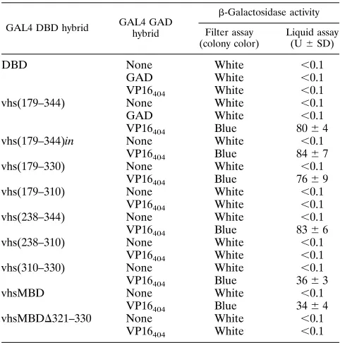
A 21-amino-acid-long region of vhs is sufficient for
interac-tion with VP16 in the two-hybrid system.
We have previously
demonstrated that a derivative of vhs encompassing residues
238 to 344 was sufficient for interaction with VP16 in vitro (48).
The yeast two-hybrid system was used to more precisely map
the minimal region of vhs sufficient for stable interaction with
on November 9, 2019 by guest
VP16 in vivo. A series of carboxyl- and amino-terminal
dele-tions of the vhs ApaI-SmaI fragment (residues 179 to 344 [Fig.
1]) were generated and cloned into a GAL4 DBD expression
vector. Plasmids encoding the DBD-vhs derivatives were
co-transformed into yeast cells with GAD-VP16
404(expresses
VP16 residues 4 to 404 as a fusion to the GAL4 GAD), and
transformants were assayed for expression of the resident
GAL4-responsive lacZ reporter gene by measuring
b
-galacto-sidase activity by both liquid and filter assays. As demonstrated
previously (48) and shown in Table 1,
b
-galactosidase activity
was observed only when the DBD-vhs(179–344) and
GAD-VP16
404plasmids were cotransformed, indicative of functional
interaction between the two expressed hybrid proteins.
Inter-action with VP16
404was also observed with DBD-vhs(179–
344)in, DBD-vhs(179–330), and DBD-vhs(238–344). Thus,
res-idues 238 to 330 contain determinants necessary for interaction
with VP16 in S. cerevisiae. Activity was not observed with
DBD-vhs(179–310) or DBD-vhs(238–310). Western
immuno-blot analysis with antiserum directed against the GAL4 DBD
demonstrated that the nonfunctional fusion proteins were
ex-pressed (46).
The above findings indicate that an important determinant
for binding to VP16 lies between residues 310 and 330. To
determine if this small region constitutes an independent and
modular binding domain, DBD-vhs(310–330) was constructed
and tested. This derivative activated expression of the lacZ
gene in the presence of VP16
404to a level of approximately 40
to 50% of that of DBD-vhs(179–344), indicating that this small
region is sufficient for interaction with VP16. The higher
en-zymatic activity with the longer vhs fusion protein may indicate
that the extended protein is more stable or that residues
out-side the minimal domain contribute to binding efficiency.
Al-ternatively, there may be differences in the activation potential
of the respective two-hybrid transactivator complexes.
The DBD-vhs(310–330) fusion protein contained three
ad-ditional amino acids at the carboxyl terminus as a result of the
cloning procedure. To ensure that these extra amino acids
were inert in the two-hybrid assay, an oligonucleotide cassette
spanning residues 310 to 330 and containing a termination
codon was synthesized and cloned into the GAL4 DBD vector
(see Fig. 1). This synthetic oligonucleotide is referred to as the
vhs minimal binding domain (MBD). Table 1 shows that
DBD-FIG. 1. (A) Schematic representation of the HSV-1 vhs gene and structure of deletion derivatives. The filled-in region between the NruI and SmaI sites corresponds to the region of vhs previously shown to be sufficient for binding to VP16 in vitro (48). Deletions were constructed in the ApaI-SmaI fragment as shown and cloned into vectors suitable for two-hybrid analysis in S. cerevisiae or expression in vitro. The arrow in vhs(179–344)in indicates the position of an XhoI linker insertion. The primary amino acid sequence between residues 310 and 330 is shown at the bottom. The published sequence of vhs from HSV-1 strain 17 contains a threonine at position 317 in place of the methionine found in vhs from the KOS strain used in this study (30). (B) Structure of the synthetic oligonucleotide spanning residues 310 to 330. The top strand of the synthetic oligonucleotide is shown in capital letters. The underlined nucleotides are silent substitutions of the wild-type sequence that were incorporated in order to generate novel restriction sites. The TAG termination codon immediately following residue 330 is generated following cloning into the BglII site. The sequence of the end of the GAL4 DNA-binding domain coding region (encoding amino acids 1 to 147 of GAL4) and the junction to the vhs coding region are shown.on November 9, 2019 by guest
http://jvi.asm.org/
[image:3.612.64.496.71.430.2]vhsMBD interacted with VP16
404as efficiently as did
DBD-vhs(310–330), indicating that the appended extra amino acids
did not influence interaction with VP16. A deletion derivative
of this region, missing residues 321 to 330 (DBD-vhsMBD
D
321–330), was inactive. Thus, a 21-amino-acid domain of vhs
encompassing amino acid residues 310 to 330 is sufficient for
interaction with VP16 in S. cerevisiae.
Specificity of interaction of the vhs minimal binding
do-main.
In contrast to the small contiguous binding domain in
vhs identified above, which suffices for interaction with VP16 in
S. cerevisiae, the vhs binding interface in VP16 appears to
consist of a large and perhaps noncontiguous domain. We
previously demonstrated that the carboxyl-terminal end of
VP16 that is necessary for interaction with vhs in vitro and in
vivo mapped to amino acid 369 and that additional elements
closer to the amino terminus were also required (48). In order
to determine if the binding characteristics and specificity of the
minimal region of vhs defined above were similar to those of
the larger vhs protein, we compared the interaction of
DBD-vhsMBD in yeast cells with a series of VP16 mutant
deriva-tives. As shown in Table 2, DBD-vhsMBD interacted with
VP16 truncated at amino acid 424, 404, 379, or 369 but not
with VP16 truncated at amino acid 335 or with VP16 that
contained an internal deletion of residues 141 to 178.
Interac-tion was more efficient with GAD-VP16
404and GAD-VP16
369than with GAD-VP16
424and GAD-VP16
379if relative
b
-ga-lactosidase activities are used as a measure. The same
spec-trum of interaction and relative activities was observed with
DBD-vhs(179–344). Thus, the minimal peptide requires
simi-lar determinants in VP16 for interaction in vivo as does the
larger vhs protein.
Interaction of vhs with VP16 in vitro.
To determine if the
results obtained with the two-hybrid system reflected direct
physical interaction between vhs and VP16, vhs derivatives
were cloned into an in vitro transcription-translation vector,
and the resultant [
35S]methionine-labeled polypeptides were
tested for their ability to bind to a VP16 fusion protein
(PA-VP16) that was covalently coupled to Sepharose beads.
Full-length vhs, vhs(179–344), and vhs(
D
Sma) (a derivative missing
residues 149 to 344) were synthesized in an unfused form;
however, because of the small size of the minimal domain (2
kDa), derivatives encompassing this region were expressed in
vitro as fusions to the GAL4 DBD. As controls, we also
con-structed two vectors that expressed the GAL4 DBD on its own
[pSP-DBD(Cla) and pSP-DBD(Xma)].
The labeled vhs peptides were incubated with the
VP16-coupled beads, and after extensive washing, the bound material
was eluted by boiling in 2% SDS and analyzed by
SDS-poly-acrylamide gel electrophoresis. As shown in Fig. 2A, vhs(179–
344) bound specifically to PA-VP16 beads (compare lanes d
and h; approximately 10 to 20% of the input vhs protein bound
to the VP16 beads), while vhs
D
Sma (lane c) and the luciferase
control (lane b) did not bind to either VP16 beads or control
beads. Similarly, vhs(310–330) (Fig. 2A, lanes e, i, and n) and
vhsMBD (Fig. 2B, lanes a and e) bound specifically to the
PA-VP16 beads, whereas binding over background levels was
not observed with the control GAL4 DBD proteins
DB-D(Xma) and DBD(Cla) (Fig. 2B, lanes g and h). Thus, amino
acids 310 to 330 of vhs appended to a heterologous protein are
sufficient for mediating direct protein-protein interaction with
VP16 in vitro.
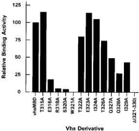
Tryptophan 321 is a critical residue for interaction.
In order
to identify individual residues in the minimal binding domain
important for interaction with VP16, we converted specific
amino acids to alanine residues by cassette mutagenesis.
Ala-nine residues were chosen because they would not be expected
to significantly affect the structure of the polypeptide backbone
(6). A total of 12 alanine substitution mutants were created;
T315A, E316A, R318A, S320A, W321A, T322A, E323A, I324A,
T326A, Q327A, Q328A, and I329A (Fig. 3). The mutant
oli-gonucleotides were cloned into the GAL4 DBD expression
vector and tested for
b
-galactosidase activity following
cotrans-formation of yeast cells with GAD-VP16
404(Fig. 3). All of the
derivatives, with one notable exception, remained capable of
inducing
b
-galactosidase activity in the presence of VP16
404and did so with an efficiency that was comparable to that of the
vhs(179–310) None White ,0.1
VP16404 White ,0.1
vhs(238–344) None White ,0.1
VP16404 Blue 8366
vhs(238–310) None White ,0.1
VP16404 White ,0.1
vhs(310–330) None White ,0.1
VP16404 Blue 3663
vhsMBD None White ,0.1
VP16404 Blue 3464
vhsMBDD321–330 None White ,0.1
VP16404 White ,0.1
aS. cerevisiae PCY2 was transformed with the indicated plasmids (DBD, GAL4 DNA-binding domain; GAD, GAL4 acidic activation domain) and as-sayed forb-galactosidase activity. For liquid assays, units ofb-galactosidase activity are averages from at least three independent transformants assayed in duplicate and normalized to cell density (OD600). For the filter assay, blue color, where indicated, was observed within 30 min following incubation of filters with buffer containing X-Gal. Where white is indicated, blue color was not observed even after overnight incubation.
VP16335 ,0.1
VP16D141–178 ,0.1 ab
-Galactosidase activity was measured as described in Table 1, footnote a.
on November 9, 2019 by guest
[image:4.612.57.298.91.334.2] [image:4.612.314.555.92.238.2]parental construct DBD-vhsMBD. The only point mutant that
was defective in this assay was W321A. Similar results were
obtained with filter assays; yeast cells transformed with W321A
remained white, even after overnight incubation, whereas all of
the other derivatives turned blue within 30 min. The alanine
substitution derivatives were also tested for activity in the
ab-sence of cotransformed VP16
404in case the introduced
muta-tions fortuitously generated a transcription activation surface
in vhs. As expected, all of the mutant derivatives were inactive
in the absence of cotransformed GAD-VP16
404. These findings
indicate that tryptophan 321 is crucial for interaction with
VP16 in vivo.
The mutant vhsMBD derivatives were expressed in vitro and
tested in the solid-phase capture assay as above. In these
ex-periments, MBP-VP16 fusion protein was used as the affinity
ligand in place of PA-VP16. Labeled proteins were incubated
with MBP-VP16-coupled beads or control beads, and the
bound material was analyzed by SDS-polyacrylamide gel
elec-trophoresis. As expected, full-length vhs, vhs(179–344), and
vhsMBD bound specifically to the VP16 beads (Fig. 4B) but
not the control beads (Fig. 4C), whereas binding was not
ob-served with vhs(
D
Sma) or the control GAL4 DBD proteins.
T315A, T322A, E323A, I324A, and T326A bound specifically
to the VP16 beads with efficiencies that were comparable to
that of vhsMBD (see Fig. 5), while E316A, Q327A, Q328A,
and I329A interacted with VP16 with a somewhat lower
effi-ciency (20 to 50% of that of vhsMBD). R318A and S320A
bound to VP16 with an efficiency of
,
5% (Fig. 4B, lanes h and
i), and specific binding of W321A and vhs(
D
321–330) was not
detected (lanes j and u). Identical results were obtained when
PA-VP16 was used as the affinity ligand rather than
MBP-VP16; thus, the nature of the fusion partner does not
differ-entially affect the binding properties of the mutant vhs
poly-peptides (46).
The above findings demonstrate that tryptophan 321 is
cru-cial for interaction with VP16 in vitro, consistent with the
results obtained in the two-hybrid assay. Moreover, residues
immediately upstream of tryptophan 321 (E-316, R-318, and
S-320) and, to a lesser extent, downstream residues (Q327A,
Q-328, and I329A) contribute to complex formation and/or
stability in vitro.
DISCUSSION
We have recently demonstrated that VP16 interacts with vhs
and downregulates vhs activity during a lytic infection, thereby
allowing the accumulation and translation of late viral mRNAs
(28, 48). In this report, we present evidence that a small
mod-ular domain in vhs encompassing amino acid residues 310 to
330 is sufficient for directing stable interaction with VP16. In
contrast, VP16 requires a large domain or multiple domains
within the amino-terminal 369 amino acid residues for
inter-action with vhs (48), implying that the overall conformation of
VP16 or the juxtaposition of spatially separate binding
deter-minants may be important for recognition of the small
inter-action domain in vhs. This situation is similar to the interinter-action
of VP16 with DNA-bound Oct-1. In this case, the relatively
FIG. 2. Amino acids 310 to 330 of vhs are sufficient for interaction with VP16 [image:5.612.60.291.72.451.2]in vitro. [35S]methionine-labeled vhs derivatives were prepared in rabbit reticu-locyte lysates programmed with the corresponding in vitro-transcribed RNAs. Vhs(310–330) and vhsMBD were expressed as fusions to the GAL4 DBD. Aliquots of the total translation mixture were incubated with PA-VP16 or pro-tein A-coupled Sepharose, as indicated. The beads were washed extensively, and bound material was eluted by boiling in 2% SDS and analyzed by SDS-polyacryl-amide gel electrophoresis. (A) Binding of vhs(330–331) to PA-VP16. vhs(179– 344) was the positive control, while vhsDSma was the negative control. (B) Binding of vhsMBD to PA-VP16. The GAL4 DBD derivatives DBD(Xma) and DBD(Cla) are negative controls. The positions of molecular size markers (lane M) are shown (in kilodaltons).
FIG. 3. Structure of alanine substitution mutants of the vhs minimal binding domain and activity in the two-hybrid system. Specific amino acids in the 310 to 330 region of vhs were altered to alanine residues (underlined) by cassette mutagenesis of the oligonucleotide shown in Fig. 1 and tested for interaction with VP16 in the two-hybrid system.b-Galactosidase activity (units) was measured as described in Table 1, footnote a, except that yeast strain Y190 was used for transformations.
on November 9, 2019 by guest
http://jvi.asm.org/
small POU homeodomain of Oct-1 is sufficient to direct
spe-cific protein-DNA complex formation with VP16 (50), whereas
a large domain within VP16 is necessary (19, 55).
There are no obvious features in the vhs minimal binding
region that are common to other well-characterized
protein-protein interaction motifs. Moreover, with the exception of the
HSV-2 vhs homolog (see below), a related domain does not
appear to exist in any other protein, including known VP16
ligands such as Oct-1 and HCF. Secondary-structure
predic-tions indicate that the region spanning residues 310 to 330
exists predominantly in a
b
-sheet conformation, flanked by
b
-turns. The amino-terminal end is primarily hydrophilic in
nature, while the carboxyl-terminal end is hydrophobic.
Re-sults from both the two-hybrid analysis and in vitro binding
assays show that mutation of the tryptophan residue at position
321 or deletion of residues downstream from this amino acid
abrogates interaction with VP16. Moreover, mutation of both
charged and uncharged polar residues (e.g., E-316, R-318, and
S-320) proximal to W-321 reduced the formation and/or
[image:6.612.147.491.70.461.2]sta-FIG. 4. Tryptophan 331 in the vhs minimal binding domain is essential for interaction with VP16 in vitro. Point mutant derivatives and a deletion derivative of the vhs minimal binding domain were transcribed and translated in vitro. Aliquots of the total translation reaction (A) were incubated with agarose beads coupled with MBP-VP16 (B) or control beads coupled with maltose-binding protein (C), and bound material was analyzed by polyacrylamide gel electrophoresis. Full-length vhs (lane b) and vhs(179–344) (lane d) were positive controls, while vhsDSma (lane c), DBD(Xma) (lane q), and DBD(Cla) (lane r) were negative controls. The autoradiograph in panel A was exposed for 6 h, and the autoradiographs in panels B and C were exposed for 24 h. The positions of molecular size markers (lane m) are shown (in kilodaltons).
FIG. 5. Relative binding efficiency of vhs minimal binding domain mutants. The relative binding activity of the vhs point mutant derivatives was quantitated by phosphorimage analysis of the gels shown in Fig. 4. The values shown rep-resent the amount of vhs protein bound to MBP-VP16 beads as a percentage of input protein, normalized to the value obtained with vhsMBD, which was taken as 100%. Binding over background was not detected with W321A or with vhsMBDD321–330.
on November 9, 2019 by guest
[image:6.612.362.505.533.668.2]bility of the vhs-VP16 complex in vitro, suggesting that these
residues constitute part of a binding pocket. Tryptophan is
principally hydrophobic in character, but the aromatic side
chain can act as a donor in charge transfer interactions (8).
Overall, the mutational sensitivity spectrum suggests that
elec-trostatic, hydrogen-bonding, and possibly hydrophobic
interac-tions may contribute to complex formation and stability.
Whether other determinants outside this minimal region that
contribute to complex formation and binding affinity exist in
the intact molecule is currently under investigation.
Interestingly, with the exception of the tryptophan mutant,
none of the alanine substitution derivatives were compromised
in the two-hybrid assay. For instance, R318A and S320A,
which interacted very poorly with VP16 in vitro, induced
b
-ga-lactosidase activity as efficiently as vhsMBD in the two-hybrid
assay. It is possible that complexes with these derivatives are
less stable under the conditions employed in the solid-phase
capture assay than in the more physiological conditions that
exist in vivo in yeast cells. It should be noted that the
two-hybrid assay is a sensitive but indirect measure of
protein-protein interaction and may not necessarily measure of the
stability of the vhs-VP16 complex. Weak and/or transient
as-sociation between vhs and VP16 that may not be detectable in
in vitro binding assays may suffice to efficiently activate the
lacZ reporter gene in yeast cells.
Vhs homologs from other alphaherpesviruses, such as
pseu-dorabies virus, equine herpesviruses, and varicella-zoster virus,
have regions of sequence identity with HSV-1 vhs throughout
their lengths (4); however, with the exception of vhs from
HSV-2, the minimal VP16-binding region is not conserved. It
is possible that HSV has evolved unique strategies and
mech-anisms of gene regulation that rely on a linkage between VP16
and vhs. Indeed, it is not known if vhs homologs participate in
host shutoff activity, and moreover, the functional properties of
VP16 differ in other viruses. For instance, transcription
acti-vation by VP16 homologs from other alphaherpesviruses
ap-pears to be mediated through distinct mechanisms (32), and at
least for varicella-zoster virus, the VP16 homolog is not
essen-tial for virus growth (7).
Transient-transfection assays reveal that vhs functions in the
absence of other viral proteins (22, 38). However, in infected
cells, other viral proteins, such as the virion-associated protein
kinase product of the UL13 gene, also play a role in shutoff
activity (20, 37). It is possible that vhs activity or VP16-vhs
association itself is regulated by additional factors, perhaps
through phosphorylation events or additional levels of
protein-protein interaction. In this regard, it is interesting that the
minimal binding domain contains a consensus casein kinase II
phosphorylation site (S/T-X-X-D/E). Further characterization
of the vhs protein interaction surface and more precise
iden-tification of the binding determinants present in VP16 will help
elucidate how combinatorial interactions between VP16, vhs,
and other viral and cellular factors contribute to the multilevel
regulation of viral gene expression.
Finally, it is conceivable that the vhs-VP16 complex is a
novel antiviral target, since prevention or disruption of
com-plex formation could lead to vhs-mediated degradation of viral
mRNA and thus termination of productive infection. The vhs
binding interface identified here provides a logical starting
point for the development of potentially useful interventive
strategies that target the VP16-vhs complex.
ACKNOWLEDGMENTS
We thank F. Jones for providing the vhs-N331 plasmid.
This work was supported by grants from the National Cancer Insti-tute of Canada (NCIC). J.S. is a recipient of a studentship from the
Medical Research Council of Canada. J.R.S. is a Terry Fox Scientist, and J.P.C. is a Senior Scientist of the NCIC.
REFERENCES
1. Ace, C. I., T. A. McKee, J. M. Ryan, J. M. Cameron, and C. M. Preston. 1989. Construction and characterization of a herpes simplex virus type 1 mutant unable to transinduce immediate-early gene expression. J. Virol. 63:2260– 2269.
2. Ausubel, F. M., R. Brent, R. E. Kingston, D. D. Moore, J. G. Seidman, J. A.
Smith, and K. Struhl (ed.).1990. Current protocols in molecular biology. Wiley, New York.
3. Batterson, W., and B. Roizman. 1983. Characterization of the herpes simplex virion-associated factor responsible for the induction ofagenes. J. Virol.
46:371–377.
4. Berthomme, H., B. Jacuemont, and A. Epstein. 1993. The pseudorabies virus host-shutoff homolog gene nucleotide sequence and comparison with alpha-herpesvirus protein counterparts. Virology 193:1028–1032.
5. Chevray, P., and D. Nathans. 1992. Protein interaction cloning in yeast: identification of mammalian proteins that react with the leucine zipper of Jun. Proc. Natl. Acad. Sci. USA 89:5789–5793.
6. Chou, P. Y., and G. D. Fasman. 1978. Empirical predictions of protein conformation. Annu. Rev. Biochem. 47:251–276.
7. Cohen, J. I., and K. Seidel. 1994. Varicella-zoster virus (VZV) open reading frame 10 protein, the homolog of essential herpes simplex virus protein VP16, is dispensable for VZV replication in vitro. J. Virol. 68:7850–7858. 8. Creighton, T. E. 1983. Proteins: structures and molecular principles. W. H.
Freeman and Co., New York.
9. Elble, R. 1992. A simple and efficient procedure for the transformation of yeasts. BioTechniques 13:18–20.
10. Elgadi, M., C. Hayes, F. Jones, D. Andrews, and J. R. Smiley. 1995. Unpub-lished observations.
11. Falcone, D., and D. A. Andrews. 1991. Both the 59untranslated regions and sequences surrounding the start site contribute to efficient initiation of trans-lation in vitro. Mol. Cell. Biol. 11:2656–2664.
12. Fenwick, M. L., and J. Clark. 1982. Early and delayed shutoff of host protein synthesis in cells infected with herpes simplex virus. J. Gen. Virol. 61:121–125. 13. Fenwick, M. L., and R. D. Everett. 1990. Inactivation of the shutoff gene (UL41) of herpes simplex virus types 1 and 2. J. Gen. Virol. 71:2961–2967. 14. Fenwick, M. L., and R. D. Everett. 1990. Transfer of UL41, the gene con-trolling virion-associated host cell shutoff, between different strains of herpes simplex virus. J. Gen. Virol. 61:121–125.
15. Fenwick, M. L., and M. M. McMenamin. 1984. Early virion-associated sup-pression of cellular protein synthesis by herpes simplex virus is accompanied by inactivation of mRNA. J. Gen. Virol. 65:1225–1228.
16. Fenwick, M. L., and S. A. Owen. 1988. On the control of immediate early (a) mRNA survival in cells infected with herpes simplex virus. J. Gen. Virol.
69:2869–2877.
17. Fields, S., and O. Song. 1989. A novel genetic system to detect protein-protein interaction. Nature (London) 340:245–246.
18. Gerster, T., and R. G. Roeder. 1988. A herpes virus transactivating protein interacts with transcription factor OTF-1 and other cellular proteins. Proc. Natl. Acad. Sci. USA 85:6347–6351.
19. Greaves, R., and P. O’Hare. 1989. Separation of the requirements for pro-tein-DNA complex assembly from those for functional activity in the herpes simplex virus regulatory protein Vmw65. J. Virol. 63:1642–1650. 20. Hardwicke, M. A., and R. M. Sandri-Goldin. 1994. The herpes simplex virus
regulatory protein ICP27 contributes to the decrease in cellular mRNA levels during infection. J. Virol. 52:99–107.
21. Herr, W., and M. A. Cleary. 1995. The POU domain: versatility in transcrip-tional regulation by a flexible two-in-one DNA-binding domain. Genes Dev.
9:1679–1693.
22. Jones, F. E., C. A. Smibert, and J. R. Smiley. 1995. Mutational analysis of the herpes simplex virus virion host shutoff protein: evidence that vhs functions in the absence of other viral proteins. J. Virol. 69:4863–4871.
23. Katan, M., A. Haigh, C. P. Verrijzer, P. C. van der Vliet, and P. O’Hare. 1990. Characterization of a cellular factor which interacts functionally with Oct-1 in the assembly of a multicomponent transcription complex. Nucleic Acids Res. 18:6871–6880.
24. Kristie, T. M., J. H. LeBowitz, and P. A. Sharp. 1989. The octamer-binding proteins form multi-protein-DNA complexes with the HSVaTIF regulatory protein. EMBO J. 8:4229–4238.
25. Kristie, T., and P. A. Sharp. 1993. Purification of the C1 factor required for the stable recognition of the Oct-1 homeodomain by the herpes simplex virus
a-trans inducing factor (VP16). J. Biol. Chem. 268:6525–6534.
26. Kwong, A. D., and N. Frenkel. 1987. Herpes simplex virus infected cells contain a function(s) that destabilizes both host and viral mRNAs. Proc. Natl. Acad. Sci. USA 84:1926–1930.
27. Kwong, A. D., J. A. Kruper, and N. Frenkel. 1988. Herpes simplex virus virion host shutoff function. J. Virol. 62:912–921.
28. Lam, Q., C. A. Smibert, K. E. Koop, C. Lavery, J. P. Capone, S. P.
Wein-heimer, and J. R. Smiley.Submitted for publication.
29. Lin, Y. S., I. Ha, E. Maldonado, D. Reinberg, and M. Green. 1991. Binding
on November 9, 2019 by guest
http://jvi.asm.org/
1 exhibits increased stability of immediate-early (alpha) mRNAs. J. Virol.
61:604–606.
36. Oroskar, A. A., and G. S. Read. 1989. Control of mRNA stability by the virion host shutoff function of herpes simplex virus. J. Virol. 63:1897–1906. 37. Overton, H., D. McMillan, L. Hope, and P. Wong-Kai-in. 1994. Production of host shutoff-defective mutants of herpes simplex virus type 1 by inactiva-tion of the UL13 gene. Virology 202:197–206.
38. Pak, A. S., D. N. Everly, K. Knight, and G. S. Read. 1995. The virion host shutoff protein of herpes simplex virus inhibits reporter gene expression in the absence of other viral proteins. Virology 211:491–506.
39. Popova, B., P. Bilan, P. Xiao, M. Faught, and J. P. Capone. 1995. Transcrip-tional activation by DNA-binding derivatives of HSV-1 VP16 that lack the carboxyl-terminal acidic activation domain. Virology 209:19–28.
40. Preston, C. M., M. C. Frame, and M. E. M. Campbell. 1988. A complex formed between cell components and an HSV structural polypeptide binds to a viral immediate early gene regulatory DNA sequence. Cell 52:425–434. 41. Read, G. S., and N. Frenkel. 1983. Herpes simplex virus mutants defective in virion-associated shutoff of host polypeptide synthesis and exhibiting abnor-mal synthesis ofa(immediate-early) viral polypeptides. J. Virol. 46:498–512. 42. Read, G. S., R. M. Karr, and K. Knight. 1993. Isolation of a herpes simplex virus type 1 mutant with a deletion in the virion host shutoff gene and identification of multiple forms of the vhs (UL41) polypeptide. J. Virol.
67:7149–7160.
50. Stern, S., M. Tanaka, and W. Herr. 1989. The Oct-1 homeo-domain directs formation of a multiprotein-DNA complex with the HSV transactivator VP16. Nature (London) 341:624–630.
51. Stringer, K. F., C. J. Ingles, and J. Greenblatt. 1990. Direct and selective binding of an acidic transcriptional activation domain to the TATA-box factor TFIID. Nature (London) 345:783–786.
52. Thompson, C. C., and S. L. McKnight. 1992. Anatomy of an enhancer. Trends Genet. 8:232–236.
53. Ward, P. L., and B. Roizman. 1994. Herpes simplex genes: the blueprint of a successful human pathogen. Trends Genet. 10:267–274.
54. Weinheimer, S. P., B. A. Boyd, S. K. Durham, J. L. Resnick, and D. R.
O’Boyle.1992. Deletion of the VP16 open reading frame of herpes simplex virus type 1. J. Virol. 66:258–269.
55. Werstuck, G., and J. P. Capone. 1989. Identification of a domain of the herpes simplex virus trans-activator Vmw65 required for protein-DNA com-plex formation through the use of protein A fusion proteins. J. Virol. 63: 5509–5513.
56. Wilson, A. C., K. LaMarco, M. G. Peterson, and W. Herr. 1993. The VP16 accessory protein HCF is a family of polypeptides processed from a larger precursor protein. Cell 74:115–126.
57. Xiao, P., and J. P. Capone. 1990. A cellular factor binds to the herpes simplex virus type 1 transactivator Vmw65 and is required for protein-DNA complex
assembly with Oct-1. Mol. Cell. Biol. 10:4974–4977.