0022-538X/96/$04.0010
Copyrightq1996, American Society for Microbiology
Targeting Human Immunodeficiency Virus Type 1 Reverse
Transcriptase by Intracellular Expression of Single-Chain
Variable Fragments To Inhibit Early Stages
of the Viral Life Cycle
FARIDA SHAHEEN, LINGXUN DUAN, MINGHUA ZHU, OMAR BAGASRA,
ANDROGER J. POMERANTZ*
The Dorrance H. Hamilton Laboratories, Section of Molecular Retrovirology, Division of Infectious Diseases, Department of Medicine, Jefferson Medical College, Thomas Jefferson University,
Philadelphia, Pennsylvania 19107
Received 8 December 1995/Accepted 21 February 1996
Novel molecular approaches to inhibit human immunodeficiency virus type 1 (HIV-1) infection have received increasing attention because of the lack of effective antiviral drug therapies in vivo. We now demonstrate that cells can be intracellularly immunized by cytoplasmic expression of single-chain variable antibody fragments (SFv) which bind to the HIV-1 reverse transcriptase (RT) enzyme. The expression of anti-RT SFv in T-lymphocytic cells specifically neutralizes the RT activity in the preintegration stage and affects the reverse transcription process, an early event of the HIV-1 life cycle. Blocking the virus at these early stages dramat-ically decreased HIV-1 propagation, as well as the HIV-1-induced cytopathic effects in susceptible human T lymphocytes, by impeding the formation of the proviral DNA. These data also demonstrate that intracellular, complete SFvs may gain access to viral proteins of the HIV-1 preintegration complex. These SFvs will provide a tool with which to better understand the molecular mechanism(s) involved in restricting viral replication in HIV-1-infected cells.
It has been over a decade since the discovery of human immunodeficiency virus type 1 (HIV-1) as the major cause of AIDS. There has been only modest progress in the develop-ment of therapy that may stop or significantly alter the ulti-mately fatal course of HIV-1 infection (19). HIV-1 infection leads to severe immunodeficiency in most infected individuals, specifically by depleting CD4-positive T lymphocytes (34). These lymphocytes appear to be the major viral reservoirs in lymphoid organs and in the peripheral blood (26).
Because of the extremely complex life cycle of HIV-1, it has been difficult to develop adequate therapeutic modalities to significantly interrupt HIV-1 replication (18). Retroviral re-verse transcriptase (RT) plays a key role in the early stages of the retroviral life cycle. The HIV-1 RT contains both RNA-and DNA-directed DNA polymerase activities RNA-and an RNase H activity. These enzymatic functions are important in the conversion of the single-stranded viral RNA genome into dou-ble-stranded DNA prior to integration of the retroviral provi-rus into the host cells’ genome (9, 18). Traditionally, antiviral approaches have been aimed at limiting viral replication. The RT enzyme of HIV-1 has been a target of several
pharmaco-logical inhibitors, which include the nucleoside analog 39
-azido-39-deoxythymidine (AZT) and the nonnucleoside
com-pound Nevirapine, both of which act on the enzyme’s RNA-dependent DNA polymerase activity (23–25, 46). Currently, antiviral therapies based on RT inhibitors, cytokines, and re-ceptor-blocking agents are not completely successful because of the exceptional ability of HIV-1 to mutate, which results in
rapid development of quasispecies which evade host defenses and become resistant to various antiviral agents (15, 41). How-ever, an alternative approach, antiviral gene therapy, whereby host cells can be genetically altered or engineered to confer long-lasting protection against viral infection or replication after infection, appears to be an attractive and convincing technology (37). Several such strategies are currently used and directed toward the inhibition of HIV-1 replication or the
elimination of the infected cells via trans-dominant negative
mutant HIV-1 protein expression, viral antisense oligonucleo-tide sequences, specific ribozymes, and HIV-1-activated sui-cide genes (7, 8, 11, 27, 28, 42, 47).
Recently, several reports described the successful use of an intracellular immunization strategy targeted against HIV-1 replication (7, 8, 12–14, 29, 30), which involved the expression of recombinant genes encoding antibody fragments within cells. Advances in the design and engineering of single-chain antigen-binding proteins promise increased utility of antibody genes (43). The single-chain variable fragment (SFv) of an antibody is the smallest structural domain which retains the complete specificity and binding site capabilities of the paren-tal antibody. Intracellular expression of SFv constructs and the synthesis of single-chain antibodies in cells have been reported to alter or block various steps in mammalian cell growth and can be used to understand both the normal and the patholog-ical cellular processes (6).
Recently, we reported that intracellular expression of SFv moieties targeted to the HIV-1 regulatory protein, Rev, po-tently inhibited HIV-1 replication in human cells (12–14). Rev function is pivotal to the viral life cycle, after integration of the viral DNA into the host genome and the establishment of the provirus, and acts by rescuing unspliced viral RNA from the nuclei of HIV-1-infected cells (9). To further expand the use of the intracellular immunization approach as a tool for gene
* Corresponding author. Mailing address: The Dorrance H. Hamil-ton Laboratories, Section of Molecular Retrovirology, Division of In-fectious Diseases, Department of Medicine, Jefferson Medical Col-lege, Thomas Jefferson University, 1020 Locust St., Suite 329, Philadelphia, PA 19107. Phone: (215) 955-8575. Fax: (215) 923-1956.
3392
on November 9, 2019 by guest
http://jvi.asm.org/
Downloaded from
on November 9, 2019 by guest
http://jvi.asm.org/
Downloaded from
on November 9, 2019 by guest
http://jvi.asm.org/
Downloaded from
on November 9, 2019 by guest
http://jvi.asm.org/
Downloaded from
on November 9, 2019 by guest
http://jvi.asm.org/
Downloaded from
on November 9, 2019 by guest
http://jvi.asm.org/
Downloaded from
on November 9, 2019 by guest
http://jvi.asm.org/
therapy in the treatment of HIV-1 infections, the RT enzyme of HIV-1 was targeted for specific intracellular SFv expression to block HIV-1 replication. We now report the construction of a complete SFv antibody fragment from a murine hybridoma, producing anti-RT immunoglobulin G (IgG). The RT enzyme of HIV-1 was chosen as a target for SFv-induced inhibition to study if early stages of HIV-1 replication, prior to the estab-lishment of provirus and integration, could be successfully blocked and would inhibit viral replication in human cells. In addition, inhibition of RT by SFvs would also demonstrate the ability of complete intracellular SFvs to interact with retroviral proteins within the HIV-1 preintegration complex.
MATERIALS AND METHODS
Construction of anti-RT SFv.The murine hybridoma cell line producing a monoclonal antibody (MAb) against HIV-1IIIB RT (called RT MAb 3) was
kindly provided by Intracel Inc. (Cambridge, Mass.). The immunogen used to generate this RT MAb was the bacterially expressed recombinant RT protein of HIV-1IIIB. The hybridoma cells were grown in RPMI 1640 medium
supple-mented with 10% fetal calf serum. Approximately 23107cells were used for
preparation of total cellular RNA by lysis with guanidinium isothiocyanate buffer and a cesium chloride gradient procedure as previously described (4). Culture supernatant containing RT MAb 3 was used in the in vitro RT activity assays (10).
The cDNAs of the heavy-chain (VH) and light-chain (VL) regions of the RT
monoclonal immunoglobulin 3 transcripts were synthesized by reverse RT-initi-ated PCR. Primer sets used to amplify mouse VHand VLregions (Ig-Primer
methodology) were obtained from Novagen. For first-strand cDNA synthesis, 1 to 3mg of total RNA was mixed with the 39primers (antisense) specific to VLand
VHregions, heated for 5 min at 658C, and then incubated at 378C for 1 h with 200
U of avian myeloblastosis virus RT in buffer containing 0.07 M KCl, 0.02 M Tris (pH 8.3), 5 mM dithiothreitol, and 1.0mM each deoxynucleoside triphosphate. After reverse transcription, 10ml of template DNA from the reverse transcrip-tion reactranscrip-tion was subjected to PCR amplificatranscrip-tion, using appropriate primers (Novagen) with 0.5ml ofTaqpolymerase (Perkin-Elmer Corp., Norwalk, Conn.) in PCR buffer containing 2.5 mM MgCl2in a 50-ml total volume. Amplification
was carried out in a thermal cycler (Perkin-Elmer) for 1 cycle of denaturation at 948C for 5 min and then for 35 cycles with the following parameters: denaturation for 1 min at 948C, annealing for 90 s at 508C, and extension for 60 s at 728C, with a final extension of 10 min. The PCR-amplified fragments were cloned into the pT7 Blue vector (Novagen) (31). Clones were screened first by PCR amplifica-tion, using primers specific to the pT7 polylinker for the correct size, and ana-lyzed further by DNA sequencing. PCR primers (59and 39ends) containing appropriate restriction enzyme sites for further cloning were used to reamplify the VLand VHfragments, with deletion of remaining secretory leader sequences.
The DNAs of light and heavy chains were joined together via a flexible linker as described by Duan et al. (12, 13). The anti-RT VLfragment was cloned viaNdeI
andApaI sites 59to the linker, and the anti-RT VHfragment was cloned as a
BglII-KpnI fragment 39to the linker, to obtain anti-RT SFv3 as a single-fragment construct.
The murine leukemia virus-based retroviral expression vector pSLXCMV, which contains the bacterial neomycin resistance gene (neo) (33, 40), was used in these experiments. The anti-RT SFv3 construct was subcloned as anMluI-SmaI fragment into theMluI-HpaI sites of the polylinker region of the pSLXCMV vector. The integrity of the SFv DNA in the vector was confirmed by restriction enzyme mapping and DNA sequencing. The details of pSLXCMV-CAT and pSLXCMV-D8SFv vector constructs, which express chloramphenicol acetyl-transferase (CAT) and an anti-Rev SFv protein, used as controls, were previously reported (14).
Cell cultures and viruses.The PA317 amphotropic retrovirus packaging cell line was maintained in Dulbecco’s modified Eagle’s medium supplemented with 10% fetal calf serum (growth medium; GIBCO-BRL) (33). SupT1, a CD4-positive human T-lymphocytic cell line susceptible to HIV-1 infection (20), was grown in RPMI 1640 medium supplemented with 10% fetal calf serum. All the cells were grown at 378C in a humidified incubator with 5% CO2.
The HIV-1 strains used in this study include NL4-3, which contains all open reading frames (2), and R7-HXB2 (38). The preparation of viral stocks and their titration, using tissue culture infectious doses, were previously described in detail (3, 14).
Neutralization of RT activity as measured by DNA polymerase assays. Exog-enous HIV-1 RT assays were performed as previously described (16, 46), using poly(rA)-oligo(dT) (Pharmacia Inc.) as a primer template. Incorporation of [3
H]dTTP was then measured. The reaction mixture contained (100-ml total volume), unless otherwise indicated, 50 mM Tris HCl (pH 7.3), 100 mM KCl, 5 mM MgCl2, 3 mM dithiothreitol, 2.5mg of poly(rA)-oligo(dT)12–18, 3.3mM
[3
H]dTTP, and 0.05 U of HIV-1 RT enzyme (1 U equals the amount of enzyme that catalyzes the incorporation of 1 nmol of [3
H]dTMP per h at 378C). The recombinant HIV-1 RT enzyme and a second murine anti-HIV-1 RT MAb
(anti-RT MAb Intracel), used in the RT assays, were obtained from Intracel. Anti-RT MAb 3 was partially purified from murine hybridoma supernatant by 40% ammonium sulfate precipitation and renaturation by dialysis in phosphate-buffered saline (PBS). Reaction mixtures with or without 0.5mg of MAbs in 25
ml of a solution containing 0.05 U of RT of enzyme in PBS were kept at room temperature for 15 min to allow the binding of MAb with the RT enzyme. Then the remaining 75ml of the mixture containing the substrate was added to each tube, and the reactions were carried out at 378C for 35 min. As a negative control, two MAbs which do not bind to HIV-1 RT, concentrated with ammonium sulfate precipitation as described above, were used. As a positive control, the RT inhibitor N3dTTP, a triphosphate derivative of AZT (Moravek, Inc.), was used
(47). The enzymatic reaction was stopped by adding 1ml of 0.5 M EDTA. A 15-ml aliquot of the reaction was spotted onto DE81 chromatography paper (Whatman International, Ltd., Madison, England), washed three times for 5 min in PBS, washed once with 100% ethanol, dried, mixed with scintillation cocktail, and analyzed in an automated scintillation counter (model LS6000K; Beckman).
Production of amphotropic retroviruses.Helper-free, recombinant murine leukemia virus-based retroviruses were produced by transfection of recombinant retroviral vector plasmids into PA317 packaging cells. Approximately 50% con-fluent PA317 cells were transfected in 100-mm-diameter dishes with 10mg of the plasmid DNA, using a standard calcium phosphate transfection method (Pro-mega), and incubated in growth medium for 14 h. This medium was then re-moved and replaced with fresh growth medium. After a 24-h incubation, cells were grown in growth medium containing G418 (750mg/ml; Sigma). The me-dium was changed every 3 to 4 days until colonies formed. The G418-resistant colonies were then pooled together and grown for 1 more week in medium containing G418.
Transduction of T-lymphocytic cells.For transduction of SupT1 cells, 5 ml of G418-free supernatant from transfected PA317-selected cell cultures was used to infect 13106to 23106target cells with Polybrene (8mg/ml) for 24 to 48 h. Cells
were then washed with serum-free medium and maintained in G418 selection for 2 days. Clonal cell lines were isolated by limiting dilution and G418 selection. Mixed cellular populations were isolated by continuously culturing the cells in G418-containing medium (750mg/ml) for 3 weeks.
HIV-1 infections. (i) One-step viral growth.One-step HIV-1 infectivity exper-iments were performed with SupT1 cells. Some cells were first treated with 10
mM AZT (Sigma) for 45 min at 378C to allow AZT to penetrate the cells. Then AZT-treated SupT1 cells or transduced SupT1 cells expressing either CAT, anti-Rev D8SFv (13), or anti-RT SFv3 were infected with HIV-1NL4-3at a very
high multiplicity of infection (MOI) of 2.0 for 2 h. Cells were then washed with PBS three times, treated with 5 U of RNase-free DNase I (Sigma) in 10 mM MgCl2, and incubated at 378C for 30 min. Cells were washed three times with
PBS to remove DNase I and then maintained in growth medium until collected at various time points postinfection (1, 3, 6, and 20 h) for isolation of total cellular DNA.
Total cellular DNA was prepared by a quick lysis method as previously de-scribed (22). The procedure includes suspension of cells in 100ml of solution A (10 mM Tris-HCl [pH 8.3], 100 mM KCl), lysis in 100ml of solution B (10 mM Tris-HCl, [pH 8.3], 1% Tween 20, 1% Nonidet P-40) containing 25 mg of proteinase K, and incubation at 608C for 60 min. The samples were then boiled for 30 min to inactivate the proteinase K. The synthesis of viral DNA was detected by PCR, using the SK38-SK39 primer pair, which is located in thegag
region of the HIV-1 genome (1). To amplifyb-globin DNA sequences, primer pair PCO3-PCO4 was used with 50-fold-diluted DNA samples as an internal control to normalize for cellular DNA (39). Quantitative DNA PCR was per-formed as previously described (49). The PCR products were separated on 1.5% agarose gels, transferred onto membranes, hybridized with 59-end-labeled32
P-SK19 probe, specific to thegag sequence, and32P-RSO6 probe, specific for b-globin sequences. The oligonucleotide probes were32P labeled with [g-32P]
dATP (3,000mCi; NEN Dupont) in a T4 kinase reaction. The hybridized prod-ucts were quantitated with a PhosphorImager (Molecular Dynamics). The DNA standard curves, used for quantitation, were derived from ACH-2 DNA, which contain one integrated copy of HIV-1 double-stranded proviral DNA per cell (49).
(ii) HIV-1 challenge studies.Viral stocks of the HIV-1 strains used in the challenge experiments were NL4-3 and R7-HXB2. The G418-selected mixed cell populations and/or clonal lines were first maintained in G418-free medium for at least 2 weeks prior to HIV-1 infection. Parental SupT1 cells alone and/or cells transduced with either CAT or the anti-RT SFv3 were incubated with infectious NL4-3 and R7-HXB2 virus at various input MOIs (0.001 to 0.012) for 4 h. Cells were then washed four times with prewarmed, serum-free medium. Cells were maintained in growth medium. Every 3 to 5 days, cells were split 1:2 to maintain a cell density of approximately 106
/ml, and the culture supernatants were col-lected for HIV-1 p24 antigen analyses. The HIV-1 p24 antigen levels in super-natants were determined by enzyme-linked immunosorbent assay (ELISA; Du-pont). Cell viability was monitored by trypan blue exclusion staining.
Immunostaining for SFv expression.Intracellular SFv protein localization was determined by indirect immunofluorescence assays (5, 12). The transduced cells were cultured overnight on eight-chambered glass slides. After removal of the culture medium, cells were fixed overnight in 95% methanol–5% acetic acid at 48C. Cells were then heat fixed for 1 min at 958C, washed twice with PBS, and blocked overnight with 3% normal goat serum in PBS at 48C. Cells were
incu-VOL. 70, 1996 ANTI-RT SFv AND HIV-1 3393
on November 9, 2019 by guest
http://jvi.asm.org/
bated with 1:100-diluted fluorescein isothiocyanate-conjugated goat anti-mouse IgG (kappa chain specific; Sigma) for 2 h at 378C. After being washed five times in PBS, cells were mounted and analyzed by epifluorescence microscopy.
RESULTS
Cloning of the anti-RT SFv into a retroviral vector.The VL
and VHchains of the anti-RT SFv were cloned from a murine
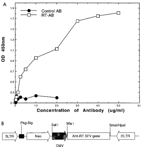
hybridoma cell line, RT 3, which produces a MAb that binds to recombinant HIV-1 RT in ELISAs and Western blots (immu-noblots). The binding constant to recombinant HIV-1 RT by
ELISA was approximately 1.731027M (Fig. 1A) (12, 13). At
least five independent clones, representing the heavy or light chain, were subjected to sequence analysis. The complete DNA sequence and predicted amino acid sequence of the anti-RT SFv were obtained (GenBank/EMBL accession
num-ber 048716). After ligation of VLand VLregions into a single
fragment by using a flexible linker (GGGGS)3 (11), the RT
SFv3 fragment was cloned into a murine leukemia virus-based retroviral expression vector, pSLXCMV, as shown in Fig. 1B. The anti-RT SFv was expressed from an internal cytomegalo-virus promoter. The complete SFv construct, including linker sequences, consist of 786 bp encoding an approximately
28.5-kDa protein. The VL and VH chains are 381 and 351 bp,
respectively.
Gene expression by a retroviral vector.Retroviral vectors are widely used because of their high transduction efficiency (33). pSLXCMV-CAT, a retroviral construct previously re-ported, has been shown to maintain stable expression levels of the CAT reporter gene in SupT1 and CEM cell lines for at
least 4 months (14). High transduction efficiencies (more than
60%) were reported for studies using the pSLXCMV-b-Gal
construct in SupT1 and CEM transduced cells by staining for
b-galactosidase expression (14). To analyze the expression of
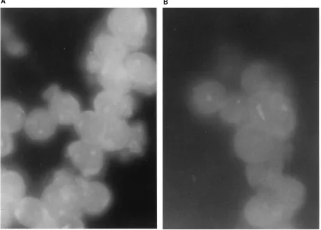
the anti-RT SFv gene in human T-lymphocyte-derived cells, transduced SupT1 cells were G418 selected, and mixed cell populations were maintained in G418-containing medium for at least 4 weeks, whereas 20 cellular clones were maintained in selection medium for approximately 2 months. After G418 selection, cells were grown in medium without antibiotics for 10 days. The expression of anti-RT SFv was analyzed in mixed cell populations and 10 cell clones by immunofluorescence staining and also by RT-initiated PCR (not illustrated). As illustrated in Fig. 2, cytoplasmic localization of the SFv protein was demonstrated, by specific immunostaining of the anti-RT SFv, only in RT SFv3-transduced cells. Mixed cell populations and all the cellular clones analyzed by immunofluorescence revealed SFv protein expression. With the same technique, these cells were shown to maintain SFv protein expression for 3 to 4 months of passage in culture. Of note, cell growth curves and viability were not altered by transduction of the anti-RT SFv3 (not illustrated).
Neutralization of HIV-1 RT activity by RT MAb 3.To ana-lyze, in vitro, if binding of MAbs to RT would neutralize its enzymatic activity (DNA polymerase activity), RT MAbs were first allowed to bind to the HIV-1 RT enzyme, then DNA
synthesis was carried out with [3H]dTTP in a mixture at 378C
for 35 min, and the polymerized products were quantitated by scintillation counting (see Materials and Methods). As nega-tive and posinega-tive controls, two murine MAbs which do not bind to HIV-1 RT (MAb M [mouse anti-hepatitis B virus surface antigen antibody] and MAb 421 [mouse anti-p53 antibody])
and N3dTTP, an RT inhibitor, were mixed with the RT
en-zyme, and then DNA synthesis was carried out. The results (Fig. 3) demonstrated a dramatic decrease (75 or 60%) in DNA polymerase activity of HIV-1 RT in the presence of either a well-characterized RT MAb (affinity purified; anti-RT MAb Intracel) or RT MAb 3. Dependence of concentration on the MAbs was not clearly demonstrable (not shown). The DNA polymerizing activity was not altered in the presence of
nonspecific MAbs M and 421, while the presence of N3dTTP
completely inhibited (by more than 90%) enzymatic activity, as it competes with dTTP and acts as a chain terminator. These results clearly show that specific binding of RT MAb 3 to HIV-1 RT decreased the activity of this retroviral enzyme in vitro.
Inhibition of HIV-1 replication in human T-lymphoid cells expressing anti-RT SFv.To determine whether the intracellu-lar expression of anti-RT SFv3 was able to prevent HIV-1 replication in susceptible T-lymphoid cells, HIV-1 challenge experiments were conducted with stably transformed SupT1-RT-SFv3 cells, using virus strains NL4-3 and R7-HXB2. The infectivity assays were performed with transduced, mixed SupT1 cell populations and with cellular clones. For HIV-1 challenge experiments, RT SFv3-transduced cells (mixed pop-ulations and clones), CAT-transduced SupT1 cells, and
non-transduced SupT1 cells were infected with HIV-1NL4-3(MOIs
of 0.012 and 0.006) and HIV-1R7-HXB2 (MOIs of 0.01 and
0.001), and the spread of HIV-1 in the cultures was determined by quantitating the HIV-1 p24 antigen levels released into the culture medium (Fig. 4). Low levels of HIV-1 p24 antigen were observed at early time points of infection in the supernatants of both SupT1-RT-SFv3 and control cells infected with NL4-3 and R7-HXB2 viruses. Parental nontransduced and CAT-transduced cells further supported the vigorous replication of HIV-1, as shown by the initial increases in HIV-1 p24 antigen,
FIG. 1. (A) Binding affinity curve for an anti-HIV-1 RT MAb. In vitro bind-ing affinity of purified anti-HIV-1 RT antibody (RT-AB) molecules (serial dilu-tions from 2.5mg per well) was analyzed via ELISA, using bacterially expressed recombinant HIV-1-RTIIIBprotein (1 mg per well; Intracel) and goat
anti-murine horseradish peroxidase-conjugated antibody (AB; Sigma) (12, 13). A nonreactive control antibody was concomitantly evaluated in the ELISA. Stan-dard deviations were less than 10%. OD, optical density. (B) Schematic repre-sentation of the anti-HIV-1 RT SFv gene construct in the retroviral expression vector pSLXCMV. 5LTR, 59long terminal repeat; Pkg-Sig, packaging signal; CMV, cytomegalovirus promoter; Neo, neomycin resistance gene; 39LTR, 39
long terminal repeat.
on November 9, 2019 by guest
http://jvi.asm.org/
[image:3.612.58.300.68.318.2]which peaked at approximately 15 to 22 days with an MOI of 0.01 (Fig. 4B and D). HIV-1 p24 antigen levels subsequently decreased in the control cells as a result of the vigorous cyto-pathic effects of virus replication in these cells and subsequent cell death (not shown). On days 15 to 22 postinfection, both SupT1-RT-SFv3 mixed cell populations and SupT1-RT-SFv3 clones 3 and 4 demonstrated approximately 80 to 97% de-creases in HIV-1 p24 antigen production compared with the nontransduced SupT1 cells or cells expressing the CAT vector without SFv (controls). Of note, as with many other gene therapeutic systems to inhibit HIV-1 (14, 37), with rare excep-tions (29), increasing the MOI to 1.0 for input virus could overwhelm much of the effect of anti-RT SFv (not shown). The challenge experiments in which the cells were infected with lower doses of virus (MOI of 0.006 or 0.001) showed slightly delayed initial increases of HIV-1 p24 antigen, which peaked at days 18 to 25. Again, levels of infection were dramatically lower in the cells expressing RT-SFv3 than in the vector-trans-duced cells (Fig. 4A and C). These results indicate that HIV-1 replication was significantly reduced or inhibited in the SupT1-RT-SFv3 cells. In addition, certain anti-Rev SFvs and isolated
VH chains to HIV-1 Rev did not significantly inhibit HIV-1
replication (not shown).
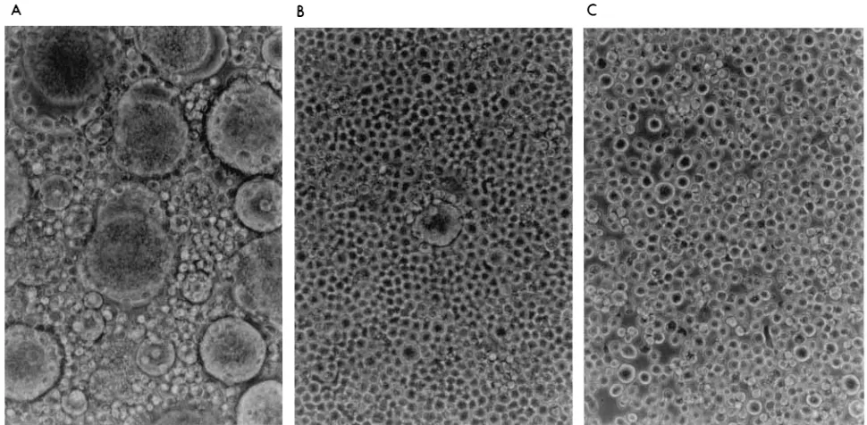
Microscopically, SupT1-RT-SFv3 cells, when infected with HIV-1 strains NL4-3 and R7-HXB2, showed delayed and weak cytopathic effects, as observed by syncytium formation and cell death (Fig. 5). This result suggests that intracellular anti-RT SFv expression protected cells against the cytopathic effects of HIV-1. High numbers of syncytia began to appear in control SupT1-CMV-CAT cells and nontransduced SupT1 cells be-tween days 9 and 15, then the level gradually decreased. In contrast, SupT1-RT-SFv3 cells were totally resistant to syncy-tium formation until days 12 to 17, when only a few syncytia began to appear. Syncytia did not significantly accumulate in the cultures transduced with the anti-RT SFv for over 4 weeks. Further studies are under way to analyze the cells and ex-pressed viruses which are present after 1 month in culture and evaluate potential viral resistance and/or transcriptional shut-down of the SFv transgene.
Intracellular mechanism of anti-RT SFv effects on inhibi-tion of HIV-1 expression.To determine whether the observed decrease in HIV-1 p24 antigen levels correlated with a specific decrease of viral reverse transcription in the cellular cytoplasm, secondary to intracellular expression of anti-RT SFv3 in T-lymphoid cells, one-step HIV-1 infection experiments were
[image:4.612.77.539.77.408.2]performed with HIV-1NL4-3, at a high MOI of 2.0, to obtain
FIG. 2. Analysis of SFv protein expression in the cytoplasm of T lymphocytes. Immunofluorescent staining of the intracellular anti-RT SFv moiety was demonstrated in situ. Mixed populations and cellular clones expressing anti-RT SFv protein or CAT were maintained in culture for 2 months and then used for immunostaining of the RT SFv protein, using a fluorescein isothiocyanate-conjugated goat anti-mouse IgG (kappa chain specific; Sigma). (A) SupT1–anti-RT SFv mixed cellular populations, transduced with pSLXCMV-anti-RT SFv3, demonstrated specific staining for cytoplasmic SFv. (B) SupT1-CAT cells, transduced with pSLXCMV-CAT (vector as negative control), showed minimal nonspecific staining (magnification,3400). Of note, fluorescein isothiocyanate-labeled antibodies to HIV-1 Rev gave no specific immunostaining in these noninfected cells and acted as a specificity control (not shown). These photomicrographs are representative of several other experiments with mixed populations and cell clones.
VOL. 70, 1996 ANTI-RT SFv AND HIV-1 3395
on November 9, 2019 by guest
http://jvi.asm.org/
one-step growth curves of viral replication (see Materials and Methods). To monitor HIV-1 DNA in the infected cells, viral DNA levels were compared by using quantitative PCR for
HIV-1gag DNA. Use of the primer pair SK38-SK39 and the
SK19 probe, which detects HIV-1gagDNA, leads to positive
results only after the synthesis of almost full-length negative-strand HIV-1 DNA (49). Of note, negative-negative-strand strong-stop HIV-1 reverse transcripts were not used for these analyses because they are abundant intravirion reverse transcripts (48, 49). Thus, little quantitative changes in strong-stop negative-strand moieties were detectable in these studies (not shown). These studies were performed on SupT1-CAT, SupT1-plus-AZT, SupT1-Rev-SFvD8, and SupT1-RT-SFv3 mixed popula-tions and cell clones at 1, 3, 6, and 20 h postinfection. The
results (Fig. 6) demonstrate that the levels of HIV-1gagDNA
sequences were significantly lower (60 to 88%) (40 or fewer
HIV-1 DNA copies per 104cells at 1, 3, 6, and 20 h
postinfec-tion) in cells expressing anti-RT SFv3 than in the control cells transfected with either CAT (vector) or pSLXCMV-D8SFv, in which the HIV-1 DNA copy numbers were more
than 1,800 to 2,500 per 104cells at 3 to 20 h after infection. Of
note, the anti-Rev SFv inhibits HIV-1 expression at a postint-egration step by altering Rev function and thus does not affect reverse transcription (13). The lack of effect of anti-Rev SFv on HIV-1 DNA synthesis demonstrates that transduction of cells with an SFv which does not bind to HIV-1 RT does not non-specifically alter intracellular reverse transcription. As dramat-ically high MOIs of HIV-1 were used in these studies to gen-erate one-step growth, viral inhibition was not appreciable because of early cellular death secondary to initial syncytium formation (not shown; see above).
As a positive control, AZT treatment demonstrated
signifi-cant decreases (68 to 85%) in viral DNA synthesis in SupT1 cells, with approximately 35 or fewer copies of HIV-1 DNA, compared with both control cells expressing either CAT or anti-Rev SFv. Thus, the intracellular expression of anti-RT SFv3 is shown to decrease viral DNA to levels comparable to those in the AZT-treated cells. This result suggests that early stages of the viral life cycle were specifically altered or inter-fered with by the anti-RT SFv.
DISCUSSION
In this report, we demonstrate that the expression of an anti-RT SFv moiety in the cytoplasm of susceptible human T-lymphocytic cells markedly decreased HIV-1 replication by specifically inhibiting HIV-1 RT activity. The importance of this approach is based on its ability to significantly decrease HIV-1 replication before viral integration into the host ge-nome and the establishment of a proviral state in the infected cell.
The HIV-1 RT enzyme is multifunctional and catalyzes the synthesis of a double-stranded DNA copy of the viral RNA (44) by RNA-dependent DNA polymerase, DNA-dependent DNA polymerase, and RNase H activities (21, 45). Therefore, it is an excellent target for antiviral therapy. RT enzymes can use a variety of RNA and DNA primers within in vitro assays. In general, the assays are based on measurements of DNA polymerase activity by using synthetic primers and templates and lead to an elongation reaction (21). For many years, the only approved antiviral treatments of AIDS in the United States were drugs which targeted HIV-1 RT, although viral protease inhibitors are now available. The drugs which inhibit HIV-1 RT are used to inhibit viral DNA synthesis by acting as chain terminators (19). Viruses resistant to RT inhibitors ap-pear very quickly during the course of AIDS treatment and in tissue culture (14, 23, 24). Therefore, novel gene therapy ap-proaches have received great attention for antiviral therapeu-tics (37). Accordingly, we are using the intracellular immuni-zation approach and analyzing anti-RT SFvs via cell culture assays for potential interference in HIV-1 RT functions as a molecular antiviral therapy.
This is the first report describing the construction of a re-combinant anti-HIV-1 RT SFv construct, in a retroviral vector, whereby the actual antigen-binding fragments of the
immuno-globulin, the VL and VH chains of an anti-RT MAb, were
joined together via a flexible linker as a single molecule. As well, we demonstrate its potential use as a gene therapeutic approach to restrict HIV-1 replication in human cells. Intra-cellular expression of anti-RT SFv renders the HIV-1-permis-sive cells resistant to wild-type growth of several strains of HIV-1. In protected cells, HIV-1 propagation was restricted at early stages of viral replication after the entry of virions into the cellular cytoplasm rather than at later stages of the HIV-1 life cycle, during which the assembly and production of virions occur (18). We observed a dramatic decrease in viral DNA synthesis within 1 to 3 h of viral infection in cells expressing anti-RT SFv, at levels similar to those found after AZT treat-ment. Nevertheless, the effects of anti-RT SFv were somewhat less intense than those of AZT, and triphosphorylated AZT may have somewhat diminished levels by 12 h postinfection. AZT selectively interacts with HIV-1 RT, blocks the RT-me-diated reverse transcription, inhibits viral replication, and blocks the cytopathic effects of HIV-1 in cell culture (19). The blockade of the reverse transcription process in the RT SFv-expressing cells suggests that the HIV-1 RT enzyme, upon binding with SFv, is not functionally available in the preinte-gration complex for completing the reverse transcription
pro-FIG. 3. Inhibition of HIV-1 RT activity by anti-RT MAb 3. A purified HIV-1 RT enzyme was treated with MAbs or N3dTTP (an RT inhibitor) as described in
Materials and Methods and assayed for exogenous RT activity. RT assays were based on the DNA polymerase activity, which catalyzes the elongation of oligo-nucleotide primer (dTTP), using a synthetic RNA template, poly(rA)12–18, in the presence of labeled [3
H]dTTP. Activity is expressed as counts per minute of [3
H]dTMP incorporation in the presence of the following: bar 1, control, time zero reaction without RT; bar 2, HIV-1 RT alone (0.05 U), 35-min reaction; bars 3 to 7, RT plus anti-RT MAb Intracel, anti-RT MAb 3, nonspecific MAb M, nonspecific MAb 421, and N3dTTP (2mM), respectively. All reactions were
carried out in duplicate. The graph represents the averages of two independent experiments.
on November 9, 2019 by guest
http://jvi.asm.org/
cess and hence interrupts the steps before the establishment of a provirus and DNA integration into the host genome. Of note, targeting the regulatory protein Rev with SFvs would not pre-vent viral integration into the host genome, as Rev SFvs inter-fere with the viral life cycle at later stages, after the establish-ment of a provirus (13).
Our results agree with certain of the findings recently re-ported by Maciejewski et al. (29), which suggested the intra-cellular formation of anti-RT Fab fragments whereby the light and heavy chains of IgG molecules were separately cloned in
episomal expression vectors (pMEP4 and pREP9) from a mouse hybridoma producing an anti-RT MAb. These studies appeared to demonstrate the inhibition of HIV-1 replication in MOLT-3 cells only when both light and heavy chains were expressed together, by separate vectors, in the cells. These were surprising results, as the reducing environment of the cytoplasm is a relatively poor milieu for heavy- and light-chain interactions from separate plasmids (6, 17, 35). In comparison to the studies of Maciejewski et al., our experiments have two distinct differences: (i) the MAb3 had a significant RT
neutral-FIG. 4. Inhibition of HIV-1 replication in anti-RT SFv-transduced SupT1 cells. The SupT1 cells transduced with CAT- or RT SFv-expressing retroviral vectors (mixed populations [Mix] or clones) and nontransduced SupT1 cells were infected with HIV-1NL4-3(MOIs of 0.012 and 0.006) (A and B) or HIV-1R7-HXB2(MOIs of
0.010 and 0.001) (C and D). HIV-1 replication was quantitated by assaying HIV-1 p24 antigen levels in the culture supernatants, using an ELISA (Dupont). The data are representative of at least two sets of independent experiments.
VOL. 70, 1996 ANTI-RT SFv AND HIV-1 3397
on November 9, 2019 by guest
http://jvi.asm.org/
[image:6.612.102.514.69.562.2]ization effect, whereas, Maciejewski et al. used a hybridoma which did not neutralize HIV-1 RT activity in vitro (29), and (ii) our anti-RT SFv was significantly smaller than the intra-cellular Fab fragments used by Maciejewski et al. Of note, though, it is not clear that extracellular neutralization of exog-enous HIV-1 RT activity by anti-RT SFvs will strictly correlate with intracellular immunization effects. The utilization of SFv
molecules may be more suitable for intracellular immuniza-tion, since they consume relatively fewer resources of the transduced cell and hence should not interfere with normal cellular functions (12, 13). In addition, a single molecule, such as an SFv, is absolutely necessary for efficient ex vivo gene therapy in humans.
Experiments were performed to determine which function
FIG. 5. Anti-RT SFv inhibition of cytopathic effects of HIV-1 infection. Shown is the microscopic morphology (syncytium formation and cell death) of SupT1 cells infected with syncytium-inducing strain HIV-1R7-HXB2(MOI of 0.001) after 17 days postinfection. (A) CAT (vector)-expressing cells; (B) mixed cell populations
[image:7.612.64.549.72.310.2]expressing anti-RT SFv; (C) clone 4 expressing anti-RT SFv. Syncytium formation was photographed at a magnification of3400. Similar results for syncytium formation were observed when the cells were infected with HIV-1NL4-3(not shown).
FIG. 6. Quantitative PCR analyses of HIV-1 DNA in cells expressing anti-RT SFv. Various SupT1 lines were infected with cell-free HIV-1NL4-3(MOI of 2.0). Total
cellular DNA was prepared from cells by using a quick lysis procedure (36, 49), and the DNA (1.43104
cell equivalents per lane) was amplified by PCR with the primer pairs SK38-SK39, complementary to thegagregion of the HIV-1 genome, and PCO3-PCO4, complementary tob-globin cellular gene, used as an internal control. As a positive control, AZT-treated SupT1 cells were also analyzed. Note that fresh AZT was readded to these cells after virion infection and washing. As well, anti-Rev SFv-transduced cells were evaluated. A standard curve of 10-fold serial dilutions of ACH-2 DNA is included. The levels of viral DNA (copy numbers) were calculated on the basis of the DNA standard curves, and the percentage changes compared with control values were calculated on the basis of quantitative measurements of hybridized products, using a PhosphorImager. The data are representative of at least two independent experiments.
on November 9, 2019 by guest
http://jvi.asm.org/
of the HIV-1 RT was interfered with by RT MAb 3 in in vitro assays (10, 16). The anti-RT MAb used in these studies binds strongly with HIV-1 RT in ELISAs and is capable of neutral-izing the DNA polymerase activity of HIV-1 RT within in vitro assays with a synthetic primer-template. Preliminary results showed binding of MAb 3 to a synthetic peptide which overlaps the DNA polymerase activity domain of HIV-1 RT (amino acids 325 to 349 of the HIV-1 polymerase) (not shown). There-fore, these experiments suggested that this particular MAb alters RT function by inhibiting DNA polymerase activity. Ex-periments are in progress to further confirm the mapping of the precise epitopes or domains to which the MAb binds on RT. Recently, we have constructed this anti-RT SFv in a bac-terial expression vector and are further characterizing the SFv protein for its abilities to bind HIV-1 RT and synthetic pep-tides of RT. In addition, primary viral isolates, with and with-out RT mutations, are now being systematically evaluated with the anti-HIV-1 RT SFv.
The results of these experiments extend our previous find-ings that single-chain antibodies can be stably expressed, func-tion in the cytoplasm, and are nontoxic to human cells (13, 14). Folding of the SFv within the cell to form functional binding sites can occur in the reducing environment of the cytoplasm (5, 12). The SFvs can also be precisely manipulated for binding to specific epitopes on the target molecules intracellularly (12– 14, 30, 32). Thus, SFvs may provide a tool for the control of intracellular infections and other diseases as well as for under-standing the biological mechanisms in cells leading to a disease state. To achieve even stronger and longer-term protection, we are constructing two or more SFvs on the same vector, target-ing different steps (e.g., RT and Rev) of the HIV-1 life cycle. We believe that such a multistep strategy may further increase the potency of this gene therapeutic approach.
ACKNOWLEDGMENTS
We thank Rita M. Victor and Brenda O. Gordon for excellent secretarial assistance.
This work was supported in part by PHS grant AI36552 and a grant from Intracel, Inc.
REFERENCES
1.Abbott, M. A., B. J. Poiesz, B. C. Byrne, S. Kwok, J. J. Sninsky, and G. D. Ehrlich.1988. Enzymatic gene amplification: qualitative and quantitative methods for detecting proviral DNA amplified in vitro. J. Infect. Dis.158:
1158–1168.
2.Adachi, A., H. E. Gendelman, S. Keonig, T. Folks, R. Willey, A. Rabson, and M. A. Martin.1986. Production of AIDS-associated retrovirus in human and non-human cells transfected with an infectious molecular clone. J. Virol.
59:284–291.
3.Aldovini, H., and B. D. Walker (ed.).1990. Techniques in HIV research. Stockton Press, New York.
4.Ausubel, F. M., R. Brent, R. E. Kingston, D. D. Moore, J. G. Seidman, J. A. Smith, and K. Struhl (ed.).1994. Current protocols in molecular biology, vol. 1. John Wiley & Sons, Inc., New York.
5.Bagasra, O., S. D. Wright, T. Seshamma, J. W. Oakes, and R. J. Pomerantz.
1992. CD14 is involved in control of human immunodeficiency virus type I expression in latently-infected cells by lipopolysaccharide. Proc. Natl. Acad. Sci. USA89:6285–6290.
6.Biocca, S., F. Ruberti, M. Tafani, P. Pierandrei-Amladi, and A. Cattaneo.
1995. Redox state of single chain Fv fragments targeted to the endoplasmic reticulum, cytosol and mitochondria. BioTechniques13:1110–1115. 7.Chen, S.-Y., J. Bagley, and W. A. Marasco.1994. Intracellular antibodies as
a new class of therapeutic molecules for gene therapy. Hum. Gene Ther.
5:595–601.
8.Chen, S.-Y., Y. Khouri, J. Bagley, and W. A. Marasco.1994. Combined intra-and extracellular immunization against human immunodeficiency virus type 1 infection with a human anti-gp120 antibody. Proc. Natl. Acad. Sci. USA
91:5932–5936.
9.Cullen, B. R.1991. Human immunodeficiency virus as a prototypic complex retrovirus. J. Virol.65:1053–1056.
10.Devico, A. L., R. Rahman, M. G. Sarngadharan, and F. Di Veronese.1994. Mechanism of enzyme inhibition mediated by anti-reverse transcriptase
an-tibodies from HIV type 1-infected individuals. AIDS Res. Hum. Retrovi-ruses10:953–960.
11. Dropulic, B., and K.-T. Jeang.1994. Gene therapy for human immunodefi-ciency virus infection: genetic antiviral strategies and targets for intervention. Hum. Gene Ther.5:927–939.
12. Duan, L.-X., O. Bagasra, M. A. Laughlin, J. W. Oakes, and R. J. Pomerantz.
1994. Potent inhibition of human immunodeficiency virus type 1 replication by an intracellular anti-Rev single-chain antibody. Proc. Natl. Acad. Sci. USA91:5075–5079.
13. Duan, L.-X., H. Zhang, J. W. Oakes, O. Bagasra, and R. J. Pomerantz.1994. Molecular and virological effects of intracellular anti-Rev single-chain vari-able fragments on the expression of various human immunodeficiency vi-rus-1 strains. Hum. Gene Ther.5:1315–1324.
14. Duan, L.-X., M. Zhu, O. Bagasra, and R. J. Pomerantz.1995. Intracellular immunization against HIV-1 infection of human T-lymphocytes: utility of anti-Rev single-chain variable fragments. Hum. Gene Ther.6:1561–1571. 15. Erice, A., and H. H. Balfour.1994. Resistance of HIV-1 to anti-retroviral
agents. A review. Clin. Infect. Dis.18:149–156.
16. Furman, P. A., M. H. St. Clair, J. A. Fyfe, J. L. Rideout, P. M. Keller, and G. B. Elion.1979. Inhibition of herpes simplex virus-induced DNA polymer-ase activity and viral DNA replication by 9-(2-hydroxyethoxymethyl)guanine and its triphosphate. J. Virol.32:72–77.
17. Glockshuber, R., M. Malia, I. Pfitzinger, and A. Pluckthun.1990. A com-parison of strategies to stabilize immunoglobulin Fv-fragments. Biochemistry 29:1362–1367.
18. Greene, W. C.1991. The molecular biology of human immunodeficiency virus type 1 infection. N. Engl. J. Med.324:307–317.
19. Hirsch, M. S., and R. T. D’Aquila.1993. Therapy for human immunodefi-ciency virus infection. N. Engl. J. Med.328:1686–1695.
20. Hoxie, J. A., J. D. Alpers, J. L. Rackowski, K. Hueber, B. S. Haggerty, A. J. Cedarbaum, and J. C. Reed.1986. Alteration in T4 (CD4) protein and mRNA synthesis in cells transfected with HIV-1. Science234:1123–1127. 21. Katz, R. A., and A. M. Skalka.1994. The retroviral enzymes. Annu. Rev.
Biochem.63:133–173.
22. Kwok, S., J. J. Lipka, N. McKinney, D. E. Kellog, B. J. Poiesz, S. K. H. Foung, and J. J. Sninsky.1990. Low incidence of HTLV infections in ran-dom blood donors with indeterminate Western blot patterns. Transfusion
30:491–494.
23. Lacey, S. F., J. E. Reardon, E. S. Furfine, T. A. Kunkel, K. Bebenek, K. A. Eckert, S. D. Kemp, and B. A. Larder.1992. Biochemical studies on the reverse transcriptase and RNase H activities for human immunodeficiency virus strains resistant to 39-azido-39-deoxythymidine. J. Biol. Chem. 267:
15789–15794.
24. Larder, B. A., K. E. Coates, and S. D. Kemp.1991. Zidovudine resistant human immunodeficiency virus selected by passage in cell culture. J. Virol.
65:5232–5236.
25. Larder, B. A., and S. D. Kemp.1989. Multiple mutations in HIV-1 reverse transcriptase confer high-level resistance to zidovudine (AZT). Science246:
1155–1157.
26. Levy, J. A.1993. Pathogenesis of human immunodeficiency virus infection. Microbiol. Rev.57:183–289.
27. Lisziewicz, J., D. Sun, D. Lisziewicz, and R. C. Gallo.1995. Antitat gene therapy: a candidate for late-stage AIDS patients. Gene Ther.2:218–222. 28. Liu, J., C. Woffendin, Z.-Y. Yang, and G. J. Nabel.1994. Regulated
expres-sion of a dominant negative form of Rev improves resistance to HIV repli-cation in T cells. Gene Ther.1:32–37.
29. Maciejewski, J. P., F. F. Weichold, N. S. Young, A. Cara, D. Zella, M. S. Reitz, Jr., and R. C. Gallo.1995. Intracellular expression of antibody frag-ments directed against HIV reverse transcriptase prevents HIV infection in vitro. Nat. Med.1:667–673.
30. Marasco, W. A., W. A. Haseltine, and S.-Y. Chen.1993. Design, intracellular expression, and activity of a human anti-human immunodeficiency virus type 1 gp120 single-chain antibody. Proc. Natl. Acad. Sci. USA90:7889–7893. 31. Marchuk, D., M. Drumm, A. Saulino, and F. S. Collins.1990. Construction
of T-vectors, a rapid and general system for direct cloning of unmodified PCR products. Nucleic Acid Res.19:1154.
32. Mhashilkar, A. B., J. Bagley, S.-Y. Chen, A. M. Szilvay, D. G. Helland, and W. A. Marasco.1995. Inhibition of HIV-1 Tat-mediated LTR transactivation and HIV-1 infection by anti-Tat single chain intrabodies. EMBO J.14:1542– 1551.
33. Miller, A. D., and G. J. Roseman.1989. Improved retroviral vectors for gene transfer and expression. BioTechniques7:980–990.
34. Pantaleo, G., C. Graziosi, and A. S. Fauci.1993. The immunopathogenesis of human immunodeficiency virus infection. N. Engl. J. Med.328:327–335. 35. Pluckthun, A.1990. Antibodies from Escherichia coli. Nature (London)
347:497–498.
36. Poiesz, B. J., G. D. Ehrlich, B. C. Byrne, K. Wells, S. Kwok, and J. Sninsky.
1990. p. 47–75.InL. M. de la Maza and E. M. Peterson (ed.), Medical virology, vol. 9. Plenum Press, New York.
37. Pomerantz, R. J., and D. Trono.1995. Genetic therapies for HIV infections: promise for the future. AIDS9:985–993.
38. Ratner, L., W. Haseltine, R. Patarca, K. J. Livak, B. Starcick, S. F. Josephs,
VOL. 70, 1996 ANTI-RT SFv AND HIV-1 3399
on November 9, 2019 by guest
http://jvi.asm.org/
E. R. Doran, J. A. Rafalski, E. A. Whitehorn, K. Baumeister, L. Ivanoff, S. R. Petteway, Jr., M. L. Pearson, J. A. Lautenberger, T. S. Papas, J. Ghrayeb, N. T. Chang, R. C. Gallo, and F. Wong-Staal.1985. Complete nucleotide sequence of AIDS virus, HTLV-III. Nature (London)313:277–284. 39.Saiki, R. S., S. Scharf, F. Faloona, K. B. Mullis, K. B. Horn, G. T. Horn, H. A.
Ehrlich, and N. Arnheim. 1985. Enzymatic amplification of beta-globin genomic sequences and restriction site analysed for diagnosis of sickle cell anemia. Science230:1350–1354.
40.Scharfmann, R., J. H. Axelrod, and L. M. Verma.1991. Long-termin vivo
expression of retrovirus-mediated gene transfer in mouse fibroblast implants. Proc. Natl. Acad. Sci. USA88:4626–4630.
41.St. Clair, M. H., J. L. Martin, G. Tudor-Williams, M. C. Bach, C. L. Vavro, D. M. King, P. Kellam, S. D. Kemp, and B. A. Larder.1991. Resistance to ddI and sensitivity to AZT induced by a mutation in HIV-1 reverse tran-scriptase. Science253:1557–1559.
42.Sun, L. Q., J. Pyati, J. Smythe, L. Wang, J. Macpherson, W. Gerlach, and G. Symonds.1995. Resistance to human immunodeficiency virus type 1 infec-tion conferred by transducinfec-tion of human peripheral blood lymphocytes with ribozyme, antisense, or polymeric trans-activation response element con-structs. Proc. Natl. Acad. Sci. USA92:7272–7276.
43.Tai, M.-S., M. Mudgett-Hunter, D. Levinson, G.-M. Wu, E. Haber, H.
Op-permann, and J. S. Huston.1990. A bifunctional fusion protein containing Fc-binding fragment B of staphylococcal protein A amino terminal to anti-digoxin single-chain Fv. Biochemistry29:8024–2030.
44. Temin, H. M., and D. Baltimore.1972. RNA directed DNA synthesis and RNA tumor viruses. Adv. Virus Res.17:129–186.
45. Skalka, A. M., and S. P. Goff.1993. Reverse transcriptase. Cold Spring Harbor Laboratory Press, Cold Spring Harbor, N.Y.
46. St. Clair, M. H., C. A. Richards, T. Spector, K. J. Weinhold, W. H. Miller, A. J. Langlois, and P. A. Furman.1987. 39-Azido-39-deoxythymidine triphos-phate as an inhibitor and substrate of purified human immunodeficiency virus reverse transcriptase. Antimicrob. Agents Chemother.31:1972–1977. 47. Yu, M., J. Ojwang, O. Yamada, A. Hampel, J. Rapapport, D. Looney, and F.
Wong-Staal.1993. A hairpin ribozyme inhibits expression of diverse strains of human immunodeficiency virus type 1. Proc. Natl. Acad. Sci. USA90:
6340–6344.
48. Zhang, H., O. Bagasra, M. Niikura, B. J. Poiesz, and R. J. Pomerantz.1994. Intravirion reverse transcripts in the peripheral blood plasma of human immunodeficiency virus type 1-infected individuals. J. Virol.68:7591–7597. 49. Zhang, H., Y. Zhang, T. P. Spicer, M. A. Abbott, and B. J. Poiesz.1993. Reverse transcription takes place within extracellular HIV-1 virions: poten-tial biological significance. AIDS Res. Hum. Retroviruses9:1287–1296.
on November 9, 2019 by guest
http://jvi.asm.org/
AUTHORS’ CORRECTION
(5 articles)
Nuclear Preservation and Cytoplasmic Degradation of Human Immunodeficiency
Virus Type 1 Rev Protein
SATOSHI KUBOTA, LINGXUN DUAN, RIKA A. FURUTA, MASAKAZU HATANAKA,
ANDROGER J. POMERANTZ
The Dorrance H. Hamilton Laboratories, Section of Molecular Retrovirology, Division of Infectious Diseases, Department of Medicine, Jefferson Medical College, Thomas Jefferson University, Philadelphia, Pennsylvania, and
Institute for Virus Research, Kyoto University, Sakyo-ku, Kyoto 606-01, Japan
Volume 70, no. 2, p. 1282–1287.
Binding of Intracellular Anti-Rev Single Chain Variable Fragments to
Different Epitopes of Human Immunodeficiency Virus Type 1 Rev:
Variations in Viral Inhibition
YONG WU, LINGXUN DUAN, MINGHUA ZHU, BAOCHENG HU, SATOSHI KUBOTA, OMAR BAGASRA,
ANDROGER J. POMERANTZ
The Dorrance H. Hamilton Laboratories, Center for Human Retrovirology, Division of Infectious Diseases, Department of Medicine, Jefferson Medical College, Thomas Jefferson University, Philadelphia, Pennsylvania 19107
Volume 70, no. 5, p. 3290–3297.
Targeting Human Immunodeficiency Virus Type 1 Reverse Transcriptase by
Intracellular Expression of Single-Chain Variable Fragments To Inhibit
Early Stages of the Viral Life Cycle
FARIDA SHAHEEN, LINGXUN DUAN, MINGHUA ZHU, OMAR BAGASRA,ANDROGER J. POMERANTZ
The Dorrance H. Hamilton Laboratories, Section of Molecular Retrovirology, Division of Infectious Diseases, Department of Medicine, Jefferson Medical College, Thomas Jefferson University, Philadelphia, Pennsylvania 19107
Volume 70, no. 6, p. 3392–3400.
Intracellular Expression of Single-Chain Variable Fragments To Inhibit
Early Stages of the Viral Life Cycle by Targeting Human
Immunodeficiency Virus Type 1 Integrase
PNINA LEVY-MINTZ, LINGXUN DUAN, HUIZHONG ZHANG, BAOCHENG HU, GEETHANJALI DORNADULA, MINGHUA ZHU, JOSEPH KULKOSKY, DIANE BIZUB-BENDER, ANNA MARIE SKALKA,
ANDROGER J. POMERANTZ
Dorrance H. Hamilton Laboratories, Center for Human Virology, Division of Infectious Diseases, Department of Medicine, Jefferson Medical College, Thomas Jefferson University, Philadelphia, Pennsylvania 19107, and Fox Chase
Cancer Center, Philadelphia, Pennsylvania 19111
Volume 70, no. 12, p. 8821–8832.
Potent Inhibition of Human Immunodeficiency Virus Type 1 in Primary T Cells
and Alveolar Macrophages by a Combination Anti-Rev Strategy
Delivered in an Adeno-Associated Virus Vector
ROGER T. INOUYE, BIN DU, DEBORAH BOLDT-HOULE, ANTHONY FERRANTE, IN-WOO PARK, SCOTT M. HAMMER,
LINGXUN DUAN, JEROME E. GROOPMAN, ROGER J. POMERANTZ,ANDERNEST F. TERWILLIGER
Divisions of Experimental Medicine and Hematology/Oncology and Infectious Disease, Beth Israel Deaconess Medical Center and Harvard Institutes of Medicine, Boston, Massachusetts 02215, and Dorrance H. Hamilton Laboratories, Center for Human Virology,
Division of Infectious Disease, Thomas Jefferson University, Philadelphia, Pennsylvania 19107
Volume 71, no. 5, p. 4071–4078.
The following correction pertains to all of the above articles.
The heavy chain of the D8 anti-Rev single-chain variable fragment (SFv) has been reanalyzed and demonstrated to be an aberrant heavy chain sequence. This heavy chain sequence is very close to the aberrant heavy chain sequence published by P. Thammana (Molecular Immunology 31:77–78, 1994), who derived it via RT-PCR directly from the RNA of the NS1 cell line which is commonly used as a fusion partner to construct mouse hybridomas. Therefore, it is likely that the aberrant D8SFv heavy chain was derived from a gene originating from the fusion partner cell line used to make the original D8 hybridoma and not from the heavy chain gene expressed by the B-cell precursor to this hybridoma. This aberrant heavy chain has a deletion in the framework region 3 (FR3) leading to a frameshift in CDR3 and downstream regions of the heavy chain gene. In addition, there were some individual nucleotide changes, on reanalysis, that brought the heavy chain sequence even closer to that described by Thammana. It should also be noted that the function of the aberrant heavy chain in the D8SFv is not known. As well, the initial 12 amino acids in the D8SFv represent a portion of the Vkleader sequence. The possible effects, if any, of this segment on subcellular localization and/or secretion have not been investigated.
There was noted to be rather minimal binding data for the D8SFv available to be reevaluated at this time, including only a single ELISA for the D8SFv to recombinant Rev and a single binding study of the activation domain peptide of Rev. Comparisons to the original D8 monoclonal antibody are not obtainable since the characteristics of the original monoclonal antibody are not fully demonstrated at the present time. As well, complete original data dealing with anti-Rev D10SFv binding to peptides of Rev are not available at the present time.
On reanalysis, inaccuracies in the D10SFv sequence were noted. D10SFv on resequencing showed the following differences compared to the published sequence: T58 to A (ACA to GCA), T106 to S (ACG to TCG), and G115 to A (GGT to GCT). Nucleotide changes which did not alter the amino acid sequence were: 81C to T, 315G to C, 354T to A, and 357T to A (numbering started at initial ATG codon). A nucleotide was demonstrated to be missing at position 771 (G) (L257), compared to the original sequence, altering the remaining amino acids (DYWGQGTSVTVSSAKTTPPPVYPLAPGS). On reevaluation, the originally reported D10 sequence would connote a frameshift in the CDR3 of Vh, but now, as resequencing revealed the missing nucleotide,
the frameshift is shown not to be present. Thus, D10SFv is, in fact, an appropriate antibody sequence.
AUTHORS’ CORRECTION*
(3 articles)
Binding of Intracellular Anti-Rev Single Chain Variable Fragments to
Different Epitopes of Human Immunodeficiency Virus Type 1 Rev:
Variations in Viral Inhibition
YONG WU, LINGXUN DUAN, MINGHUA ZHU, BAOCHENG HU, SATOSHI KUBOTA, OMAR BAGASRA,
ANDROGER J. POMERANTZ
The Dorrance H. Hamilton Laboratories, Center for Human Retrovirology, Division of Infectious Diseases, Department of Medicine, Jefferson Medical College, Thomas Jefferson University, Philadelphia, Pennsylvania 19107
Volume 70, no. 5, p. 3290–3297, 1996.
Targeting Human Immunodeficiency Virus Type 1 Reverse Transcriptase by
Intracellular Expression of Single-Chain Variable Fragments To Inhibit
Early Stages of the Viral Life Cycle
FARIDA SHAHEEN, LINGXUN DUAN, MINGHUA ZHU, OMAR BAGASRA,ANDROGER J. POMERANTZ
The Dorrance H. Hamilton Laboratories, Section of Molecular Retrovirology, Division of Infectious Diseases, Department of Medicine, Jefferson Medical College, Thomas Jefferson University, Philadelphia, Pennsylvania 19107
Volume 70, no. 6, p. 3392–3400, 1996.
Intracellular Expression of Single-Chain Variable Fragments To Inhibit
Early Stages of the Viral Life Cycle by Targeting Human
Immunodeficiency Virus Type 1 Integrase
PNINA LEVY-MINTZ, LINGXUN DUAN, HUIZHONG ZHANG, BAOCHENG HU, GEETHANJALI DORNADULA, MINGHUA ZHU, JOSEPH KULKOSKY, DIANE BIZUB-BENDER, ANNA MARIE SKALKA,
ANDROGER J. POMERANTZ
Dorrance H. Hamilton Laboratories, Center for Human Virology, Division of Infectious Diseases, Department of Medicine, Jefferson Medical College, Thomas Jefferson University, Philadelphia, Pennsylvania 19107, and Fox Chase
Cancer Center, Philadelphia, Pennsylvania 19111
Volume 70, no. 12, p. 8821–8832, 1996.
The following correction applies to all of the above articles and to the Authors’ Correction published in the April 1998 issue of the Journal of Virology (72:3505–3506):
In attempts to re-evaluate our previously reported data on inhibition of HIV-1 by intracellular single chain variable fragments (SFvs), some original data were not able to be located and some other data might not be fully consistent with certain previously published graphs. Therefore, we have now completed extensive new experimentation involving the anti-HIV-1 single chain variable fragments (SFvs), including Rev (D8), Integrase (IN33), and Reverse Transcriptase SFvs, described by our laboratories. In these repeat studies, the multiplicity of infections (mois) used in viral challenge studies were estimated by tissue culture infectious dose 50% (TCID50) per target cell, calculated via a described technique (Techniques in HIV Research, Eds. Aldovini, A. and Walker,
B., 1990). The mois of viral input which led to complete or near complete protection of SUPT1 T-cells transduced with the
anti-HIV-1-SFvs varied between 0.00004 to 0.00002. These were somewhat lower than those described in the initial reports (utilizing HIV-1NL4-3). Nevertheless, the levels of viral growth in control cultures, in the previously published reports and in the
new studies, were very similar. Many of the new studies actually showed higher viral growth in control cultures, with complete viral
recently shown the specificity of post-integration inhibition of HIV-1 by the RevD8-SFv in latently infected cells (AIDS Res. & Human Retro. 14:1573, 1998). Binding studies for D8RevSFv against recombinant Rev protein were also repeated by ELISA and showed binding above the background and negative control levels. Intracellular specific binding was also demonstrated by another group utilizing our IN33SFv (PNAS 96:11723, 1999). Our recent results are consistent with our previously published studies in that they confirm that SFvs to HIV-1 Rev, Reverse Transcriptase and Integrase are able to specifically and significantly inhibit HIV-1 replication compared to control cultures.
![FIG. 4. Inhibition of HIV-1 replication in anti-RT SFv-transduced SupT1 cells. The SupT1 cells transduced with CAT- or RT SFv-expressing retroviral vectors(mixed populations [Mix] or clones) and nontransduced SupT1 cells were infected with HIV-1 (MOIs of 0](https://thumb-us.123doks.com/thumbv2/123dok_us/1277883.80308/6.612.102.514.69.562/inhibition-replication-transduced-transduced-expressing-retroviral-populations-nontransduced.webp)