STUDY OF EXPRESSION OF p16INORAL SQUAMOUS CELL
CARCINOMA, POTENTIALLY MALIGNANT DISORDERS
ANDNORMAL MUCOSA
Dissertation submitted to
THE TAMILNADU Dr.M.G.R. MEDICAL UNIVERSITY
In partial fulfilment for the Degree of
MASTER OF DENTAL SURGERY
BRANCH VI
ORAL PATHOLOGY AND MICROBIOLOGY
ACKNOWLEDGEMENT
My heartfelt gratitude to my post graduate teacher Dr.K.Ranganathan, MDS, MS (Ohio), PhD, Professor and Head of Department of Oral and Maxillofacial Pathology, Ragas Dental College and Hospital for his constant, support and encouragement and for guiding me throughout my dissertation.
I extend my sincere gratitude to Dr.UmaDevi.K.Rao, Professor, Department of Oral and Maxillofacial Pathology, Ragas Dental College and Hospital for her constant guidance and support and her advice in completion of my work.
My sincere thanks to Dr.Elizabeth Joshua, Professor, Department of Oral and Maxillofacial Pathology, Ragas Dental College and Hospital for her constant support and encouragement throughout my study.
My sincere thanks to the principal Dr.S.Ramachandranand Chairman Mr.Kanakaraj, Ragas Dental College and Hospital for their permission to use the facilities of the institution.
I earnestly thank Professor, Dr.T.Rooban, Department of Oral and Maxillofacial Pathology, Ragas Dental College and Hospital for his encouragement and concern in helping me to complete this study.
I extend my sincere thanks to Reader Dr.N.Lavanya and Senior Lecturer Dr.C.Lavanya, Department of Oral and Maxillofacial Pathology, Ragas Dental College and Hospital for their encouragement.
I am very grateful to our Research Assistant Mrs.Kavitha, Biostatistician Mrs.Deepa, Lab Technician Mr.Rajan, Department of Oral and Maxillofacial Pathology, Attender Mrs.Vasanthi, Ragas Dental College and Hospital for their constant help in completion of my study.
I also thank Professor and Head Dr.Rajkumar, Department of Oral and Maxillofacial Pathology, SRM Dental college and Professor and Head Dr.Sathish Kumar, Department of Oral and Maxillofacial Pathology, KarpagaVinayaga Institute of Dental Sciences for their encouragement and Motivation.
I thank Dr.JayGanesh, General pathologist, Institute of Obstetrics and Gynaecology Hospital, Dr.Janaki, Prosthodontist and Mr.Venkatesh, Biotechnologist for their immense support and encouragement.
I acknowledge gratefully the help of my batch-mates Dr.AiswaryaLekshmy S U, Dr.Jaishlal M S, Dr.Nithya S, Dr.ShanmugaPriya
P and Dr.Vaishnavi S. I also thank my seniors and juniors for their support and encouragement.
ABSTRACT
BACKGROUND:It has been hypothesized that inactivation of p16 plays a vital role in evolution of potentially malignant disorders [Epithelial Dysplasia, Oral SubmucousFibrosis (OSF)] and in Oral Squamous Cell Carcinoma (OSCC). We wanted to ascertain the role of p16 in OSF, epithelial dysplasia and OSCC as an early marker for malignant transformation and its liability in OSCC.
AIM:
To study the expression of p16 in normal mucosa (Group I), OSF (Group II), epithelial dysplasia (Group III) and OSCC (Group IV).
MATERIALS AND METHODS:
A total sample size of 70 which includes 10 controls of Group I, 20 cases of Group II, 20 cases of Group III, 20 cases of Group IV were studied for p16 expression by immunohistochemistry.We have employed p16 monoclonal antibody of clone G175-405 in our study. The positive control sample of our study is squamous cell carcinoma of uterine cervix.
RESULTS:
Out of 70 samples 16 cases (22.9%) of cases were p16 positive which includes five in group I, seven in group II, one in group III, three in group IV. Out of sixteen p16 positive samples four cases showed cytoplasmic staining (12,1 in Group I,II,IV respectively) , eleven cases showed nuclear and cytoplasmic staining (3,5,1,2 in Group I,II,III,IV respectively) and one showed cytoplasmic and membrane staining.
CONCLUSION:
In this study we addressed the association particularly between p16 and OSF, epithelial dysplasia and OSCC. The results of this study with respect to OSF data, highlights that p16 could play a role in malignant transformation of OSF and we hypothesize that it could be associated with HPV.Further studies should ascertain the HPV status of the cases to be included, with a larger sample size to establish and understand if p16 expression could have a role in oral potentially malignant lesions and OSCC.
CONTENTS
1. INTRODUCTION 1
2. AIM AND OBJECTIVES 3
3. MATERIALS AND METHODS 5
4. REVIEW OF LITERATURE 15
5. RESULTS 46
6. DISCUSSION 53
7. SUMMARY AND CONCLUSION 62
8. BIBLIOGRAPHY
65
ANNEXURE I
ANNEXURE II
ANNEXURE III
1
INTRODUCTION
Oral squamous cell carcinoma (OSCC), is the sixth most common cancer worldwide and the third most common form of cancer in the developing countries. Squamous cell carcinoma occurs due to multiple genetic changes leading to formation of either abnormal proteins or altered amount of normal proteins1.
Clinical OSCC is often preceded by stepwise transition from potentially malignant states like leukoplakia and Oral Submucous Fibrosis (OSF) to the metastatic tumour phenotype. A variety of alterations accumulate to potentiate this transition to malignancy2.
Leukoplakia, the most common potentially malignant lesion of the oral mucosa is defined by WHO as “a white patch or plaque that cannot be characterized clinically or pathologically as any other disease”. The malignant transformation rate for leukoplakia ranges from 5-20% and is particularly correlated with the degree of dysplasia3. The transition from normal oral epithelium to oral dysplasia and cancer results from accumulated genetic and epigenetic alterations4. The grading of epithelial dysplasia remains subjective as it relies on cellular atypia and architectural disturbances.
2
Given this aggressive nature of the potentially malignant lesions, identification of a suitable biomarker is imperative for timely diagnosis, prognosis and treatment6. Mutations in tumour suppressor genes namely p53, pRb, p16 and pro-apoptotic genes namely bcl2, bax have been variously attributed to the development and transformation of precancer to cancer. It has been found that inactivation of p16 occurs early in the development of OSCC7.
Homozygous deletion, point mutation, loss of heterozygosity and the more common aberrant methylation are the frequently reported alterations in the p16 gene8. p16 is a cyclin dependent kinase inhibitor(CKI), a tumour suppressor gene, and is the second commonly affected gene next to p53 in OSCC. The main function of p16 is to control the phosphorylation of retinoblastoma (Rb) gene and block the progression of cell cycle.
Human papilloma virus (HPV) has been reported to initiate carcinogenesis in cervical cancers and OSCC. HPV causes oral carcinogenesis by acting on viral oncoproteins E6 and E7. P16 inactivates RB protein67. Similarly overexpression of p16 in cervical cancer is due to functional inactivation of Rb by HPV E7 oncoprotein. Thereby, it clearly shows that HPV causes inactivation of p16 pathway which leads to malignant transformation and carcinogenesis. There is a strong association between HPV presence and p16 in certain neoplasms.
3
AIM AND OBJECTIVES
AIM:
To assess the expression of p16 in normal oral mucosa, potentially malignant disorders (Leukoplakia, OSF) and OSCC.
OBJECTIVES:
1. To study the expression of p16 in formalin fixed paraffin embedded tissue specimens of normal oral mucosa by immunohistochemistry (IHC). 2. To study the expression of p16 in formalin fixed paraffin embedded tissue
specimens of epithelial dysplasia (Leukoplakia) by IHC.
3. To study the expression of p16 in formalin fixed paraffin embedded tissue specimens of OSF by IHC.
4. To study the expression of p16 in formalin fixed paraffin embedded tissue specimens of OSCC by IHC.
5. To compare the expression of p16 between normal oral mucosa, Leukoplakia, OSF and OSCC.
STUDY SETTING:
4
specimens. The approval from Institutional Review Board (IRB) of Ragas Dental College and Hospital, Chennai was obtained. (ANNEXURE I)
HYPOTHESIS: (NULL)
There is no difference in expression of p16 in potentially malignant disorders (Leukoplakia, OSF), OSCC when compared to normal oral mucosa.
STUDY SUBJECTS:
The study material comprised of 70 formalin fixed, paraffin embedded tissue specimens. The samples were divided into 4 groups namely: Group I, Group II, Group III and Group IV.
Group I: 10 normal mucosa tissue specimens.
Group II: 20 histopathologically confirmed OSF fibrosis tissue specimens.
Group III: 20 histopathologically confirmed epithelial dysplasia (Leukoplakia) tissue specimens.
5
METHODOLOGY
1. Tissue samples of Normal mucosa (n=10), OSF (n=20), Leukoplakia (n=20) and OSCC (n=20) were taken from the patients and from the archival blocks.
2. A detailed case history including age, gender and occupation, past medical history & past dental history, history of drugs and trauma were recorded. 3. General examination and intra oral examination was done.
4. Biopsy was done from the lesion site. Normal mucosa was taken when the patients were undergoing minor surgery for impacted teeth cases.
5. The tissue biopsied was immediately transferred to 10 % buffered formalin.
6. After adequate fixation, tissues were embedded in paraffin.
7. From the paraffin embedded blocks 4 microns thick, sections were cut and used for routine Hematoxylin and Eosin (H&E) staining and immunohistochemical (IHC) staining.
8. Tissue sections of squamous cell carcinoma of cervix were used as positive controls for p16 positivity.
HEMATOXYLIN & EOSIN (H&E) STAINING:
REAGENTS:
6 PROCEDURE:
The slides were dewaxed in xylene and hydrated through graded alcohol to water. The sections on the slides were flooded with Harris’s hematoxylin for 5 minutes. The slides were washed in running tap water for 5 minutes. The slides were differentiated in 1% acid alcohol for 5 minutes.
The slides were washed well in running tap water for 5 minutes. The tissue sections on the slides were then stained in eosin for 30 seconds. The slides were washed in running tap water for 1 minute. The slides were then dehydrated through alcohol, cleared, mounted and viewed under light microscope (LM).
IHC:
ARMAMENTARIUM:
Microtome
Autoclave
Hot air oven
Slide warmer
Couplin jars
Measuring jar
Weighing machine
APES coated slides
Slide carrier
Aluminium foil
Micro-pipettes
7
Electronic timer
Beakers
Rectangular steel tray with glass rods
Sterile gauze
Cover-slips
Light microscope
REAGENTS USED:
Concentrated HCl
Laxbro solution
APES (3 amino propyl tri ethoxy silane)
Acetone
Citrate buffer
Phosphate Buffer Saline (PBS)
3% H2O2
Deionized distilled water
Absolute alcohol
Xylene
ANTIBODIES USED:
1. Primary antibody – p16 monoclonal antibody, BIOGENEXTM Clone: G175 – 405
(FIGURE:1)
8
immunoglobulins labelled with enzyme polymer, super enhancer reagent, antimouse monoclonal negative control serum, and liquid DAB Diamino-benzidine-chromogen) (FIGURE: 1)
IHC PROCEDURE:
PRETREATMENT OF THE SLIDES:
The slides were first washed in tap water for few minutes.
The slides were then soaked in detergent solution for 1 hour.
After 1 hour, each slide was brushed individually using the detergent
solution and were transferred to distilled water.
The slides were washed in two changes of distilled water.
The slides were washed in autoclaved distilled water.
The slides were immersed in 1 N HCL (100 ml HCl in 900 ml distilled
water) overnight.
The following day slides were taken out of acid and washed in two
changes of autoclaved distilled water.
All the slides were then transferred to slide trays, wrapped in aluminium
foil and baked in hot air oven for 4 hours at 180 degrees Centigrade.
APES (3 Amino propyl tri ethoxy silane) coating:
Slides first dipped in couplin jar containing acetone for 2 minutes ↓
Dipped in APES for 5 minutes ↓
Dipped in two changes of distilled water for 2 minutes each ↓
9
PREPARATION OF PARAFFIN SECTIONS:
After the slides were dry, tissue section of 5 micron thickness were made in a rotary manual microtome. The ribbons of tissue section were transferred onto the APES coated slide from the tissue float bath such that two tissue bits come on to the slide with a gap in between. One of the tissue sections was labelled positive (P) and the other negative (N).
PROCEDURE:
10
three changes of cold PBS for 5 minutes in each. The sections were washed in 3 changes of cold PBS for 5 minutes in each. Then the slides were wiped carefully without touching the tissue section to remove excess PBS. Then a drop of DAB was added to the sections for 5 minutes. Slides were then washed in distilled water to remove excess chromogen and counter stained with hematoxylin. Then the slides were transferred to 70% alcohol, 100% alcohol and one change of xylene. The tissue sections were mounted with DPX. The slides were then observed under the microscope. Throughout the procedure care was taken not to dry the tissues.
IHC PROCEDURE FLOW CHART:
APES coated slides with 2 paraffin embedded tissues ↓
Placed in xylene thrice (5 minutes each) ↓
Placed in 100% isopropanol (5 minutes) ↓
Placed in 70% isopropanol (5 minutes) ↓
Washed in distilled water thrice (5 minutes each) ↓
Placed in 3% hydrogen peroxide (20 minutes) ↓
Kept in citrate buffer at pH 6 and autoclaved for antigen retrieval and bench cooled for 40 minutes
11
Washed in distilled water thrice (5 minutes) ↓
Protein blocking serum added and incubated for one hour ↓
Primary antibody added to the specimen and incubated for one hour ↓
Washed in PBS thrice (5 minutes each) ↓
Secondary antibody added and incubated in an enclosed hydrated container (30 minutes)
↓
Washed in PBS thrice (5 minutes each) ↓
HRP secondary reagent added and incubated (30 minutes) ↓
Washed in PBS thrice (5 minutes each) ↓
DAB added and incubated in an enclosed hydrated container (5 minutes) ↓
Washed in PBS thrice (5 minutes each) ↓
Stained with hematoxylin (20 seconds) ↓
12
Placed in 70% isopropanol (1minute) ↓
Placed in 90% isopropanol (1minute) ↓
Placed in 100% isopropanol (1 minute) ↓
Placed in xylene (1 dip) ↓
Slides were mounted using DPX ↓
Slides were observed under the Light Microscope and graded
Criteria for detecting the expression of p16:
1. H&E sections:
- The H&E stained sections were thoroughly examined.
- Dysplastic lesions were categorised as mild dysplasia, moderate dysplasia and severe dysplasia. The koilocytic changes were also noted in dysplastic sections.
- Oral squamous cell carcinoma was graded as well differentiated, moderately differentiated, poorly differentiated.
2. Corresponding sections as examined by H&E were stained by IHC to detect p16 expression.
13 4. For IHC
- The positive control used for IHC was squamous cell carcinoma of cervix. - The stained slides were screened, examined systematically for p16 expression
in the nucleus, cytoplasm and also for membrane staining.
- Positive cells were counted in the basal, suprabasal layers of normal mucosa. - Positive cells were counted in the basal, suprabasal layers in dysplasia, basal
and suprabasal layers in oral submucous fibrosis, in the invasive front of oral squamous cell carcinoma.
- Percentage of positive cells were also counted in each case and it was categorised as
0 = negative; 1+ = 1% to 25% of cells positive; 2+ = 26% to 50%; 3+ = 51% to 75%; 4+ = 76% to 100%.
- Connective tissue was also examined in all the lesions.
- Nuclear staining was expressed as percentage of cells and the positivity were noted as mean labelling index (MLI) and cytoplasmic staining was also recorded.
- 0, 1+, 2+, 3+, 4+ staining with reference to the positive control was observed with reference to nuclear and cytoplasmic staining grading.
- The MLI for all the positive groups were calculated using the formula: Number of positive cells
14 STATISTICAL ANALYSIS:
Statistical analysis was done using SPSSTM version 17. Proportion of p16 staining was compared between and within the study groups using 'chi square' test. 't' test and 'oneway ANOVA' was done for continuous variables. A p value of 0.05 was considered statistically significant.
SAMPLE SIZE:
15
REVIEW OF LITERATURE
CELL CYCLE:
The basic cell cycle is divided into four stages: G1, S, G2, and M. G1 is the gap phase during which cells prepare for the process of DNA replication. It is during the 'G1' phase that the cell integrates mitogenic and growth inhibitory signals and then decides to pause, or exit the cell cycle. An important checkpoint in 'G1' has been identified in which the cell becomes committed to DNA replication and completes a cycle. 'S' phase is defined as the stage in which DNA synthesis occurs. 'G2' is the second gap phase during which the cell prepares for the process of division. M stands for mitosis, the phase in which the replicated chromosomes are segregated into separate nuclei and cytokinesis occurs to form two daughter cells. In addition to G1, S, G2, and M, the term G0 is used to describe cells that have exited the cell cycle and become quiescent9.
CDK INHIBITORS: INK4 and Cip/Kip FAMILY:
16
p21Cip1, p27Kip1 and p57Kip2. The CKIs function as a break for the premature entry of the cell to the S phase by inhibiting the CDKs10.
INK4/ARF LOCUS AND p16INK4a:
The INK4 gene family encodes for p16INK4a, p15INK4B, p18INK4C, and p19INK4D, all of which bind to CDK4 and CDK6 and inhibit their kinase activities by interfering with their association with D-type cyclins. Specific polypeptide inhibitors of CDK4 and CDK6, so-called the INK4 proteins (inhibitors of CDK4), can directly block cyclin D-dependent kinase activity and cause G1 phase arrest.
17
proteins and progression into the S phase. pRb is a key gatekeeper in cell cycle transition. In normal cycling cells, entry to the S-phase from G1 phase is related to the functional inactivation of pRb by phosphorylation by cyclin dependent kinases11.
INK4A/ARF locus encodes two distinct products. Exon1α encodes part of p16INK4a protein which is a cyclin dependent kinase inhibitor. Exon1β is spliced onto an acceptor site common to 1α but encodes a nonhomologous protein called p19ARF, in an alternative reading frame. p16INK4a inhibits the CDK 4 and 6 thus causing inhibition of E2F transcription factors. INK4A/ARF locus encodes two distinct proteins functioning upstream of both Rb and p53 pathways in the cell. The INK4A/ARF locus represents a unique phenomenon by encoding two distinct tumour suppressor proteins inhibiting the cell cycle progression in a different manner. p16INK4a acts by inhibiting CDK4 and 6 and keeping pRb active while p19ARF leads to stabilization of p53. Functions of these proteins upstream of both pRb and p53 make this locus a ‘keystone gene’ in cell cycle12.
p16 AND CARCINOGENESIS:
18
in oral cancer. In some tumours, such as cervical cancer, p16INK4a is frequently silenced by hypermethylation of the gene. The other CKIs also function as tumour suppressors and are frequently mutated or otherwise silenced in many human malignancies, including familial melanomas, sporadic pancreatic adenocarcinomas, and squamous cell carcinomas of the esophagus13.
Hypophosphorylated Rb in complex with the E2F transcription factors binds to DNA, recruits chromatin-remodeling factors (histone deacetylases and histone methyl transferases), and inhibits transcription of genes whose products are required for the S phase of the cell cycle. When Rb is phosphorylated by the cyclin D–CDK4, cyclin D–CDK6 and cyclin E–CDK2 complexes, it releases E2F. The latter then activates transcription of S-phase genes. The phosphorylation of Rb is inhibited by CKIs, because they inactivate cyclin-CDK complexes. Virtually all cancer cells show dysregulation of the G1-S checkpoint as a result of mutation in one of four genes that regulate the phosphorylation of Rb; these genes are Rb1, CDK4, the genes encoding cyclin D proteins, and CDKN2A(p16)14.
Tsantoulis PK et al (2007) emphasized the importance of cancer transforming from the transitional precursor lesions. They also elucidated the genetic implications of the molecular mediators in cancer which includes cyclin dependent kinase inhibitors, TP53 and Rb1 and oncogenes like the cyclin family. They also summarised the tumorigenic effects of HPV and EBV15.
19
generation of endocrine and hematopoietic cells. They found that CDK2 is important for the first meiotic division of male and female germ cells. These suggested that CDKs are important for the regulation of cell cycle and that they can be utilised for therapeutic strategies in cancer cells16.
p16 GENE REGULATION AND INACTIVATION:
Serrano M et al (1993) stated that the cell division of eukaryotic cells is regulated by a family of protein kinases which were latter classified as cyclin dependent kinases. The sequential activation of individual members of the family and their consequential phosphorylation of critical substrates promotes orderly progression through cell cycle. They concluded that p16INK4A acted at both the upstream and downstream function in the cell cycle of Rb to form a negative feedback loop which regulates the ability of Rb to prevent cell proliferation17.
20
Mao L et al (1995) used cell lines derived from head and neck cancers, HeLA cell lines and normal lymphoblastic cells to investigate the presence of alternative promoter or initiator site showing methylation. p16INK4A AND p15INK4B were localized to chromosome 9p21, and pl6 was subsequently found to be mutated in familial melanoma and deleted in a wide variety of sporadic cancers. This finding emphasized the denovo methylation of 5' CpG island resulting in transcriptional block of full length p16 in many neoplasms19.
Stone S et al (1995) studied the genomic structure and transcriptional regulation of p16 gene in humans by using genomic cloning, cDNA clones, in cell lines, lymphocytes by flow cytometry. From this two types of promoters α and β were found and were of similar size. These results showed p16 gene is complex with α and β promoters with different coding potential. They concluded that genetic evidence of p16 and Rb are members of growth regulatory pathway often inactivated leading to tumour progression. They also stated that if the role of β transcript is regulation of cell growth negatively, it could probably be another pathway that could be independent of p16 and Rb. They also used molecular genetic techniques to explore the role of p16 in normal development and cancer. They found that p16 derived mRNAs are probably generated from separate promoters and transcription from one of the promoters appears to be regulated, atleast in part, by the retinoblastoma gene product20.
21
tumorigenesis by establishing an essentially irreversible growth arrest that requires activities of the p53 and pRb tumour suppressor proteins. They showed that, depending on expression of the pRb regulator p16, replicative senescence is not necessarily irreversible. Expression of telomerase did not reverse the senescence arrest. Their results indicated that the senescence response to telomere dysfunction is reversible and is maintained primarily by p53. However, p16 provides a dominant second barrier to the unlimited growth of human cells21.
Ohtani N et al (2004) summarised the mechanisms involved in the senescence of p16 which leads to tumour suppression. They summarised the importance of telomere shortening which leads to senescence and telomere independent mechanisms which involves oxidative stress and inactivation of p16-Rb pathway which leads to cancer22.
p16 IN OTHER NEOPLASMS:
Herman J et al (1995) studied the promoter methylation of p16 in breast cancer, prostate cancer, renal cancer and colon cancer. They found aberrant denovo methylation of p16 in all the cancer cell lines. They concluded that p16 promoter methylation plays an important diagnostic and prognostic factor for all the common human cancers23.
22
carcinomas. They also signified the low frequency of p16 mutations in common tumours such as ovarian cancer, breast cancer and colon cancer24.
p16 IN VASCULAR SMOOTH MUSCLE CELLS:
Tanner F et al (2000) studied the expression of p27, p21 and p16 in vascular smooth muscle cells. Their results showed that p27 and p21 inhibits the proliferation of vascular smooth muscle cells while p16 has no interference in it. This suggested that p16 has no influence in growth of vascular smooth muscle cells25.
p16 IN OESOPHAGEAL CANCERS:
Mathew R et al (2001) studied the immunohistochemical expression of p5, pRb, p21, p16 to analyse the prognostic significance of these proteins and their alterations in the molecular basis of oesophageal cancers. They analysed 100 oesophageal squamous cell carcinomas and found overexpression of p53, MDM2 and cyclin D1 proteins in 73, 42 and 67% of the cases, respectively, and loss of expression of p21, p16 and pRb in 36, 45 and 75% of the cases, respectively. From the results, they concluded that p16 cannot be used as a specific marker for oesophageal cancer26.
p16 IN CERVICAL CANCERS:
23
neoplastic cervical epithelium and had a negative expression in normal tissues. Their results suggested that p16INK4a is reliable for early diagnosis of cervical neoplasm27.
p16 IN ODONTOGENIC CYSTS:
Artese L et al (2008) studied the expression of p16 in different odontogenic cysts to correlate the clinical pathology of these cysts. Their results showed overexpression of p16 in radicular or follicular cysts and loss of expression in keratocystic odontogenic tumours. These differences suggested that aggressive potential of keratocystic odontogenic tumour may be related to decrease in p16 expression28.
LEUKOPLAKIA:
24
Proliferative verrucous leukoplakia (PVL), is characterized by widespread, multiple sites of involvement, usually in patients without known risk factors29.
MALIGNANT TRANSFORMATION OF EPITHELIAL DYSPLASIA:
Hall G et al (2008) studied the malignant transformation potential of epithelial dysplasia in 284 biopsies. They used pyrosequencing assays along with methylation specific PCR. They analysed 24 non transforming cases, 14 transforming cases and 38 samples of epithelial dysplasia. Their results showed 57 of the cases which transformed to oral squamous cell carcinoma showed p16 promoter methylation and 2% of the cases which does not undergo malignant transformation were negative for p16 methylation. From their results they proposed that p16 can be used as a predictor for malignant transformation30.
Wang Z et al (2009) studied the pathways of oral epithelial dysplasia transforming into a malignancy. They used two dimensional electrophoresis to study the proteins involved in the malignant transformation. From their results they found that varying levels of differentially expressed proteins were responsible for the malignant transformation. In particular three homologs of PA28 are significant in malignant transformation31.
p16 IN EPITHELIAL DYSPLASIA:
25
and interpretable staining in 74 biopsy specimens and by direct sequencing analysis they found loss of heterozygosity in one specimen out of 10 biopsy specimens. The staining in dysplastic are mostly nuclear and cytoplasmic staining is not significant. They proposed that the loss of expression may be due to 3 possible mechanisms. One mechanism is the use of only one marker to characterize the chromosomal region was not sufficient to detect a small deletion which may lead to the loss of protein expression. Second possibility is that normal contaminating DNA may cause retention pattern in cases of homozygous deletion. Third mechanism may be due to DNA methylation of p16INK4a causing loss of protein expression7.
26
Angiero F et al (2008) studied the expression of the cell-cycle proteins p16 and p53 in the dysplastic epithelium, in association with Ki-67 which may represent significant markers to recognize evolution of precancerous disease in the oral cavity and to improve identification of the degree of dysplasia. The nuclear expression of p53 and Ki- 67 and nuclear and/or cytoplasmic expression of p16 protein was examined in 54 biopsy specimens from the oral cavity obtained over a period of 3 years. Results showed p53 and p16 expression respectively in 81.8% and 54.5% of cases, while Ki-67 was elevated in all the cases. The expression of the cell-cycle proteins p16 and p53 in the dysplastic epithelium, in association with Ki-67, may represent significant markers to recognize evolution of precancerous disease in the oral cavity and to improve identification of the degree of dysplasia33.
27 HPV IN EPITHELIAL DYSPLASIA:
Fregonesi PA et al (2003) studied about the presence of HPV in epithelial dysplasia, hyperplasia and squamous cell carcinoma by insitu hybridisation and immunohistochemistry in 46 oral biopsy specimens which were oral hyperplasias, Oral squamous papilloma, oral premalignant lesions, oral squamous cell carcinoma respectively. From their results they found that 18 of the 46 biopsy specimens were HPV positive and 28 were HPV negative. They proposed that p16 protein expression in oral carcinogenesis may occur in relation to the functional inactivation of Rb protein by HPV infection. They also suggested that HPV pathogenesis by E6 and E7 oncoproteins deactivates Rb which thereby causes malfunction of p16. They concluded that there is a strong correlation between the presence of HPV and their overexpression in potentially malignant and malignant lesions of the oral mucosa35.
28 ORAL SUBMUCOUS FIBROSIS:
Pindborg et al (1966) proposed that OSF is an insidious, chronic disease affecting any part of the oral cavity and sometimes the pharynx. Although occasionally preceded by and/or associated with vesicle formation, it is always associated with a juxta-epithelial inflammatory reaction followed by a fibroelastic change of the lamina propria, with epithelial atrophy leading to stiffness of the oral mucosa and causing trismus and inability to eat. There are connective tissue changes present in OSF.
The early stage is characterized by a finely fibrillar collagen, dispersed with marked edema. The fibroblastic response is produced by fibroblasts which are plump of young cells with abundant cytoplasm. In later stages, the collagen present in connective tissue is moderately hyalinized, the amorphous change starting from the juxta-epithelial basement membrane. Occasionally, thickened collagen bundles are still seen separated by slight residual edema. The fibroblastic response is less marked and the fibroblasts are mostly having elongated spindle-shaped nuclei and scanty cytoplasm. Oral epithelium in the affected areas is markedly atrophic as compared with the thickness of normal oral epithelium. The rete pegs are completely lost. The buccal mucosa, normally non-keratinised are showing. The atrophy of the oral epithelium is probably secondary to the connective tissue changes5.
PATHOGENESIS OF OSF:
29
pathogenesis of OSF. This study evaluates the copper-staining pattern of buccal epithelial cells in oral cytological smears of non-chewers, chewers and OSF. Copper appeared as shades of pale red within the cytoplasm of chewers and did not show any stain in non-chewers. Intense red stain was seen in OSF smears as dark granules within the cytoplasm. They concluded that intense staining of copper in OSF buccal smears in areca nut chewers support the role of copper in the pathogenesis of OSF37.
30 MOLECULAR PATHOGENESIS OF OSF:
Initial events of the disease include chronic irritation of the oral mucosa due to constant betel quid chewing habit. This further leads to chronic inflammation. The normal physiologic events include activation of T cell and macrophages at the irritation site. Then there will be increase in cytokines IL6, TNF, IFα and increase in growth factor TGFβ. In normal physiologic process there will be constant production of collagen and degradation. In OSF there will be increase in collagen production and decrease in collagen degradation. In collagen production pathway TGFβ activates N procollagen peptides, procollagen precursor, procollagen genes. These in turn increase procollagen which further releases collagen in soluble and insoluble form. As a result there will be total increase in collagen production. In collagen degradation pathway TGFβ activates genes, tissue inhibitor of matrix metalloproteinase (TIMP) and plasminogen activator inhibitor (PAI). TIMP inhibits activated collagenase and PAI inhibits conversion of plasminogen into plasmin. Plasmin also inhibits conversion of procollagenase into collagenase as a result there will be decreased collagenase activity and collagen degradation. This leads to fibrosis of the oral mucosa and OSF results39.
MALIGNANT TRANSFORMATION OF OSF:
31
15-yr observation period (median 8yrs). These findings impart a high degree of malignant potential of OSF40.
Pillai R et al (1992) proposed that OSF is a condition with a high risk of malignant transformation. They suggested a multifactorial causation for the malignant transformation of OSF namely; genetic, carcinogenic, immunologic, viral, nutritional, and autoimmune possibilities, all of which also have been implicated in the development of oral cancer41.
Jeng JH et al (2001) found that areca nut products induce mutagenic and genotoxic effects, in addition to inducing preneoplastic as well as neoplastic lesions in experimental animals. Areca nut should, thus, be highly suspected as a human carcinogen. The mutagenicity and genotoxicity of areca alkaloids has been detected by many short-term assays. It appears that areca nut toxicity is not completely due to its polyphenol, tannins and alkaloid content. Reactive oxygen species produced during auto-oxidation of areca nut polyphenols in the Betel Quid chewer's saliva, are crucial in the initiation and promotion of oral cancer. Nitrosation of areca alkaloids also produces areca nut-specific nitrosamines, that have been demonstrated to be mutagenic, genotoxic and are capable of inducing tumours in experimental animals42.
32
Pundir S et al (2010) found the development of cancer from OSF in 2 cases. In patients with OSF, the oral epithelium becomes atrophic and thereby becomes more vulnerable to carcinogens. It is now accepted that chewing areca is the most important aetiological factor for developing OSF. The atrophic epithelium shows first an intercellular edema and later epithelial atypia associated with moderate epithelial hyperplasia. From then on, carcinoma may develop any time. It is suggested that OSF should be regarded as a condition that causes predisposition to the development of oral cancer44.
p16 IN OSF:
Takeshima M et al (2008) studied the occurrence of hypermethylation in OSF. They found high frequency hypermethylation of p14, p15 and p16 in OSF and no hypermethylation in normal epithelium. No significant correlation was observed between p53 positive reactions and hypermethylation in any lesions. The hypermethylation was highly detectable even in p53 negative lesions, suggesting that hypermethylation of p14, p15 and p16 occur regardless of whether the lesions have p53 mutations or not. From all these findings they concluded that hypermethylation may be involved in the pathogenesis of OSF45.
33
carcinogenesis of OSF and the positive correlation between FHIT and p16 demonstrates that they may act together promoting the carcinogenesis of OSF46.
ORAL SQUAMOUS CELL CARCINOMA:
Williams HK et al (2000) postulated that OSCC, the most common cancer of the oral cavity accounts for over 90% of malignant neoplasms. The incidence of oral cancer remains high and is associated with many deaths in both Western and Asian countries. Several risk factors for the development of oral cancer are now well known, including smoking, drinking and consumption of smokeless tobacco products. Genetic predisposition to oral cancer has been found in certain cases but its components are not yet entirely clear. In accordance with the multi-step theory of carcinogenesis, the natural history of oral cancer seems to be gradually evolving through transitional precursor lesions from normal epithelium to a full-blown metastatic phenotype. A number of genomic lesions accompany this transformation. Furthermore, several key genes have been implicated, especially well-known tumour suppressors like the cyclin-dependent kinase inhibitors, TP53 and RB1 protein and oncogenes like the cyclin family, Epidermal Growth Factor Receptor (EGFR) and Rouse Avian Sarcoma (RAS). Viral infections, particularly with oncogenic HPV subtypes and Epstein Barr Virus (EBV), can have a tumorigenic effect on oral epithelia47.
34
genome anatomy project (HN-CGAP), characterisation of malignant, potentially malignant and normal cells by means of DNA microarray technology using laser capture mirodissection (LCM) which plays a vital role in detection of head and neck squamous cell carcinomas and their pathogenesis48.
Perez OB et al (2006) summarised the alterations in genetic basis and disease progression of squamous dysplasia to cancer. They discussed the loss of heterozygosity (LOH) of 9p21 chromosomal region in the early stages of cancer. They discussed the genetic alterations involving in inactivation of tumour suppressor genes viz. p16INK4a, p53, cyclin D1, p14ARF and activation of proto-oncogenes viz FHIT, Rb, EGFR and RASSF1A respectively49.
Massano J et al (2006) reviewed the relevant published data into 3 groups patient-, tumour-, treatment related factors. Tumour related factors included some less commonly studied factors viz. disease staging, extracapsular dissemination, resection margin free of disease and tumour thickness. Tumour molecular factors included several genes which includes tumour suppressor genes p16 and p53, overexpression of oncogenes PRAD1, EGFR and cyclins and cyclin dependent kinases involved in the cell cycle. They concluded that large scale study of the factors involved in prognosis of cancer must be done to evaluate the therapeutic strategies of cancer50.
35
oral cancer diagnostics. They suggested certain other diagnostic tools like laser and light induced fluorescence spectroscopy, photo acoustic imaging, quantum dots, narrow band imaging, 2 photon fluorescence, tetrahertz imaging for the detection of oral cancer. They proposed that along with well-established imaging techniques other adjunct detection methods must be used to evaluate the early diagnosis and treatment of cancer51.
Campo TJ et al (2008) summarised the importance of cytogenetic alterations which included LOH, microsatellite instability, epigenetic alterations like aberrant methylation of chromosomes involving cancer and precancer. They proposed that p16 methylation and mutation involves in 70% of head and neck squamous cell carcinomas. They also stated that the tumour-stroma interactions and the intercellular signalling pathways were involved in OSCC adding that signalling molecules, stromelysin, E-cadherin, oncogenes, tumour suppressor genes and viruses were responsible for tumorigenesis52.
p16 IN ORAL SQUAMOUS CELL CARCINOMA:
36
squamous cell carcinomas and the inactivation may be due to homozygous deletions, point mutations and methylation of CpG islands8.
Lazarus P et al (1998) studied the genetic mutations of p16 and p53 in oral squamous cell carcinomas and their association with specific genotypes using PCR. They found mutations in p53 exons 5-9 and p16 exons1-2 mutations. They found significant association of p53 with genotypes CYP1A1 and GSTM1 and no association of p16 with these genotypes. They concluded that mutations of these genes may be due to specific carcinogens and their association with specific genotypes which helps in the prognosis53.
37
Poi MJ et al (2001) studied the somatic mutations of INK4a-ARF locus in squamous cell carcinomas of head and neck which includes samples from pharynx, larynx, oral cavity and unclassified samples. They used PCR nonradioactive modification of single stranded conformational polymorphism (SSCP) termed cold SSCP in 100 squamous cell carcinomas. They found microdeletions, insertions, sequence alterations in 27 cases of squamous cell carcinoma in all the analysis. They found that many microdeletions and microinsertions are present along with the usual point mutations involved in p16 in cancer. They also proposed that these genetic alterations should be studied in a large scale to evaluate the importance of p16 in cancer. From the results they concluded that alterations in p16 and p14 resulted in complete mutation of INK4a-ARF locus which is prevalent in the squamous cell carcinoma of head and neck55.
Yuen PW et al (2002) studied p16 expression in 225 squamous cell carcinoma using immunohistochemistry. They found decreased expression in 48% of the cases and more intense expression in tumours with aggressive nature. The expression of p16 gradually reduced as the staging of the tumour advanced. They proposed that p16 is not significantly related to sex, age but being an important factor in cell cycle its reduced expression in tumours with high rate of proliferation is obvious. They concluded that from their data p16 cannot be used as a prognostic factor and for detecting nodal metastasis and survival56.
38
alterations in both p16 and p53 were of no diagnostic importance. They concluded that p16 cannot be used as a prognostic indicator for survival and recurrence of cancer57.
Viswanathan M et al (2003) studied promoter hypermethylation of p16, p15, hMLH1, MGMT and E-Cadherin in 51 oral squamous cell carcinomas using sensitive restriction-multiplex PCR method. They found aberrant methylation of atleast one gene in 75% of the cases and no methylation was observed in 25 oral squamous tissues. They proposed that the methylation may be due to somatic sequence alterations in the genes. They also emphasised the importance of methylation in cancers. The methylation in human cancer is distinct and need not be in the same region on subsequent formation. Particularly in p16, there is higher incidence of methylation in the promoter site of the gene which gives more information regarding the significance of p16. They concluded that detection of promoter methylation of any of these genes may help in the diagnosis of OSCC58.
39
Nilsson K et al (2004) studied about the staining patterns and characterized the expression of p16INK4a, Rb-phosphorylation and proliferation in actinic keratosis, squamous cell carcinoma in situ and invasive squamous cell carcinoma based on the infiltrative behaviour. From their results they showed the expression of p16INK4a were weak and cytoplasmic p16INK4a staining and functional Rb in actinic keratosis, Strong nuclear and cytoplasmic staining in all carcinomas in situ in parallel with inactive Rb, invasive squamous carcinoma with a mixed p16 staining pattern where some tumours had strong cytoplasmic staining, large fraction of Rb-phosphorylated cells and high proliferation. They highlighted the upregulation of cytoplasmic p16INK4a in infiltrative cells compared to tumour cells. Similarly a strong and combined nuclear and cytoplasmic staining in infiltrative cells, was present in other invasive squamous cancers. They proposed that there is an independent potential behaviour of p16 to infiltrate or upregulate into the cytoplasm or nucleus which is also independent of tissue localisation or potential to affect proliferation60.
40
Lee JK et al (2004) analysed inactivation patterns of p16INK4a genes to evaluate the role of p16INK4a inactivation in the development of oral squamous cell carcinoma in 6 different cell lines. 3 kinds of inactivation pattern were examined (by homozygous deletion, promoter hypermethylation, point mutation). They used methylation specific PCR for their analysis. Their study concluded that inactivation patterns of p16INK4a were mainly homozygous deletion, promoter hypermethylation rather than point mutation in oral cancer cell lines. They also suggested that the therapeutic modalities of oral squamous cell carcinoma should be focussed on the type of inactivation62.
Muirhead DM et al (2006) studied the p16 and retinoblastoma (Rb) gene expression in patient with 39 squamous cell carcinomas with resected margins using immunohistochemistry. They found retinoblastoma expression in 39 cases and p16 expression was seen in 6 cases. They found that there is no correlation between anatomic site and p16 changes instead they proposed that most of the oral premalignant lesion with reduced p16 expression is due to loss of heterozygosity. They proposed that reduced expression was seen in cases of nonkeratinised areas, poorly differentiated morphology and Rb gene expression in keratinised areas. They concluded that p16 and Rb expression were related to morphological findings and presence of keratinisation. They also proposed that changes in p16 also attribute to histologic grading and metastatic potential of the tumour cells63.
41
induced carcinogenesis and sacrificed at 10, 14, 19 weeks. They tumour sections under histopathologic study showed decreased expression of p16 in hyperplasia, dysplasia and cancer in decreasing order. They proposed that increased amount of p16 protein during hyperplasia observed may be due to unscheduled transactivation of the p16 gene. Increased accumulation of p16 mRNA and protein is attributed to cellular senescence, oncogenic hyperactivity of RAS and inactivation of Rb. They concluded that p16 can be used as a prognostic indicator for cancer64.
Dragomir LP et al (2012) studied the p53, p16 and Ki67 expression in 34 squamous cell carcinomas and dysplastic lesions using immunohistochemistry. Their results showed increased expression of Ki67 in all the cases, increased expression of p16 in dysplastic lesions and increased expression of p53 in tumour front of the dysplastic lesions. They suggested that p16 expression was mainly due to histopathologic prognostic factors. They concluded that these genes can be used as a marker for detecting the aggressive nature of the lesions65.
HPV IN ORAL SQUAMOUS CELL CARCINOMA:
42
metaplastic capability as in uterine cervix, the presence of deep crypts and invaginations on its surface which facilitates the contact of HPV with the basal cells and lastly the immediate availability of cytokines from the underlying lymphocytes. They concluded that HPV plays an important role in head and neck tumorigenesis with or without use of tobacco and alcohol consumption66.
Kreimer AR et al (2005) reviewed a series of cases to detect the HPV subtypes in the head and neck cancers. They did their study in 5,046 head and neck squamous cell carcinomas using PCR based detection methods and genotype determination. They found out that prevalence of HPV subtypes 16 and 18 was higher in oropharyngeal cancers and laryngeal cancers and in squamous cell carcinomas respectively in descending order. From the results they proposed that association of HPV in oropharyngeal cancers are much significant and needs further investigation for detailed prognostic factors67.
Lewis JS et al (2010) studied the presence of HPV and p16 expression in squamous cell carcinomas using PCR and immunohistochemistry respectively. From their results they found the presence of HPV and p16 expression in most of the oropharyngeal squamous cell carcinomas. They proposed that there is a complete association of HPV in oropharyngeal cancers and p16 immunohistochemistry and that this can be used as the best diagnostic criteria for oropharyngeal cancers68.
43
They reviewed the complete epidemiology of HPV associated HNSCC. They proposed that there is an increase in HPV-HNSCC in the tonsils and/or base of tongue. They also emphasized that HPV-HNSCCs are completely unique from cancers which are associated with the risk factors like tobacco- and alcohol-related and shows a different tumour behaviour. They emphasized the importance of routine examination of HPV status in clinical setting which gives prognostic information and shows patients eligible for clinical trials for better treatment strategies69.
O’Rorke MA et al (2012) conducted a metaanalysis of HPV association in head and neck cancer and their survival. They found reduction in mortality in HPV associated head and cancers compared to cancers not associated with HPV. They proposed that there was an increase in the association of HPV in head and neck cancers which gave way to detailed study in their molecular involvement70.
44
Dufour X et al (2012) reviewed the complete epidemiology, pathogenesis and biomarkers for HPV associated head and neck cancers. They emphasized the importance of different biomarkers in detecting the early involvement of HPV subtypes in head and neck cancers. The most significant biomarkers for HPV are p16, p53, EGFR and Cyclin D172.
Isayeva T et al (2012) reviewed the treatment outcomes in HPV positive and negative oropharyngeal cancer. They also presented the data on HPv16/18 transcriptional status in oral cavity carcinomas, salivary gland neoplasms using nested reverse transcription PCR. They detected HPV DNA in 4,195 oral cavity cancer patients, 1,712 laryngeal cancer patients, 120 sinonasal cancer patients, 154 nasopharyngeal cancer patients. Their data revealed significant association of HPV in orolaryngeal neoplasm. From the results they proposed that there is a strong viral interaction with orolaryngeal tumours which leads to progression of carcinoma73.
HPV and p16:
45
proposed that HPV also may involve in the alterations in p16 gene which further leads to malignancy74.
Kong CS et al (2009) studied the relationship between human papilloma viruses and head and neck squamous cell carcinomas to correlate the prognostic markers. They studied 82 squamous cell carcinomas by pyrosequencing analysis. They found that 44% of the tumours showed strong HPV positive signals. From their results they showed that there is a strong relationship between HPV and head and neck squamous cell carcinomas75.
46
RESULTS
SAMPLE CHARACTERISTICS:
10 controls of normal mucosa (Group I), 20 cases of OSF (Grousp II), 20 cases of leukoplakia (Group III) and 20 cases of oral squamous cell carcinoma (Group IV) were analysed for immunoreactivity of p16 protein.
Distribution of Age among Total Study Groups:
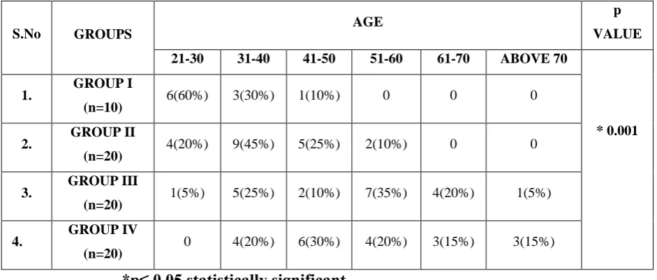
The cases were divided into six groups based on age. The groups were 21- 30 years, 31-40 years, 41-50 years, 51-60 years, 61-70 years and above 70. In Group I out of 10 cases 6(60%) belonged to 21-30 yerars, 3(30%) belonged to 31- 40 years and one (10%) belonged to 41-50 years. In Group II out of 20 cases 4(20%) belonged to 21-30 years, 9(45%) belonged to 31-40 years, 5(25%) belonged to 41-50 years, and 2 cases (10%) belonged to 51-60 years. In Group III out of 20 cases one (5%) belonged to 21-30 years, 5(25%) belonged to 31-40 years, 2(10%) belonged to 41-50 years, 7(35%) belonged to 51-60 years, 4(20%) belonged to 61-70 years and 1(5%) belonged to above 70 years. In Group IV out of 20 cases 4(20%) belonged to 31-40 years, 6(30%) belonged to 41-50 years, 4(20%) belonged to 51- 60 years, 3(15%) belonged to 61-70 years and 3(15%) belonged to above 70 years. There was a significant difference between the groups with respect to age (p = 0.001).(Table 1, Graph 1)
Distribution of Gender among Total Study Groups:
47
The was a significant difference between the groups with respect to gender (p = 0.455). (Table 2, Graph 2).
Distribution of Site among Total Study Groups:
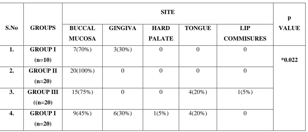
In Group I, 7(70%) incisional biopsies were from buccal mucosa and 3(30%) were from gingiva. In Group II, all the 20(100%) cases were from buccal mucosa. In Group III, 15(75%) were from buccal mucosa, 4(20%) were from tongue and one (5%) was from lip commissures. In Group IV, 9(45%) were from the buccal mucosa, 6(30%) were from gingiva, one (5%) was from hard palate and 4(20%) were from tongue. There was a significant difference between the groups with respect to site (p = 0.022). (Table 3, Graph 3)
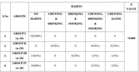
Distribution of Habits among Total Study Groups:
48
DISTRIBUTION OF p16 STAINING AMONG STUDY GROUPS:
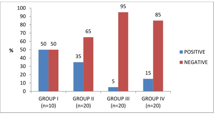
Of the total number of cases subjected to p16 staining, 16(22.9%) cases showed positive staining and 54(77.1%) of the cases showed negative staining. In Group I, out of 10 cases 5(50%) showed positivity and 5(50%) showed negative staining. In Group II, out of 20 cases only 7(35%) were positive for p16 expression and 13(65%) of the cases showed negative staining. In Group III, out of 20 cases only one (5%) showed positivity and 19(95%) cases showed negative staining. In Group IV, out of 20 cases only 3(15%) showed positivity and 17(85%) of the cases showed negative staining. There was a significant difference between the groups with respect to p16 positivity (p = 0.017). (Table 5, Graph 5) The following parameters were used to evaluate p16 staining in all the 4 groups:
- Staining intensity - Staining pattern
- Percentage of cells stained - Tissue localisation of the stain
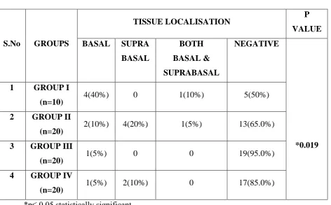
Tissue Localisation of the Stain:
49
layers of the epithelium. In Group IV 1(5%) showed staining in the basal layers of the epithelium and 2(10%) showed staining in the suprabasal layers of the epithelium. There was a significant difference between the groups with respect to staining pattern (p = 0.019). (Table 6, Graph 6)
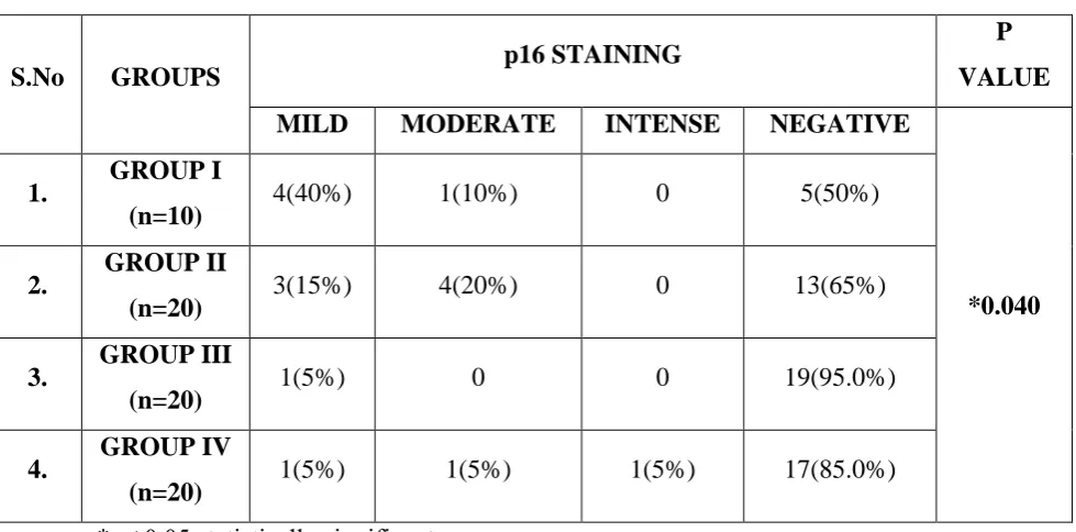
Staining Intensity:
Each of the positively stained cases were graded as mild, moderate and intense based on the intensity of p16 immunostaining. Among the 4 groups, 9(12.9%) cases had mild intensity, 6(8.6%) cases had moderate intensity and 1(1.4%) case showed intense staining.
In Group I, 4(40%) showed mild intensity and 1(10%) showed moderate intensity of p16 stain. In group II, 3(15%) showed mild intensity, 4(20%) showed moderate intensity. In Group III, 1(5%) showed mild intensity. In Group IV, one (5%) showed mild intensity, one (5%) showed moderate intensity and one (5%) showed intense staining. There was a significant difference between the groups with respect to staining intensity (p = 0.040). (Table 7, Graph 7)
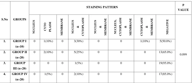
Staining Pattern:
p16 staining within each cell was localised to nucleus or cytoplasm or membrane or in combinations between them. Out of 16 cases, 4(5.7%) cases revealed cytoplasmic staining. 11(15.7%) revealed staining in both nucleus and cytoplasm and one (1.4%) showed staining in both cytoplasm and membrane.
50
staining in both cytoplasm and membrane. In Group II, 2(10%) cases showed staining in cytoplasm and 5(25%) cases showed staining in both nucleus and cytoplasm. In Group III, only one (5%) case showed staining in nucleus and cytoplasm. In Group IV, one (5%) case showed staining in cytoplasm and 2(10%) cases showed staining in nucleus and cytoplasm. There was no significant difference between the groups with respect to staining pattern (p = 0.099). (Table 8, Graph 8)
Nuclear and Cytoplasmic Staining Grading:
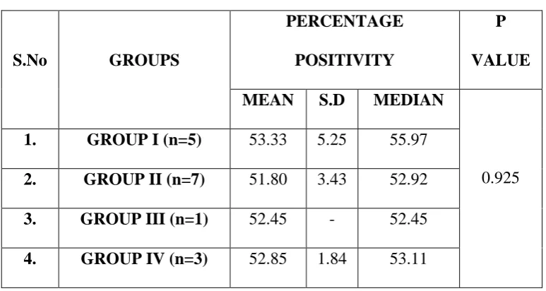
Out of 5 cases in Group I 5 cases had negative cells per 100 cells examined and 2 cases had 1% to 25% of positive cells, 2 cases had 26% to 50% of positive cells and one case had 51% to 75% of positive cells. In group II out of 20 cases 13 cases had negative cells per 100 cells examined, 3 cases had 1% to 25% of positive cells and 4 cases had 26% yo 50% of positive cells. In Group III 19 cases had negative cells per 100 cells and one case had 1% to 26% of positive cells. In Group IV17 cases had negative cells per 100 cells examined, 2 cases had 1% to 25% of positive cells and one case had 76% to 100% of positive cells. (Table 9)
Staining Intensity Comparison between I And Group II:
51
Staining Intensity Comparison between Group II And Group IV:
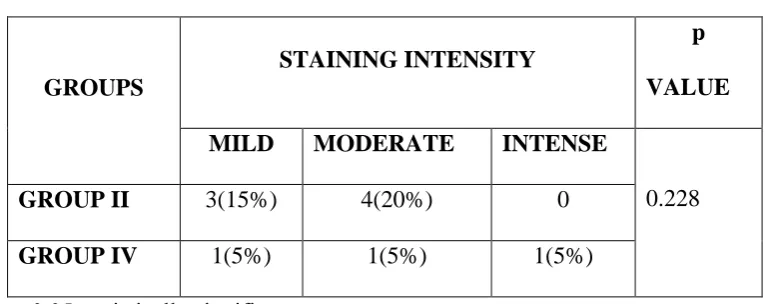
Out of 7 cases in Group II, 3(15%) showed mild intensity and 4(20%) showed moderate intensity. Out of 3 cases in Group IV one (5%) showed mild intensity, one (5%) showed moderate intensity and one (5%) showed intense staining. There was no significant difference between the groups with respect to staining pattern (p = 0.228). (Table 11)
Staining Intensity Comparison between Group I And Group IV:
Out of 5 cases in Group I, 4(40%) showed mild intensity and one (10%) showed moderate intensity. Out of 3 cases in Group IV, one (5%) showed mild intensity, one (5%) showed moderate intensity and one (5%) showed intense staining. There was no significant difference between the groups with respect to staining pattern (p = 0.080). (Table 12)
Staining Pattern Comparison between Group I And Group II:
Out of 5 cases of Group I, one (10%) showed cytoplasmic staining, 3(30%) cases showed both nuclear and cytoplasmic stainingand one (10%) showed staining in cytoplasm and membrane. Out of 7 cases of Group II, 2(10%) showed cytoplasmic staining and 5(25%) staining in nucleus and cytoplasm. There was no significant difference between the groups with respect to staining pattern (p = 0.510). (Table 16)
Staining Pattern Comparison between Group II And Group IV:
52
in cytoplasm, two (10%) showed nuclear and cytoplasmic staining. There was no significant difference between the groups with respect to staining pattern (p = 0.341). (Table 17)
Staining Pattern Comparison between Group I And Group IV:
TABLES AND GRAPHS
TABLE 1: Age Distribution Among Total Study Groups
*p≤ 0.05 statistically significant
GRAPH 1: Age Distribution Among Total Study Groups
GROUP I – NORMAL MUCOSA GROUP II – OSF
GROUP III – EPITHELIAL DYSPLASIA
GROUP IV – ORAL SQUAMOUS CELL CARCINOMA 60
30
10
0 0 0
20 45 25 10 5 25 10 35 20 5 0 20 30 20
15 15
0 10 20 30 40 50 60 70 80 90 100
21-30 31-40 41-50 51-60 61-70 ABOVE
70 %
GROUP I (n=10) GROUP II(n=20) GROUP III (n=20) GROUP IV(n=20)
S.No GROUPS AGE
p
VALUE
21-30 31-40 41-50 51-60 61-70 ABOVE 70
* 0.001 1. GROUP I
(n=10) 6(60%) 3(30%) 1(10%) 0 0 0
2. GROUP II
(n=20) 4(20%) 9(45%) 5(25%) 2(10%) 0 0
3. GROUP III
(n=20) 1(5%) 5(25%) 2(10%) 7(35%) 4(20%) 1(5%)
4. GROUP IV
TABLE 2: Gender Distribution Among Total Study Groups
S.No GROUPS
GENDER p
VALUE
MALE EMALE
1. GROUP I (n=10) 8(80%) 2(20%)
0.455 2. GROUP II (n=20) 19(95%) 1(5%)
3. GROUP III (n=20) 16(80%) 4(20%) 4. GROUP IV (n=20) 18(90%) 2(10%) *p≤ 0.05 statistically significant
GRAPH 2: Gender Distribution Among Total Study Groups
GROUP I – NORMAL MUCOSA GROUP II – OSF
GROUP III – EPITHELIAL DYSPLASIA
GROUP IV – ORAL SQUAMOUS CELL CARCINOMA 80 95 80 90 20 5 20 10 0 10 20 30 40 50 60 70 80 90 100
GROUP I (n=10) GROUP II (n=20) GROUP III (n=20) GROUP IV (n=20)
% MALE
TABLE 3: Site Distribution Among Total Study Groups
S.No GROUPS
SITE
p
BUCCAL
MUCOSA
GINGIVA HARD
PALATE
TONGUE LIP
COMMISURES
VALUE
1. GROUP I
(n=10)
7(70%) 3(30%) 0 0 0
*0.022
2. GROUP II
(n=20)
20(100%) 0 0 0 0
3. GROUP III
((n=20)
15(75%) 0 0 4(20%) 1(5%)
4. GROUP I
(n=20)
9(45%) 6(30%) 1(5%) 4(20%) 0
*p≤ 0.05 statistically significant
GRAPH 3: Site Distribution Among Total Study Groups
GROUP I – NORMAL MUCOSA GROUP II – OSF
GROUP III – EPITHELIAL DYSPLASIA
GROUP IV – ORAL SQUAMOUS CELL CARCINOMA 70 30 100 75 0 20 5 45 30 5 20 0 10 20 30 40 50 60 70 80 90 100
% GROUP I (n=10)
TABLE 4: Habit Distribution Among Total Study Groups
S.No GROUPS
HABITS p
VALUE NO HABITS CHEWING & DRINKING DRINKING & SMOKING CHEWING, DRINKING & SMOKING CHEWING ALONE *0.000 1. GROUP I
(n=10) 10(100%) 0 0 0 0
2. GROUP II
(n=20) 0 6(30%) 0 9(45%) 5(25%)
3. GROUP III
(n=20) 13(65%) 0 5(25%) 1(5%) 1(5%)
4. GROUP IV
(n=20) 16(80%) 0 0 3(15%) 1(5%)
*p≤ 0.05 statistically significant
GRAPH 4: Habit Distribution Among Total Study Groups
GROUP I – NORMAL MUCOSA GROUP II – OSF
GROUP III – EPITHELIAL DYSPLASIA
GROUP IV – ORAL SQUAMOUS CELL CARCINOMA 100 30 45 20 65 25
5 5
80 15 5 0 10 20 30 40 50 60 70 80 90 100
NO HABITS CHEWING & DRINKING DRINKING & SMOKING CHEWING, DRINKING & SMOKING CHEWING ALONE
% GROUP I (n=10)
TABLE 5: Distribution Of p16 Staining Among Total Study Groups
S.No DIAGNOSIS
p16 STAINING p
VALUE POSITIVE NEGATIVE
1. GROUP I (n=10) 5(50%) 5(50%)
*0.017 2. GROUP II (n=20) 7(35%) 13(65%)
3. GROUP III (n=20) 1(5%) 19(95%) 4. GROUP IV (n=20) 3(15%) 17(85%) *p≤ 0.05 statistically significant
GRAPH 5: Distribution Of p16 Staining Among Total Study Groups
GROUP I – NORMAL MUCOSA GROUP II – OSF
GROUP III – EPITHELIAL DYSPLASIA
TABLE 6: Tissue Localisation In p16 Positive Study Groups
S.No GROUPS
TISSUE LOCALISATION P
VALUE
BASAL SUPRA
BASAL
BOTH
BASAL &
SUPRABASAL
NEGATIVE
*0.019 1 GROUP I
(n=10) 4(40%) 0 1(10%) 5(50%)
2 GROUP II
(n=20) 2(10%) 4(20%) 1(5%) 13(65.0%) 3 GROUP III
(n=20) 1(5%) 0 0 19(95.0%)
4 GROUP IV
(n=20) 1(5%) 2(10%) 0 17(85.0%)
*p≤ 0.05 statistically significant
GROUP I – NORMAL MUCOSA GROUP II – OSF
GROUP III – EPITHELIAL DYSPLASIA
GRAPH 6: Tissue Localisation In p16 Positive Study Groups
GROUP I – NORMAL MUCOSA GROUP II – OSF
GROUP III – EPITHELIAL DYSPLASIA
GROUP IV – ORAL SQUAMOUS CELL CARCINOMA 40 0 10 50 10 20 5 65 5
0 0
95
5 10
0 85 0 10 20 30 40 50 60 70 80 90 100
BASAL SUPRABASAL BOTH BASAL
AND SUPRABASAL
NEGATIVE
% GROUP I (n=10)