1
The Tamil Nadu
Dr M.G.R. Medical University
Chennai
A Study on clinical and pathological aspects of
Parotid Gland Tumours
Dissertation submitted for
M.S. Degree (General Surgery)
2
DEPARTMENT OF SURGERY,
MADURAI MEDICAL COLLEGE AND GOVERNMENT RAJAJI HOSPITAL, MADURAI.
CERTIFICATE
This is to certify that the dissertation entitled “A STUDY ON CLINICAL AND PATHOLOGICAL ASPECTS OF PAROTID
GLAND TUMOURS is a bonafide record of work done by Dr.R.RENGANATHAN., in the Department of Surgery, Government Rajaji Hospital, Madurai Medical College, Madurai., under the direct guidance of me.
Professor of Surgery Professor and HOD
and unit chief Department of surgery, Madurai Medical College and
Government Rajaji Hospital, Madurai.
DEAN
3
DECLARATION
I, Dr.R.Renganathan, solemnly declare that the dissertation titled “A study on clinical and pathological aspects of Parotid Gland Tumours” has been prepared by me.
This is submitted to The Tamil Nadu Dr. M.G.R. Medical University, Chennai, in partial fulfillment of the rules and regulations for the award of MS degree General Surgery.
Place: Madurai Dr.R.Renganathan.
4
ACKNOWLEDGEMENT
I owe my sincere and profound gratitude to my unit chief
Prof. Dr. K.V. MAHESWARAN, M.S., Professor of Surgery,
Madurai Medical College , for his constant guidance and objective direction to my work on this subject.
I thank Prof. Dr. S.M. SIVAKIMAR, M.S., DEAN, Madurai Medical College and Govt. Rajaji Hospital, Madurai, for allowing me to utilize the hospital materials for this study.
My sincere thanks to assistant professors Dr.S.Selva Chithambaram, M.S., Dr.C.Balasubramaniam, M.S., Dr.Subbiah
Chandhirasekarji, M.S., Dr.S.Babu,M.S., Dr.Celine Foustina
Mary,M.S., and to all staffs, Madurai Medical College and Govt., Rajaji Hospital,who helped me to make this study a success.
5
CONTENTS
1. INTRODUCTION 1
2. REVIEW OF ANATOMY AND PATHOPHYSIOLOGY 3
3. AIM OF STUDY 35
4. MATERIALS AND METHODS 36
5. PROFORMA 37
6. OBSERVATIONS 42
7. DISCUSSION 54
8. SUMMARY 58
9. CONCLUSSION 60
10.BIBLIOGRAPHY 62
6
INTRODUCTION
Tumours of the salivary glands form a fascinating subject for the head and neck surgeons. The relative infrequency combined with considerable histologic and Behavioural diversity and their regional anatomic relationship make them unusually interesting and challenging. The parotid tumours are more so because of their intimate relationship to the facial nerve, the presence of intra and paraparotid lymph nodes, and presence of a lobe.
Salivary gland tumours comprise less than 3 % of all neoplasms of head and neck and no more than 1 % of all neoplasms. Approximately 80-85% of all salivary gland neoplasm occurs in the parotid gland. Although 80% of these are benign, earlier reports indicated a high recurrence rate after limited resection. This has led to a more aggressive approach to the treatment of these lesions in the recent years in the form of either subtotal or total conservative parotidectomy with excellent results.
7
8
REVIEW OF ANATOMY AND PATHOPHYSIOLOGY
DEVELOPMENT OF PAROTID
The parotid gland arises from the epithelial of the mouth. The frequent finding of sebaceous glands in the parotid suggests an ectodermal origin. Parotid glands are the first salivary glands to appear. When the embryo is about 4-6 weeks of development, parotid appears as an epithelial ingrowth near the angle of the mouth on inner surface of either cheeks. As it grows backwards, towards the ramus of the mandible, it becomes a hollowed tube at this level, branches intensively into the primodial ducts and acinar cells. The tube persists as the parotid duct. Condensation of the mesenchyme surrounding the developing parotid gland occurs, later in embryonic life. So lymphnodes may become entrapped in the gland. The origin of myoephithelial cells is unknown.
SURGICAL ANATOMY
9
usually more or less detached, lies between the zygomatic arch above and the parotid duct below. This is named the accessory parotid.
The investing layer of deep cervical fascia splits between the angle of the mandible and mastoid to enclose the parotid gland.
The parotid gland is like an inverted, flattened, three sided pyramid. The surfaces are a small superior surface, super antromedial and posteromedial surface. The lower part of the gland tapers to form a blunt apex. The superior surface is concave and is related to the external acoustic meatus and to the posterior surface of temporomandibular joint. Superficial temporal vessels and auriculotemporal nerve emerge out from the gland at this surface.
The superficial surface is covered with skin, superficial fascia, parotid fascia which is thick and adherent to the gland and a few deep parotid lymph nodes embedded in the gland.
10
The posteromedial surface is moulded to the mastoid and the styloid process. Thus it is related to the sternomastoid, posterior belly of digastric, stylohyoid muscle and ligament, stylopharyngeus and the stylomandibular ligament. The external carotid artery grooves this surface before it enters the gland. The anteromedial and the posteromedial surfaces meet along a medial margin which may be in contact with the side wall of the pharynx.
The apex of the gland extends into the carotid triangle. The cervical branch of the facial nerve and the two divisions of the retromandibular vein emerge through it.
The parotid duct is 5 cm long. It crosses the masseter and at the anterior border of this muscle, it turns inwards, pierces the buccinator, runs deep to the mucosa and opens into the oral cavity opposite the upper second molar tooth.
11
The external carotid artery the gland through its posteromedial surface and divides into its terminal branches.
The facial nerve is associated with the parotid gland for an important part of its course during which it divides into its external branches. After it exits the stylomastoid foramen, it passes forwards around the neck of the condyle of the mandible and enters the parotid gland, passing superficial to the external carotid artery and the posterior facial vein. At this stage, the nerve usually divides into two main divisions the temperofacial and cervicofacial, these in turn subdivide into at least five main branches-temporal, zygomatic, buccal, mandibular and cervical.
In 70% of cases, anastomotic connections between the two main divisions exist. Anastomotic between the terminal branches of three temperofacial divisions are frequent, anastomosis between the two branches of the cervicofacial is likely to result in permanent paralysis than injury to the cervicofacial division.
SURFACE ANATOMY
12
condyle to a joint just above the middle of the massester and then downwards and backwards to a point about 2 cm below and behind the angle to the mandible. Its upper border, concave upwards and backwards corresponds to a curved line drawn from the upper border of the mandibular condyle across the lobule straight line joining the ends of the anterior and upper borders.
The parotid duct corresponding to-the middle third of the line drawn from the lower border of the tragus to a point midway between the ala of the nose and the red margin of the upper lip.
BLOOD SUPPLY
13
LYMPH DRAINAGE
Lymph drains to the intra and extra glandular nodes and then along the external carotid artery to the jugulodigatric and other glands of the anterior superior group of upper deep cervical nodes.
COMPARTMENTS OF THE GLANDS
The parotid may be considered to have 3 compartments. The superficial nerve compartment contains greater auricular nerve, auriculotemporal nerve and facial nerve. The middle venous compartment contains the superficial temporal vein joining the internal maxillary vein to form the posterior facial vein which joins the post auricular vein to form the external jugular vein. The deep arterial compartment contains the external carotid artery, internal maxillary artery and superficial temporal artery.
STRUCTURE OF THE GLAND
15
PHYSIOLOGY OF THE PAROTID GLAND
The total quantity of saliva produced during a 24hours period is about 1000-1500ml. About 90% of this fluid is derived from the parotid and submandibular glands in more or less equal amounts.
Parotid gland secretion is controlled by physical and psychic stimulation via the parasympathetic system. Afferent impulses are relayed to the superior and inferior salivatory nucleus with the tympanic branch of the 9th cranial nerve to the tympanic plexus (Jacobson’s nerve). From here the lesser superficial petrosal nerve carries impulses to the otic ganglion. Post ganglion fibres reach the parotid via the auriculotemporal nerve. Sympathetic fibres leave the ventral roots of the upper three thoracic ganglions. Post ganglionic fibres reach the parotid via the carotid plexus, traveling with the arterial supply to the gland.
PAROTID NEOPLASMS
Incidence
16
AETIOLOGY
There is no evidence either clinical or experimental to suggest that in the salivary glands a pre-existing inflammatory, obstructive or traumatic condition predisposes to malignant change. In experimental animals parotid tumours can be induced by carcinogenic hydrocarbons, ionizing radiations and viruses. Vitamin D deficiency may be important.
PATHOGENESIS
An origin of salivary gland tumours from epithelial cells appears to be the current majority view. Some workers have postulated that the cell of origin may be individual cell types like the myoephithelial cells. Various tumours may arise from either intercalated or excretory ducts and this theory is based upon salivary gland embryogenesis and the potential duct epithelium during reactive process.
17
CLASSIFICATION
The difficulty of histopathological interpretation, the presence of a wide range of histopathological patterns and the variable biological behaviour of salivary gland tumours led to a profusion of classification none of which have been universally accepted until the international classification of salivary tumours was formulated in 1972.
Some of the important classifications are:
I. CLASSIFICATION BY F.W.FOOTE AND E.L.FRAZELL
1. Mixed tumour
a) Benign mixed tumour b) Malignant mixed tumour
2. Mucoepidermoid tumours
a) Low grade tumours b) High grade tumours
3. Squamous cells carcinoma
4. adeno carcinoma
a) Adenoid cystic
b) Miscellaneous-Trabecular of solid, or anaplastic mucous cells
18
5. Papillary cystadenoma lymphomatosum
a) Oxphil adenoma
b) Sebaceous cell adenoma
c) Benign lymphoepithelial lesions
d) Unclassified tumours-Benign and malignant II. CLASSIFICATION BY DAVID H.PATEY (1969)
A.PRIMARY
1. Epithelial
a) Mixed tumours
b) Mucoepidermoid tumour c) Cylindroma (adenoid cystic)
d) Carcinoma – squamous cell carcinoma - Adeno carcinoma
- Undifferentiated carcinoma
e) Aden lymphoma (papillary cystadenoma lymphomatosum) f) Oxphil cell carcinoma
2. Connective tissue
a) Haemangioma
19
B.SECONDARY
From various sources such as breast, bronchus, malignant melanoma etc.,
III. W.H.O. CLASSIFICATION OF SALIVARY GLAND
TUMOURS (THACKRAY AND SOBIN 1972)
1. EPITHELIAL TUMOURS
A. Adenomas
1. Pleomorphic adenoma (mixed tumour)
2. Monomorphic adenoma. a) Aden lymphoma
b) Oxphilic adenoma
c) Others
B. Mucoepidermoid tumours
C. Acinic cell tumours
D. Carcinoma
1.Adenoid cystic carcinoma 2. Adeno carcinoma
3. Undifferentiated carcinoma 4. Epidermoid carcinoma
20
II. NON EPITHELIAL TUMOURS.
III. UNCLASSIFIED TUMOURS.
IV. ALLIED CONDITIONS.
a. Benign lympho-epithelial lesions b. Sialosis
c. Oncocytosis
PLEOMORPHIC ADENOMA (Mixed tumour)
It is most common tumour of the salivary gland. It is of epithelial origin and benign. The term pleomorphic was coined by Willis in 1967. it forms 60-70% of all parotid tumours and 80% of all benign tumours. It is usually unilateral. It is purely of epithelial origin. Material in chondromyxoid areas has been considered to be a modified type of mucin, made up of chondroitin, a mucopolysaccaride elaborated by the myoephithelial cells. The maximum incidence is seen in the 5th decade and sex distribution is equal.
Gross pathology
21
and varies from firm to soft. The tumour expands by focal growth producing nodular extensions into the capsule. Operative enucleation of the tumour is frequently responsible for leaving satellite nodules this accounts for frequency and multiplicity of recurrent nodules after such operations.
Microscopic pathology
A mixed tumour consists of epithelial and mesenchyme like elements. In the epithelial component, the cells may be arranged in sheets or lie sparsely in a mucoid of chondromyxomatous matrix. In the mucoid and myxoid areas, myoephithelial cells are present. The cartilaginous areas may calcify or ossify. 10% tumours are highly cellular and such tumours are liable to recur. The tumour is relatively radio resistant and careful local excision with a margin of normal tissue is the treatment of choice.
22
2. MONOMORPHIC ADENOMAS
These benign tumours are characterized by the regularity of their cell structure and pattern
a) ADENOLYMPHOMA (Warthin’s tumour)
This is the second most benign tumour of the parotid gland. The Aden lymphoma is a distinctive tumour which arises almost constantly in relation to the parotid gland in a superficial location, especially at the lower pole. This benign tumour is believed to arise from heterotypic gland tissue which has become enclosed developmentally in a lymph node. It is the only functional tumour arising from parotid gland.
The lesion appears as a slow growing soft tissue mass in males over the age of 40 years. Commonly the lesion arises multifocally and a striking clinical feature is that bilateral gland involvement occurs in 7 to 10% of cases.
Gross Pathology
23
Microscopic Pathology
Histologically, the tumour is composed of glandular and often cystic structures having a papillary cystic pattern lined by columnar cells. A variable amount of lymphoid tissue with follicle formation characterizes the stroma.
Aden lymphoma concentrates 99 TC predestinate at much greater degree than normal salivary tissue.
b)Oxphil Adenoma (Oncocytoma)
This tumour is very uncommon in the parotid. They are round or ovoid, encapsulated, solid tumours of slow growth which usually occurs in a higher age group. The tumour cells resemble oncocytes which are cell derives from the duct epithelium. This tumour has a predilection for older females.
c) Basal Cell Adenoma
24
d) Clear Cell Adenoma
This is also a rare monomorphic adenoma. In this tumour, duct like structures surrounded by clear cells in the position normally occupied by myoephithelial cells is seen.
B. MUCOEPIDERMOID TUMOUR
Mucoepidermoid tumours form 2-5% of all salivary tumours out of which 70-90% occur in the parotid tumours and 25% of malignant parotid tumours. It is commonly seen in the 3rd to 5th decade with an equal withy an equal sex incidence. It is the commonest salivary gland tumour in childhood. It is slow growing and not encapsulated.
Gross Pathology
These tumours are ovoid and well circumscribed. Infiltration may be evident with ulceration of the overlying surface. These tumour always show an attachment to fascia. The cut surface shows solid elements and cyst filled with a viscid mucin.
Microscopic Pathology
25
Mucoepidermoid tumours infiltrate locally and though they may metastasise, this ability is limited.
C. ACINIC CELL TUMOUR
This tumour constitutes about 1% of all salivary gland tumours and accounts for 2-4% of all parotid tumours. Similar to warthin’s tumour, it may be bilateral. The peak incidence is in the 5th decade. These are solitary lesions, compact, hard and knobby and mobile.
Gross Pathology
This tumour is apparently encapsulated, hard and presents a most grey white cut surface flecked with brown areas. Small cyst containing serous and blood stained fluid may also be found.
Microscopic Pathology
26
A 5 year survival rate of around 90% makes it a much more benign tumour than Mucoepidermoid tumour. Rarely the tumour may infiltrate locally or metastasis.
D. CARCINOMAS
Malignant diseases of the salivary glands are rare. Half of them occur in the parotid. About 25% of parotid tumour are malignant.
The natural course of many of these neoplasms was characterised by long duration, repeated local recurrences, occasional metastasis to the regional lymph nodes and lungs. All these neoplasms tend to present at an early stage to produce obvious clinical signs of malignancy.
1. Adenoid Cystic Carcinoma
This tumour is relatively rare in the parotid gland. It forms 2% of parotid tumours. The peak incidence is in the 6th decade (18%). The incidence of lymph node metastasis from direct or contiguous invasion rather than emboli is low (15%).
Gross Pathology
27
MICROSCOPIC PATHOLOGY
This tumour is composed of basal type epithelial cells and of a stroma which is variably hyaline. The epithelial cells may be arranged in solid, granular, cribriform or cystic masses. The adenoid cystic carcinoma is isomorphic lacking the marked and varied stromal changes so characteristic of pleomorphic adenoma. Adenoid cystic carcinoma metastasizes distantly especially to the lungs.
2. ADENOCARCINOMA
Aden carcinoma form 3% of parotid tumours. There is 23% of incidence of preoperative facial paralysis. They are all highly malignant tumours that metastasis widely. Their prognosis is determined by the grade and stage of the tumours rather than by the extent of surgery. Local recurrence of this most lethal tumour exceeds 50%.
3. SQUAMOUS CELL CARCINOMA
28
Gross Pathology
This tumour consists of epithelial cells which form keratin or exhibit intercellular bridges. It arises from the ductal system and the prognosis is grave.
4. UNDIFFERENTIATED CARCINOMA
These are poorly differentiated malignant tumours of epithelial origin. These include the solid undifferentiated carcinoma, Trabecular carcinoma and salivary duct carcinoma. The presence of epithelial and myoephithelial cells in these tumours have been shown by electron microscopy.
5. CARCINOMA IN PLEOMORPHIC ADENOMA
This tumour is one in which there is areas of invasive growth and cellular atypia together with histological areas of typical pleomorphic adenoma.
29
II NON EPITHELIAL TUMOURS
Non epithelial stromal tumours of the parotid are rare. They include haemangioma, lymphangioma, nuerofibroma, lipoma, sarcoma, metastatic tumours and lymphoma. Junaid et. al., has reported a rare incidence of dermatofibrosarcoma protruberans of the parotid gland.
CLINICAL FEATURES:
Tumours of the parotid gland often present with painless or asymptomatic swelling. Usually the mass is situated behind the angle of the jaw or overlying the ramus of the mandible, below and in front of the ear, often lifting the tragus and the ear lobe.
30
Normal histology Pleomorpic adenoma
Warthin s Tumour Oncocytoma
Basal cell adenoma Mucoepidermoid
31
Adeno carcinoma
Malignant pleomorpic
Squamous cell carcinoma
32
STAGING OF PAROTID GLAND CANCERS
The American joint committee staging of salivary gland tumours is as follows:
PRIMARY TUMOUR
Tx - Primary tumour cannot be assessed
T0 - No evidence of primary tumour.
T1 - Tumour less than 2 cms in greatest dimension without
extraparenchymal extension
T2- Tumour >2 CM<4cm in greatest dimension without
extraparenchymal extension
T3- Tumour more than 4 cm and / or tumour having
extraparenchymal extension
T4a - Tumour invades skin, mandible, ear canal,and/or facial nerve
T4b - Tumour invades skull base and / or pterygoid plates and/or
encases carotid artery.
NODAL INVOLVEMENT
Nx - Regional lymph nodes cannot be assessed
N0 - No regional lymph node metastasis
33
N2a - Metastasis in single ipsilateral LN, > 3 cm but < 6 cm N2b- Metastasis in multiple ipsilateral LN, none> 6 cm N2c- Metastasis in bilateral or contralateral LN, none > 6 cm N3 – Metastasis in a lymph node, more than 6 cm
METASTASIS
MX - Distant metastasis cannot be assessed
M0 - No distant metastasis
M1 - Distant metastasis to lungs, bone etc.,
STAGE GROUPING T N M
STAGE I T1 N0 M0
STAGE II T2 N0 M0
STAGE III T3 N1 M0
T1 N1 M0
T2 N1 M0
T3 NO M0
STAGE IVA T4a N1 M0
T4a N2 M0
T1 N2 M0
T2 N2 M0
T3 N2 M0
T4a N2 M0
STAGE IVB Tb Any N M0
Any T N3 M0
34
INVESTIGATIONS
A number of diagnostic tests are available. They are,
1. Sialography
It is done by injecting 1-2cc of a various contrast medium (Lipoidol), over distension is avoided by stopping the injection when the patient complains of pain. Sialography fails to detect parotid neoplasms less than 1cm in diameter. It can however give an idea of deep lobe involvement. Benign lesions displace the ductal system whereas malignant lesions may infiltrate it causing ductal obstruction.
2. Radiosialography
For Radiosialography 1 Mci of 99 TcM labeled sodium pertechtinate is injected intravenously and the parotids are scanned using a gamma camera. All parotid neoplasms other than Warthin’s tumour and oncocytoma are radionegative.
3. Ultrasonography
35
4. Computed Tomography (CT) and MRI
CT and MRI is currently the radiographic study favoured in the investigations of parotid neoplasms. CT with IV contrast is useful in separating intrinsic parotid masses from paraparotid masses as well as differentiating deep lobe masses from other parapharyngeal space masses. In addition during the same scan axial sections of the neck may be taken to investigative metastatic adenopathy. It is also useful in detecting distant metastasis.
5. Angiography
This is useful in tumours of the parapharyngeal in order to differentiate salivary gland tumour from chemodectoma of the cartotid sheath or a neoplasm of the nerve shealth, both of which have a characteristic tumour circulation.
6. Fine Needle Aspiration Biopsy (FNAC)
36
especially accurate in pleomorphic adenoma. This information obtained from this test can be extremely valuable in planning the operative procedure and in information the patient of the relative likelihood of having to sacrifice all or part of the facial nerve.
7. Incisional Biopsy
Incisional biopsy of the parotid is not without risk. Damage to important neurovascular structures, the creation of salivary fistulas and the contamination of tissue planes by neoplastic cells are the serious risks involved, furthermore, fibrosis at the site of biopsy can make later surgery difficult. On no account should a discrete parotid mass be subjected to Incisional biopsy since there is a 9 cut off 10 chances that the mass is a pleomorphic adenoma, incising which will almost certainly lead to a late recurrence.
8. Frozen section biopsy
37
9. Needle biopsy
Needle biopsy using tru-cut or vim silvermann needle can be done under local analgesia, but carries all he risks of Incisional biopsy through of lesser degree. Some authors claim very little spillage along the needle tract.
DIFFERENTIAL DIAGNOSIS OF PAROTID TUMOURS
Some of the rarities that mimic parotid swellings are:
Hypertrophic masseter
Winged mandible (in the 1st arch syndrome)
Dental cysts
Branchial cysts
Myxoma of the masseter
Mandibular tumours
Lymphadenitis of the preauricular lymph node
Secondaries of neck.
TREATMENT
TREATMENT OF BENIGN TUMOURS
38
and so it was followed by enucleation and post operative irradiation. But this carried on with it risk of radiation induced cancer in the young. So removal of the growth with a good cuff of normal parotid tissue was practiced later. However this resulted in a facial weakness in some cases. A superficial conservative parotidectomy became the accepted surgery for benign lesions confined to the superficial lobe. For lesions in the deep lobe, a total parotidectomy with conservation of the facial nerve is advisable. In all cases, the patent must be informed preoperatively of the possibility of injury to the nerve and should give his knowledge consent for the treatment.
TREATMEN OF MALIGNANT LESIONS
TREATMENT GROUP
TUMOUR TYPE TREATMENT
I T1&T2 low grade
Mucoepidermoid& acinic cell tumour
Superficial / total conservative parotid dectomy
II T1&T2 low grade
Mucoepidermoid& all other tumours
Total parotidectomy (conservative)+ Node dissection for N1
III T3N0 or N1
IV T4
39
SURGERY
PAROTIDECTOMY
Parotidectomy may be conservative when all the main branches of the facial nerve are preserved or radical when the entire trunk and portion of the facial nerve is sacrificed. Depending upon the part removed, it may be superficial, deep or total. Deep parotidectomy is not recommended because of risk of fistula formation, moreover, it is difficult to approach the deep lobe with the superficial lobe in situ .Incision used : lazy S.
The basic operation for parotid neoplasm is the superficial parotidectomy. This procedure will suffice for all superficial lobe benign neoplasms. The structure of paramount importance in a parotidectomy is the facial nerve. The only constant location of the nerve has to be traced from this point. There are other approaches which involve identifying a peripheral branch and following it retrograde to the main trunk. Facial nerve function should be checked immediately prior to skin closure, should be performed. Because of the overlying nerve fibres, any effort to remove all of the deep lobe will necessarily be in piecemeal only.
40
intracranically. The facial nerve should be removed well into the mastoid drilling it out of its bony canal.
NECK DISSECTION
Radial neck dissection should be carried out in the presence of involved nodes and this should be done prophylactically for squamous cell carcinoma and undifferentiated carcinoma. Goonde at all (1960) found it helpful to remove a ‘sentinel node’ located at the junction of the anterior and posterior facial vein for frozen early in the operation. If the node is involved by the tumour enblock neck dissection is performed. However the study conducted in the University of Virginia recommends neck dissection only for palpable lymph node Secondaries.
COMPLICATIONS OF PARATIDECTOMY
1. Reactionary Haemorrhage 2. Facial nerve paresis / paralysis
41
6. Infection 7. Keloid
8. Cosmetic disfigurement 9. Paraesthesia of ear lobule
10.Seeding and spillage of tumour cells and consequent recurrence.
RADIO THERAPY
Unfortunately most salivary neoplasms are relatively radioresistant. However radiotherapy has been useful in reducing locoregional recurrence significantly in patients with stage II and stage III disease. A dose of 5000-6000 rads is given to the parotid bed. It is given within 6 weeks after surgery. Most adenoid cystic carcinoma requires post operative radiotherapy irrespective of their site of origin.
The general indications for radiotherapy are: 1. Residual malignant tumour after surgery.
2. Microscopic malignant tumour at resected margin. 3. Unresectable primary carcinoma
4. Unresectable recurrent malignant tumour 5. Malignant lymphoma
42
Major complications of irradiation include soft tissue ulceration, orocutaneous fistulas and osteoradionecrosis of the mandible.
CHEMOTHERAPY
Recently objective responses with cis-platin or Adriamycin based chemotherapy in some patients with inoperable or metastatic parotid cancer have been observed. Schramm et al., (1981) has shown good response for adenoid cystic carcinoma to cis-platin 80-100mg/m2 at 4-6 weeks intervals with subjective relief of pain and regression of the primary lesion. Responses might be prolonged with addition of radiotherapy.
PROGNOSIS
43
AIM OF STUDY
1. To review the literature of parotid gland tumours in the light of their histologic classification.
2. To analyse the various modes of clinical presentation and their correlation with histopathology.
3. To compare the incidence, clinicopathological presentation and method of treatment of this study with various other reports in the literature.
4. To infer whether a more radical procedure is associated with a greater risk to the facial nerve.
5. To study whether recurrence after a conservative procedure is controllable by further surgery without undue risk to the patient. 6. To study the effectiveness of radiotherapy in the post operative
management of malignant parotid tumours.
44
MATERIALS AND METHODS
54 cases of parotid tumours treated in Govt.Rajaji Hospital, Madurai during the period of 24 months (Oct 2006 to Oct 2008) have been taken for the present study. It has been taken and recorded according to the proforma given below.
The diagnosis of parotid tumour was made with the aid of clinical findings. A special investigation like fine needle aspiration cytology was done in most cases. 43 out 54 cases were subjected to surgical treatment. The confirmation of diagnosis and identification of specific type of tumour was done by histopathological examination of the excised specimen. Radiotherapy was given for appropriate cases following surgery.
45
PAROTID GLAND NEOPLASMS
A CLINICOPATHOLOGICAL ANALYSIS
PROFORMA
SI. NO OP/IP NO. WARD NO.
NAME AGE SEX:
ADDRESS RELIGION
OCCUPATION INCOME
Date of Admission : Date of Operation : Date of Discharge :
Complaints and history of present illness:
1. Swelling in the parotid region a. Duration
b. Unilateral / Bilateral
c. Mode of onset- Acute / insidious d. Progress of symptoms
2. Pain-Present / Absent
46
4. Discharge from stenson’s duct – Present / Absent
If present-nature of discharge- Purulent/Watery-Quantity little/profuse
5. Any other swellings 6. Loss of appetite 7. Loss of weight 8. Abdominal pain 9. Others
PAST HISTORY
1. Mumps 2. Trauma to parotid area 3. Diabetes mellitus 4. Hypertension
5. Jaundice 6. Similar illness 7. Treatment-if any 8. Operations- if any
FAMILY HISTORY
1. Members in family with similar illness
PERSONAL HISTORY
Diet Addictions -smoking, alcohol, tobacco chewing
LOCAL EXAMINATION
Inspection
47
3. Size 4. Shape
5. Diffuse / Localised
6. Surface-Smooth / irregular 7. Skin over the swelling
8. Visible pulsations-present / absent 9. Ear lobe lifted or not
10. Oral cavity, Stenson’s duct orifice 11.Tonsils and oropharynx
12.Stenson’s duct orifice
13.Mandibulo mastoid groove obliterated or not
Palpation
1. Temperature over the swelling- Normal/raised 2. Tenderness-Present/absent
3. Consistency-Soft/firm/hard/variable 4. Mobility-Absent/present/restricted
5. Fixity to the surrounding structures-Present/absent 6. Plane of swelling / Bimanual palpation
7. Pulsation-present/absent 8. Thrill-present/absent
48
Auscultation Bruit-present/absent
B.STENSON’S DUCT:
a. discharge-present/absent
If present-nature-purulent/watery-quantity b. Stones-present/absent
c. Strictures-if any
d. Thickening of the duct-if any C. Fistula if any
D. Movement of the jaw E. Involvement of facial nerve F. Enlargement of lymphnodes G. Other relevant of lymphnodes H. Examination of Oral cavity Oro pharynx
Systematic examination
49
TREATMENT
A)Surgery a. Type b. Findings
c. Post operative period
d. Post operative complications if any
1. Wound infection 2. Fistula
3. Injury to facial nerve 4. Recurrence
5. Any other complications B)Radio therapy
C)Chemotherapy
Post surgical histopathology report
50
OBSERVATIONS
TABLE I
INCIDENCE OF PAROTID TUMOURS DURING THE PERIOD
Type of patients Total number of admissions
Total number of parotid tumours
% of parotid tumours All cases 72113 54 0.0748
TABLE II
INCIDENCE OF EPITHELIAL AND NON-EPITHELIAL TUMOURS OF PAROTID GLAND IN THE STUDY S.NO Type of tumour Number of cases Percentage
1 Epithelial 54 100.0
2 Non epithelial 0 0
Total 54 100.0
TABLE III
INCIDENCE OF BENIGN AND MALIGNANT EPITHELIAL TUMOURS OF THE PAROTID GLAND IN THE STUDY
S.NO Type of tumour Number of cases Percentage
1 Benign 39 72.22
2 Malignant 15 27.77
51
INCIDENCE OF BENIGN AND MALIGNANT EPITHELIAL
TUMOURS OF THE PAROTID GLAND IN THIS STUDY
28%
72%
benign
52
TABLE IV
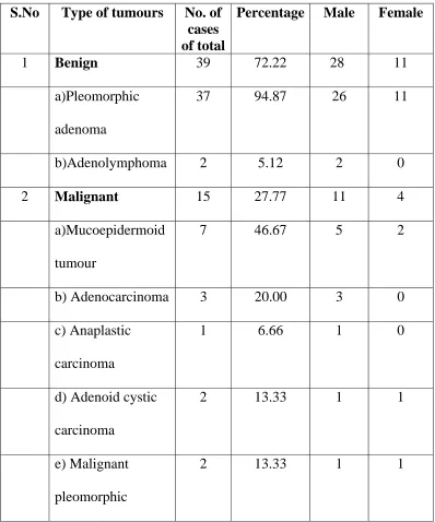
INCIDENCE OF VARIOUS HISTOLOGICAL TYPES OF
EPITHELIAL TUMOURS OF THE PAROTID GLAND IN THE
STUDY
S.No Type of tumours No. of cases of total
Percentage Male Female
1 Benign 39 72.22 28 11
a)Pleomorphic adenoma
37 94.87 26 11
b)Adenolymphoma 2 5.12 2 0
2 Malignant 15 27.77 11 4
a)Mucoepidermoid tumour
7 46.67 5 2
b) Adenocarcinoma 3 20.00 3 0 c) Anaplastic
carcinoma
1 6.66 1 0
d) Adenoid cystic carcinoma
2 13.33 1 1
e) Malignant pleomorphic
53
histological types of parotid gland tumours
pleomorpic adenoma
adenolymphoma
mucoepidermoid
adenocarcinoma
anaplastic ca
adenoid cystic ca
malignant pl.ad
Pleomorpic adenoma 66.66%
Adenolymphoma 3.7%
Mucoepidermoid 11.11%
Adenocarcinoma 5.55%
Anaplastic ca 1.85%
Adenoid cystic ca 3.7%
54
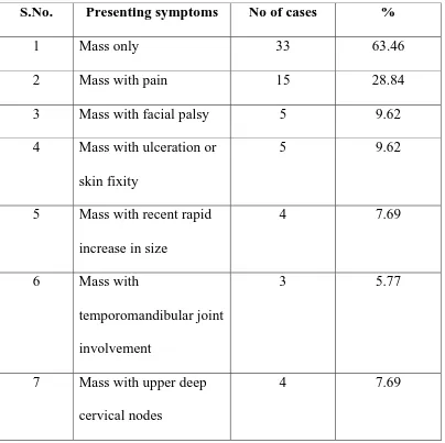
TABLE V
PRESENTING SYMPTOMS AND SIGNS OF 52 PATIENTS WITH
PAROTID GLAND TUMOUR IN THE STUDY
S.No. Presenting symptoms No of cases %
1 Mass only 33 63.46
2 Mass with pain 15 28.84 3 Mass with facial palsy 5 9.62 4 Mass with ulceration or
skin fixity
5 9.62
5 Mass with recent rapid increase in size
4 7.69
6 Mass with
temporomandibular joint involvement
3 5.77
7 Mass with upper deep cervical nodes
4 7.69
55
Among the malignant tumours the pre-operative diagnosis was correct in 10 cases. 2 cases that presented only with a mass were diagnosed as pleomorphic adenoma and FNAC was inconclusive. The histological type-Mucoepidermoid carcinoma was proved by the histopathological report of the surgical specimen.
Pleomorphic adenoma
56
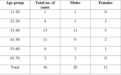
TABLE VI
AGE AND SEX DISTRIBUTION OF 36 CASES OF PLEOMORPHIC ADENOMA
Age group Total no. of cases
Males Females
11-20 1 1 0
21-30 4 1 3
31-40 13 11 5
41-50 11 9 2
51-60 4 3 1
61-70 3 3 0
Total 36 28 11
TABLE VII
PRESENTING SYMPTOMS AND SIGNS OF 37 PATIENTS WITH PLEOMORPHIC ADENOMA IN THE STUDY
S.NO Presenting symptom
and signs
No. of cases Percentage
1 Mass only 30 81.08
[image:56.612.123.524.143.393.2]57
An asymptomatic parotid lump was the most common presenting feature. Most of the lesions were present for 1 to 4 years prior to diagnosis. One patient had involvement of both lobes while in all the rest, only the superficial lobe was involved. The diagnosis was based mainly on clinical examination. FNAC was done in all cases. It offered positive rate 91.89% (34 out of 37 cases).
TABLE VIII
SURGICAL TREATMENT OF 32 CASES OF PLEOMORPHIC
ADENOMA
S.No. Surgery Number of cases Percentage
1 Superficial parotidectomy 29 78.37 2 Total parotidectomy
(conservative)
3 8.19
3 No surgical treatment 5 13.51
58
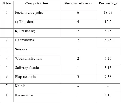
TABLE IX
POST OPERATIVE COMPLICATIONS SEEN IN THE 32
OPERATED CASES
S.No Complication Number of cases Percentage
1 Facial nerve palsy 6 18.75
a) Transient 4 12.5
b) Persisting 2 6.25
2 Haematoma 2 6.25
3 Seroma - -
4 Wound infection 2 6.25
5 Salivary fistula 1 3.13
6 Flap necrosis 3 9.38
7 Keloid - -
8 Recurrence 1 3.13
59
done, presented after 12 months with recurrence of the lesion. He refused any further intervention by way of surgery.
Adenolymphoma (Warthin’s tumour)
In this series, there were two cases of Adenolymphoma. Both were male patients, one 58 years old and the other 62 years old. The duration of symptoms was 6 years and 3 years respectively. Mass was the only presenting symptom. It was soft in consistency and mobile. Both did not have facial palsy. FNAC was done in both cases. The report came as benign epithelial cells. The surgical treatment were as follows:
Superficial parotidectomy-1 Enucleation-1
The patient for whom enucleation was done developed post operative facial palsy.
MALIGNANT TUMOURS
60
TABLE X
THE INCIDENCE OF VARIOUS TYPES OF MALIGNANT
TUMOURS OF THE PAROTID GLAND IN THE STUDY
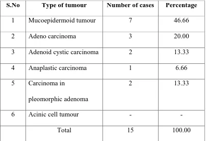
S.No Type of tumour Number of cases Percentage
1 Mucoepidermoid tumour 7 46.66 2 Adeno carcinoma 3 20.00 3 Adenoid cystic carcinoma 2 13.33 4 Anaplastic carcinoma 1 6.66 5 Carcinoma in
pleomorphic adenoma
2 13.33
6 Acinic cell tumour - -
Total 15 100.00
Mucoepidermoid tumour
61
THE INCIDENCE OF VARIOUS TYPES OF MALIGNANT
TUMOURS OF THE PAROTID GLAND IN THIS STUDY
47%
20% 13%
7%
13% 0%
Mucoepidermoid tumour Adeno carcinoma
Adenoid cystic carcinoma Anaplastic carcinoma
62
misdiagnosed as pleomorphic adenoma and a superficial parotidectomy was done. The post operative histological report came as mucoepidermoid carcinoma. These patients were given postoperative radio therapy. For 4 patients total conservative parotidectomy was done, post operative radio therapy was not given to the patients. There has been no recurrences in the short follow up. Two patients developed wound infection in the post operative period. 1 patient left the hospital when adviced surgery.
Adeno carcinoma
63
Anaplastic carcinoma
The incidence of anaplastic carcinoma in this series is 6.66%. A 50 years old male patient presents with a mass, facial nerve involvement, upper cervical nodes, temporomandibular joint involvement and skin fixity. FNAC showed dysplastic epithelial cells. Neoadjuant CT, followed by debunking parotidectomy and post operative radiotherapy was given;
Adenoid cystic carcinoma
The incidence of adenoid cystic carcinoma in this study was 13.33% a 40 year old female patient presented with a recurrent ulceroproliferative growth in the right parotid region. The histopathological report was adenoid cystic carcinoma. She was put on chemotherapy cis-platin, cyclophosphamide and 5-flurouracil, initial response to chemotherapy was satisfactory with reduction of pain and in the size of lesion. The patient was lost to follow up.
Malignant pleomorphic adenoma:
64
cervical nodes and the temporomandibular joint. This patient was put on chemotherapy with cis-platin.
[image:64.612.123.527.400.690.2]The other patient had skin involvement and a mass. A radical parotidectomy with post operative radiotherapy was given to the patient. The histopathological report was pleomorphic adenoma turning malignant, squamous cell type. Tumour recurrence was not noted at follow up. (2 months)
TABLE XI
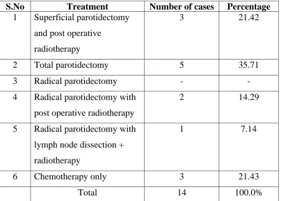
TREATMENT OFFERED TO MALIGNANT PAROTID
TUMOUIRS IN THE STUDY
S.No Treatment Number of cases Percentage
1 Superficial parotidectomy and post operative
radiotherapy
3 21.42
2 Total parotidectomy 5 35.71 3 Radical parotidectomy - - 4 Radical parotidectomy with
post operative radiotherapy
2 14.29
5 Radical parotidectomy with lymph node dissection +
radiotherapy
1 7.14
6 Chemotherapy only 3 21.43
65
DISCUSSION
The present study is based on a small series and short follow up Hence it is a difficult task to compare the present study with those found in the literature and arrive at a meaningful conclusion. However a sincere effort is made to do so in the following pages.
PLEOTROPHIC ADENOMA
66
TABLE XII
COMPARTIVE STUDY OF DIFFERENT PAROTID TUMOURS
IN THE STUDY WITH THE REPORTS OF VARIOUSAUTHORS
S. No.
Type of tumour Thanckaray & Lintus Toraya et al Sindha et al Saksela et al Present study 1 Pleomorphic adenoma
74.0 70.5 60.0 60.4 68.52
2 Monomorphic adenoma
8.6 12.7 18.5 6.6 3.70
3 Mucoepidermoid 3.2 2.1 6.8 7.5 12.96 4 Acinic cell 2.3 - 2.0 1.9 - 5 Carcinoma 11.9 14.7 12.7 23.6 14.81
In this study mixed tumour has got a male preponderance ratio of 2:1. The other studies in the literature showed a female preponderance.
67
showed a high recurrence rate (25%) and a greater risk of injury to the facial nerve (50%).
FNAC has true positive rate 86.9% in diagnosing this condition and it must be performed in all cases. No complications were noted following this investigation.
ADENOLYMPHOMA
In this series, there were only 2 cases (5.12%) in contrast to other studies that report a higher incidence of this tumour. Both the patients are males which are in accordance to the other studies which showed a male predominance. A swelling was the presenting complaint in both cases. Soft, cystic parotid swelling (Benign) is almost diagnostic of the condition except in confirming that it is a benign swelling. Superficial parotidectomy is curative.
MALIGNANT TUMOURS
68
The presence of a mass is the most consistent finding in all cases. Malignant pleomorphic adenoma has the same natural history of its benign counterpart, but is showed an initial slow growth followed by rapid increase in size. Pain was present in 9 patients. Facial nerve was involved in 5 patients. Secondary lymph node involvement was present in 4 cases. Temporomandibular joint was involved in 3 cases.
70
Rt pleomorpic adenoma
72
SUMMARY
The clinical findings and histological features of 54 cases of parotid gland tumours treated at Govt.Rajaji hospital, Madurai from Oct 2006 to Oct 2008 were studied. Parotid gland tumours were uncommon tumours forming only 0.073% of all cases admitted in the hospital. The study illustrates a wide variation in the natural history and degree of malignancy of parotid tumours. The clinical features of benign tumours had definite bearing on the final histopathological with no complication. FNAC is a reliable investigation for benign tumours with no complication. FNAC is useful in malignant tumours also, being positive in 11 out of 15 cases (73.33%). But FNAC could not provide the histopathological type of tumour in most case. So is there a role for Tru-cut biopsy in these cases doubtful cases.
73
74
CONCLUSION
1. Incidence of parotid tumours was 0.074% of all cases admitted to Govt.Rajaji hospital, Madurai during the twenty four months of study. So they are an uncommon pathology.
2. Benign tumours form the majority 72.22% of parotid neoplasms pleomorphic adenoma being the commonest 68.52%
3. Superficial lobe was the commonest site for tumour formation. 4. There was a male preponderance of 2:1in parotid tumours.
5. Higher incidence of parotid tumour was found in the 4th and 5th decades of life.
6. Mass in the parotid region was the commonest mode of presentation of parotid tumours.
7. The clinical features of benign tumours have a definite bearing on the final histopathological diagnosis and are therefore a reliable guide for the preoperative diagnosis and treatment of benign parotid tumours.
8. Maximum incidence of pleomorphic adenoma was between 31-40 years of age with a male preponderance
75
10.Adenolymphoma showed a 100% male predominance in this study with both the cases in the 6th decade.
11.FNAC is a useful diagnostic tool in the diagnosis of benign parotid tumours with virtually no complication.
76
BIBLIOGRAPHY
1. Bailey & Love Text book of Surgery.
2. The New Aird’s companion in surgical studies 3. Oxford Text book of surgery
4. Sabiston Text book of surgery
77
s.no. name age sex mass pain
F a cianerve er ve Lym ph no de ski n tmj t recu rr en ce fnac su rg
ery Final diagnosis
1 Alagu 16 m + - PA
2 Bomiraj 20 m + SP PA
3 Mustafa 36 m + SP PA
4 Shanmugam 37 m + SP PA
5 Kavitha 25 f + - PA
6 Ganesan 39 m + SP PA
7 Arupadai 37 m + SP PA
8 Vetrivel 33 m + SP PA
9 Mariammal 29 f + SP PA
10 Rajakumari 28 f + SP PA 12 Karuppaiah 37 m + SP PA
13 Lenin 38 m + SP PA
14 Arivoli 37 m + SP PA
15 Palaniamma 39 f + - PA 16 Anbarasan 45 m + SP PA
17 Subburaj 46 m + SP PA
18 Arbudaraj 42 m + SP PA
19 Palani 41 m + SP PA
78
s.n
o
name age sex
mass pain
Facial N
LN ski
n tmj t recu rr en ce fnac su rge
ry Final
diagnosis
21 Mariamma 37 f + SP PA
22 Kalirajan 55 m + SP PA
23 Nallusamy 54 m + CTP PA
24 Siluvai 36 m + SP PA
25 Karpagavalli 40 f + - AdCC 26 Thangapandi 63 m + CTP PA
27 Sivasamy 36 m + - PA
28 Tirutani 66 m + CTP PA
29 Pitchaiamal 36 f + SP PA
30 Md yusuf 50 m + + + + - An.C
31 Rajappa m + + TP MEC
32 Mariappan 58 m SP AL
33 Guru m + PA SP MEC
34 Nallammal f + - + - MPA
35 Santhanam m CT AdCC
36 Erulappan m + + - AC
37 Natrayan m + + TP MEC
38 Mookan m + + + + CT AC
39 Duraisamy m + + + CT AC
79
s.no name age sex mass pain
Facial
N
LN skin tmj
t recu rre nce fnac surge
ry Final
diagnosis
41 Kamarnisha 45 f + SP PA
42 Maria 46 f + SP PA
43 Subramani 48 m + SP PA
44 Muthulaxmi f + PA SP MEC
45 Dandapani 46 m + SP PA
46 Ramachetiar 47 m + SP PA
47 Vasuki 55 f + SP PA
48 Deivani f + PA SP MEC
49 Rasudevar 52 m + SP PA
50 Ponnammal 37 f + + SP PA
51 Vellai 62 m E AL
52 Paramu m + + + - MPA
53 Chinnan m DC TP MEC
54 kutrallam m DC TP MEC
PA- pleomorpic adenoma MPA- malignant pleomorpic adenoma
AL- adenolymphoma MEC- mucoepidermoid carcinoma
AC- adeno carcinoma AdCC- adenoid cystic carcinoma
AnC- anaplastic carcinoma SP -Superficial parotidectomy
TP -Total parotidectomy DC - Dysplastic changes