0022-538X/96/$04.0010
Copyrightq1996, American Society for Microbiology
Characterization of a 120-Kilodalton Pre-S-Binding Protein as a
Candidate Duck Hepatitis B Virus Receptor
JI-SU LI, SHU-PING TONG,ANDJACK R. WANDS*
Molecular Hepatology Laboratory, Massachusetts General Hospital Cancer Center and Harvard Medical School, Charlestown, Massachusetts 02129
Received 27 November 1995/Accepted 5 June 1996
Infection by human and animal hepadnaviruses displays remarkable host and tissue tropism. The infection cycle probably initiates with binding of the pre-S domain of viral envelope protein to surface receptors present on the hepatocyte. Three types of neutralizing monoclonal antibodies against duck hepatitis B virus (DHBV) have their binding sites clustered within residues 83 to 107 of the pre-S protein, suggesting that this region may constitute a major receptor binding site. A 170- or 180-kDa duck protein (p170 or gp180) which binds DHBV particles through this part of the pre-S sequence has been identified recently. Although the p170 binding protein is host (duck) specific, its distribution is not restricted to DHBV-infectible tissues. Using the pre-S
protein fused to glutathione S-transferase and immobilized on Sepharose beads, we have now identified an
additional binding protein with a size of 120 kDa (p120). p120 expression is restricted to the liver, kidney, and pancreas, the three major organs of DHBV replication. While optimal p170 binding requires an intact pre-S protein, binding to p120 occurs much more efficiently with a few N- or C-terminally truncated forms. The p120 binding site was mapped to residues 98 to 102 of the pre-S region, which overlaps with a cluster of known virus-neutralizing epitopes. Site-directed mutagenesis revealed residues 100 to 102 (Phe-Arg-Arg) as the critical p120 contact site; nonconservative substitution in any of the three positions abolished p120 binding. Double mutations at positions 100 to 102 markedly reduced DHBV infectivity in cell culture. Short pre-S peptides covering the clustered neutralizing epitopes (also p170 and p120 binding sites) reduced DHBV infectivity in primary duck hepatocyte cultures. Thus, p120 represents a candidate component of the DHBV receptor complex.
Interaction between a viral envelope protein and its cell surface receptor(s) initiates the virus infection cycle and often determines the host range and tissue tropism (for a recent review, see reference 6). Virus-receptor interactions may be complex processes. For example, binding of human immuno-deficiency virus to CD4 molecules is insufficient to allow viral infection of mouse T lymphocytes (18), and adenovirus attach-ment to cell surface and subsequent internalization require two separate steps involving distinct cellular proteins (29). We are interested in identifying the cellular protein(s) required for attachment and penetration of duck hepatitis B virus (DHBV), an avian relative of the human hepatitis B virus. Accumulating evidence supports the idea that the pre-S region of the DHBV large envelope protein mediates receptor binding and deter-mines species specificity (7, 10, 26). Binding of DHBV particles to Pekin duck hepatocytes has recently been demonstrated (22), but the exact receptor contact sites have not been iden-tified. Binding sites for pre-S-neutralizing antibodies are lo-cated in residues 58 to 66, 83 to 90, 91 to 99, 100 to 107, and 139 to 145 (2, 30), with three neutralizing epitopes between residues 83 and 107. On the other hand, all linker substitutions in the N-terminal two-thirds of the pre-S protein abolished viral infectivity without affecting virion formation (15). Since each of these mutants had about a dozen amino acid residues replaced by irrelevant residues, the significance of these results to the receptor binding site(s) has been difficult to evaluate. Construction of DHBV-heron hepatitis B virus chimeric enve-lope proteins revealed DHBV pre-S residues 22 to 90 as
im-portant in determining the host range (7). Nevertheless, these studies do not formally exclude the possibility that sequences outside this region are critical for receptor binding but inter-changeable between the two avian viruses.
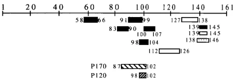
We and others have identified a 170- or 180-kDa duck pro-tein (p170 or gp180) which binds to the clustered neutralizing epitopes (residues 87 to 102) of DHBV pre-S envelope protein (11, 28). This protein binds native DHBV particles with species specificity, suggesting that it may be involved in the initial stage of virus-hepatocyte interaction. However, this binding protein is expressed in a variety of other tissues not infectible with DHBV, and transfection of the gp180 cDNA into the LMH chicken hepatoma cell line did not confer susceptibility to DHBV infection (12). It seems therefore that p170/gp180 is not the sole receptor molecule for DHBV. We now report the identification of an additional pre-S-binding protein with a size of 120 kDa (p120). The possible role of p120 as part of the DHBV receptor complex is suggested by its restricted expres-sion in DHBV-infectible tissues, by colocalization of its bind-ing site with three virus-neutralizbind-ing epitopes, and by markedly decreased infectivity of DHBV mutants constructed with an impaired p120 binding motif.
MATERIALS AND METHODS
Pre-S fusion protein constructs.To facilitate the identification of binding proteins for the DHBV pre-S molecule and mapping of the binding site, full-length and truncated forms of the pre-S protein were expressed as fusion con-structs with glutathione S-transferase (GST) and immobilized on glutathione-Sepharose beads. Radiolabeled liver proteins which bound to the pre-S protein were retained on the beads and could be subsequently visualized by sodium dodecyl sulfate-polyacrylamide gel electrophoresis (SDS-PAGE) and fluorogra-phy. Some of the truncated pre-S constructs used in the present study have been described in detail previously (28). Additional deletional mutants were generated by 20 cycles of PCR amplification of DHBV clone 16 (19) and inserted into the
* Corresponding author. Mailing address: Molecular Hepatology Laboratory, MGH Cancer Center, Bldg. 149, 13th St., Charlestown, MA 02129. Phone: (617) 726-5601. Fax: (617) 726-5609.
6029
on November 9, 2019 by guest
http://jvi.asm.org/
metabolic labeling, and detection of pre-S-binding proteins have been described previously (28). To examine the availability of p120 on the cell surface, plated hepatocytes on petri dishes were washed three times with phosphate-buffered saline (PBS), incubated at room temperature (RT) for 30 min with 1 mM sulfo-LC-biotin (Pierce) in PBS, and washed again three times with PBS before lysis of cells. The composition of the lysis buffer has been described previously (28). The precleared lysates were incubated with various constructs of the pre-S fusion protein immobilized on Sepharose beads, and the retained materials were separated by SDS-PAGE. Proteins were transferred to nitrocellulose membrane, and nonspecific binding sites were blocked by incubation at RT for 1 h with 5% bovine serum albumin (BSA) in PBS–0.05% Tween 20 (PBST). After incubation at RT for 1 h with a 1:2,000 dilution of streptavidin-conjugated with horseradish peroxidase (HRP; Pierce), biotinylated proteins were visualized by enhanced chemiluminescence. For the negative control, immunoprecipitation of a cyto-plasmic protein (Golgib-COP) was done with both surface-labeled hepatocyte lysates and labeled total liver proteins. For biotinylation of total liver proteins, 1 ml of liver tissue lysate (corresponding to 100 mg of liver tissue) was incubated with 2.5 mg of sulfo-LC-biotin for 1 h at 48C. The reaction was terminated by the addition of glycine to 100 mM followed by further incubation for 3 h. A total of 6 mg of surface-labeled or labeled total liver proteins was incubated with a 1:50 dilution of a monoclonal antibody against Golgib-COP protein (M3A5; Sigma) for 2 h at 48C. After the addition of 100ml of protein G-Sepharose beads (50% slurry), samples were incubated overnight. The retained proteins were analyzed as described above.
To study the tissue distribution of p120 by means of its affinity with truncated pre-S protein, 0.5 g of frozen tissue was homogenized in 10 ml of lysis buffer, precleared twice with Sepharose-GST beads, and then incubated with 2mg of the immobilized GST fusion protein of pre-S construct 80-102. After separation of bound proteins with an SDS–6% PAGE minigel (BioRad), protein bands were visualized by staining with Coomassie blue. To detect the small amount of p120 retained by the intact pre-S protein (construct 1-161), the bound proteins sepa-rated by SDS–8% PAGE were transferred to nitrocellulose filter. The blot was incubated successively with a 1:1,000 dilution of rabbit polyclonal anti-p120 antiserum (16a) in PBST and a 1:1,000 dilution of a donkey anti-rabbit immu-noglobulin conjugated with HRP, and positive bands were revealed with Sigma Fast DAB (3,39-diaminobenzidine) tablets dissolved in water.
To detect p120 in duck tissues by direct Western blotting (immunoblotting), 50
mg of protein was separated by SDS–6% PAGE and transferred to a nitrocellu-lose filter. The filter was blocked with 3% BSA in PBST and incubated with a 1:1,000 dilution of rabbit polyclonal anti-p120 antibody at RT overnight. After a thorough wash, the filter was incubated with a 1:1,000 dilution of a donkey anti-rabbit immunoglobulin conjugated with HRP, and positive bands were re-vealed with Sigma Fast DAB.
Inhibition of DHBV infectivity by pre-S peptides.Primary duck hepatocytes were seeded at a density of 33105cells per well into 12-well plates. The
experiments were performed within 1 week after plating. Pre-S polypeptides 80-102 and 80-104 expressed as GST fusion proteins were purified onto Sepha-rose beads. The GST portion was removed by digestion with thrombin (Sigma) and centrifugation. Thrombin (molecular mass, 55 kDa) was not removed in the initial experiments but was removed in repeat experiments through a Centricon 30 filter (cutoff, 30 kDa), and similar results were obtained. Because of the nature of gene fusion with the pGEX-2TK vector, all the peptides contained at their N termini nine irrelevant amino acid residues (GSRRASVGS) contributed by the thrombin recognition site, the protein kinase domain, and the BamHI site. Incubation of the hepatocytes in the plates with the peptides at three different concentrations at RT for 1 h was followed by the addition of 1ml of viremic serum and a further incubation for 3 h. After an extensive washing, the cells were maintained in L15–1% dimethyl sulfoxide medium supplemented with a neutral-izing rabbit polyclonal anti-pre-S antiserum (16a) to suppress virus spread. Cells were harvested at day 8 postinfection. The experiments were performed in duplicate, and the harvested cells were pooled for hybridization experiments.
Transfection and infection with pre-S substitution mutants.pDHBV3.5 was constructed to contain a 3.5-kb-overlength DHBV genome (NcoI-NsiI fragment of DHBV clone 16 [19]) inserted between the EcoRI and PstI sites of pUC18
cessive digestions with DNase I and proteinase K followed by phenol-chloroform extraction, the amounts of DHBV DNA were determined by Southern blotting. To selectively detect virion particles, the pelleted material was digested with pronase and DNase I as described previously (15).
Virion particles concentrated from 3 to 6 ml of culture medium were used to infect primary duck hepatocytes cultured in 6-well plates. After incubation at 378C for 6.5 h, cells were washed and cultured for an additional 7 days before extraction of total cellular DNA for Southern blot analysis.
RESULTS
A 120-kDa binding protein with high affinity for truncated
forms of pre-S protein. Nine N-terminal deletion constructs
and five C-terminal constructs had been made as GST fusion proteins to map the binding site for pre-S-interacting protein p170 (28) (Fig. 1A). Interestingly, two N-terminal constructs and one C-terminal construct strongly recognized a new 120-kDa protein (p120), while the intact pre-S protein and other deletion mutants did not (Fig. 1). p120 is not a glycosylated protein since labeling of the primary duck hepatocytes in the presence of tunicamycin (1mg/ml) did not modify the mobility of the binding protein (data not shown). The three p120-bind-ing constructs contained pre-S sequences from residues 92 to 161, 98 to 161, and 1 to 102, respectively. These results suggest that the p120 binding motif is normally hidden by the sur-rounding pre-S sequences but can be made accessible by sub-stantial truncation at either the N or C terminus. Further deletion removed the p120 binding motif, thereby abrogating p120 binding. Thus, the sequence bracketed by residues 98 and 102 would be the putative p120 binding motif. Consistent with this interpretation, the p120 binding capacity was maintained in the three double-deletion constructs with a fixed C terminus at residue 102 but different N termini at residues 25, 59, and 80 (Fig. 1). Construct 80-102, which retained only 23 amino acid residues of the pre-S region, bound p120 at least as efficiently as the longer construct 1-102. To test whether residues 98 to 102 can bind p120 in the absence of any surrounding DHBV sequence, mutant E98S-102 was constructed. Indeed, this con-struct was able to retain p120 efficiently (Fig. 1).
Interestingly, the p120 binding site overlaps extensively with the binding site (residues 100 to 107) of virus-neutralizing monoclonal antibody SD20 (2, 13). This neutralizing epitope is in fact one of the three clustered neutralizing epitopes (Fig. 2). Recently, we also isolated a monoclonal antibody which at a 1:200 dilution of the hybridoma culture supernatant inhibited DHBV infection in primary duck hepatocytes by more than 90% (data not shown). With the use of GST-tagged deletion mutants, the binding site of this neutralizing monoclonal anti-body was mapped to pre-S residues 98 to 104, which entirely cover the p120 binding site. However, unlike p120, the mono-clonal antibody requires pre-S residues 103 and 104 for binding (data not shown).
on November 9, 2019 by guest
Retention of low-level p120 by intact pre-S construct.The fact that intact pre-S protein failed to retain p120 raised the issue of the significance of the p120–pre-S interaction. To test the possibility that intact pre-S protein can immobilize small amounts of p120, we resorted to a more sensitive detection method. Unlabeled liver proteins from 0.5 g of tissue were incubated with the GST-fused intact pre-S protein (construct 1-161), and the bound p120 protein was identified by Western blot analysis with a rabbit polyclonal antiserum raised against gel-purified p120. With this improved-sensitivity detection sys-tem, p120 was found in the retained material, although with a retention that was much less efficient than that by construct 80-102 (Fig. 3, compare lanes 1 and 2). p120 retention by the intact pre-S protein seems independent of p170, since it occurs in DHBV-infected liver tissue (left panel) in which p170 bind-ing has been previously shown to be sequestered by endoge-nous viral pre-S protein (28).
Efficient p120 binding requires precise truncation at pre-S
C terminus. While all constructs terminating at residue 102
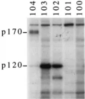
(constructs 1-102, 25-102, 59-102, and 80-102) bound large amounts of p120, those ending at residue 104 (constructs 1-104, 25-104, and 80-104) did not (Fig. 1). To further define the boundary between p120-binding and nonbinding
[image:3.612.144.474.79.423.2]C-termi-nal-deletion constructs, three additional constructs with a fixed N terminus at residue 80 but different C termini at residues 103, 101, and 100 were constructed. As shown in Fig. 4, p120 did not bind the mutant truncated at amino acid 104 but could bind mutants terminating at either residue 103 or 102. Further FIG. 1. Recognition of 120-kDa duck hepatocyte protein by several truncated forms of DHBV pre-S protein. The pre-S part of DHBV large envelope protein and its truncated forms (N-terminal, C-terminal, and double deletions) were expressed as GST fusion proteins and purified on glutathione-Sepharose beads. They were incubated at 48C with35S-labeled primary duck hepatocyte lysates which had been precleared with Sepharose beads. After extensive washing with the lysis buffer, bound
proteins were fractionated by reducing SDS–8% PAGE.35S-labeled proteins were revealed by fluorography. (A) Schematic representation of pre-S constructs and their
affinities for p120. (B) Fluorograph. Molecular size markers (in kilodaltons) are indicated on the left, and the positions of p170 and p120 are indicated on the right.
FIG. 2. Colocalization of p120 and p170 binding sites and clustered neutral-izing epitopes. The pre-S region is schematically shown at the top (amino acids 1 to 161). p170 and p120 binding sites and the epitopes recognized by neutral-izing (black bar) and nonneutralneutral-izing (white bar) monoclonal antibodies are indicated. The epitopes for amino acids 58 to 66, 91 to 99, 127 to 138, and 139 to 145 are according to Yuasa et al. (30); those for amino acids 83 to 90 and 100 to 107 are according to Chassot et al. (2); and those for amino acids 98 to 104 and 112 to 126 are according to Li et al. (16a). A nonessential region for viral infectivity is shown (open bar with dots) (16).
on November 9, 2019 by guest
http://jvi.asm.org/
[image:3.612.317.554.571.653.2]truncation at residue 101 or 100 abrogated binding. Therefore, terminal truncation has to occur at residue 102 (with a C-terminal Phe-Arg-Arg sequence) or 103 (with a C-C-terminal Arg-Arg-Gln sequence) in order to gain affinity for p120.
p120 binding motif is composed of a tripeptide sequence of
Phe-Arg-Arg.To further elucidate the contribution of
individ-ual amino acid residues to p120 binding, site-directed mu-tagenesis was carried out for residues 97 through 102 (Fig. 5A). Most mutants were generated in the 80-102 construct, because of its strong reactivity with p120 in the wild-type sequence as well as for simplicity in the construction of mutations. The binding results of the pre-S mutants in construct 80-102 are shown in the left panel of Fig. 5B. The mutation of glutamic acid at position 98 to valine (E98V) maintained binding. In-deed, an increased retention of p120 was observed (Fig. 5B). With the change of alanine at position 99 to aspartic acid (A99D), reactivity to p120 was also retained. This residue is not conserved in nature, as it was found to be changed to tyrosine in a goose hepatitis B virus strain which can infect ducks (24). As for the phenylalanine residue at position 100, substitution by either valine or leucine (F100V and F100L) abolished binding, while mutation to another aromatic amino acid such as tryptophan (F100W) was tolerated. For the pen-ultimate arginine, mutation to neither leucine nor histidine (R101L and R101H) was tolerated, but with the substitution by lysine (R101K), the residual binding capacity was retained. Most stringent for p120 binding was the terminal arginine residue, for which substitutions by glycine, histidine, and even lysine (R102G, R102H, and R102K) totally abolished p120 binding. Therefore, the triplex of Phe-Arg-Arg residues at po-sitions 100 to 102 is critical for p120 binding and very likely constitutes the p120 contact site. These three residues are conserved in all DHBV strains (and a goose hepatitis B virus strain) that have been sequenced.
Residues 100 to 102 are positioned at the C terminus in construct 80-102. To rule out the possibility that the impor-tance of these residues was merely a result of a positional effect, three mutants of these residues, F100V, R101L, R102G, and three mutants of residues 97 and 98, R97C, E98A and E98V, were introduced into another p120-binding construct, construct 92-161, and the affinity for p120 was tested. All the three mutants of residues 100 to 102 failed to retain p120 while mutants of residues 97 and 98 could bind to the protein (Fig. 5B, right panel).
p120 is detectable on hepatocyte cell surface.If p120 is the
p170 was retained by constructs 1-161 and 80-104, though the signal obtained was weaker. To rule out the possibility that the p120 and p170 detected were derived from a small amount of intracellular protein leaked from dead cells, immunoprecipita-tion of Golgib-COP, a Golgi microtubule-associated protein (4), was carried out using monoclonal antibody M3A5. This monoclonal antibody recognizes an epitope shared by the Golgi b-COP protein (110 kDa) and a high-molecular-mass doublet of the microtubule-associated protein (MAP). As a result, both the 110-kDa protein band (data not shown) and a doublet high-molecular-mass band (;300 kDa) were precipi-tated only from total labeled duck liver lysate (Fig. 6, lane 6) and not from the surface-labeled lysate (lane 5), suggesting that contamination by cytoplasmic proteins was insignificant in these experiments.
Disruption of p120 binding motif reduces DHBV infectivity.
The crucial role of pre-S residues 100 to 102 in mediating p120 interaction enabled us to test the significance of p120 in the DHBV life cycle by genetic approaches. Double amino acid substitutions were introduced into p120-binding residues 100 to 102 and succeeding residues 103 and 104 of the replication-competent DHBV genome pDHBV3.5: F100V-R101L, R101I-R102D, R101L-R102L, and Y103C-Q104F. As a control, a mutant with triple amino acid substitutions, K95S-R97L-E98A, was used (the mutations cover the p170 binding site but do not abolish p170 binding). Although some of the mutations caused amino acid changes in the overlapping polymerase gene
(R101I-R012D: S311Y-P312R; F100V-R101L: F310C;
Y103C-Q104F: S314F; and K95S-R97L-E98A: S306C), this portion of the polymerase is a spacer region tolerant of sub-stantial sequence alterations (1, 16). After the mutants were transfected into LMH cells, the secretion of pelletable particles into culture medium at different time points was measured. In by SDS–8% PAGE and blotted onto a nitrocellulose filter. After incubation with a
[image:4.612.117.236.73.181.2]rabbit anti-p170 antibody (upper panel) (16a) or an anti-p120 antibody (lower panel), the protein bands were revealed by HRP-conjugated anti-rabbit serum and DAB as described in Materials and Methods. The positions of p170 and p120 are indicated.
FIG. 4. p120 retention by C-terminal deletion constructs of pre-S protein requires an exact truncation at residue 102 or 103. The five constructs used had a common N terminus at residue 80 but different C termini at residues 104, 103, 102, 101, and 100, respectively. Detection of p120 from35S-labeled primary duck
hepatocyte lysates was done as described in the legend to Fig. 1. The positions of p170 and p120 are indicated.
on November 9, 2019 by guest
[image:4.612.392.477.574.673.2]general, no major variations in the secretion of virion-core particles were found (Fig. 7A, analysis at day 3), except that mutant Y103C-Q104F produced fewer particles at day 7 post-transfection (data not shown). All the mutants displayed sim-ilar ratios of virion/core particles, since removal of core parti-cles by Pronase-DNase I (15) decreased the hybridization signal similarity for all the mutants (Fig. 7B). Indeed, no major difference in the levels of covalently closed circular DNA, total and core DHBV RNA (data not shown), and core DNA (Fig. 7C) could be detected at day 7 posttransfection.
Equal amounts of virion particles concentrated from the culture medium were used to infect primary duck hepatocytes for 6.5 h. The infectivity was determined by Southern blot hybridization of intracellular DHBV DNA at day 8 postinfec-tion. All the mutants except the triple mutant K95S-R97L-E98A exhibited significantly reduced amounts of viral DNA in infected cells (Fig. 7D). According to results from four inde-pendent transfection-infection experiments, the degree in re-duction of infectivity follows the order Y103C-Q104F . F100V-R101L and R101I-R102D.R101L-R102L. Immuno-fluorescence staining of infected cells with an pre-S anti-body (16a) revealed a corresponding reduction in the number of cells infected, although the intensities of fluorescence in the positive cells were not significantly different between cells in-fected with the wild-type virus and the mutants (data not shown).
Synthetic pre-S peptides covering the p170 and p120
bind-ing sites interfere with DHBV infectivity.The p170 binding site
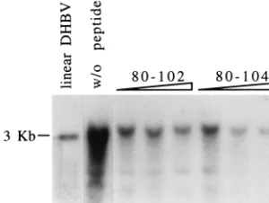
entirely covers the three clustered neutralizing epitopes, and p120 binding site overlaps the C-terminal epitope. If the clus-tered epitopes are indeed part of the receptor binding site, then pre-S peptides covering this region might compete for receptor binding and interfere with DHBV infection. Two pre-S peptides were used for this experiment: 102 and 80-104. The GST fusion protein of peptide 80-102 binds p120 (but not p170) efficiently, while peptide 80-104 binds p170 (but not p120) with low efficiency. The two pre-S peptides were purified from GST by thrombin cleavage. Peptides were preincubated with hepatocyte monolayers for 1 h before infection with 1ml of viremic duck serum. As a result, both peptides reduced DHBV infectivity (Fig. 8). At a concentration of 10 mg/ml, peptides 80-102 and 80-104 reduced the degree of DHBV infectivity similarly. Increasing the peptide concentration to 1 mg/ml enhanced the inhibitory effect significantly for peptide 80-104 but only slightly for peptide 80-102. The strong
inhibi-tory effect of peptide 80-104 lends support to the hypothesis that the clustered neutralizing epitopes are a contact site of DHBV receptor.
p120 expression is restricted to DHBV-infectible tissues.To
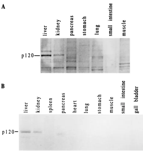
study the tissue distribution of p120, tissue lysates were pcleared and incubated with the construct 80-102, and the re-tained proteins were revealed by an SDS-PAGE minigel fol-lowed by Coomassie blue staining. p120 was clearly found in the liver and kidney (Fig. 9A). It was weakly detected in the pancreas but not in the other tissues examined, including stom-ach, lung, small intestine, skeletal muscle (Fig. 9A), spleen, heart, and gall bladder (data not shown). As an independent confirmation of this result, a direct Western blotting detection method for p120 was developed. In accordance with the affinity approach, p120 was found most abundantly in the liver fol-lowed by the kidney but least abundantly in the pancreas (Fig. 9B). No p120 was found in the other tissues tested. This pat-tern of the tissue-specific distribution of p120 coincides with the known tissue tropism of DHBV infection (5).
DISCUSSION
In the present study, we have identified and characterized p120, a second binding protein for the DHBV pre-S envelope FIG. 5. Critical role of pre-S residues 100 to 102 for p120 binding. Single amino acid substitutions were introduced into either construct 80-102 or construct 92-161, and the effects on p120 binding were examined with35
[image:5.612.66.553.73.227.2]S-labeled primary duck hepatocyte lysates. (A) Schematic representation of mutants and binding results. (B) Fluorograph showing binding results of mutants in construct 80-102 (left) or 92-161 (right). The position of the p120 band is indicated. WT, wild type.
FIG. 6. Detection of p120 in lysates of cell-surface biotinylated primary duck hepatocytes. Primary duck hepatocytes cultured for 2 days in petri dishes were labeled with sulfo-LC-biotin. Cells were washed three times with PBS before and after labeling. The cell lysates were incubated with the following pre-S constructs 80-102 (lane 1), 80-104 (lane 2), 25-102 (lane 3), and 1-161 (lane 4). The positions of p120 and p170 are indicated. As a negative control, 6 mg of either surface (lane 5) or total (lane 6) biotinylated liver proteins was immunoprecipi-tated with monoclonal antibody M3A5, which recognizes an epitope shared by Golgib-COP, a Golgi membrane protein, and microtubule-associated protein (MAP). The positions of the MAP doublet are indicated.
on November 9, 2019 by guest
http://jvi.asm.org/
protein. Compared with p170, p120 exhibits remarkable tissue-specific expression in the liver and the kidney, the major sites of DHBV replication (5). The data indicating that p120 is a candidate DHBV-binding protein of a putative receptor com-plex is supported by (i) restricted tissue distribution coincident with the tissue tropism of DHBV replication, (ii) colocalization of the binding site with virus-neutralizing epitopes, (iii) inter-ference of DHBV infection by short pre-S peptides covering its (and p170’s) binding site, (iv) reduced infectivity of DHBV mutants with a disrupted p120 binding motif, and (v) cell surface localization on primary duck hepatocytes. On the other hand, the following observations may or may not be compatible with the assumption that p120 is a DHBV receptor.
For example, it is not exclusively a cell-surface protein. Al-though we could detect p120 by cell surface labeling with sulfo-LC-biotin, the protein is also located within the hepato-cytes. Whether the amount of p120 present on the cell surface is sufficient to allow viral entry is not presently known. Alter-natively, p120 could play a role in the intracellular trafficking of internalized DHBV particles and if this proves to be the case, it does not need to be located principally on the cell surface. It was determined that a homologous binding protein was detect-able in non-DHBV-infectible hosts. Using pre-S construct 80-102, we could detect p120-related proteins in chicken and human livers (16a). If the host and tissue specificities of DHBV infection are determined at the receptor level, the double spec-ificities may be explained by postulating that p170 and p120 proteins are part of a DHBV receptor complex.
The p120-reactive peptide 80-102 does not inhibit DHBV
highest peptide concentration.
As shown in Fig. 3, the intact pre-S protein expressed in
Escherichia coli retains only a small amount of p120. The
addition of DHBV viremic duck serum into a tube containing a metabolically labeled hepatocyte lysate and immobilized pre-S construct 80-102 failed to significantly reduce the amount of p120 that adhered to the beads (16a), suggesting that DHBV particles do not have a large amount of truncated pre-S protein, similar to those in vitro which bind p120 effi-ciently. However, considering the fact that residues 101 and 102 are both arginine, the p120-reactive pre-S protein can be generated by proteolytic cleavage through a trypsin-like pro-tease or an endopeptidase specific for dibasic residues (3, 8, 25). Proteolytic cleavage of viral envelope protein after di- or tetrabasic residues is required for infectivity of myxoviruses and retroviruses, although in these instances the cleavage (i) occurs during virion maturation and (ii) exposes a new hydro-phobic N terminus required for virus-cell fusion (9, 20, 21, 23). Most interestingly, Lu and colleagues recently demonstrated that protease treatment of HBV particles enhanced its infec-tivity in a hepatoma cell line (17), thus reinforcing the hypoth-esis that hepadnavirus infection may require the action of proteases.
[image:6.612.121.228.70.314.2]The simplest model to account for p170 and p120 as com-ponents of the DHBV receptor is as follows: virion particles are attracted onto the hepatocyte surface initially by p170. This event is followed by either direct conformational change of the pre-S protein or by proteolytic cleavage at Arg-102 to activate p120 binding. p120 binding subsequently allows viral entry or participates in intracellular trafficking. Irrespective of how the FIG. 7. Impairment of DHBV infectivity by mutations at p120 binding site.
Various DHBV mutants in an overlength DHBV genome were transfected into LMH cells in duplicate. DHBV particles were concentrated from pooled media, and equal amounts of virion particles were used to infect primary duck hepato-cyte cultures for 6.5 h. Cells were harvested at day 7 postinfection. (A) Southern blot analysis of viral particles secreted in LMH culture medium at day 3 post-transfection. (B) Southern blot analysis of another aliquot of day 3 viral particles pretreated with Pronase and DNase I. (C) DHBV DNA associated with intra-cellular core particles at day 7 posttransfection. (D) Intraintra-cellular DHBV DNA in primary duck hepatocytes infected for 6.5 h with virus particles produced in LMH cells. Lanes: 1, R101I-R102D; 2, R101L-R102L; 3, F100V-R101L; 4, Y103C-Q104F; 5, K95S-R97L-E98A; 6, wild type.
FIG. 8. Inhibition of DHBV infection of primary duck hepatocytes by short pre-S peptides. The pre-S peptides 80-102 and 80-104 were mass produced as GST fusion proteins and removed from the GST partner by thrombin cleavage. They were incubated at RT for 1 h with primary duck hepatocytes at three different concentrations: 10mg/ml, 100mg/ml, and 1 mg/ml. DHBV-positive duck serum (1ml) was then added, and incubation continued for an additional 3 h. Cells were harvested at day 8 postinfection, and intracellular DHBV DNA was studied by Southern blot analysis. w/o peptide, without peptide.
on November 9, 2019 by guest
[image:6.612.357.507.544.657.2]p120-DHBV interaction occurs, the role of p120 in the DHBV life cycle can be tested by two key experimental approaches: namely, whether an antibody against p120 inhibits DHBV in-fection of primary duck hepatocytes and/or whether transfec-tion of p120 cDNA (together with p170 cDNA) into nonper-missive cells renders them susceptible to DHBV infection.
ACKNOWLEDGMENTS
We thank R. Carlson for synthesis of oligonucleotides and J. Lavine of Children’s Hospital, Boston, Mass., for providing LMH cells.
This work was supported by grants CA-35711 and AA-08169 from the National Institutes of Health and the Tan Yan Kee Foundation, Inc.
REFERENCES
1. Bartenschlager, R., and H. Schaller. 1988. The amino-terminal domain of the hepadnaviral P-gene encodes the terminal protein (genome-linked pro-tein) believed to prime reverse transcription. EMBO J. 7:4185–4192. 2. Chassot, S., V. Lambert, A. Kay, C. Godinot, B. Roux, C. Trepo, and L. Cova.
1993. Fine mapping of neutralizing epitopes on duck hepatitis B virus (DHBV) pre-S protein using monoclonal antibodies and overlapping pep-tides. Virology 192:217–223.
3. Davey, J., K. Davis, Y. Imai, M. Yamamoto, and G. Matthews. 1994. Isolation and characterization of Krp, a dibasic endopeptidase required for cell via-bility in the fission yeast Schizosacharomyces pombe. EMBO J. 13:5910– 5921.
4. Duden, R., G. Griffiths, R. Frank, P. Argos, and T. E. Kreis. 1991.b-COP, a 110 kd protein associated with non-clathrin-coated vesicles and the Golgi complex, shows homology tob-adaptin. Cell 64:649–665.
5. Halpern, M. S., J. M. England, D. I. Deery, D. J. Petcu, W. S. Mason, and K. L. Molnar-Kimber.1983. Viral nucleic acid synthesis and antigen accu-mulation in pancreas and kidney of Pekin ducks infected with duck hepatitis B virus. Proc. Natl. Acad. Sci. USA 80:4865–4869.
6. Haywood, A. M. 1994. Virus receptors: binding, adhesion strengthening, and changes in viral structure. J. Virol. 68:1–5.
7. Ishikawa, T., and D. Ganem. 1995. The pre-S domain of the large viral envelope protein determines host range in avian hepatitis B virus. Proc. Natl. Acad. Sci. USA 92:6259–6263.
8. Julius, D., A. Brake, L. Blair, R. Kunisawa, and J. Thorner. 1984. Isolation of the putative structural gene for the lysine-arginine-cleaving endopeptidase required for processing of yeast prepro-a-factor. Cell 37:1075–1089. 9. Kawaoka, Y., and R. G. Webster. 1988. Sequence requirements for cleavage
activation of influenza virus hemagglutinin expressed in mammalian cells. Proc. Natl. Acad. Sci. USA 85:324–328.
10. Klingmuller, U., and H. Schaller. 1993. Hepadnavirus infection requires interaction between the viral pre-S domain and a specific hepatocellular receptor. J. Virol. 67:7414–7422.
11. Kuroki, K., R. C. Cheung, P. L. Marion, and D. Ganem. 1994. A cell surface protein that binds avian hepatitis B virus particles. J. Virol. 68:2091–2096. 12. Kuroki, K., F. Eng, T. Ishikawa, C. Turck, F. Harada, and D. Ganem. 1995.
gp180, a host cell glycoprotein that binds duck hepatitis B virus particles, is encoded by a member of the carboxypeptidase gene family. J. Biol. Chem. 270:15022–15028.
13. Lambert, V., D. Fernholz, R. Sprengel, I. Fourel, G. Deleage, G. Wildner, C. Peyret, C. Trepo, L. Cova, and H. Will.1990. Virus-neutralizing monoclonal antibody to a conserved epitope on the duck hepatitis B virus pre-S protein. J. Virol. 64:1290–1297.
14. Lantz, L. M., and K. L. Holmes. 1995. Improved nonradioactive cell surface labeling technique for immunoprecipitation. BioTechniques 18:58–60. 15. Lenhoff, R. J., and J. Summers. 1994. Coordinate regulation of replication
and virus assembly by the large envelope protein of an avian hepadnavirus. J. Virol. 68:4565–4571.
16. Li, J.-S., L. Cova, R. Buckland, V. Lambert, G. Deleage, and C. Trepo. 1989. Duck hepatitis B virus can tolerate insertion, deletion, and partial frameshift mutation in the distal pre-S region. J. Virol. 63:4965–4968.
16a.Li, J.-S., S.-P. Tong, and J. R. Wands. Unpublished data.
17. Lu, X., T. Block, and W. F. Gerlich. 1996. Protease-induced infectivity of hepatitis B virus for a human hepatoblastoma cell line. J. Virol. 70:2277– 2285.
18. Maddon, P. J., A. G. Dalgleish, J. S. McDougal, P. R. Clapham, R. A. Weiss, and R. Axel.1986. The T4 gene encodes the AIDS virus receptor and is expressed in the immune system and the brain. Cell 47:333–348. 19. Mandart, E., A. Kay, and F. Galibert. 1984. Nucleotide sequence of a cloned
duck hepatitis B virus genome: comparison with woodchuck and human hepatitis B virus sequences. J. Virol. 49:782–792.
20. McCune, J. M., L. B. Rabin, M. B. Feinberg, M. Lieberman, J. C. Kosek, G. R. Reyes, and I. L. Weissman.1988. Endoproteolytic cleavage of gp160 is required for the activation of human immunodeficiency virus. Cell 53:55–67. 21. Perez, L., and E. Hunter. 1987. Mutations within the proteolytic cleavage site of the Rous sarcoma virus glycoprotein that block processing to gp85 and pg37. J. Virol. 61:1609–1614.
22. Pugh, J. C., D. Qu, W. S. Mason, and H. Simmons. 1995. Susceptibility to duck hepatitis B virus infection is associated with the presence of cell surface receptor sites that efficiently bind viral particles. J. Virol. 69:4814–4822. 23. Scheid, A., and P. W. Choppin. 1977. Two disulfide-linked polypeptide
chains constitute the active F protein of paramyxoviruses. Virology 80:54–66. 24. Shi, H., J. M. Cullen, and J. E. Newbold. GenBank accession no. M95589. 25. Smeekens, S. P., and D. F. Steiner. 1990. Identification of a human insuli-noma cDNA encoding a novel mammalian protein structurally related to the yeast dibasic processing protease Kex2. J. Biol. Chem. 265:2997–3000. 26. Summers, J., P. M. Smith, M. Huang, and M. Yu. 1991. Morphogenetic and
regulatory effects of mutations in the envelope proteins of an avian hepad-navirus. J. Virol. 65:1310–1317.
27. Tong, S.-P., J.-S. Li, L. Vitvitski, and C. Trepo. 1992. Replication capacities of natural and artificial precore stop codon mutants of hepatitis B virus: relevance of pregenome encapsidation signal. Virology 191:237–246. 28. Tong, S.-P., J.-S. Li, and J. R. Wands. 1995. Interaction between duck
hepatitis B virus and a 170-kilodalton cellular protein is mediated through a neutralizing epitope of the pre-S region and occurs during viral infection. J. Virol. 69:7106–7112.
29. Wickham, T. J., P. Mathias, D. A. Cheresh, and G. R. Nemerow. 1993. Integrins avb3 and avb5 promote adenovirus internalization but not virus attachment. Cell 73:309–319.
[image:7.612.57.295.67.318.2]30. Yuasa, S., R. C. Cheung, Q. Pham, W. S. Robinson, and P. L. Marion. 1991. Peptide mapping of neutralizing and nonneutralizing epitopes of duck hep-atitis B virus pre-S polypeptide. Virology 181:14–21.
FIG. 9. Tissue distribution of p120. (A) Detection by pre-S construct. About 0.5 g of tissue was homogenized in lysis buffer, precleared, and incubated with 2
mg of GST fusion protein of construct 80-102 immobilized on Sepharose beads. Bound proteins were separated by SDS–6% PAGE and visualized by Coomassie blue staining. (B) Detection by direct Western blotting. Proteins (50mg) were separated by SDS-PAGE and transferred to nitrocellulose filter. The blot was incubated with a rabbit polyclonal anti-p120 antibody, and a positive signal was revealed by HRP-conjugated anti-rabbit serum and DAB. The position of the p120 band is indicated in each panel.