0022-538X/96/$04.0010
Copyrightq1996, American Society for Microbiology
Detection of Borna Disease Virus (BDV) Antibodies and BDV
RNA in Psychiatric Patients: Evidence for High Sequence
Conservation of Human Blood-Derived BDV RNA
CHRISTIAN SAUDER,1,2ANJA MU¨ LLER,1BEATRICE CUBITT,2JENS MAYER,1JU¨ RGEN STEINMETZ,3
WOLFGANG TRABERT,3BA¨ RBEL ZIEGLER,3KLAUS WANKE,3NIKOLAUS MUELLER-LANTZSCH,1
JUAN CARLOSDE LATORRE,2*ANDFRIEDRICH A. GRA¨ SSER1*
Abteilung Virologie, Institut fu¨r medizinische Mikrobiologie und Hygiene,1and Institut fu¨r Neurologie und Psychiatrie,3
Universita¨tskliniken des Saarlandes, 66421 Homburg, Germany, and Division of Virology, Department of
Neuropharmacology, The Scripps Research Institute, La Jolla, California 920372
Received 25 April 1996/Accepted 6 August 1996
In several vertebrate species, Borna disease virus (BDV), the prototype of a new group of animal viruses, causes central nervous system disease accompanied by diverse behavioral abnormalities. Seroepidemiological data indicate that BDV may contribute to the pathophysiology of certain human mental disorders. This hypothesis is further supported by the detection of both BDV antigens and BDV RNA in peripheral blood mononuclear cells (PBMCs) of patients with psychiatric disorders and the isolation of BDV from such PBMCs. Here we describe serological and molecular epidemiological studies on psychiatric patients and healthy individuals from the area of Homburg, Germany. Using a novel Western blot (immunoblot) assay, we found a BDV seroprevalence of 9.6% among 416 neuropsychiatric patients, which is significantly higher than the 1.4% found among 203 healthy control individuals. Human sera displayed a prominent immunoreactivity against the virus nucleoprotein, the p40 antigen. Reverse transcriptase-mediated PCR analysis of RNA extracted from PBMCs of a subset of 26 of the neuropsychiatric patients revealed that 50% were BDV RNA positive. Three of the 13 BDV RNA-positive patients also had BDV-positive serology, whereas one patient with serum antibodies to BDV p40 antigen did not harbor detectable BDV RNA in PBMCs. BDV p40 and p24 sequences derived from human PBMCs exhibited both a high degree of inter- and intrapatient conservation and a close genetic relationship to animal-derived BDV sequences.
Clinical and epidemiological evidence together with virolog-ical studies indicate that viruses can contribute to the patho-physiology of neuropsychiatric disorders whose etiology re-mains elusive (35, 56, 60).
Borna disease virus (BDV) is an enveloped nonsegmented, negative-stranded RNA virus (8, 13, 14). Replication and tran-scription of the BDV genome take place in the nuclei of in-fected cells (7, 13), and RNA splicing contributes to BDV gene expression regulation (15, 48). These features, unique among
the members of theMononegavirales, signal BDV as the
pro-totype of a new group of animal viruses (16, 46). The BDV genome contains five open reading frames (ORFs), I to V (8, 13). As yet, viral polypeptides with predicted molecular masses of 40, 24, and 16 kDa corresponding to ORFs I, II, and III, respectively, have been detected in BDV-infected cells and tissues.
In several vertebrate species, BDV causes central nervous system disease characterized by behavioral abnormalities and diverse pathological manifestations, depending on the species, age, and immune status of the host, as well as the particular virus strain and route of infection (32, 33, 40, 41). This wide
host range and the observation that BDV-induced behavioral disturbances in animals resemble some types of affective dis-orders in humans (52) prompted studies aimed at determining a possible association between BDV and human mental disor-ders.
Seroepidemiological studies show a significantly higher se-roprevalence of BDV in patients with neuropsychiatric disor-ders than in healthy individuals (2, 5, 18, 43, 57). The detection of BDV RNA in peripheral blood mononuclear cells (PBMCs) from neuropsychiatric (6, 26) and chronic fatigue syndrome (39) patients, together with the recent isolation of infectious BDV from PBMCs of three psychiatric cases (17), provides additional support for the hypothesis that BDV may contribute to certain human mental disorders. However, little is known about which viral antigens are recognized by BDV-positive human sera and the correlation between detection of anti-BDV antibodies in serum and viral RNA in PBMCs. More-over, there is only very limited information on BDV sequences derived from PBMCs of psychiatric patients from different geographic areas and on the level of divergency between hu-man- and animal-derived BDV sequences (4, 6, 17, 24, 47).
Here we describe the development of a new Western blot (immunoblot) assay for the detection of human anti-BDV an-tibodies, using as target antigens recombinant BDV proteins expressed in insect cells. Using this assay, 40 (9.6%) of 416 serum specimens from psychiatric patients from the area of Homburg, Germany, were found to be BDV seropositive, com-pared with only three (1.4%) of 203 serum specimens of a control group from the same area. The majority of these hu-man sera positive for BDV recognized only the BDV p40 antigen. Reverse transcriptase (RT)-mediated PCR (RT-PCR)
* Corresponding author. Mailing address for J. C. de la Torre: Divi-sion of Virology, Department of Neuropharmacology, The Scripps Research Institute, 10666 N. Torrey Pines Rd., La Jolla, CA 92037. Phone: (619) 784-9462. Fax: (619) 784-9981. Electronic mail address: juanct@riscsm.scripps.edu. Mailing address for F. A. Gra¨sser: Institut fu¨r medizinische Mikrobiologie und Hygiene, Abteilung Virologie, Universita¨tskliniken des Saarlandes, Haus 47, 66421, Homburg/Saar, Germany. Phone: 49-6841-163983. Fax: 49-6841-163980. Electronic mail address: graesser@med-rz.uni-sb.de.
7713
on November 9, 2019 by guest
http://jvi.asm.org/
analysis of RNA extracted from PBMCs of a subset of 26 psychiatric patients revealed 13 of them to be BDV RNA positive. Among the BDV RNA-positive patients, only three had serum antibodies to BDV antigens detectable by Western blotting. In contrast to a recent report (24), we found that BDV p40 and p24 sequences derived from PBMCs of neuro-psychiatric patients exhibit a high degree of inter- and intrapa-tient conservation and also are genetically very closely related to BDV sequences of animal origin.
MATERIALS AND METHODS
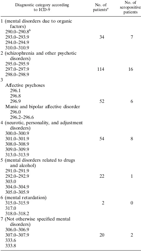
Subjects.Serum specimens from 416 unselected and consecutively admitted psychiatric in patients from the Department of Psychiatry of the University Hospital in Homburg, treated for a variety of psychiatric diseases (Table 1), as well as from 203 surgery patients (matched by age and sex to the first 203 psychiatric patients) from the same hospital, were evaluated for BDV serology. The group of psychiatric patients consisted of 203 males and 213 females. The mean age was 44.85 years (standard deviation, 40.01 years; range, 18 to 85 years).
Diagnostic classification was made at discharge according to ICD-9 (10) and was blind regarding BDV data. The four-digit ICD-9 subclasses were condensed to seven main diagnostic categories (Table 1). All categories had comparable num-bers of patients.
RT-PCR studies were performed on 28 blood samples from 26 of the psychi-atric patients (21 males and 5 females) and 23 blood samples from healthy volunteers from Homburg. Patient blood samples used for RT-PCR and serology were taken at admission.
Generation of recombinant baculoviruses.BDV p40, p24, and p16 genes were amplified by PCR from the respective cDNAs (14). The primers used for PCR amplification were p40 (59-ATGCCACCCGGGAGACGCCTGATTGAT-39, 59 -CGGGATCCCGGGCTAGTTTAGACCAGTCACTCC-39), p24 (59-GGCCAT ATGCGCCCGGGCCCATCGAGTCTGGTCGACTCCCTG-39, 59-CTCGAG CCCGGGTTATGGTATGATGTCCCACTCATC-39), and p16 (59 -CGAATC-CCCGGGAATTCAAAGCATTCCTA-39, 59-TCCCCCCGGGCAGTATTGC AACTAACGG-39). Underlined nucleotides indicate BDV-specific sequences. Amplified DNA fragments were digested withXmaI and ligated into the bacu-lovirus transfer vectors pAC409 (p40 and p16) and pAC401 (p24) (54) linearized with XmaI. Recombinant baculoviruses were generated by cotransfection of
Spodoptera frugiperdaSF158 insect cells with the respective recombinant bacu-lovirus transfer vectors and linearized BaculoGold DNA (Pharmingen, La Jolla, Calif.) as a source of baculovirus DNA, using Lipofection as described previously (21, 23). SF158 cells were maintained in TC100 medium supplemented with penicillin (40 IU/ml) and streptomycin (50mg/ml) (54). The expression of the different recombinant proteins in insect cells was verified by Western blot anal-ysis using polyclonal rabbit sera specific for each protein (see below).
Generation of specific rabbit antisera.BDV p40, p24, and p16 polypeptides were expressed as TrpE fusion proteins inEscherichia coliBL21/DE3 (53), using the pATH vector system (28). An N-terminal 826-bp fragment of the BDV p40 gene was excised from pCRII-p40 with XbaI-HindIII and ligated into
XbaI-HindIII-digested pATH2 DNA. The complete p24 ORF was excised from pCRII-p24 with EcoRI and ligated toEcoRI-digested pATH1 DNA.Xma I-digested PCR-amplified DNA corresponding to full-length p16 ORF was in-serted intoXmaI-digested pATH2 DNA. Protein extracts from bacteria express-ing the TrpE fusion proteins were separated by sodium dodecyl sulfate-polyacrylamide gel electrophoresis (SDS-PAGE), and the fusion proteins were recovered from the gel by using 50 mM NH4HCO3–3%b-mercaptoethanol as
the elution buffer. Freeze-dried proteins (25 to 50mg) were emulsified in 1 ml of phosphate-buffered saline (PBS) and 1 ml of complete or incomplete Freund’s adjuvant. For each BDV protein, two rabbits were immunized by intradermal injection and subsequently boosted subcutaneously at 3-week intervals with 25 to 50mg of antigen in Freund’s incomplete adjuvant. Blood was collected three times during weeks 3 and 14, and the serum was analyzed for the presence of BDV-specific antibodies by Western blotting.
Detection of BDV-specific antibodies.Lysates of insect cells infected with either recombinant baculoviruses or wild-type baculovirus (Autographa califor-nicanuclear polyhedrosis virus) as a negative control were separated by SDS-PAGE (12 or 15% gel) as described previously (20). After electrophoretic trans-fer, the nitrocellulose membrane (0.2-mm pore size; Schleicher & Schuell Inc., Keene, N.H.) was preincubated at 258C for 30 min in blocking solution (5% nonfat dry milk in PBS). After being cut in strips, the membranes were incubated with the human sera (diluted 1:20) or the rabbit sera (diluted 1:100) in blocking solution overnight at 48C. Membranes were washed three times in PBS, incu-bated for 1 h at 258C with the appropriate secondary antibody (horseradish peroxidase-conjugated goat anti-human or goat anti-rabbit immunoglobulin G [Sigma, Munich, Germany]), diluted 1:500 in blocking solution. After three washes with PBS, bound antibodies were visualized either with diaminobenzidine in the presence of hydrogen peroxide or by the enhanced chemiluminescence method, using an ECL kit (Amersham Buchler, Braunschweig, Germany).
For competition experiments, approximately 107baculovirus-infected SF158
cells were washed twice in ice-cold PBS and resuspended in 1 ml of lysis buffer (100 mM Tris-HCl [pH 9.0], 100 mM NaCl, 5 mM KCl, 0.5 mM MgCl2, 1 mM
CaCl2, 1 mM dithiothreitol, 0.5% Triton X-100, 1 mM phenylmethylsulfonyl
fluoride, 50mM leupeptin). After 15 min on ice, debris were pelleted (30 min, 48C, 10,0003g), and 300ml of clarified lysate was added to 100ml of serum. The mixture was kept at 48C for 8 to 10 h, mixed with blocking solution, and incu-bated overnight with the nitrocellulose membrane strip. The membrane was then processed as described above.
Preparation of PBMCs.Blood samples (15 to 20 ml) collected in the presence of EDTA as anticoagulant were used to purify PBMCs, using Ficoll-Paque Plus (Pharmacia, Freiburg, Germany). PBMC samples were washed twice with RPMI and finally resuspended in 3 to 4 ml of 90% fetal calf serum–10% dimethyl sulfoxide on ice. Aliquots of 1 to 1.5 ml were stored frozen in liquid nitrogen until use.
Preparation of RNA.Total RNA extraction from PBMCs was performed by using either an RNA Easy Purification kit (Qiagen, Hilden, Germany) in the Homburg laboratory as instructed by the manufacturer or TRI reagent (MRC Inc., Cincinnati, Ohio) in the La Jolla laboratory. Briefly, cell pellets were lysed in 1 ml of TRI reagent, and 20mg of oyster glycogen was added as carrier prior to extraction of the RNA. RNA samples were dissolved in 10ml of chelexed diethylpyrocarbonate-treated double-distilled H2O and stored at2708C until
[image:2.612.57.295.94.518.2]use. TABLE 1. Assignment of psychiatric patients’ diagnoses to seven
main categories and diagnoses of seropositive patients
Diagnostic category according to ICD-9 No. of patientsa No. of seropositive patients
1 (mental disorders due to organic factors)
290.0–290.8b
293.0–293.9 34 7
294.0–294.9 310.0–310.9
2 (schizophrenia and other psychotic disorders)
295.0–295.9
297.0–297.9 114 16
298.0–298.9 3
Affective psychoses 296.1
296.8
296.9 52 6
Manic and bipolar affective disorder 296.0
296.2–296.6
4 (neurotic, personality, and adjustment disorders)
300.0–300.9
301.0–301.9 54 8
308.0–308.9 309.0–309.9 313.0–313.9
5 (mental disorders related to drugs and alcohol)
291.0–291.9
292.0–292.9 22 1
303.0 304.0–304.9 305.0–305.9 6 (mental retardation)
315.0–315.9 2 0
317.0 318.0–318.2
7 (Not otherwise specified mental disorders)
306.0–306.9
307.0–307.9 20 2
333.6 333.8
a
Diagnoses from 298 of 416 patients were available. b
Four-digit ICD-9 code for diagnosis.
7714 SAUDER ET AL. J. VIROL.
on November 9, 2019 by guest
http://jvi.asm.org/
Detection of BDV RNA sequences in PBMCs by RT-PCR.Total RNA (1 to 2 mg) was reverse transcribed by using 200 U of Superscript II RNase H2RT (Gibco BRL, Gaithersburg, Md.) in a total volume of 20ml using either random hexanucleotides at 14 pmol/ml or a BDV p40-specific primer, BV829R (Table 2), at 10 mM. Reverse transcription reactions were also primed with oligo(dT) primers at 2.5mM in the Homburg laboratory as described previously (29).
PBMC cDNAs were screened for the presence of BDV p40 and p24 sequences by nested PCR. Primer pairs used for first and second rounds of BDV p40 and p24 PCR are indicated in Table 2. The conditions for both BDV p40 PCR rounds were as follows: 948C for 2 min (1 cycle) and 948C for 1 min, 578C for 1 min, and 728C for 1 min (40 cycles in the La Jolla laboratory; 50 cycles in the Homburg laboratory). The conditions for both BDV p24 PCR rounds were as follows: 948C for 3 min (1 cycle) and 948C for 1 min, 588C for 1 min, and 728C for 1 min (50 cycles). PCRs were completed by a final extension round of 10 min at 728C. For the first round of p24 and p40 PCR, 4 to 10ml of the cDNA product was used; for the second round of p24 and p40 PCR, 2ml of the first-round PCR product was used. Each PCR used primers at 0.2mM, deoxynucleoside triphosphates at 200mM, 1.5 mM MgCl2, 10 mM Tris-HCl (pH 8.3), 50 mM KCl, and 2.5 U of Taqpolymerase (Boehringer Mannheim) in a final volume of 50 or 100ml.
Total RNA was also used to generate cDNAs with either a glyceraldehyde-3-phosphate dehydrogenase (GAPDH)-specific primer or oligo(dT) primers, fol-lowed by PCR amplification with specific primers to generate a 192-bp GAPDH fragment as described previously (9).
PCR products were separated by 2% agarose gel electrophoresis, stained with ethidium bromide, and then subjected to Southern blot hybridization using a BDV p40- or BDV p24-specific probe. BDV p40 sequences (528 bp) were detected with a32P-labeled probe corresponding to an internal 415-bp p40
fragment (nucleotides 345 to 760 in the BDV RNA genome). BDV p24 se-quences (391 bp) were detected with a p24-specific oligonucleotide (nucleotides 1627 to 1647 of the BDV genome)32P labeled with terminal
deoxynucleotidyl-transferase. BDV p40 and p24 sequences were also detected with a p40 fragment (nucleotides 277 to 805 of the BDV genome) or a p24 fragment (nucleotides 1443 to 1834 of the BDV genome), both amplified by PCR in the presence of digoxigenin-dUTP for nonradioactive detection using a nonradioactive DNA labeling and detection kit (Boehringer Mannheim).
In vitro transcription of RNA.A plasmid containing a full-length p24 ORF (strain C6BV) cloned in pCRII was linearized withBamHI and in vitro tran-scribed to generate a sense p24 RNA, using an mMESSAGE mMACHINE-T7 kit (Ambion Inc., Austin, Tex.). In vitro-transcribed p24 RNA was analyzed by agarose gel electrophoresis, and its concentration was estimated by ethidium bromide staining and comparison with calibrated markers.
Nested p24 RT-PCR using rTthDNA polymerase.Approximately 104RNA
molecules of in vitro-transcribed p24 RNA mixed with 5 mg of total RNA prepared from the human histiocytic lymphoma cell line U-937 (ATCC CRL 1593 [55]) was used for a reverse transcription reaction, followed by an ampli-fication reaction of p24 cDNA sequences by using recombinantTth(rTth) DNA polymerase and an EZ buffer pack (Perkin-Elmer, Branchburg, N.J.) with p24-specific primers BV1387F and BV1865R (Table 2) and conditions as described previously (26). A second-round PCR using 5ml of the RT-PCR product, also using rTthDNA polymerase, the EZ buffer pack, and primers BV1443F and BV1834R (Table 2), was performed as described previously (26). The same amount of in vitro-transcribed p24 RNA, also mixed with 5mg of U-937 total RNA, was used for a p24 RT-PCR using the conditions described above for the analysis of PBMCs.
Cloning and sequencing of PCR products.p24 and p40 PCR products were cloned in the pCRII vector by using a TA cloning kit (Invitrogen, San Diego, Calif.), in a pMOS vector by using a pMOSBlue T-Vector kit (Amersham Buchler), or in a pNOTA/T7 vector by using the Prime PCR Cloner cloning system (5prime/3prime Inc., Boulder, Colo.). One to three clones of each cloned product were sequenced with Sequenase version 2.0 (U.S. Biochemical Corpo-ration, Cleveland, Ohio). Sequencing of uncloned PCR product was performed by using a Abi Prism Dye Terminator Cycle Sequencing Ready Reaction kit (Perkin-Elmer).
RESULTS
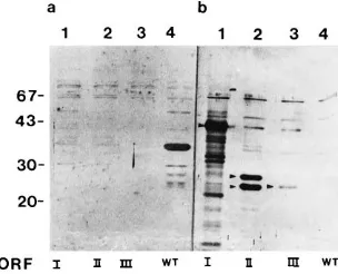
Establishment of a Western blot assay to detect antibodies to BDV antigens. To detect anti-BDV antibodies present in human sera, we developed a Western blot assay using as target antigens BDV p40, p24, and p16 proteins expressed in insect cells by using the baculovirus system. The expression of these proteins was verified by using rabbit antisera raised against
each of the three BDV proteins synthesized inE. colias TrpE
[image:3.612.61.568.82.183.2]fusion proteins (Fig. 1). The p40 and p24 proteins migrated with their expected electrophoretic mobilities, although in the case of p24 protein, we observed a doublet. Whether the lower p24 band represents a degradation product or differences in posttranslational modifications is unknown. The p16 protein encoded by BDV ORF III has recently been shown to be glycosylated in vivo, thus migrating with an apparent molecular mass of 18 kDa (27). However, in our system, p16 exhibited an apparent molecular mass of about 22 kDa, probably as a result of a glycosylation pattern characteristic for insect cells (23, 36). The specificity of the immunoreactivity exhibited by the sera from rabbits immunized with recombinantly expressed BDV proteins was supported by results obtained in assays using a serum of a rabbit acutely infected with BDV. This serum rec-ognized both p40 and p24 antigens, but it did not react with
[image:3.612.360.512.481.604.2]FIG. 1. Expression of BDV p40, p24, and p16 proteins in insect cells. ORFs I to III of BDV, corresponding to proteins with predicted molecular masses of 40, 24, and 16 kDa, respectively, were expressed in insect cells by using the baculovirus system. Lysates of infected SF158 insect cells were separated by SDS-PAGE (12% gel) and transferred to nitrocellulose, and the recombinant proteins were visualized by using specific polyclonal rabbit antibodies at a dilu-tion of 1:100. (a) Preimmune rabbit sera were mixed and incubated with a membrane containing extracts of cells infected with a wild-type (WT) baculovirus (lane 4) or with recombinant baculoviruses expressing the indicated BDV pro-teins. (b) In parallel, a second membrane strip containing the same set of samples was incubated with a mixture of specific polyclonal rabbit antisera directed against BDV p40, p24, and p16 proteins. The positions of the different recom-binant BDV proteins are indicated by arrowheads. The electrophoretic mobili-ties and sizes (in kilodaltons) of molecular mass marker proteins (Pharmacia) are indicated on the left.
TABLE 2. Primers used for nested PCR for detection of BDV p40 and p24 cDNAs
Gene PCR
round Primer
a Sequence Nucleotide positions in BDV RNA genome
p40 1st BV259F 59-TTCATACAGTAACGCCCAGC-39 259–278
BV829R 59-GCAACTACAGGGATTGTAAGGG-39 829–808
2nd BV277F 59-GCCTTGTGTTTCTATGTTTGC-39 277–297
BV805R 59-GCATCCATACATTCTGCGAG-39 805–766
p24 1st BV1387F 59-TGACCCAACCAGTAGACCA-39 1387–1405
BV1865R 59-GTCCCATTCATCCGTTGTC-39 1865–1847
2nd BV1443F 59-TCAGACCCAGACCAGCGAA-39 1443–1461
BV1834R 59-AGCTGGGGATAAATGCGCG-39 1834–1816
a
F and R indicate the antigenomic and genomic, respectively, polarities.
on November 9, 2019 by guest
http://jvi.asm.org/
p16 (data not shown). In addition, recombinant p40 and p24 BDV antigens were recognized by a serum from a mouse infected with BDV (data not shown).
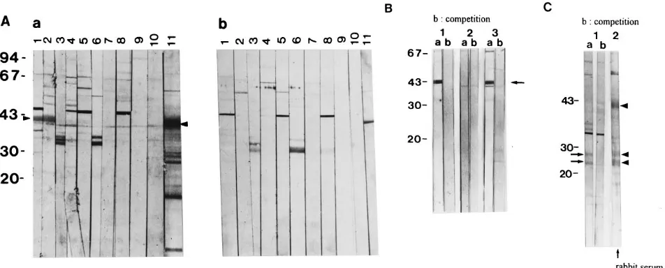
Detection of antibodies to BDV proteins in human sera.We tested a total of 416 serum specimens from patients treated at the Department of Psychiatry of the University Hospital in Homburg for a variety of mood disorders (Table 1) for the presence of BDV-specific antibodies. Serum specimens were scored as positive only when their corresponding immunore-activities in Western blots could be competed against by pincubation with extracts from insect cells infected with a re-combinant baculovirus expressing the respective viral antigen (Fig. 2). Extracts from insect cells infected with wild-type bac-ulovirus were used as a negative control for the competition experiments (data not shown). Of the 416 serum specimens tested, we found 40 (9.6%) positive for BDV. One serum reacted exclusively with p24, two sera reacted with both p40 and p24, and 37 sera showed reactivity only with p40. No serum specimen with antibodies to p16 was detected. Of the 40 sero-positive patients, 24 were male and 16 were female. This sex
ratio difference is not significant (x250.136). Figure 2 shows
representative examples of Western blot analyses. As a control group, we tested sera of 203 age- and sex-matched control patients treated in the University Hospital in Homburg for surgical procedures or diseases unrelated to viral infections or mood disorders. Among these sera, we detected only three (1.4%) BDV-positive samples, and these reacted exclusively with p40. The diagnoses of all seropositive psychiatric patients and all diagnoses available from the seronegative patients are listed in Table 1. The sevenfold-increased BDV seroprevalence
found in psychiatric patients is statistically significant (x2 5
0.001), but there was no correlation between BDV seroposi-tivity and any specific diagnosis.
Detection of BDV RNA sequences in human PBMCs. To investigate the prevalence of BDV RNA in PBMCs from psy-chiatric patients, we used RT-PCR procedures to detect BDV p40 and p24 RNA sequences. Twenty-eight blood samples from 26 patients with various psychiatric diseases (Table 3) were collected. From one patient, we examined three blood samples taken at admission and 16 and 19 weeks after hospi-talization. In addition, blood samples from 23 healthy volun-teers from the Homburg area were also collected. PBMCs from all 51 blood samples were prepared in the Department of Virology of the University of the Saarland in Homburg and coded, and aliquots from each PBMC sample were indepen-dently analyzed by RT-PCR in Homburg and at The Scripps Research Institute in La Jolla. Total RNA isolated from PBMCs was reverse transcribed and then analyzed by nested PCR for the presence of BDV p40 and p24 RNA sequences (Table 3). As a control for RNA quality, we analyzed all PBMC samples for the presence of GAPDH RNA.
[image:4.612.70.546.70.262.2]After performance of the p40-specific RT-PCR, a predicted 528-bp DNA fragment was detected by ethidium bromide staining in 8 of the 28 PBMC samples analyzed in the La Jolla laboratory (Fig. 3A) and in six of 25 samples analyzed in the Homburg laboratory (Table 3). Southern blot hybridization confirmed the specificity of the RT-PCR products (Fig. 3A and data not shown). RT-PCR for p40 on sample 30 resulted in amplification of a p40-specific 528-bp DNA fragment detect-able only by Southern blot hybridization (Fig. 3A, lower panel,
FIG. 2. Detection in human sera of antibodies directed against BDV proteins. (A) Extracts from wild-type (b) and BDV p40 recombinant (a) baculovirus-infected cells were separated by SDS-PAGE (12% gel) and analyzed by Western blotting. Pairs of membrane strips, corresponding to extracts from wild-type and p40 recombinant baculovirus-infected cells, were reacted in parallel with human sera at a dilution of 1:20. Lanes 1 to 11 show immunoreactivities of 10 representative human sera (lanes 1 to 10) and an anti-p40 rabbit serum (lane 11). Lanes 1 and 2 illustrate representative immunoreactivities of p40-positive human sera; lanes 3 to 10 illustrate BDV-negative human sera. Electrophoretic migration of p40 is indicated by arrowheads. (B) Specificity of human sera p40 immunoreactivity. The specificity of the human sera was demonstrated by competition experiments using soluble p40 antigen. Lanes 1 and 3 show the reactions of two positive sera; lane 2 shows the reaction of a negative serum. The position of p40 is indicated by the arrow. Lanes a and b show the immune reactivity before and after, respectively, preincubation of the sera with soluble p40 antigen. Preincubation of the sera with extract from wild-type virus-infected insect cells did not compete for the binding of the human antibodies to p40 (data not shown). (C) Human serum p24 immunoreactivity. A mixture of extracts from BDV p40 and p24 recombinant baculovirus-infected cells was separated by SDS-PAGE (12.5% gel) and analyzed by Western blotting. The positions of the recombinantly expressed p40 and p24 proteins were detected by using a mixture of p40- and p24-specific rabbit antisera (lane 2). Lanes 1a and 1b show the immunoreactivities of a human serum with recombinantly expressed p24 before and after, respectively, preincubation of the serum with soluble p24 antigen. Preincubation of the serum with extract from wild-type virus-infected insect cells did not compete for the binding of the antibodies to p24 (data not shown). Incubation of the human serum with a membrane strip containing electrophoretically separated extract from wild-type baculovirus-infected cells did not show any immunoreactivity with a protein of 24 kDa (data not shown). Molecular mass marker proteins (positions indicated in kilodaltons) were the same as used for Fig. 1. Positions of BDV p24 (double band) and p40 proteins are indicated by arrows on the left and arrowheads on the right, respectively.
7716 SAUDER ET AL. J. VIROL.
on November 9, 2019 by guest
http://jvi.asm.org/
lane 30). In case of sample 10, shown as p40 negative in Fig. 3A, lane 10, additional independent RT-PCR assays detected by Southern blot hybridization a specific signal that corre-sponded to a deleted form of p40 (Table 3). In addition, rep-etition of the p40 nested PCR with the RT product of sample 1 (Fig. 3A) in La Jolla, using 50 instead of 40 cycles, amplified a 528-bp fragment (Table 3) confirmed to be p40 specific by Southern blot hybridization (data not shown). Only two PBMC samples, 11 and 19, were found p40 positive solely in one of the two laboratories (Table 3).
The p24-specific RT-PCR yielded a predicted 391-bp DNA fragment detected by ethidium bromide staining in 11 of 26 PBMCs analyzed in the Homburg laboratory (Fig. 3B). All signals were shown to be p24 specific by Southern blot hybrid-ization (Fig. 3B). All seven PBMCs samples found p40 positive by ethidium bromide staining in La Jolla were also found to be positive for p24 in Homburg. In addition, all six p40-positive samples analyzed in Homburg were also p24 positive at the same location. PBMC samples 2, 18, and 31, obtained at dif-ferent times from the same patient, were positive for both BDV p40 and p24 (Table 3).
In all RNA samples analyzed by RT-PCR, we could amplify a 192-bp GAPDH fragment (Fig. 3A and data not shown).
Some minor discrepancies found between RT-PCR results ob-tained in the two laboratories (Table 3) are likely related to differences in experimental procedures and quality of the RNA samples analyzed.
As a control group, we analyzed by RT-PCR 23 PBMC samples from healthy volunteers. All samples were analyzed for p40 in both laboratories and also for p24 in the Homburg laboratory. All samples were found to be negative for both p40 and p24 by ethidium bromide staining of the gel, whereas we could amplify the 192-bp GAPDH fragment in all RNA sam-ples analyzed (data not shown).
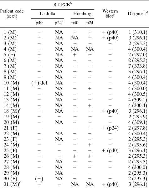
Sera from the 28 blood samples from the psychiatric patients investigated by RT-PCR were also analyzed in the Western blot assay for the presence of antibodies against BDV p40 or BDV p24 (Table 3). Only 6 of the 28 samples had anti-BDV antibodies. All three blood samples from the same patient positive by RT-PCR were also positive for anti-p40 antibodies. We found that a high proportion (9 of 13) of the patients found to be positive for BDV RNA did not have detectable serum antibodies to BDV (Table 3).
The diagnoses of the 26 patients could be mainly assigned to three (Table 3) of the categories indicated in Table 1. BDV RNA-positive patients were found in all three groups. A high percentage (7 of 11) of patients diagnosed with schizophrenia (category 2) were BDV RNA positive. There was no significant difference in the sex ratio among RT-PCR-positive patients.
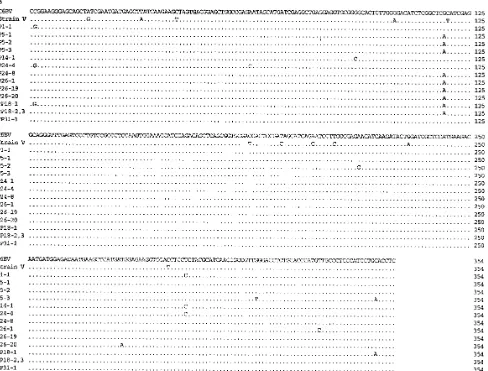
Sequence analysis of the p40 and p24 RT-PCR products derived from human PBMCs.To determine the sequences of the amplified 528-bp p40 and 391-bp p24 fragments derived from human PBMCs, PCR products were cloned and the nu-cleotide sequences of one to three clones of each PCR product were determined. Results of sequence analyses are presented in Fig. 4. We sequenced 21 different p40 clones derived from eight different PBMC samples and 13 different p24 clones derived from seven different PBMC samples. In addition, one p24 sequence was derived by sequencing an uncloned PCR product (Fig. 4B). BDV sequences derived from human PBMCs displayed a high degree of conservation with respect to sequences of BDV strains V (8) and C6BV (14), two previously characterized viral sequences derived originally from two nat-urally BDV-infected horses (Table 4). In cases in which two or more clones of the same PCR product were sequenced, single-clone-specific substitutions and changes that affected all clones derived from the same PCR product were found (Fig. 4), with intrapatient divergencies shown in Table 4. In case of p40, no common mutation between two or more patient samples was found. We identified a mutation in p24 at nucleotide position 1579 in the BDV genome which was common to many but not all patients. Interpatient divergencies for p40 and p24 were only slightly higher than the respective intrapatient divergen-cies (Table 4). Substitutions in PBMC derived p40 and p24 sequences appeared randomly distributed, with no detectable clusters. To rule out the possibility of mutations introduced via nested PCR, p40 plasmid DNA of the C6BV sequence was amplified via nested PCR and cloned, and three individual clones were sequenced. PCR experimental conditions were identical to those described above for the analysis of PBMC samples. All three sequences were 100% identical to the ex-pected C6BV sequence; thus, mutations due to experimental procedures seemed unlikely.
[image:5.612.59.297.91.393.2]Compared with C6BV, 21 (72.4%) of 29 and 8 (80%) of 10 nucleotide substitutions found in PBMC p40 and p24 se-quences, respectively, involved amino acid substitutions (Table 5), whereas compared with strain V, 45.7% of the 46 and 38.1% of the 21 nucleotide substitutions found in PBMC-derived p40 and p24 sequences, respectively, corresponded to
TABLE 3. Summary of RT-PCR and Western blot analyses of 28 blood samples from 26 psychiatric patients
Patient code (sexa
)
RT-PCRb
Western
blotc Diagnosisd La Jolla Homburg
p40 p24e p40 p24
1 (M) 1 NA 1 1 1(p40) 1 (310.1)
2 (M)f 1 NA NA 1 1(p40) 3 (296.1)
3 (M) 1 NA 1 1 2 2 (295.3)
4 (M) 1 NA NA NA 2 4 (300.4)
5 (F) 1 NA 1 1 2 2 (297.0)
6 (M) 2 NA 2 2 2 2 (295.3)
7 (M) 2 NA 2 2 2 7 (333.8)
8 (M) 2 NA 2 2 2 3 (296.1)
9 (M) 2 NA 2 2 2 4 (300.4)
10 (M) (1) del NA 2 2 2 4 (300.4)
11 (M) 1 NA 2 1 2 4 (300.0)
12 (M) 2 NA 2 2 2 4 (300.5)
13 (M) 2 NA 2 2 2 4 (309.1)
14 (M) 2 NA 2 1 2 4 (300.4)
18 (M)f 1 NA 1 1 1(p40) 3 (296.1)
19 (M) 2 2 1 1 2 2 (295.9)
20 (M) 2 NA 2 2 2 4 (309.1)
21 (F) 2 2 2 1 1(p24) 2 (297.8)
22 (M) 2 NA 2 2 2 4 (300.4)
23 (F) 2 NA 2 2 2 2 (295.3)
24 (M) 2 2 2 1 2 2 (295.6)
25 (F) 2 2 2 2 1(p40) 3 (296.1)
26 (M) 1 2 1 1 2 2 (295.3)
27 (M) 2 NA 2 2 2 2 (295.3)
28 (M) 2 NA 2 2 2 4 (300.0)
29 (M) 2 NA 2 2 2 2 (295.3)
30 (F) (1) NA 2 2 2 2 (295.3)
31 (M)f 1 1 NA NA 1(p40) 3 (296.1)
a
M, male; F, female. b
(1), signal visible only after Southern blot hybridization; del, deleted p40 DNA fragment; NA, not analyzed.
c
Antibody reaction against BDV p40 or p24 is indicated in parentheses. d
Diagnosis as assigned to the seven main categories (Table 1) and, in paren-theses, ICD-9 code number.
e
In six of the PBMC samples analyzed for p40 in La Jolla, random hexamers were used to prime the RT. These RT products were reanalyzed in a p24-specific nested PCR.
f
Samples taken from the same patient at different time points.
on November 9, 2019 by guest
http://jvi.asm.org/
7718 SAUDER ET AL. J. VIROL.
on November 9, 2019 by guest
http://jvi.asm.org/
amino acid exchanges. Strains V and C6BV are identical at the amino acid level within the regions of p40 and p24 analyzed in this study. Hence, PBMC-derived sequences exhibited the same amino acid divergency with respect to both strains V and C6BV. The determined intra- and interpatient divergencies of the deduced amino acid sequences were slightly higher than those at the nucleotide level for both p40 and p24 (Table 4). Some of the amino acid exchanges found are nonconservative. The mutation in p24 at nucleotide position 1579 in the BDV genome found in the sequences of many patients corresponded to a conservative amino acid exchange (Arg to His). Amino acid substitutions common to all sequenced clones indicative of a specific human BDV strain could not be observed. Intra-and interpatient divergencies determined for both p24 Intra-and p40 did not reveal significant differences in divergence rates be-tween the two BDV genes. p40 RT-PCR products of samples 2, 18, and 31, and p24 RT-PCR products of samples 18 and 31, all from the same patient, were sequenced. These samples were taken in intervals of 4 months (samples 2 and 18) or 3 weeks (samples 18 and 31). Comparison of the different quences revealed minor fluctuations in the distribution of se-quences present at a given time within the patient PBMCs. However, during the period of time separating the first and last sample analyzed, the consensus sequences of both p40 and p24 remained unchanged. Thus, no evolution of BDV p40 se-quence was observed during 19 weeks of infection in the same patient.
DISCUSSION
Seroepidemiological studies indicate a significantly higher prevalence of BDV antibodies in neuropsychiatric patients than in healthy individuals (5). However, these studies have relied mostly on immunofluorescence assays (IFA) using BDV-infected cells (5), impeding the identification of the viral antigens recognized by the human sera. Moreover, very low BDV antibody titers in human sera (2, 5, 42, 43), together with the frequent finding in neuropsychiatric patients of autoanti-bodies that often have a staining pattern very similar to that characteristically associated with BDV infection (19, 30, 51), have contributed to misleading results in studies using IFA (58). Although there is a lack of consensus on a reliable sero-logical method, a recent report suggested that Western blot assays may be more reliable than IFA for evaluating BDV serology (58). Therefore, we developed a Western blot assay using recombinant BDV p40, p24, and p16 polypeptides as sources of target antigens to detect antibodies in human sera directed against these viral proteins.
We detected antibodies that recognized BDV antigens in 40 9.6%) of 416 serum specimens from neuropsychiatric patients, whereas only 3 (1.4%) of 203 samples from the control group had BDV-positive serology. These findings are consistent with the previously reported higher seroprevalence of BDV among
neuropsychiatric patients (5). Most of the BDV-positive hu-man sera (37 of 40) recognized only BDV p40. Two serum specimens recognized both p40 and p24 antigens, whereas only one serum sample recognized exclusively p24, and none of the sera examined recognized BDV p16. Consistent with previ-ously documented studies (2, 5, 18, 42, 43), our results indi-cated that positive human sera had very low titers of BDV antibodies. The possible contribution of immune complexes to this phenomenon cannot be excluded (11). The low serum dilutions (1:20) used in the Western blot assay probably con-tributed to the nonspecific staining of some proteins present in the extracts used as sources of target antigens. However, in all cases, the immunoreactivity of human sera with p40 and p24 BDV antigens could be blocked in competition experiments using soluble BDV antigens (Fig. 2 and data not shown). The very low number of BDV antibody-positive sera found in the control group provides additional support for the specificity of the reaction of the human antibodies with BDV p40 and p24 proteins.
In contrast to our findings, two other studies also using a Western blot assay documented that 6.5% and 14.4% of sera from patients with affective disorders (18) or schizophrenia (58), respectively, recognized more than one BDV antigen. Nevertheless, these studies also showed that a high percentage of control sera recognized a single BDV antigen. Sera from BDV-infected animals recognize most frequently both p40 and p24 BDV antigens and less often also p16 (33, 34, 44, 58). The reasons for the prominent immune response against BDV p40 in neuropsychiatric patients described here are unknown. It should be noted that p40 is the BDV counterpart of the nu-cleoprotein (NP) in other nonsegmented, negative-stranded RNA viruses (8, 14, 16, 46). In many cases, the NP is the virus gene product expressed at the highest levels during infection, and the infected host normally mounts a good antibody re-sponse against these viral NPs. We could not detect p16 im-munoreactivity with any of the human sera analyzed. However, we cannot rule out the possibility that abnormal glycosylation of the p16 protein produced in the baculovirus expression system could alter or mask epitope recognition by human sera. Nevertheless, detection of human anti-p16 antibodies in a Western blot assay has been reported only in one study (58). Our results did not reveal a distinct association of BDV seropositivity with a specific neuropsychiatric diagnosis. The diagnoses from 118 seronegative patients were not available; however, assuming that the distribution of diagnoses among these 118 patients would be similar to that found for the 298 characterized patients, we could then estimate a BDV sero-prevalence of approximately 3% among patients with mental disorders related to drugs and alcohol abuse, which was not significantly different from that found in the control group (1.4%). The percentage (10%) of BDV-seropositive patients that we estimated among schizophrenic patients is similar to that recently reported by others (58).
FIG. 3. Detection of BDV p40 and p24 RNAs in PBMCs of psychiatric patients. Twenty-eight PBMC samples from 26 patients from the Homburg area with diverse psychiatric diseases were independently analyzed for the presence of BDV p40 RNA sequences in La Jolla (A) or BDV p24 RNA sequences in Homburg (B). RNA was extracted from PBMCs and reverse transcribed, and nested PCR was performed with BDV p40- or p24-specific sets of primers (Table 2). As a control for RNA quality, RT-PCR analysis for detection of the housekeeping cellular GAPDH mRNA was performed. RT-PCR products were analyzed by 2% agarose gel electro-phoresis and ethidium bromide staining (A, upper panels; B, first and third panels). PCR products were analyzed by Southern blot hybridization with a32P labeled p40
probe (A, lower panels) or a p24 probe labeled with digoxigenin (Boehringer Mannheim) (B, second and fourth panels). PCR products derived from patient PBMCs were applied in lanes 1 to 14 and 18 to 31, respectively. Lane numbers correspond to patients code numbers in Table 3. In lanes 4 and 31 (B), no PCR products were loaded, since the corresponding PBMC samples were not analyzed in the Homburg laboratory. Lanes 15 and 32 show negative RT-PCR controls, for which water instead of RNA was used. RT-PCR products obtained by using total RNA prepared from BDV-negative C6 cells (lanes 16 and 33) or from BDV persistently infected C6BV cells (lanes 17 and 34) were loaded as controls. In the upper panels of panel A, p40 nested RT-PCR products were mixed with the GAPDH RT-PCR products prior to agarose gel electrophoresis. The 528-bp p40 specific, 391-bp p24-specific, and the 192-bp GAPDH-specific fragments are indicated by arrows. Lanes M, (A, upper panels), 100-bp DNA ladder (Gibco BRL). Positions of coelectrophoresed DNA markers are indicated.
on November 9, 2019 by guest
http://jvi.asm.org/
7720 SAUDER ET AL. J. VIROL.
on November 9, 2019 by guest
http://jvi.asm.org/
BDV RNA has been detected in PBMCs of naturally and experimentally infected animals (37, 38, 49) and also in hu-mans (6, 17, 26, 39). This finding provides another diagnostic marker to assess whether BDV prevalence is higher in neuro-psychiatric patients than in healthy individuals. Therefore, we analyzed by RT-PCR RNA extracted from PBMCs from psy-chiatric patients and healthy volunteers from the same geo-graphic area (Homburg). PBMCs from 25 psychiatric patients were analyzed in both laboratories (La Jolla and Homburg) for the presence of BDV p40 and/or p24 RNA sequences. Six (24%) of these patients were found to be BDV positive in both laboratories. Six other patients were found to be BDV RNA positive in only one of the two laboratories, and in two of these cases, BDV sequences were detected only by Southern blot hybridization of the RT-PCR product. One additional patient analyzed only in the La Jolla laboratory was also BDV RNA positive. On the basis of the total number of samples found to be BDV RNA positive in both or only one of the laboratories, 50% (13 out of 26) of the neuropsychiatric patients analyzed carried BDV RNA sequences in their PBMCs. In contrast,
none of the 23 PBMC samples from healthy volunteers was positive for p40 or p24 by ethidium bromide after RT-PCR. One of them was found to be positive for p40 after Southern blot hybridization in La Jolla. Attempts to clone this PCR product have failed, and independent nested PCRs using the same RT product did not reproducibly score this sample as BDV RNA positive. Reconstruction experiments indicated that under our PCR conditions, reproducible positive RT-PCR for p40 RNA could not be achieved with 300 or fewer
RNA target molecules in a background of 5mg of total RNA
[image:9.612.67.555.74.445.2](45). It is possible that the p40 RNA-positive sample found among the healthy controls and the two patient PBMC samples found to be p40 positive only after Southern blot hybridization correspond to cases of very low viral load within the PBMCs. RT-PCR control experiments consisting of the omission of the RT enzyme in the RT step consistently failed to amplify any BDV sequences, ruling out the possibility of contamina-tion with plasmid DNA or amplicons containing BDV se-quences analyzed in our studies. In addition, water samples and BDV-negative RNA samples included as controls in each
FIG. 4. Nucleotide sequence alignment of BDV p40 (A) and p24 (B) among C6BV (14), strain V (8), and cDNA clones derived from psychiatric patient PBMCs. Partial p40 and p24 sequences shown correspond to nucleotides 298 to 785 and 1462 to 1815, respectively, of the C6BV genome (antigenomic polarity). Dots indicate nucleotide identity with respect to C6BV. Numbers on the left indicate the patient code numbers and numbers of the clones sequenced (i.e., p4-1 corresponds to the sequence derived from clone 1 of patient 4). Different clones derived from the same PBMCs with the same nucleotide sequence were summarized in one single sequence (i.e., p3-3 and p3-45p3-3,4). The p24 sequence p18-1 was derived from an uncloned PCR product. Sequences derived from different PBMC samples from the same patient are marked with asterisks.
on November 9, 2019 by guest
http://jvi.asm.org/
RT-PCR assay were also always negative. Moreover, we rigor-ously observed well-established guidelines (50) to avoid con-tamination problems during the performance of RT-PCR as-says. In addition, no work with BDV-infected cells has been conducted in the Homburg laboratory. Furthermore, the unique nucleotide changes found in the p40 and p24 RNA sequences derived from the patient PBMCs clearly
distin-guished them from known animal BDV sequences. Taking all of these findings into account together with the good concor-dance observed between results independently obtained in both laboratories, we can rule out contamination with a labo-ratory source of BDV sequences.
[image:10.612.57.298.108.233.2]Our results did not show a correlation between BDV RNA prevalence in PBMCs of psychiatric patients and specific diag-noses. However, it is worth noting that 7 of 11 patients with diagnoses of schizophrenia were found to be positive for BDV RNA. We observed that only a small subset of patients found to be positive for BDV RNA also had serum antibodies to BDV antigens (Table 3). This lack of correlation between RNA and serological markers has also been found recently by others in humans (26, 39) and animals (37, 38). Seropositive but BDV RNA-negative cases may represent BDV infections that have been cleared by the host immune system. Conversely, detection of viral RNA in the absence of antibodies against BDV may reflect a recent infection with a delayed humoral immune response, as described, for instance, in the case of hepatitis C virus infection (12). BDV could also interfere with the host immune response by using strategies similar to those exploited by other viruses with the ability to persist without eliciting a noticeable humoral response (1). These findings suggest that both serology and RT-PCR should be used to diagnose BDV infection.
TABLE 4. Degree of conservation of human BDV p40 and p24 sequences with respect to animal sequences (C6BV and strain V) and percentages of inter- and intrapatient divergencies of human
BDV p40 and p24 sequences
Protein
Range (%) of:
Sequence divergency in comparison with C6BV
(or strain V)
Interpatient divergency
Intrapatient divergency
p40
Nucleotide level 0–1.03 (3.48–4.57) 0.2–2.05 0–1.43 Amino acid
level
0–1.84 (0–1.84) 0–3.68 0–2.45
p24
Nucleotide level 0–0.85 (3.11–3.95) 0–1.69 0–1.13 Amino acid
level
0–2.56 (0–2.56) 0–4.27 0–2.56
TABLE 5. Nucleotide mutations involving amino acid exchanges found in p40 and p24 sequences derived from patient PBMCs
Protein Patient
code Clone(s)
Nucleotide position in BDV genome RNA
Nucleotide changea
Amino acid position in BDV ORF
Amino acid changea
p40 5 3, 4, 6 328 A3C 92 His3Pro
26 1, 2, 3 333 G3U 94 Gly3Trp
31 1 336 G3C 95 Val3Leu
18 1 402 A3G 117 Lys3Glu
26 2 418 A3G 122 Lys3Arg
18 1, 2, 11 493 U3C 147 Val3Ala
11 3 531 G3A 160 Glu3Lys
2 6 543 A3G 164 Lys3Glu
5 6 565 C3U 171 Ala3Val
5 6 571 U3C 173 Leu3Ser
4 1, 2 579 C3U 176 Pro3Ser
4 1, 2 586 A3U 178 His3Leu
5 3 592 A3G 180 Glu3Gly
11 2 610 A3U 186 Gln3Leu
5 4 625 A3G 191 His3Arg
11 2 630 G3U 193 Ala3Ser
4 1, 2 636 G3U 195 Asp3Tyr
31 4 649 G3U 199 Gly3Val
31 1 651 C3A 200 Gln3Lys
31 1 673 U3A 207 Val3Glu
18 1 756 A3U 235 Met3Leu
p24 24 4 1524 A3C 85 Asn3His
14 1 1554 G3C 95 Gly3Arg
5 1, 2, 3
18 1, 2, 3 1579 G3A 103 Arg3His
24 8
26 1, 19, 20
5 2 1680 A3G 137 Asn3Asp
26 20 1738 U3A 156 Met3Lys
1 2
14 1 1756 U3C 162 Leu3Pro
24 4
5 3 1776 G3U 169 Gly3Trp
5 3 1810 C3A 180 Ala3Glu
18 1
a
Change found in PBMCs (right position) compared with C6BV and strain V sequences (left position).
7722 SAUDER ET AL. J. VIROL.
on November 9, 2019 by guest
http://jvi.asm.org/
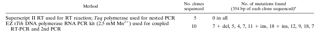
[image:10.612.62.555.362.708.2]Analysis of BDV p40 and p24 RNA sequences derived from patient PBMCs revealed a high degree of sequence conserva-tion compared with known animal BDV sequences. This find-ing is consistent with our previous results obtained from se-quencing of p24, p16, and p56 genes of BDV isolates from three psychiatric patients by cocultivation of patient PBMCs with BDV-susceptible cells (17). Because of the lack of proof-reading activity associated with their polymerases, RNA vi-ruses exhibit high mutation frequencies, which provides the potential for the frequently observed rapid RNA virus evolu-tion (22). However, RNA viruses can also display long-term stasis both in nature and in laboratory conditions as a result of selection for fit master sequences in rather constant environ-ments (22, 59). A high level of sequence conservation has been also found among BDV sequences derived from naturally in-fected horses from different geographic areas and separated by more than 10 years (47), as well as for BDV sequences derived from naturally infected animals of different species (4). Whether this high sequence conservation is due to a hypothet-ical, as yet unidentified proofreading function of the BDV RNA polymerase, or the result of selective pressure acting on tight structure-function constraints, remains to be determined. Sequence variability of BDV p24 in PBMCs significantly higher than that here reported has been recently found in Japanese neuropsychiatric patients (24). It is conceivable that different strains of BDV are present in Japan. Differences in the characteristics of patient populations analyzed in Japan and Germany could also account for the differences in the rate of sequence variation. Nevertheless, we have evidence that different experimental procedures used for the RT-PCR anal-ysis of human PBMCs also significantly contributed to these differences. Studies conducted on Japanese patients (26) used
the EZ rTthRNA PCR system (Perkin-Elmer). This method is
based on the use of rTthpolymerase, which allows both reverse
transcription and DNA amplification to be conducted in a single buffer system which contains manganese. Because the negative effect of manganese on the fidelity of DNA synthesis is well documented (3, 31), we compared the experimentally induced mutation frequencies found under our RT-PCR
con-ditions and by the EZ rTthRNA PCR method. For this
pur-pose, p24 RNA was generated by in vitro transcription via T7 polymerase from a plasmid containing the C6BV p24 ORF. Equal amounts of p24 RNA were used to amplify the same
fragment within p24, using the EZ rTthRNA PCR method as
described previously (26) or our RT-PCR conditions. PCR products generated by both methods were cloned, and the sequences of several independent clones were determined (Ta-ble 6). Sequence analysis of five clones derived from PCR products obtained with our RT-PCR conditions showed 100% identity with respect to the C6BV p24 plasmid sequence. In contrast, 10 clones derived from PCR products generated with
the EZ rTth RNA PCR method showed 4 to 18 nucleotide
substitutions per clone with respect to the template C6BV p24 RNA, representing an intrasample divergency as high as 10.2%. Two of these clones had also single-base insertions or
deletions. These results indicate that the higher BDV sequence variation previously reported (24) may have been overesti-mated because of technical procedures.
The route of BDV transmission has not been clearly estab-lished, and how humans could be infected with BDV is un-known. The high conservation observed between human and animal BDV sequences suggests the possibility of zoonosis. However, transmission of BDV to humans via blood seems also plausible. In this regard, a prevalence of 4.2 to 5% of BDV RNA in PBMCs has been recently reported among blood do-nors in Japan (25). Our data provide strong support for the contention that BDV is associated with certain human psychi-atric disorders, possibly contributing to the pathophysiology of these disorders. On the basis of such a hypothesis, it would be predicted that similar associations should be found with blood samples from psychiatric patients from different geographic areas. As yet there is only limited information from psychiatric patients from Germany and Japan. Hence, there is a need for comprehensive molecular epidemiological studies to rigorously evaluate the contribution of BDV to human mental disorders.
ACKNOWLEDGMENTS
C. Sauder and A. Mu¨ller contributed equally to this work. We thank Barbara Ga¨rtner, Daniel Gonzalez-Dunia, and Michael Teng for helpful discussions, Dorothee Gra¨sser for analysis of the patients’ diagnoses, Anne Bang for the sequence alignments, and Si-grid Ko¨nig for excellent technical assistance. We thank Georg Pauli for the generous gift of serum from a BDV-infected rabbit, Steven Rubin for serum from a BDV-infected mouse, and Peter Staeheli for the generous gift of RNA isolated from BDV-infected and uninfected C6 cells.
This work was supported by a grant from the University of the Saarland to N. Mueller-Lantzsch and by grant RO1 NS/AI 32355 to J. C. de la Torre; C. Sauder is a recipient of a fellowship sponsored by the Bundesministerium fu¨r Forschung und Technologie.
REFERENCES
1.Ahmed, R., L. A. Morrison, and D. M. Knipe.1996. Persistence of viruses, p. 219–249.InB. N. Fields, D. M. Knipe, P. M. Howley, et al. (ed.), Fields virology, 3rd ed., Lippincott-Raven Publishers, Philadelphia.
2.Bechter, K., S. Herzog, and R. Schuettler.1992. Possible significance of Borna disease for humans. Neurol. Psychiatry and Brain Res.1:23–29. 3.Beckman, R. A., A. S. Mildvan, and L. A. Loeb.1985. On the fidelity of DNA
replication: manganese mutagenesis in vitro. Biochemistry24:5810–5817. 4.Binz, T., J. Lebelt, H. Niemann, and K. Hagenau.1994. Sequence analyses of
the p24 gene of Borna disease virus in naturally infected horses, donkey and sheep. Virus Res.34:281–289.
5.Bode, L.1995. Human infections with Borna disease virus and potential pathogenic implications, p. 103–130.InH. Koprowski and I. Lipkin (ed.), Borna disease. Springer-Verlag, Berlin.
6.Bode, L., W. Zimmermann, R. Ferszt, F. Steinbach, and H. Ludwig.1995. Borna disease virus genome transcribed and expressed in psychiatric pa-tients. Nat. Med.1:232–236.
7.Briese, T., J. C. de la Torre, A. Lewis, H. Ludwig, and W. I. Lipkin.1992. Borna disease virus, a negative-strand RNA virus, transcribes in the nucleus of infected cells. Proc. Natl. Acad. Sci. USA89:11486–11489.
8.Briese, T., A. Schneemann, A. J. Lewis, Y. S. Park, S. Kim, H. Ludwig, and W. I. Lipkin.1994. Genomic organization of Borna disease virus. Proc. Natl. Acad. Sci. USA91:4362–4366.
[image:11.612.49.555.83.137.2]9.Buesa-Gomez, J., J. C. de la Torre, T. Dyrberg, M. Landin-Olsson, R. S.
TABLE 6. Comparison of two different RT-PCR methods
Method No. clones sequenced
No. of mutations found (354 bp of each clone sequenced)a
Superscript II RT used for RT reaction;Taqpolymerase used for nested PCR 5 0 in all EZ rTthDNA polymerase RNA PCR kit (2.5 mM Mn21) used for coupled
RT-PCR and 2nd PCR 10 71del, 5, 4, 7, 111ins, 181ins, 12, 9, 18, 7
ains and del, single-base-pair insertion and deletion, respectively.
on November 9, 2019 by guest
http://jvi.asm.org/
Mauseth, A. Lernmark, and M. B. Oldstone.1994. Failure to detect genomic viral sequences in pancreatic tissues from two children with acute-onset diabetes mellitus. J. Med. Virol.42:193–197.
10. Bundesminister fu¨r Jugend, Familie und Gesundheit.1988. Manual of the international statistical classification of diseases, injuries, and causes of death (ICD-9) (German version), 9th version. Verlag W. Kohlhammer GmbH, Cologne, Germany.
11. Casali, P., and M. B. A. Oldstone.1983. Immune complexes in viral infec-tion, p. 7–48.InP. A. Bachmann (ed.), New developments in diagnostic virology, Springer-Verlag, Berlin.
12. Chien, D. Y., Q. L. Choo, A. Tabrizi, C. Kuo, J. McFarland, K. Berger, C. Lee, J. R. Shuster, T. Nguyen, D. L. Moyer, M. Tong, S. Furuta, M. Omata, T. G., H. Alter, E. Schiff, L. Jeffers, M. Houghton, and G. Kuo.1992. Diagnosis of hepatitis C virus (HCV) infection using an immunodominant chimeric polyprotein to capture circulating antibodies: reevaluation of the role of HCV in liver disease. Proc. Natl. Acad. Sci. USA89:10011–10015. 13. Cubitt, B., and J. C. de la Torre.1994. Borna disease virus (BDV), a
nonsegmented RNA virus, replicates in the nuclei of infected cells where infectious BDV ribonucleoproteins are present. J. Virol.68:1371–1381. 14. Cubitt, B., C. Oldstone, and J. C. de la Torre.1994. Sequence and genome
organization of Borna disease virus. J. Virol.68:1382–1396.
15. Cubitt, B., C. Oldstone, J. Valcarcel, and J. C. de la Torre.1994. RNA splicing contributes to the generation of mature mRNAs of Borna disease virus, a non-segmented negative strand RNA virus. Virus Res.34:69–79. 16. de la Torre, J. C.1994. Molecular biology of Borna disease virus: prototype
of a new group of animal viruses. J. Virol.68:7669–7675.
17. de la Torre, J. C., L. Bode, R. Du¨rrwald, B. Cubitt, and H. Ludwig. Molec-ular characterization of Borna disease virus isolated from peripheral blood mononuclear cells of psychiatric patients. Virus Res., in press.
18. Fu, Z. F., J. D. Amsterdam, M. Kao, V. Shankar, H. Koprowski, and B. Dietzschold.1993. Detection of Borna disease virus-reactive antibodies from patients with affective disorders by Western immunoblot technique. J. Af-fective Disord.27:61–68.
19. Ganguli, R., B. S. Rabin, K. N. R. Chengappa, V. L. Nimgaonkar, C. G. McAllister, Z. W. Yang, and J. S. Brar.1992. Autoimmune phenomena in schizophrenic patients. Abstr. Schizophr. Res.6:140.
20. Gra¨sser, F. A., P. Haiss, S. Goettel, and N. Mueller-Lantzsch.1991. Bio-chemical characterization of Epstein-Barr virus nuclear antigen 2A. J. Virol.
65:3779–3788.
21. Groebe, D. R., A. E. Chung, and C. Ho.1990. Cationic lipid-mediated co-transfection of insect cells. Nucleic Acids Res.18:4033.
22. Holland, J. J. (ed.).1992. Genetic diversity of RNA viruses. Curr. Top. Microbiol. Immunol.176.
23. King, L. A., and R. D. Possee.1992. The baculovirus expression system. Chapman and Hall, London.
24. Kishi, M., Y. Arimura, K. Ikuta, Y. Shoya, P. K. Lai, and M. Kakinuma.
1996. Sequence variability of Borna disease virus open reading frame II found in human peripheral blood mononuclear cells. J. Virol.70:635–640. 25. Kishi, M., T. Nakaya, Y. Nakamura, M. Kakinuma, T. A. Takahashi, S.
Sekiguchi, M. Uchikawa, K. Tadokoro, K. Ikeda, and K. Ikuta.1995. Prev-alence of Borna disease virus RNA in peripheral blood mononuclear cells from blood donors. Med. Microbiol. Immunol.184:135–138.
26. Kishi, M., T. Nakaya, Y. Nakamura, Q. Zhong, K. Ikeda, M. Senjo, M. Kakinuma, S. Kato, and K. Ikuta.1995. Demonstration of human Borna disease virus RNA in human peripheral blood mononuclear cells. FEBS Lett.364:293–297.
27. Kliche, S., T. Briese, A. H. Henschen, L. Stitz, and W. I. Lipkin.1994. Characterization of a Borna disease virus glycoprotein, gp18. J. Virol.68:
6918–6923.
28. Koerner, T. J., J. E. Hill, A. M. Myers, and A. Tzagoloff.1991. High-expression vectors with multiple cloning sites for construction of trpE fusion genes: pATH vectors. Methods Enzymol.194:477–490.
29. Krug, M. S., and S. L. Berger.1987. First-strand cDNA synthesis primed with oligo(dT). Methods Enzymol.152:316–325.
30. Legros, S., J. Mendlewicz, and J. Wybran.1985. Immunoglobulins, autoan-tibodies and other serum fractions in psychiatric disorders. Eur. Arch. Psy-chiatry Neurol. Sci.235:9–11.
31. Leung, D. W., E. Chen, and D. V. Goeddel.1989. A method for random mutagenesis of a defined DNA segment using a modified polymerase chain reaction. Technique1:11–15.
32. Lipkin, W. I., T. Briese, and J. C. de la Torre.1992. Borna disease virus: molecular analysis of a neurotropic infectious agent. Microb. Pathog.13:
167–170.
33. Ludwig, H., L. Bode, and G. Gosztonyi.1988. Borna disease: a persistent virus infection of the central nervous system. Prog. Med. Virol.35:107–151.
34. Ludwig, H., V. Koester, G. Pauli, and R. Rott.1977. The cerebrospinal fluid of rabbits infected with Borna disease virus. Arch. Virol.55:209–223. 35. Morozov, P. V. (ed.).1983. Research on the viral hypothesis of mental
disorders. Adv. Biol. Psychiatry12. 36. Mu¨ller, A.Unpublished results.
37. Nakamura, Y., S. Asahi, T. Nakaya, M. K. Bahmani, S. Saitoh, K. Yasui, H. Mayama, K. Hagiwara, C. Ishihara, and K. Ikuta.1996. Demonstration of Borna disease virus RNA in peripheral blood mononuclear cells derived from domestic cats in Japan. J. Clin. Microbiol.34:188–191.
38. Nakamura, Y., M. Kishi, T. Nakaya, S. Asahi, H. Tanaka, H. Sentsui, K. Ikeda, and K. Ikuta.1995. Demonstration of Borna disease virus RNA in peripheral blood mononuclear cells from healthy horses in Japan. Vaccine
13:1076–1079.
39. Nakaya, T., H. Takahashi, Y. Nakamura, S. Asahi, M. Tobiume, H. Kurat-sune, T. Kitani, K. Yamanishi, and K. Ikuta.1996. Demonstration of Borna disease virus RNA in peripheral blood mononuclear cells derived from Japanese patients with chronic fatigue syndrome. FEBS Lett.378:145–149. 40. Richt, J. A., S. VandeWoude, M. C. Zinc, J. E. Clements, S. Herzog, L. Stitz, R. Rott, and O. Narayan.1992. Infection with Borna disease virus: molecular and immunobiological characterization of the agent. Clin. Infect. Dis.14:
1240–1250.
41. Rott, R., and H. Becht.1995. Natural and experimental Borna disease in animals, p. 17–30.InH. Koprowski and W. I. Lipkin (ed.), Borna disease, Springer-Verlag, Berlin.
42. Rott, R., S. Herzog, K. Bechter, and K. Frese.1991. Borna disease, a possible hazard for man? Arch. Virol.118:143–149.
43. Rott, R., S. Herzog, B. Fleischer, A. Winokur, J. Amsterdam, W. Dyson, and H. Koprowski.1985. Detection of serum antibodies to Borna disease virus in patients with psychiatric disorders. Science228:755–756.
44. Rubin, S. A., R. 2. Waltrip, J. R. Bautista, and K. M. Carbone.1993. Borna disease virus in mice: host-specific differences in disease expression. J. Virol.
67:548–552.
45. Sauder, C.Unpublished results.
46. Schneemann, A., P. A. Schneider, R. A. Lamb, and W. I. Lipkin.1995. The remarkable coding strategy of borna disease virus: a new member of the nonsegmented negative strand RNA viruses. Virology210:1–8.
47. Schneider, P. A., T. Briese, W. Zimmermann, H. Ludwig, and W. I. Lipkin.
1994. Sequence conservation in field and experimental isolates of Borna disease virus. J. Virol.68:63–68.
48. Schneider, P. A., A. Schneemann, and W. I. Lipkin.1994. RNA splicing in Borna disease virus, a nonsegmented, negative-strand RNA virus. J. Virol.
68:5007–5012.
49. Sierra-Honigmann, A. M., S. A. Rubin, M. G. Estafanous, R. H. Yolken, and K. M. Carbone.1993. Borna disease virus in peripheral blood mononuclear and bone marrow cells of neonatally and chronically infected rats. J. Neu-roimmunol.45:31–36.
50. Simmonds, P.1995. Polymerase chain reaction, p. 107–142.InU. Dessel-berger (ed.), Medical virology. Oxford University Press, Oxford. 51. Sirota, P., K. Schild, M. Firer, A. Tanay, A. Elizur, D. Meytes, and H. Slor.
1991. Autoantibodies to DNA in multicase families with schizophrenia. Biol. Psychiatry29:2715.
52. Solbrig, M. V., J. H. Fallon, and W. I. Lipkin.1995. Behavioral disturbances and pharmacology of Borna disease, p. 93–101.InH. Koprowski and W. I. Lipkin (ed.), Borna disease. Springer-Verlag, Berlin.
53. Studier, F. W., and B. A. Moffatt.1986. Use of bacteriophage T7 RNA polymerase to direct selective high-level expression of cloned genes. J. Mol. Biol.189:113–130.
54. Summers, M. D., and G. E. Smith.1987. A manual of methods for baculo-virus vectors and insect cell culture procedures. Bulletin 1555. Texas Agri-culture Experimental Station, College Station, Tex.
55. Sundstrom, C., and K. Nilsson.1976. Establishment and characterization of a human histiocytic lymphoma cell line (U-937). Int. J. Cancer17:565–577. 56. ter Meulen, V.1991. Virus-cell interactions in the nervous system. Semin.
Neurosci.3:81–171.
57. VandeWoude, S., J. A. Richt, M. C. Zink, R. Rott, O. Narayan, and J. E. Clements.1990. A Borna virus cDNA encoding a protein recognized by antibodies in humans with behavioral diseases. Science250:1278–1281. 58. Waltrip, R. W., II, R. W. Buchanan, A. Summerfelt, A. Breier, W. T.
Car-penter, N. Bryant, S. A. Rubin, and K. M. Carbone.1995. Borna disease virus and schizophrenia. Psychiatry Res.56:33–44.
59. Weaver, S. C., T. W. Scott, and R. Rico-Hesse.1991. Molecular evolution of Eastern equine encephalomyelitis virus in North America. Virology182:774– 784.
60. Yolken, R. H., and E. F. Torrey.1995. Viruses, schizophrenia, and bipolar disorders. Clin. Microbiol. Rev.8:131–145.
7724 SAUDER ET AL. J. VIROL.