0022-538X/96/$04.00
1
0
Copyright
q
1996, American Society for Microbiology
Targeted Adenovirus Gene Transfer to Endothelial and Smooth
Muscle Cells by Using Bispecific Antibodies
THOMAS J. WICKHAM,
1* DAVID M. SEGAL,
2PETER W. ROELVINK,
1MIGUEL E. CARRION,
1ALENA LIZONOVA,
1GAI MI LEE,
1ANDIMRE KOVESDI
1GenVec, Inc., Rockville, Maryland 20852,
1and Experimental Immunology, National Cancer
Institute, Bethesda, Maryland 20892-1360
2Received 17 April 1996/Accepted 24 June 1996
A major hurdle to adenovirus (Ad)-mediated gene transfer is that the target tissue lacks sufficient levels of
receptors to mediate vector attachment via its fiber coat protein. Endothelial and smooth muscle cells are
primary targets in gene therapy approaches to prevent restenosis following angioplasty or to promote or inhibit
angiogenesis. However, Ad poorly binds and transduces these cells because of their low or undetectable levels
of functional Ad fiber receptor. The Ad-binding deficiency of these cells was overcome by targeting Ad binding
to
a
vintegrin receptors that are sufficiently expressed by these cells. In order to target
a
vintegrins, a bispecific
antibody (bsAb) that comprised a monoclonal Ab to the FLAG peptide epitope, DYKDDDDK, and a
mono-clonal Ab to
a
vintegrins was constructed. In conjunction with the bsAb, a new vector, AdFLAG, which
incorporated the FLAG peptide epitope into its penton base protein was constructed. Complexing AdFLAG
with the bsAb increased the
b
-glucuronidase transduction of human venule endothelial cells and human
intestinal smooth muscle cells by seven- to ninefold compared with transduction by AdFLAG alone. The
increased transduction efficiency was shown to occur through the specific interaction of the complex with
a
vintegrins. These results demonstrate that bsAbs can be successfully used to target Ad to a specific cellular
receptor and thereby increase the efficiency of gene transfer.
Adenovirus has been widely used as a vector to deliver genes
to a number of tissues in vivo, including lung, vascular,
neuro-nal, and muscle tissue (7, 11, 16, 24). Attributes of the
adeno-virus system include its ability to deliver and express genes in
nonproliferating cells, a well-characterized genome, and its
ability to be grown to high titers. One potential improvement
to this system would be to target the vector to receptors on
specific cell types. This improvement would increase the
effi-ciency of gene delivery to the target tissue and reduce
nonspe-cific transduction of untargeted tissue, thus allowing lower
vector doses to be administered with fewer unwanted side
effects (7, 20). A further benefit of targeting adenovirus would
be the increased transduction efficiency of tissues poorly
trans-duced because of their low expression of adenovirus receptors
(8).
In order to understand how to target adenovirus to new
receptors and tissues, it is first necessary to understand the
involvement of the viral coat proteins with cellular receptors
during the normal infection process. There are two adenovirus
coat proteins which interact with distinct cellular receptors
during the infection process (36). The fiber coat protein, alone,
mediates viral attachment to an, as yet, unidentified cellular
receptor (14, 22). While adenovirus is known to bind via the
fiber to epithelial-derived cells used to propagate adenovirus,
the distribution of the adenovirus fiber receptor in tissues
targeted for gene therapy has not been established.
Unlike the fiber receptor, the identity and tissue distribution
of the penton base receptor are generally known (36).
Follow-ing fiber-mediated attachment to cells, the penton base binds
to
a
vb
3and
a
vb
5integrin receptors which mediate virus
inter-nalization into cells via receptor-mediated endocytosis (21, 32,
36). The
a
vintegrins are cell adhesion receptors expressed by
a number of cell types, with the exception of unstimulated
hematopoietic cells (13). These receptors bind matrix proteins,
including fibronectin and vitronectin, via the amino acid motif,
RGD, present in many matrix proteins (6, 13, 29). The penton
base, likewise, binds
a
vintegrins via an RGD sequence (2, 18,
36). The penton base of adenovirus type 5 (Ad5) plays no role
in virus attachment (36). The lack of involvement of the penton
base in the initial binding event is likely due to the steric
hindrance imposed by the long fiber protein and/or the
over-10-fold-higher affinity of the fiber-fiber receptor interaction
compared with that of the penton-
a
vintegrin interaction (36).
Many tissues including, endothelial (17), lung epithelial (25),
liver (15), muscle (23), and neural (16) tissues have been
shown to be transduced by adenovirus. However, the role of
the adenovirus fiber protein in the relative transduction
effi-ciencies of these tissues has not been established. In this study
we demonstrate that the low level of fiber-mediated,
adenovi-rus binding is a primary factor that limits the transduction
efficiency of endothelial and smooth muscle cells relative to
epithelial-derived cell lines. The low adenovirus binding level
on these cells is presumably due to their low or absent
expres-sion of fiber receptor, despite their known expresexpres-sion of the
second adenovirus receptor,
a
vintegrins (5, 9). We also
dem-onstrate that vector binding to endothelial and smooth muscle
cells can be dramatically enhanced by circumventing fiber
re-ceptor-mediated binding and targeting the attachment of a
modified recombinant adenovirus to these cells via
a
vinte-grins. Targeting adenovirus attachment to these cells is
accom-plished through the use of a bispecific antibody (bsAb) with
primary specificity to
a
vintegrins and a second specificity to an
epitope incorporated into the penton base coat protein (34).
With this approach, the steric or affinity restraints imposed on
penton base-mediated attachment could be simultaneously
overcome through complexing the bsAb to an epitope on the
penton. Immunoglobulin G Abs typically have high affinity for
* Corresponding author. Mailing address: GenVec, Inc., 12111
Parklawn Dr., Rockville, MD 20852. Phone: (301) 816-0396. Fax: (301)
816-0440.
6831
on November 9, 2019 by guest
http://jvi.asm.org/
their epitopes, while the extra length of the bsAb (up to 40 nm)
could potentially overcome any steric inhibition imposed by
the long length of the fiber protein (29 nm). The penton was
chosen for incorporating an Ab epitope because the RGD
binding domain can be readily manipulated (34) without
im-pairing the ability to make viable adenovirus vectors (2). By
increasing vector binding to smooth muscle and endothelial
cells via a bsAb, the transduction efficiencies of these cells are
significantly enhanced.
MATERIALS AND METHODS
Viruses and cell lines.A549 human alveolar carcinoma cells, 293 human embryonic kidney cells, primary human venule endothelial cells (HuVEC), and primary human intestinal smooth muscle cells (HISMC) were obtained from the American Type Culture Collection (Rockville, Md.). Primary aortic smooth muscle cells were obtained from Cell Systems (Kirkland, Wash.). A549 and 293 cells were maintained in Dulbecco’s modified Eagle’s medium (DMEM) supple-mented with 5% calf serum (Gibco, BRL, Grand Island, N.Y.). The primary cells, HuVEC, HISMC, and aortic smooth muscle cells, were maintained in MCDB medium (Gibco BRL) supplemented with 10% fetal bovine serum and bovine pituitary extract (Gibco BRL). Adenovirus with E1 and E3 deletions, AdCMV.Gus, contains theb-glucuronidase gene under the control of a cyto-megalovirus promoter. AdCMV.Gus was grown in 293 human embryonic kidney, cells which contain the complementary E1 region for virus growth. The virus was purified from infected cells at 2 days postinfection (p.i.) by 3 freeze-thaw cycles followed by two successive bandings on CsCl gradients. Purified virus was dia-lyzed into 10 mM Tris–150 mM NaCl (pH 7.8) containing 10 mM MgCl2and 3%
sucrose and frozen at2808C until required for use. Radiolabeled adenovirus was made by adding 50mCi of [3H]thymidine (Amersham, Arlington Heights, Ill.)
per ml to the medium of infected cells at 20 h p.i. at a multiplicity of infection of 5. The infected cells were then harvested at 60 h p.i., and the virus was purified as described above. The activity of the labeled viruses was approximately 104
virus particles per cpm. The titer of the infectious particles, expressed in fluo-rescence focusing units (FFU), was determined by a fluofluo-rescence focusing assay with 293 cells (30).
MAbs.The blocking monoclonal antibody (MAb) for all avintegrins was
obtained from the American Type Culture Collection (33). The blocking MAb to the integrinavb5, P1F6, was kindly provided by Dean Sheppard (University of
California—San Francisco) (33). The non-function-blocking 1B1.3.2 MAb to the
avsubunit ofavintegrins was provided by Sarah Bodary (Genentech, San
Francisco, Calif.). The M2 MAb directed against the FLAG peptide, DYKDDDDK, was obtained from Kodak (New Haven, Conn.), and the hem-agglutinin (HA) MAb, 12CA5, was directed against the influenza HA peptide, YPYDVPCYA.
Preparation of bsAbs.The bsAb L230:FLAG was prepared by chemically cross-linking the L230 MAb with the M2 FLAG MAb by previously described methods (27). Briefly, each Ab was dialyzed into borate-buffered saline (pH 8.5) and concentrated to greater than 2 mg/ml with Centricon 30 concentrators (Amicon, Beverly, Mass.). Succinimidyl-3-(2-pyridyldithiol)-propionate (SPDP) (Pierce Chemicals, Rockford, Ill.) was dissolved in absolute ethanol to 2 mg/ml, and a fourfold molar excess was added to 1 mg of each Ab. After 30 min at room temperature, the pH of the anti-FLAG was lowered by the addition of 0.1 volume of 1 M sodium acetate (pH 4.5), and 1 mg of solid dithiothreitol was added. After about 5 min at room temperature, the reduced anti-FLAG was passed over a Pharmacia PD10 disposable Sephadex G25 column in
borate-buffered saline and added to 1 mg of the L230 MAb. Cross-linking was allowed to proceed overnight at room temperature; the protein was concentrated to 0.3 ml and then fractionated on an HR 10/30 Superose 12 column (Pharmacia) in borate-buffered saline. Monomeric immunoglobulin G (approximately 30%) was discarded, and the cross-linked material was pooled. It consisted of about 60 to 70% dimer plus trimer, with the rest being higher-molecular-weight aggregates.
Construction of baculovirus and adenovirus transfer plasmids.The base baculovirus transfer plasmid, pRcPB5(DRGD), was constructed as previously described (34). This plasmid encodes a penton base deletion mutant encoding an 8-amino-acid deletion of the RGDavintegrin binding domain, which is replaced
by a uniqueSpeI restriction site. The chimeric FLAG and HA penton genes were created as follows. A single sense oligomer and two partially overlapping, com-plementary antisense DNA oligomers were synthesized to generate the amino acid sequences TSEAAAHAIRGDTYADYKDDDDKGSS for FLAG and TSEAAAHAIRGDTYPYDVPDYAGSS for HA. The sense oligomer contained a 59SpeI site. It encoded the TSEAAAHAIRGDTY amino acid sequence shared by both constructs and contained an 18-nucleotide sequence at its 39end which overlapped and complemented the final 18 nucleotides in the antisense oli-gomers. The two different antisense oligomers were used in combination with the single sense primer to construct the FLAG and HA inserts. Both antisense oligomers contained a 59XbaI site and 18 nucleotides which overlapped and complemented the corresponding nucleotides in the sense oligomer. The sense and antisense oligomers for each insert were mixed and filled in with Klenow fragment. The complete double-stranded inserts were then purified and digested
withXbaI andSpeI for cloning into theSpeI site of pRcPB5(DRGD). Orientation was assessed bySpeI-AscI digests of the clones. Clones in the correct orientation were then sequenced in the region of the insert. The sequences of the sense and antisense primers used to create the FLAG and HA chimeric pentons are as follows: GGACTAGTGAGGCGGCGGCCCACGCCATCCGCGGCGACACCTAC, the sense primer for both constructs; GCTCTAGACCCGGCGTAGTCGGGCACGT CGTAGGGGTAGGTGTCGCCGCGGAT, the HA antisense primer; and GCTC TAGACCCCTTGTCGTCGTCGTCCTTGTAGTCGGCGTAGGTGTCGCCGC GGAT, the FLAG antisense primer. The 18-nucleotide complementary regions in the sense and antisense strands are underlined.
Production of recombinant penton protein with baculovirus.Recombinant penton chimeras were produced by using the baculovirus expression system as previously described (34). Briefly, the recombinant baculovirus used to express the pentons was produced by transfecting Tn5insect cells with the linearized baculovirus DNA along with the baculovirus transfer plasmids described above by using standard protocols. The resultant recombinant baculovirus was used to infect Tn5cells to produce the recombinant protein. Following infection, the cells were harvested 3 days p.i., and the penton protein was purified by using a POROS HQ anion exchange high-performance liquid chromatography (HPLC) column as previously described (34). The recombinant fiber protein was pro-duced as previously described (36).
Immunoprecipitation of recombinant penton proteins.One million Tn5cells for each sample were infected with the baculovirus vectors, producing wild-type penton base, PB:WT, or chimeric FLAG penton base, PB:FLAG. The cells were harvested and pelleted 3 days p.i. The cells were then resuspended in 0.5 ml of phosphate-buffered saline (PBS) containing 10mg of leupeptin per ml, 10mg of aprotinin per ml and 1 mM phenylmethylsulfonyl fluoride and subjected to 3 freeze-thaw cycles and a final clearing by centrifugation at 15,0003gfor 15 min. A portion of the lysate was then rocked for 2 h at 48C with agarose-coupled protein A beads (Sigma, St. Louis, Mo.) that contained prebound FLAG MAb or a control L230 MAb. The agarose complexes were washed three times with PBS and then resuspended in 13nonreducing running buffer. The samples were boiled for 3 min and then run on a sodium dodecyl sulfate (SDS)–10% poly-acrylamide gel. The resulting gel was then stained with Coomassie blue, destained, and photographed.
Construction of recombinant adenovirus. The adenovirus transfer vector pAd37-59 was created by cloning thePmeI-to-BamHI fragment (37 to 59 map units) of Ad5 into the cloning vector pNEB193 (New England Biolabs, Beverly, Mass.). The penton chimera genes from the baculovirus transfer vectors, pAcSG2 PB:FLAG and pAcSG2 PB:HA, were each cloned into the pAd37-59 vector to make the adenovirus transfer vectors pAd37-59 PB:FLAG and pAd37-59 PB:HA, respectively. Recombinant adenovirus vectors were made by first isolating viral DNA from the adenovirus vector AdCMV.Gus. AdCMV.Gus is a derivative ofdl324 (31) with theb-glucuronidase gene (gus) under the control of the cytomegalovirus promoter in the E1 region. Purified vector DNA was isolated by digesting the purified virus overnight in 0.5% SDS–10 mM EDTA–10 mM Tris-buffered saline (pH 7.8) containing 1 mg of proteinase K per ml. The solution was then extracted three times with phenol-chloroform and ethanol precipitated. A portion of the purified DNA was then cut overnight with the restriction enzymeXmnI, which cuts wild-type Ad5 at positions 14561 and 15710 within the Ad5 genome. The DNA was then purified and transfected into 293 cells with either linearized pAd 37-59 PB:FLAG or pAd 37-59 PB:HA DNA with the use of calcium phosphate. The resultant recombinant vectors, AdFLAG and AdHA, were plaque-purified three times before being used to grow high-titer viral stocks. PCR and additional functional analyses were used to verify that each vector contained the proper Ab epitope (described below).
Immunofluorescence assays of adenovirus-infected cells.Adenovirus-infected cells that produced chimeric penton base proteins were detected by a standard fluorescence focusing assay with minor modifications (30). 293 cell monolayers in 6-well polystyrene plates were infected with AdCMV.Gus (referred to as AdWT), AdFLAG, or AdHA at a multiplicity of infection of 0.015. The cells were then incubated at 378C for 2 days and then fixed with methanol. The cells were preblocked with PBS containing 5% bovine serum albumin and then incu-bated with a 1:500 dilution of a rabbit polyclonal Ab to the penton base, mouse MAb FLAG to the FLAG peptide, or mouse MAb 12CA5 to the HA peptide. Cells were then washed three times and incubated with a 1:200 dilution of either an anti-mouse or anti-rabbit polyclonal, fluorescein isothiocyanate-labeled Ab (Sigma). The cells were washed three times, visualized, and photographed with a Nikon (Tokyo, Japan) Diaphot 200 inverted microscope.
Transduction assays with recombinant adenovirus vectors.HISMC, HuVEC, or A549 or 293 cells (23105per well) were seeded onto 24-well plates 1 to 2
days prior to the experiments. In assays evaluating the bsAb dose response in fiber receptor-expressing cells, 293 cells were first incubated for 1 h in the presence or absence of 5 mg of recombinant soluble fiber protein per ml. AdFLAG or AdHA (108FFU/ml) were incubated with 0.1 to 10mg of bsAb per
ml for 45 min at room temperature in DMEM–20 mM HEPES (N-2-hydroxy-ethylpiperazine-N9-2-ethanesulfonic acid). Each vector-bsAb sample (20ml) was then added to the fiber-blocked or unblocked 293 cells and incubated for 1 h at room temperature. The cells were then washed two times with DMEM and cultured in DMEM–5% calf serum for 18 h at 378C. The medium was then aspirated, and the cells were lysed in 300ml of 13reporter lysis buffer–10 mM EDTA (Promega, Madison, Wis.). Glucuronidase activity in the cell lysates was
on November 9, 2019 by guest
http://jvi.asm.org/
then assayed with the glucuronidase fluorometric assay kit (Tropix, Bedford, Mass.). In transduction experiments using HuVEC and HISMC, the AdFLAG or AdHA vector (108
FFU/ml) was preincubated for 45 min at room temperature with 3mg of the L230 MAb, the FLAG MAb, or the L230:FLAG bsAb per ml or no Ab. Each sample (20ml) was then added to HuVEC or HISMC for 1 h at room temperature. The cells were then washed twice and incubated for 18 h at 378C. Glucuronidase activity was then assayed as described above. In specificity experiments using HuVEC and HISMC, the AdFLAG or AdHA vector (108
FFU/ml) was preincubated in the presence or absence of 50mg of FLAG MAb per ml for 30 min at room temperature. The L230:FLAG bsAb was then added at 3mg/ml for an additional 30 min at room temperature. Each vector sample (20
ml) was then added to HuVEC or HISMC preblocked in the presence or absence of 50mg of competing L230 MAb per ml. The cells were incubated for 1 h at room temperature and treated as described above to determine theb -glucuron-idase activity.
Binding assays with recombinant adenovirus vectors.Confluent monolayers of 293 cells in 24-well plates were preincubated in the presence or absence of 300
ml of a 5-mg/ml concentration of the recombinant fiber protein for 1 h at 48C. The [3
H]thymidine-labeled (24,000 cpm) AdFLAG or AdWT vector was preincu-bated with 3mg of the L230:FLAG bsAb, the FLAG MAb, or the L230 MAb per ml for 1 h at 48C in a total volume of 0.3 ml. Each sample was added to the fiber-blocked or unblocked 293 cells and incubated for 1 h at 48C. The cells were then washed three times with PBS and solubilized in 200ml of a 1% SDS solution, and the radioactivity was determined in a scintillation counter.
RESULTS
Adenovirus binds poorly to smooth muscle and endothelial
cells relative to A549 cells.
Although many tissues have been
reported to be transduced by adenovirus, the relative
transduc-tion efficiencies of different tissues compared with those of the
host epithelial cells are not generally known. Smooth muscle
cells and endothelial cells are clinically relevant targets for
gene therapy in order to prevent restenosis of angioplastied
arteries and to promote or to prevent angiogenesis (19). Even
though these cell types have been previously reported to be
transducible by adenovirus (17, 26), direct comparison of
smooth muscle and endothelial cells to epithelial-derived cells
showed a significant reduction in transduction efficiency by
adenovirus.
The relative transduction efficiencies of primary HISMC and
primary HuVEC were compared with the transduction
effi-ciency of A549 alveolar carcinoma cells by using a recombinant
adenovirus vector (Fig. 1A). The recombinant vector, AdCMV
.Gus, contains the
b
-glucuronidase gene in the deleted E1
region under control of a cytomegalovirus promoter. Although
gene delivery to smooth muscle and endothelial cells did occur,
the relative expression levels were 690- and 130-fold lower,
respectively, than the expression levels in A549 cells. To assess
the role of the fiber receptor in transduction efficiencies of the
cells, the relative levels of total and fiber-specific Ad5 binding
in the three cell lines were determined (Fig. 1b). A549 cells
bound 30- and 10-fold-larger amounts of labeled Ad5 than did
smooth muscle and endothelial cells, respectively.
Further-more, while over 95% of the A549 binding was inhibitable by
5
m
g of the recombinant fiber protein per ml, the binding of
Ad5 to HISMC was unaffected by competing fiber protein.
Targeting adenovirus binding to
a
vintegrins with bsAbs.
It
was hypothesized that by increasing vector binding to HISMC
and HuVEC, their transduction efficiencies could be
simulta-neously increased. One way of increasing vector binding to
these cells was through the use of bsAbs (27). A bsAb was
constructed by chemically linking the M2 FLAG MAb with the
L230 MAb (Fig. 2). The FLAG MAb binds the FLAG
polypeptide, DYKDDDDK, while the L230 MAb binds
a
vintegrins. The L230 MAb was chosen because smooth muscle
and endothelial cells express
a
vintegrins and because
adeno-virus normally uses
a
vintegrins to enter cells (5, 9, 36).
Incor-poration of the FLAG peptide into the penton base protein
would thereby allow recognition of the adenovirus vector by
the L230:FLAG bsAb.
[image:3.612.318.556.72.467.2]Characterization of recombinant FLAG penton.
A
baculo-virus transfer plasmid containing the penton base gene with an
8-amino-acid deletion of the RGD integrin-binding domain
was used to create the penton base-FLAG chimera (PB:
FLAG). In this plasmid the RGD deletion is replaced by an
SpeI restriction cloning site. The PB:FLAG chimeric gene was
created by cloning into the penton plasmid an insert containing
the original RGD coding sequence juxtaposed to the FLAG
peptide coding sequence (Table 1) (34). Additional spacer
amino acids were also incorporated into the penton insert to
assure availability of the FLAG epitope to the FLAG MAb. A
FIG. 1. Direct correlation between adenovirus-mediated cell transduction and binding. (A) Adenovirus-mediated transduction of A549, primary endothe-lial, and primary smooth muscle cells. Confluent monolayers of A549 carcinoma cells, primary HuVEC, or primary HISMC in 35-mm wells were incubated for 1 h in the absence (mock) or presence of 53106FFU of AdCMV.Gus vector at
378C in DMEM culture medium. Cells were then washed three times with PBS and further incubated for 1 day at 378C. Glucuronidase activity was determined as described in Materials and Methods. The results are the averages of duplicate measurements. (B) Fiber-specific adenovirus binding to epithelial, endothelial, and smooth muscle cells. One million A549 carcinoma cells, primary HuVEC, or primary HISMC were preincubated for 1 h in the absence or presence of 5mg of recombinant soluble fiber protein per ml. [3H]thymidine-labeled (24,000 cpm)
adenovirus was then added to each well and incubated for 1 h at 48C in a total volume of 0.3 ml of culture medium. Cells were then washed three times, and the total cell-associated counts were measured in duplicate in a scintillation counter.
on November 9, 2019 by guest
http://jvi.asm.org/
similar penton chimera incorporating the HA peptide was also
cloned into the baculovirus transfer plasmid to create the
chi-mera PB:HA. This penton chichi-mera served as a control in these
studies.
Recombinant PB:FLAG protein was produced by using the
baculovirus expression system in Tn5
cells. The recombinant
protein was first immunoprecipitated to determine whether
PB:FLAG was recognized by the FLAG MAb and was the
correct size (Fig. 3). The FLAG MAb immunoprecipitated the
PB:FLAG that was just slightly larger than the purified
wild-type penton base standard because of the 18 additional amino
acids present in the PB:FLAG chimera. The FLAG MAb did
not precipitate PB:WT, and the control L230 MAb did not
precipitate either PB:FLAG or PB:WT. Cell adhesion studies
performed to assess the functionality of the RGD sequences in
PB:FLAG and PB:HA showed that both penton chimeras
could mediate cell adhesion in an
a
vb
3- and
a
vb
5-dependent
manner as previously shown for PB:WT (data not shown) (36).
Construction of recombinant adenovirus containing the
FLAG penton chimera.
After the functionality of the
recom-binant FLAG penton was demonstrated, the FLAG penton
gene was then used to construct a new adenovirus vector,
AdFLAG, which contained the chimeric FLAG penton
pro-tein. AdFLAG was derived directly from the vector
AdCMV-.Gus and is identical to it except for the penton base gene. A
similar vector, AdHA, containing the HA epitope in the
pen-ton base gene was also constructed. Immunofluorescence
de-tection of AdFLAG- or AdHA-infected 293 cells was done to
verify that AdFLAG contained the functional FLAG epitope
(Fig. 4). Cells infected with either the AdFLAG or the base
adenovirus vector lacking the FLAG epitope (AdWT) were
both detected by immunofluorescence with the use of a
poly-clonal Ab against the penton base. However, only cells infected
with AdFLAG were detected with the FLAG MAb. The
sim-ilar vector, AdHA, was specifically recognized by the HA MAb
but not the FLAG MAb. These results demonstrate that the
FLAG MAb recognizes the virus-derived penton chimera.
Transduction and binding of fiber receptor-blocked 293
cells by using AdFLAG complexed with L230:FLAG bsAb.
Fur-ther studies were performed to determine wheFur-ther the L230:
FLAG bsAb complexed to the AdFLAG vector can mediate
transduction of fiber receptor-containing cells via
a
vintegrins.
Cell transduction by the AdFLAG-bsAb complex was then
determined in the presence or absence of competing soluble
fiber protein (Fig. 5). AdFLAG or a control vector, AdHA, was
incubated with increasing concentrations of L230:FLAG bsAb.
The virus-antibody solutions were then incubated with fiber
receptor-containing 293 cells that had been preincubated in
medium with or without soluble competing fiber protein. In the
absence of any bsAb, the soluble fiber protein blocked over
95% of the transduction of both the AdHA and AdFLAG
vectors (Fig. 5). In the presence of fiber, increasing
concentra-tions of L230:FLAG did not increase AdHA-mediated
b
-gluc-uronidase transduction (Fig. 5B). However, increasing
concen-trations of L230:FLAG added to AdFLAG significantly
increased
b
-glucuronidase transduction of the fiber-blocked
cells in a dose-dependent manner (Fig. 5A). Furthermore,
L230:FLAG was able to completely restore transduction to the
same levels as in the absence of competing fiber. Half of the
original transduction in the absence of fiber was restored in the
presence of fiber at 0.1
m
g of bsAb per ml. Under the
[image:4.612.137.484.73.265.2]condi-tions of the experiment (10
8FFU/ml, 100 AdFLAG particles
FIG. 2. Targeting adenovirus binding toavintegrins with bsAbs. The L230 MAb directed againstavintegrins is chemically linked to the FLAG MAb directed
against the FLAG peptide epitope as described in Materials and Methods. The purified bsAb is then added to purified recombinant adenovirus containing the FLAG peptide epitope incorporated into the receptor binding domain in the penton base. The resulting bsAb-adenovirus complex imparts the virus with high-affinity binding toavintegrin cellular receptors. The dimensions are shown approximately to scale: antibody, approximately 20 nm long; fiber protein, 29 nm long.
TABLE 1. Antibody recognition motifs incorporated into penton chimeras
aAb Sequence
PB:WT...
N D– – – – – – H A I R G D T Y – – – – – – – – – – – – R A
PB:FLAG...
N D T S E A A A H A I R G D T Y A D Y K D D D D K G S S R A
PB:HA...
N D T S E A A A H A I R G D T Y P Y D V P D Y A – G S S R A
aBoldface amino acids represent the wild-type penton base sequence; underlined amino acids represent motifs recognized by the anti-FLAG or anti-HA MAbs.
on November 9, 2019 by guest
http://jvi.asm.org/
per FFU, and 60 FLAG epitopes per AdFLAG particle), this
concentration of bsAb represents an approximately 1:3 ratio of
bsAb molecules to the available FLAG epitopes, or 20
bispe-cific antibodies per virus particle. At 0.3
m
g of bsAb per ml, the
block by fiber is completely restored, corresponding to 60 bsAb
molecules per virus particle. These results strongly suggest that
the AdFLAG vector when complexed with L230:FLAG can
mediate transduction of cells via
a
vintegrins. Furthermore,
they demonstrate that there is no loss in L230:FLAG-mediated
transduction efficiency relative to the efficiency of purely
fiber-mediated adenovirus transduction of cells.
To provide further evidence that the increased transduction
efficiency by the AdFLAG-bsAb complex was due to increased
binding, radiolabeled vector binding to 293 cells in the
pres-ence or abspres-ence of the L230:FLAG bsAb was determined (Fig.
6). Radiolabeled AdFLAG or its parental vector, AdCMV.Gus
(AdWT), was preincubated with the L230:FLAG bsAb, control
FLAG MAb, or control L230 MAb. The vector-antibody
com-plexes were then added to 293 cells which had been
preincu-bated in the presence or absence of competing fiber. AdWT
binding to the fiber-treated cells was blocked regardless of Ab
(Fig. 6A). In contrast, fiber did not block the binding of the
AdFLAG vector preincubated with the L230:FLAG bsAb.
Neither the FLAG MAb nor the L230 MAb alone was able to
overcome the block. Furthermore, the binding level achieved
by the AdFLAG-bsAb complex was the same as the binding
level in the absence of competing fiber protein (Fig. 6B). These
findings demonstrate that the increase in transduction
effi-ciency of the AdFLAG-bsAb complex results from increased
vector binding. In addition, these results show that the
Ad-FLAG-bsAb complex is just as efficient in binding to
fiber-blocked cells as the vector alone in binding via the fiber.
The AdFLAG-L230:FLAG complex transduces cells lacking
fiber receptor more efficiently than does vector alone.
Further
experiments were performed to determine whether the L230:
FLAG bsAb complexed to the AdFLAG vector can increase
transduction of endothelial and smooth muscles cells which
lack high levels of fiber-mediated virus binding (Fig. 7). Cell
FIG. 3. Immunoprecipitation of PB:FLAG but not PB:WT with FLAG an-tibody. Samples of 106
Tn5 cells infected 3 days prior with the PB:WT or PB:FLAG baculovirus vectors were pelleted, resuspended in 0.5 ml of PBS plus protease inhibitors, and freeze-thawed three times. The samples were cleared by centrifugation and then incubated for 2 h at 48C with protein-A agarose beads containing approximately 5mg of prebound FLAG MAb or control L230 MAb. The beads were then washed three times with PBS, and 40ml of 13Laemmli buffer was added to each sample. The samples were boiled for 3 min, and then 20ml of each sample was run on a 10% polyacrylamide–0.5% SDS gel. The gel was stained with Coomassie blue, destained, and then photographed. Lanes: 1, 5
mg of HPLC-purified PB:WT standard; 2, 5mg of purified FLAG MAb; 3, FLAG MAb plus PB:WT Tn5cell lysate; 4, FLAG MAb plus PB:FLAG Tn5cell lysate; 5, L230 MAb plus PB:WT; 6, L230 MAb plus PB:FLAG.
FIG. 4. Immunofluorescence of AdFLAG and AdHA virus. 293 cells were plated at 50% confluency in 6-well plates 1 day prior to infection with AdFLAG or AdHA vectors at a multiplicity of infection of 0.01. After 2 days of incubation at 378C, the cells were washed, fixed, and preblocked as described in Materials and Methods. The cells were probed with a polyclonal Ab against the penton base, the FLAG MAb, or the HA MAb. The cells were then washed and probed with the appropriate fluorescein isothiocyanate-labeled anti-mouse or anti-rabbit polyclonal Abs.
on November 9, 2019 by guest
http://jvi.asm.org/
transduction by either the AdFLAG or AdHA vector was
de-termined following preincubation with the L230:FLAG bsAb,
the FLAG MAb, or the L230 MAb. Neither the L230:FLAG,
FLAG, or L230 Abs caused any significant increase in
trans-duction by the AdHA vector. However, the L230:FLAG bsAb,
but not the FLAG or L230 MAb alone, significantly increased
the transduction of smooth muscle (Fig. 7A) and endothelial
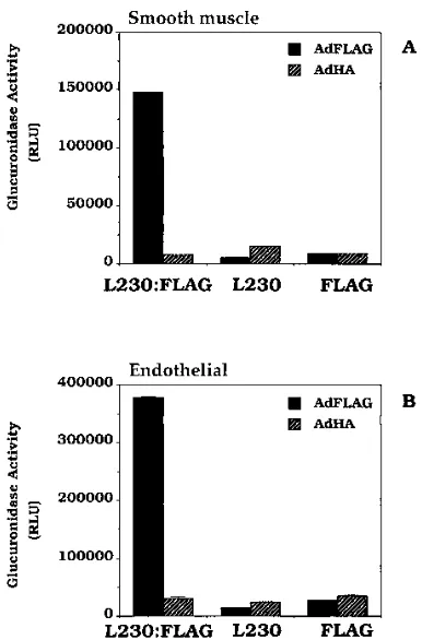
(Fig. 7B) cells by the AdFLAG vector. Transduction of the
endothelial and smooth muscle cells by the AdFLAG-bsAb
complex was increased nine- and sevenfold, respectively,
com-pared with that with the AdFLAG vector alone.
The specificity of transduction by the AdFLAG-bsAb
com-plex was evaluated by preincubation of the cells with L230
MAb or preincubation of the virus with FLAG MAb (Table 2).
Any resulting block in transduction would show that the
in-creased transduction by the AdFLAG-bsAb complex was
spe-cific to the dual interaction of the L230:FLAG bsAb with the
FLAG epitope on the virus and with
a
vintegrins on the cells.
Preincubation of the control vector, AdHA, with FLAG MAb
prior to its incubation with the L230:FLAG bsAb caused no
significant reductions in transduction by AdHA in either
HISMC or HuVEC. However, preincubation of AdFLAG with
FLAG MAb prior to incubation with L230:FLAG bsAb
blocked the increased transduction by the AdFLAG-bsAb
complex alone in both the endothelial and smooth muscle cells.
Likewise, preincubation of the cells with the L230 MAb
blocked transduction of the cells by the AdFLAG-bsAb
com-plex but had no significant effect on the transduction by AdHA
preincubated with the L230:FLAG bsAb. These results
dem-onstrate that the dual interactions of the L230:FLAG bsAb
with AdFLAG and with
a
vintegrin cell receptors are necessary
[image:6.612.66.292.64.465.2]to achieve the increase in transduction of HISMC and
HuVEC.
FIG. 5. Dose response ofb-glucuronidase expression by targetingav
inte-grins on 293 cells blocked with soluble fiber. Confluent monolayers of 293 cells in 24-well plates were preincubated in the absence or presence of 300ml of 5-mg/ml solution of Ad5 fiber protein for 1 h at room temperature. The AdFLAG (A) or AdHA (B) adenovirus vector (108
FFU/ml) was preincubated with the indicated concentrations of the L230:FLAG bsAb for 45 min at room tempera-ture in DMEM–20 mM HEPES. Each vector sample (20ml) was then added to the fiber-blocked or unblocked 293 cells and incubated for 1 h at room temper-ature. The cells were then washed twice with DMEM and incubated for 18 h at 378C in DMEM–5% calf serum. The medium was then aspirated, and the cells were lysed in 300ml of 13lysis buffer containing 10 mM EDTA. Each sample (3
ml) was then assayed fluorometrically with a luminometer. The results are the averages for duplicate samples.
FIG. 6. Increased binding of radiolabeled AdFLAG complexed with bsAb to 293 cells. Confluent monolayers of 293 cells in 24-well plates were preincubated in the presence (A) or absence (B) of 300ml of a 5-mg/ml solution of Ad5 fiber protein for 1 h at 48C. [3H]thymidine-labeled (24,000 cpm) AdFLAG or AdWT
vector was preincubated with 3mg of the L230:FLAG bsAb, the FLAG MAb alone, or the L230 MAb alone per ml for 45 min at 48C in a total volume of 20
ml. Each sample was then added to the fiber-blocked or unblocked 293 cells and incubated for 1 h at 48C. The cells were then washed three times with PBS. The cells were then solubilized in a 1% SDS solution and counted in a scintillation counter. The reported results are the averages for duplicate samples.
on November 9, 2019 by guest
http://jvi.asm.org/
[image:6.612.337.530.70.387.2]DISCUSSION
A general paradigm in gene therapy is that relative to other
vector systems virtually all cells are efficiently transduced by
adenovirus. Similar to previous reports, we have been able to
demonstrate the transduction of endothelial and smooth
mus-cle cells by adenovirus (17, 26). However, we have shown that
these two cell types, which are prime targets for gene therapy,
lack significant levels of fiber-mediated vector binding (Fig. 1).
Their low levels of binding are directly correlated with their
poor transduction efficiencies compared with those of A549
and other epithelial-derived cell lines which show
compara-tively high levels of fiber-mediated adenovirus binding. It is
also likely that other tissues targeted for adenovirus-mediated
gene therapy may likewise lack high expression levels of the
fiber receptor. The significant increases in binding and
trans-duction of cells lacking the high fiber receptor expression levels
shown here demonstrate that bsAbs can be successfully used to
expand the range of tissues amenable to efficient
adenovirus-mediated gene therapy.
Smooth muscle cells are targets for gene therapy to prevent
their proliferation and resultant obstruction of coronary
arter-ies following balloon angioplasty (19). While data for primary
intestinal smooth muscle is presented here, similarly, we have
found undetectable levels of the fiber receptor as determined
by fiber-specific adenovirus binding in primary human aorta
smooth muscle cells. These cells are, likewise, more efficiently
transduced with the AdFLAG-bsAb complex than with an
un-modified vector.
The
a
vintegrins are promising tissue-specific receptors for
targeted gene therapy.
a
vintegrin expression is activated in a
majority of melanomas (1) and glioblastomas (12). Targeting
therapeutic adenovirus to the
a
vintegrins on these cells would
allow delivery of a toxic gene, for example, while avoiding gene
delivery to healthy, surrounding tissue. Furthermore, the
inte-grin
a
vb
3is expressed on proliferating endothelial cells (3, 4).
Targeting the
a
vb
3receptor on these cells may be useful in
preventing their proliferation, such as in tumor growth or
ret-inal disease, or to promote further vascularization, such as the
revascularization of ischemic tissue.
bsAb-mediated transduction with adenovirus demonstrates
the feasibility of targeting vectors to specific tissues via
tissue-specific receptors. However, potential drawbacks of this system
may include complement activation, clearance by Fc receptors,
virus-antibody aggregation, and immune responses against the
Ab components. Despite these potential drawbacks of the
present system, they can all presumably be overcome. For
example, a fusion protein comprised of a single-chain Ab
rected towards adenovirus and a second single-chain Ab
di-rected towards a targeted cellular receptor would avoid Fc
receptor recognition and any virus-antibody aggregation
prob-lems. Furthermore, the use of human or humanized Abs will
likely prevent immune responses against the Abs (although
immune responses against the viral coat proteins are probably
of greater concern).
[image:7.612.83.276.69.362.2]Targeting gene delivery via the penton base also
demon-strates that attachment via the fiber, per se, is not required for
the efficient transduction of cells by adenovirus. Transduction
of fiber-blocked 293 cells by using the vector-antibody complex
was just as efficient as the fiber-mediated transduction of the
FIG. 7. L230:FLAG bsAb increases transduction of HuVEC (A) and HISMC (B). HuVEC or HISMC were plated in 24-well plates 2 days prior to use. AdFLAG or AdHA vectors (108 FFU/ml) were preincubated with 3mg of
L230:FLAG bsAb, L230 MAb, or FLAG MAb per ml or no Ab for 45 min at room temperature. Each vector sample (20ml) was then added to the HuVEC or HISMC and incubated for 1 h at 378C. The cells were then washed twice with DMEM and then further incubated at 378C for 18 h in DMEM–10% fetal bovine serum. The cells were then aspirated and assayed forb-glucuronidase activity as described in the legend to Fig. 5. RLU, relative light units.
TABLE 2. Specificity of glucuronidase transduction by AdFLAG-L230:FLAG bsAb complex
Condition
Glucuronidase activity ina:
Smooth muscle cells Endothelial cells
AdFLAG AdHA AdFLAG AdHA
Complex added directly to unblocked cells
b148.1 (0.3)
7.8 (0.1)
378.2 (1.6)
31.5 (1.5)
FLAG MAb added to vector prior to complex formation
c8.4 (0.1)
9.2 (0.1)
27.5 (0.7)
35.5 (0.7)
L230 MAb added to cells prior to complex addition
d5.3 (0.1)
15.5 (0.1)
14.6 (0.5)
24.2 (2.1)
aHuVEC or HISMC were plated in 24-well plates 2 days prior to transduction. For details, see Materials and Methods. AdFLAG or AdHA vectors (108FFU/ml)
were incubated with cells for 1 h. The cells were then washed and incubated at 378C for 18 h. Glucuronidase activity was determined; the results, expressed as relative light units (103), are the averages for duplicate measurements. Standard errors are given in parentheses.
bAdFLAG or AdHA was preincubated with 3mg L230:FLAG bsAb 45 min prior to addition to the cells.
cAdFLAG or AdHA was preincubated in the presence or absence of 50mg FLAG MAb per ml prior to the addition of 3mg of the L230:FLAG bsAb per ml. dCells were preincubated for 45 min with 50mg of L230 MAb per ml prior to the addition of the complex as described in footnoteb.
on November 9, 2019 by guest
http://jvi.asm.org/
[image:7.612.56.556.602.677.2]uncomplexed vector. Therefore, in the absence of
fiber-medi-ated binding, the transducing activity of an adenovirus particle
is not compromised by attachment through the bsAb.
Al-though many tissues may lack fiber receptor expression, a
num-ber of tissues are known to express the finum-ber receptor, including
certain lymphocyte cell lines (28), melanoma cells (35), and
many epithelial-derived cell lines (10, 22). Therefore, the
effi-cient targeting and limitation of transduction to a particular
cell or tissue will necessitate adenovirus vectors which lack
fiber receptor-binding activity. Such vectors may likewise
re-quire special receptor-expressing cell lines in order to
propa-gate them. In any case, it will be important to determine the
levels of adenovirus receptors in tissues targeted for gene
ther-apy. If the level of fiber receptor expression is low in a given
tissue, using bsAbs to target adenovirus to receptors that are
expressed by the tissue is likely to increase the efficiency and
specificity of gene transfer.
ACKNOWLEDGMENTS
We thank Zachary Skelding, Faith Beams, Vicki Kulesa, and Angela
Appiah for excellent technical assistance. We also thank Megan
Ma-hony, Doug Brough, Duncan McVey, and Joe Bruder for critical
re-view of the manuscript.
REFERENCES
1.Albelda, S. M., S. A. Mette, D. E. Elder, R. Stewart, L. Damjanovich, M. Herlyn, and C. A. Buck.1990. Integrin distribution in malignant melanoma: association of theb3 subunit with tumor progression. Cancer Res.50:6757– 6764.
2.Bai, M., B. Harfe, and P. Freimuth.1993. Mutations that alter an Arg-Gly-Asp (RGD) sequence in the adenovirus type 2 penton base protein abolish its cell-rounding activity and delay virus reproduction in flat cells. J. Virol.
67:5198–5205.
3.Brooks, P. C., R. A. F. Clark, and D. A. Cheresh.1994. Requirement of vascular integrinavb3for angiogenesis. Science264:569–571.
4.Brooks, P. C., A. M. Montgomery, M. Rosenfeld, R. A. Reisfeld, T. Hu, G. Klier, and D. A. Cheresh.1994. Integrin alpha v beta 3 antagonists promote tumor regression by inducing apoptosis of angiogenic blood vessels. Cell
79:1157–1164.
5.Cheresh, D. A.1987. Human endothelial cells synthesize and express an Arg-Gly-Asp-directed adhesion receptor involved in attachment to fibrino-gen and von Willebrand factor. Proc. Natl. Acad. Sci. USA84:6471–6475. 6.Cheresh, D. A., and R. C. Spiro.1987. Biosynthetic and functional properties
of an Arg-Gly-Asp-directed receptor involved in human melanoma cell at-tachment to vitronectin, fibrinogen and von Willebrand factor. J. Biol. Chem.
262:17703–17711.
7.Crystal, R. G., N. G. McElvaney, M. A. Rosenfeld, C. S. Chu, A. Mastrangeli, J. G. Hay, S. L. Brody, H. A. Jaffe, N. T. Eissa, and C. Canel.1994. Admin-istration of an adenovirus containing the human CFTR cDNA to the respi-ratory tract of individuals with cystic fibrosis. Nat. Genet.8:42–51. 8.Curiel, D. T., E. Wagner, M. Cotten, M. L. Birnstiel, S. Agarwal, C. Li, S.
Loechel, and P. Hu.1992. High-efficiency gene transfer mediated by adeno-virus coupled to DNA-polylysine complexes. Hum. Gene Ther.3:147–154. 9.Damjanovich, L., S. M. Albelda, S. A. Mette, and C. A. Buck.1992.
Distri-bution of integrin cell adhesion receptors in normal and malignant lung tissue. Am. J. Respir. Cell Mol. Biol.6:197–206.
10. Defer, C., M. T. Belin, M. L. Caillet-Boudin, and P. Boulanger.1990. Human adenovirus-host cell interactions: comparative study with members of sub-groups B and C. J. Virol.64:3661–3673.
11. French, B., W. Mazur, N. Ali, R. Geske, J. Finnigan, G. Rodgers, R. Roberts, and A. Raizner.1994. Percutaneous transluminal in vivo gene transfer by recombinant adenovirus in normal porcine coronary arteries, atherosclerotic arteries, and two models of coronary restenosis. Circulation90:2402–2413. 12. Gladson, C. L., and D. A. Cheresh.1991. Glioblastoma expression of vitro-nectin and theavb3 integrin: adhesion mechanism for transformed glial cells. J. Clin. Invest.88:1924.
13. Gladson, C. L., and D. A. Cheresh.1994. Theav integrins, p. 83–99.InY. Takada (ed.), Integrins: the biological problems. CRC Press, Boca Raton, Fla.
14. Henry, L. J., D. Xia, M. E. Wilke, J. Deisenhofer, and R. D. Gerard.1994. Characterization of the knob domain of the adenovirus type 5 fiber protein expressed inEscherichia coli. J. Virol.68:5239–5246.
15. Huard, J., H. Lochmuller, G. Acsadi, A. Jani, B. Massie, and G. Karpati.
1995. The route of administration is a major determinant of the transduction efficiency of rate tissues by adenoviral recombinants. Gene Ther.2:107–115. 16. Le Gal La Salle, G., J. J. Robert, S. Berrard, V. Ridoux, L. D. Stratford-Perricaudet, M. Stratford-Perricaudet, and J. Mallet.1993. An adenovirus vector for gene transfer into neurons and glia in the brain. Science259:988–990. 17. Lemarchand, P., H. A. Jaffe, C. Danel, M. C. Cid, H. K. Kleinman, L. D.
Stratford-Perricaudet, M. Perricaudet, A. Pavirani, J.-P. Lecocq, and R. G. Crystal.1992. Adenovirus-mediated transfer of a recombinant humana 1-antitrypsin cDNA to human endothelial cells. Proc. Natl. Acad. Sci. USA
89:6482–6486.
18. Mathias, P., T. Wickham, M. Moore, and G. Nemerow. 1994. Multiple adenovirus serotypes useav integrins for infection. J. Virol.68:6811–6814. 19. Mazur, W., N. Ali, A. Raizner, and B. French.1994. Coronary restenosis and
gene therapy. Tex. Heart Inst. J.21:104–111.
20. McCoy, R. D., B. L. Davidson, B. J. Roessler, G. B. Huffnagle, S. L. Janich, T. J. Laing, and R. H. Simon.1995. Pulmonary inflammation induced by incomplete or inactivated adenoviral particles. Hum. Gene Ther.6:1553– 1560.
21. Nemerow, G. R., T. J. Wickham, and D. A. Cheresh.1993. The role ofav
integrins in adenovirus infection. Elsevier Science Publishers, New York. 22. Philipson, L., K. Lonberg-Holm, and U. Pettersson.1968. Virus-receptor
interaction in an adenovirus system. J. Virol.2:1064–1075.
23. Quantin, B., L. D. Perricaudet, S. Tajbakhsh, and J. L. Mandel.1992. Adenovirus as an expression vector in muscle cells in vivo. Proc. Natl. Acad. Sci. USA89:2581–2584.
24. Ragot, T., N. Vincent, P. Chafey, E. Vigne, H. Gilgenkrantz, D. Couton, J. Cartaud, P. Briand, J. C. Kaplan, M. Perricaudet, and A. Kahn.1993. Efficient adenovirus-mediated transfer of a human minidystrophin gene to skeletal muscle of mdx mice. Nature (London)361:647–650.
25. Rosenfeld, M. A., K. Yoshimura, B. C. Trapnell, K. Yoneyama, E. R. Rosenthal, W. Dalemans, M. Fukayama, J. Bargon, L. E. Stier, L. Stratford-Perricaudet, M. Stratford-Perricaudet, W. B. Guggino, A. Pavirani, J. P. Lecocq, and R. G. Crystal.1992. In vivo transfer of the human cystic fibrosis transmem-brane conductance regulator gene to the airway epithelium. Cell68:143–155. 26. Schulick, A. H., G. Dong, K. D. Newman, R. Virmani, and D. A. Dichek.1995.
Endothelium-specific in vivo gene transfer. Circ. Res.77:475–485. 27. Segal, D. M., and B. J. E. G. Bast.1995. Production of bispecific antibodies,
p. 2.13.1–2.13.16.InJ. E. Coligan, A. M. Kruisbeek, D. H. Margulies, E. M. Shevach, and W. Strober (ed.), Current protocols in immunology, vol. 1. John Wiley & Sons, Inc., New York.
28. Silver, L., and C. W. Anderson.1988. Interaction of human adenovirus serotype 2 with human lymphoid cells. Virology165:377–387.
29. Smith, J. W., D. J. Vestal, S. V. Irwin, T. A. Burke, and D. A. Cheresh.1990. Purification and functional characterization of integrinavb5. J. Biol. Chem.
265:11008–11013.
30. Thiel, J. F., and K. O. Smith.1967. Fluorescent focus assay of viruses on cell monolayers in plastic petri plates. Proc. Soc. Exp. Biol. Med.125:892–895. 31. Thimmappaya, B., C. Weinberger, R. J. Schneider, and T. Shenk.1982.
Adenovirus VAI RNA is required for efficient translation of viral mRNAs at late times after infection. Cell31:543–551.
32. Varga, M. J., C. Weibull, and E. Everitt.1991. Infectious entry pathway of adenovirus type 2. J. Virol.65:6061–6070.
33. Weinacker, A., A. Chen, M. Agrez, R. I. Cone, S. Nishimura, E. Wayner, R. Pytela, and D. Sheppard.1994. Role of the integrinavb6 in cell attachment to fibronectin. J. Biol. Chem.269:6940–6948.
34. Wickham, T. J., M. E. Carrion, and I. Kovesdi.1995. Targeting of adenovirus penton base to new receptors through replacement of its RGD motif with other receptor-specific peptide motifs. Gene Ther.2:750–756.
35. Wickham, T. J., E. J. Filardo, D. A. Cheresh, and G. R. Nemerow.1994. Integrinavb5 selectively promotes adenovirus mediated cell membrane per-meabilization. J. Cell Biol.127:257–264.
36. Wickham, T. J., P. Mathias, D. A. Cheresh, and G. R. Nemerow.1993. Integrinsavb3 andavb5 promote adenovirus internalization but not virus attachment. Cell73:309–319.