Recent trends in reperfusion in STEMI (ST
elevation myocardial infarction) in a South
Indian tier-3 city
A dissertation submitted in partial fulfillment of
DM-Branch II Cardiology Examination of the
CERTIFICATE
This is to certify that this dissertation entitled ‘
Recent trends in
reperfusion in STEMI (ST elevation myocardial infarction) in
a South Indian tier-3 city
’ is a bonafide work done by Dr.
Brajesh kumar kunwar in partial fulfillment of rules and regulation
for DM (Branch II-Cardiology) examination of the Tamil Nadu Dr.
M. G. R. Medical University, to be held in July/August 2011.
66
Acronyms
PCI - Percutaneous coronary intervention
ACS - Acute coronary syndrome
UA - Unstable angina
NSTEMI - Non ST- segment elevation myocardial infarction
STEMI - ST- segment elevation myocardial infarction
MI - Myocardial infarction
PW - Posterior wall
LW - Lateral wall
SD - Standard deviation
IWI - Inferior wall myocardial infarction
STK - Streptokinase
TNK - Tenecteplase
CVA - Cerebrovascular accident
DM- Diabetes Mellitus
HTN- Hypertension
ADR- Adverse drug reaction
ACKNOWLEDGEMENT
I place on record my deep sense of gratitude to Dr. George Joseph,
professor, Department of cardiology, for his able guidance and valuable
suggestions complete without his support and encouragement.
I also thank Dr. Sunil Thomas Chandy, Professor and Head, Department of
Cardiology, for his constant help during the course of studies. The study
would never have been completed without his constant support.
I am also grateful to all my teachers and colleges in department for there
invaluable support throughout the course of studies. I also thank Mrs.
Nandini, Department of Cardiology, for her constant endeavor to finish the
tedious work on time.
My special thanks to the entire senior faculty, all my colleagues and the
office staff in the department of Cardiology, for their moral support and
timely help rendered at crucial stages.
I also wish to thank Mrs. Nithya, Department of Biostatistics, for his help in
the statistical analysis.
At last, but not the least, I thank all the patients who agreed to be a part of
this study and helped me in completing this research work.
DECLARATION
I, Dr. Brajesh Kumar Kunwar, hereby declare that this dissertation entitled
‘Recent trends in reperfusion in STEMI (ST elevation myocardial
infarction) in a South Indian tier-3 city’ has been prepared by me under
the direct supervision and guidance of Dr. George Joseph MD, DM,
Professor, Department of Cardiology, Christian Medical College, Vellore.
This is being submitted to Dr. M. G. R. Medical University in partial
fulfillment of regulations for the DM (Cardiology) examination to be held in
July/August 2011.
This dissertation has not been submitted by me either in part or in full on any
previous occasion to any university or institution for the award of any degree
or diploma.
Place: Vellore Dr. Brajesh kumar kunwar
Date: Postgraduate Student
Department of Cardiology
Christian Medical College
Vellore
1
ABSTRACT
Recent trends in reperfusion in STEMI (ST elevation myocardial
infarction) in a South Indian tier-3 city.
Background: Coronary artery disease is the leading cause of death in both
the developing and developed countries. India is going to have a large
proportion of the total number of such cases in the world in the near future.
In India, larger proportions of patients with acute coronary syndromes
(ACS) present with ST-elevation myocardial infarction (STEMI) compared
to patients in the developed countries. The best treatment for STEMI is
reperfusion with percutaneous coronary interventions (PCI). Facilities for
PCI are not available in many tertiary care centres in India. We sought to
study the recent trends of reperfusion in patients of acute STEMI in Vellore,
a tier-3 South Indian city.
Methods: 1459 consecutive patients presenting with acute STEMI to our
centre were enrolled. Their demographic profile, risk factors and mode of
reperfusion were recorded. The study period from August 2008 to July 2010
was divided into four half-years and trends of reperfusion were compared
over this study periods.
Results: Mean age of study population was 56.3+11.8 years. There were
1214 (83%) males and 245 (17%) females. The largest number of patients
were in the age group of 40-59 years (313, 50%).
1255 (86%) of patients
received some form of reperfusion therapy either by thrombolytic agent or
PCI. Thrombolysis with streptokinase in 1019 (69.8%) patients was the most
2
PCI which was used in of 137 patients (9.4%) and tenecteplase in 99 (6.7%)
patients. 204 (13.8%) did not receive any form of reperfusion therapy for
various reasons, the most common being late presentation in 175 (85.7%).
Number of patients presenting with STEMI increased from 297 in the first
half-year to 465 in the last half-year. The PCI numbers also increased from
19 in the first half year to 45 in the last half year. Use of tenecteplase as a
thrombolytic agent remained constant inspite of rising STEMI case load.
The most common territory involved in STEMI was anterior wall
593(58.2%).
Conclusion: Patients presenting with STEMI in this study were on an
average a decade younger than western population. There was about 20%
increase in STEMI every year in the present study. Younger patients are
least likely to receive primary PCI or tenecteplase as reperfusion therapy.
86% of patients received some form of reperfusion therapy and streptokinase
was the most common type of reperfusion therapy used. Primary PCI rates
increased at 20% per year during this study but still <10% of STEMI
3
INTRODUCTION
Coronary artery disease (CAD) is the most common cause of death in both
developing & developed countries
1
. By 2020 IHD is expected to increase by more
than 120% in the developing countries as compared to 30-40% in the developed
countries
2
. 60% of the world’s heart disease is expected to occur in India
3
. This
will place a huge strain on healthcare resources which are already inadequte and
overstretched. Providing IHD patients optimal care in the Indian setting requires
specific and relevant data applicable to the general population.
The spectrum of IHD in the developed countries appears to be different from
that in the Indian population. Fewer than 40% of the patients with acute coronary
syndrome present with ST-segment elevation myocardial infarction (STEMI) in the
developed countries
4,5
and this is also showing a decreasing trend with time. On
the other hand 60% of patients with ACS present with ST segment elevation
myocardial infarction in India. In patients with ST elevation myocardial infarction,
the main consideration in treatment is reperfusion which is influenced by duration
from onset of symptoms. The best treatment for patients presenting with STEMI is
reperfusion either in the form of primary percutaneous coronary intervention (PCI)
or by means of thrombolytic agents. Primary PCI re-establishes blood flow in the
culprit coronary artery by mechanically dislodging the thrombus obstructing the
4
Most data on patients with acute coronary syndromes are derived from several
large registries
8,9,10,11,12,13,14,15,16
from developed countries. There are a few small
studies from India restricted to a few hospitals
17,18,19
. We present data from a
5
AIMS AND OBJECTIVES
1:-
To evaluate the current trends in revascularization
in STEMI in a tertiary care centre in South India.
2:- To assess the number and proportion of patients
receiving pharmacological or invasive reperfusion
treatment of STEMI.
3;- To assess the number of patients not receiving any
form of reperfusion therapy and the reasons for the
same.
6
REVIEW
OF
LITERATURE
EPIDEMIOLOGY
The second half of the 20th century witnessed a global spread of the coronary
artery disease (CAD) epidemic especially in developing countries, including
India. The underlying pathology of CAD is atherosclerosis, which develops over
many years and is usually advanced by the time symptoms occur, generally in
middle age. Acute coronary and cerebrovascular events frequently occur
suddenly, and are often fatal before medical care can be given.
Of an estimated 58 million deaths globally from all causes in 2005,
cardiovascular disease accounted for 30%
23, 24.
This proportion is equal to that due
to infectious diseases, nutritional deficiencies, and maternal and perinatal
conditions combined
20
. It is important to recognize that a substantial proportion of
these deaths (46%) were in people under 70 years of age, in the more productive
period of life
21
. Globally, CAD was the leading killer in the age group > 60 years,
and, with 1332000 deaths in adults aged 15-59 years, CAD was ranked behind
HIV/AIDS only
22
. The principal cardiovascular disorder responsible for the
7
CARDIOVASCULAR DISEASE IN INDIA
At the threshold of the new millennium the threat of CAD is looming large as this
new epidemic afflicts Indians at a relatively younger age with severe and diffuse
forms of lesions. Recently, the subject of CAD in Indians (referred to as immigrants
or Asian Indians or South Asians when outside India) has become a challenge for
many research centres worldwide
25,26
. The prevalence of CAD has progressively
increased in India during the latter half of the last century, The prevalence of CAD
is two times higher (10%) in urban than in rural India
29,30
. South Indians have
higher prevalence, 7% in rural and 14% in urban areas. The vulnerability of urban
Indians to CAD is possibly related to different nutritional, environmental, and
life-style factors. Unfortunately, the on-going urbanization of rural India is likely to
narrow down these differences
31
.
India is in the midst of a demographic transition. The average life expectancy at
birth in India is 63.7 years, being 63.1 for males and 64.4 for female, compared
with the national average of 41.2 years in 1951-1961
22
. The demographic transition
has led to an increase in the number of older people (aged >60 years), from 19.61
million in 1950 to 75.93 million in 2000. The increase in life expectancy has
brought a large section of population to an age where cardiovascular disease starts
manifesting itself. In India CAD rates have increased during last 30 years, whereas
declining trends have been noticed in developed western countries
22
.
Reports on CAD in Indians from different parts of the world have shown that
Indians are at 3-4 times higher risk of CAD than white Americans, 6 times higher
than Chinese, and 20 times higher than Japanese
25,26,27
. The exact prevalence of
8
CVD. Heart diseases occur in Indians 5 to 10 years earlier than in other populations
around the world
28
. According to the INTERHEART study, the age at first
presentation of acute MI in the south Asian (Bangladesh, India, Nepal, Pakistan, Sri
Lanka) population is 53 years, whereas that in western Europe, China, and Hong
Kong is 63 years, with men more than women affected
32
. The first myocardial
infarction (MI) occurs in 4.4% of Asian women and 9.7% of men at age less than
40 years, which is 2 to 3.5 fold higher than in the west European population and is
third highest of all the regions studied worldwide
32
.
The INTERHEART study
32
, involving 52 countries, established an association
between conventional modifiable risk factors for MI in all regions of the world,
including South Asia, and in both sexes and at all ages. In South Asians,
apolipoprotein (Apo) B/ApoA1 ratio and smoking were the important risk factors,
as in the rest of the world. However, hypertension, abdominal obesity, and diabetes
had more severe effects in South Asians. The study also showed that hypertension
and diabetes were more important risk factors in younger Indian women than men.
It was also observed that the risk of CAD increased incrementally with smoking and
it was a greater risk factor in younger men than in women.
Several factors appear likely to have contributed to the acceleration of CAD
epidemic in India in recent times. These are:
(i)
Demographic transition to an older population, as a result of
increasing life expectancy
(ii)
Confluence of both conventional risk factors and
non-conventional risk factors in Indians
28
.
9
Non-conventional risk factors like hyper-insulinemia, insulin resistance,
lipoprotein A etc. are determined by genes or other 'programming' factors and
their high prevalence amongst Indians probably explain the malignant, precocious
nature of CAD that typically affects Indians. Recently a relationship between low
birth-weight, which is widely prevalent amongst Indian newborns, and enhanced
susceptibility to CAD in adult life ('Barker hypothesis') has been found. These
multiplicative effects of conventional and emerging risk factors appear to provide
a plausible explanation for the excess burden of CAD among Indians, many of
whom are lean, non-smoking, vegetarian
33
.
Cardinal features of coronary artery disease among Indians compared to other
populations
33
:
Higher rates
•
2 to 4 fold higher prevalence, incidence, hospitalization, mortality
Greater prematurity
•
5 to 10 years earlier onset of first Ml
•
5 to 10 fold higher rate of Ml and death in young <40 years of age)
Greater severity
33
•
three vessel disease common even among young premenopausal women
•
large Ml with greater muscle damage
Higher prevalence of glucose intolerance
•
insulin resistance syndrome, diabetes, central obesity
Lower prevalence of conventional risk factors
•
hypertension, obesity, cigarette smoking
•
cholesterol levels: similar to Whites but higher than other Asians
Higher prevalence of emerging (thrombogenic) risk factors
10
•
high levels of triglycerides, fibrinogen
•
low levels of HDL
•
small dense LDL
Higher rates of clinical events for a given degree of atherosclerosis
•
double that of whites
•
four fold higher than Chinese
•
higher proportion of unstable or vulnerable plaques
33
The 4 main risk factors, which showed consistently significant associations across
all south Asian countries in both sexes were current and former smoking, high
ApoB100/Apo-I ratio, history of hypertension and history of diabetes.
Between 1990 and 2020, CAD is expected to increase by 120% in women and
137% in men in developing countries, compared to 30-60% in developed countries
2
.
In addition, the proportion of patients with STEMI at 60% of the acute coronary
syndrome patients is much higher than the proportion observed in developed
countries where fewer than 40% had STEMI
5
. This suggest that patients with acute
11
Myocardial Infarction
Atherosclerosis is the gradual buildup of cholesterol and fibrous tissue in plaques
in the wall of arteries (in this case, the coronary arteries), typically over decades
35
.
Blood stream column irregularities visible on angiography reflect artery lumen
narrowing as a result of decades of advancing atherosclerosis
37
. Plaques can become
unstable, rupture, and additionally promote a thrombus that occludes the artery; this
can occur in minutes. When plaque rupture occurs, it exposes the intimal layer and
initiates a cascade of platelet activation and thrombosis resulting in occlusion of the
vessel and infarction of the subjacent myocardium
38
. Acute myocardial infarction
refers to two subtypes of acute coronary syndrome: non-ST-elevation myocardial
infarction and ST-elevation myocardial infarction, which are most frequently (but
not always) a manifestation of atherosclerotic coronary artery disease
34
. Myocardial
infarctions are usually classified by size: microscopic (focal necrosis), small (<10%
of the left ventricle myocardium), moderate (10-30% of the left ventricle
myocardium), and large (>30% of the left ventricle myocardium), and by location
38
.
Pathology of myocardial infarction
12
Myocardial infarction can be classified temporally as evolving (<6 h), acute (6
h-7 days), healing (h-7-28 days), and healed (29 days and beyond)
32
.
Reference
40
A 2007 consensus document classifies myocardial infarction into five main types
39
:
Type 1
- Spontaneous myocardial infarction related to ischaemia due to a primary
coronary event such as plaque erosion and/or rupture, fissuring, or dissection
Type 2
- Myocardial infarction secondary to ischemia due to either increased
oxygen demand or decreased supply, e.g. coronary artery spasm, coronary
embolism, anemia, arrhythmias, hypertension, or hypotension
Type 3
- Sudden unexpected cardiac death, including cardiac arrest, often with
13
ST elevation, or new left bundle branch, or evidence of fresh thrombus in a
coronary artery by angiography and/or at autopsy, but death occurring before
blood samples could be obtained, or at a time before the appearance of cardiac
biomarkers in the blood.
Type 4
- Associated with coronary angioplasty or stents:
Type 4a
- Myocardial infarction associated with PCI
Type 4b
- Myocardial infarction associated with stent thrombosis as
documented by angiography or at autopsy
Type 5
- Myocardial infarction associated with coronary artery bypass grafting.
The ST segment of the cardiac cycle represents the period between
depolarization and repolarization of the left ventricle. In the normal state, the
ST-segment is isoelectric relative to the PR ST-segment & most ST-ST-segment elevation is
a result of non-AMI causes. A study showed that of 123 adult chest pain patients
with ST-segment elevation
≥
1mm, 63 patients (51%) did not have myocardial
infarctions. These non-MI patients had were mainly left bundle branch block
(LBBB) (21%) and left ventricular hypertrophy (LVH) (33%).
Causes of ST- Segment Elevation
41
Acute Pericarditis
Benign Early Repolarization
Left bundle branch block with acute MI (Sgarbossa et al’s criteria)
Left ventricular hypertrophy
14
Brugada syndrome
Hyperkalemia
Hypothermia
Central nervous system pathologies
Prinzmetal angina
Post electrical cardioversion
Acute Myocardial Infarction
ST- segment elevation is measured:
At J point – if relative to PR segment
At 0.06 – 0.08s from J point – if relative to TP segment
15
ST Segment Elevation Criteria
42
Study Minimum
Consecutive
Leads
Minimum ST Elevation
(mm) Limb leads
Minimum
ST
Elevation
(mm)
Precordial
leads
AHA/ACC 2
1
1
GISSI-1 1
1
2
GISSI-2 1
1
2
GUSTO 2
1
2
TIMI 2
1
1
TAMI 2
1
1
Minnesota
Code
1
1mm:I,II,III,Avl,avf,V5-6
2mm:V1-4
Irrespective of which definition is used, ST elevation has poor sensitivity for AMI
where up to 50% of patients exhibit ‘atypical’ changes at presentation including
isolated ST depression, T inversion or even a normal ECG.
16
infarction (NSTEMI), which result from non-occlusive thrombus, small risk area,
brief occlusion, or an occlusion with adequate collaterals
Morphology of ST-segment elevation
Convex ST-segment is more suggestive of myocardial infarction as compared to
concave ST- segment elevation
Benign Early Repolarization
Benign early repolarization is seen most commonly in young black population. It
is often confused with ST elevation myocardial infarction (STEMI).
ECG characteristics of benign early repolarization
43
1: ST- segment elevation <2 mm
2: Concavity of initial portion of the ST- segment
3: Notching or slurring of the terminal QRS complex
4: Symmetrical, concordant T wave of large amplitude
5; Widespread or diffuse distribution of STE (does not demonstrate
territorial distribution)
6: Relative temporal stability
Distribution
ST- segment elevation due to acute myocardial infarction (MI)
usually demonstrate regional or territorial pattern
o
Anterior MI – V3-V4
o
Septal MI – V2-V3
o
Anteroseptal MI – V1/2 – V4/5
17
o
Inferior MI – II, III, aVF
Diffuse ST-segment elevation suggestive of non MI causes, e.g. pericarditis
43
.
ST-elevation myocardial infarction is difficult to diagnose in presence of left bundle
branch block. Sgarbossa criteria was put forward to diagnose myocardial
infarction in presence of left bundle branch block (LBBB)
43.
Sgarbossa Criteria
42
ST Elevation
≥
1 mm and
concordant with QRS complex
Score 5 points
Odds Ratio (OR) 25.2
ST Depression
≥
1 mm in V1, V2,
V3
Score 3 points
OR 6.0
ST Elevation
≥
5 mm and discordant
with QRS complex
Score 2 points
OR 4.3
A total score of 3 or more suggests that the patient is likely experiencing an
AMI based on the ECG criteria. With a score less than 3, the ECG diagnosis is
less certain requiring additional evaluation. Subsequent publications have
suggested that Sgarbossa’s criteria is less useful than reported, with studies
demonstrating decreased sensitivity and inter-rater reliability
Reperfusion Strategy in Myocardial Infarction
18
occludes the vessel, preventing the circulation of oxygenated blood. Irreversible
ischemia-induced myocardial necrosis may occur within 20-60 minutes of
occlusion. The mainstay of treatment is reperfusion therapy through administration
of fibrinolytics (pharmacologic reperfusion) or primary percutaneous coronary
intervention ‘(PCI) (mechanical reperfusion). Evidence exists that expeditious
restoration of flow in the obstructed infarct artery after the onset of symptoms in
patients with STEMI is a key determinant of short- and long- term outcomes
regardless of whether reperfusion is accomplished by fibrinolysis or PCI
42,43,44
. A
critically important goal of reperfusion is to restore flow in the infarct artery as
quickly and completely as possible, but the ultimate goal of reperfusion in STEMI
is to improve myocardial perfusion in the infarct zone. Despite adequate restoration
of flow in the epicardial infarct artery, perfusion of the infarct zone may still be
compromised by a combination of microvascular damage and reperfusion injury
45,46
.
Microvascular damage occurs as a consequence of downstream embolization of
platelet microemboli and thrombi followed by the release of substances from
activated platelets that promote occlusion or spasm in the microvasculature.
Reperfusion injury results in cellular edema, free radical formation, calcium
overload, and acceleration of the apoptotic process. Cytokine activation in the
infarct zone leads to neutrophil accumulation and inflammatory mediators that
contribute to tissue injury.
Time from onset of symptoms to fibrinolytic therapy is an important predictor of
MI size and patient outcome
47
. The efficacy of fibrinolytic agents in lysing
thrombus diminishes with the passage of time
48
. Fibrinolytic therapy administered
within the first 2 hours (especially the first hour) can occasionally abort MI and
19
directly related to the time from symptom onset, treatment benefit is maximized by
the earliest possible application of therapy.
The present recommendations for fibrinolytic therapy say that: In the absence of
contraindications, fibrinolytic therapy should be administered to STEMI patients
with symptom onset within the prior 12 hours and ST elevation greater than 0.1 mV
in at least 2 contiguous precordial leads or at least 2 adjacent limb leads or patients
with symptom onset within the prior 12 hours and new or presumably new LBBB.
In the absence of contraindications, it is reasonable to administer fibrinolytic
therapy to ST-segment elevation MI (STEMI) patients with symptom onset within
the prior 12 hours and 12-lead ECG findings consistent with a true posterior MI. It
is also reasonable to administer fibrinolytic therapy to patients with symptoms of
STEMI beginning within the prior 12 to 24 hours who have continuing ischemic
symptoms and ST elevation greater than 0.1 mV in at least 2 contiguous precordial
leads or at least 2 adjacent limb leads
7.
Contraindications and Cautions For Fibrinolytic Use In ST-Elevation
Myocardial Infarction
7
Absolute contraindications
•
Any prior intra-cerebral haemorrhage
•
Known structural cerebral vascular lesion (eg, arterio- venous malformation)
•
Known malignant intracranial neoplasm (primary or metastatic)
•
Ischemic stroke within 3 months except acute ischemic stroke within 3 hours
•
Suspected aortic dissection
20
•
Significant closed head or facial trauma within 3 months
Relative Contraindications
•
History of chronic severe, poorly controlled hypertension
•
Severe uncontrolled hypertension on presentation (systolic blood pressure
greater than 180 mm Hg or diastolic blood pressure greater than 110mm Hg)
•
History of prior ischemic stroke greater than 3 months, dementia, or known
intracranial pathology not covered in contraindications
•
Traumatic or prolonged (greater than 10 minutes) cardiopulmonary
resuscitation or major surgery (less than 3 weeks)
•
Recent (within 2 to 4 weeks ) internal bleeding
•
Non-compressible vascular punctures
•
For streptokinase/anistreplase : prior exposure (more than 5 days ago) or
prior allergic reaction to these agents
•
Pregnancy
•
Active peptic ulcer
•
Current use of anticoagulants : the higher the INR, the higher the risk of
bleeding
Efficacy of Intravenous Fibrinolytic Therapy In STEMI
It has been well established that fibrinolytic therapy provides a survival benefit for
patients with STEMI, based on large, well-controlled clinical trials
47,48,49.
The
21
An overview of the results of 9 trials by the ‘Fibrinolytic Therapy Trialists’
Collaborative Group comparing the outcome of patients undergoing fibrinolytic
therapy and those of controls demonstrated a highly significant 18% relative
reduction in 35 days mortality (9.6% fibrinolysis versus 11.5% control), which
corresponds to absolute reductions in 35-day mortality rates of approximately 30
per 1000 for patients who arrived at the hospital within 6 hours of the onset of
symptoms and of approximately 20 per 1000 for patients who arrived 7-12 hours
after the onset of symptoms
8
.This survival benefit is maintained over the long
term (up to 10 years).
THROMBOLYTIC AGENTS
The fibrinolytic agents currently approved for treating patients with STEMI
include streptokinase, alteplase, reteplase, and tenecteplase. Thrombolytic agents
available today are serine proteases that work by converting plasminogen to the
natural fibrinolytic agent plasmin. Plasmin lyses clot by breaking down the
fibrinogen and fibrin contained in a clot.
Streptokinase
The history of thrombolytic therapy began in 1933 when Tillet and Carner
discovered that filtrates of broth cultures of certain strains of Streptococcus
bacteria (beta-hemolytic streptococci) could dissolve a fibrin clot
51.
Streptokinase
found its initial clinical application in combating fibrinous pleural exudates,
hemothorax, and tuberculous meningitis
52.
In 1958, streptokinase was first used in
22
(GISSI) trial in 1986, which validated streptokinase as an effective therapy and
established a fixed protocol for its use in acute myocardial infarction
52.
STK has
no proteolytic activity of its own and thus activates PG to PN indirectly by first
forming a high affinity equimolar complex with PG (STK-PG activator complex)
51
. It forms a 1:1 complex with plasminogen causing conversion to plasmin. It is
non specific, activating circulating as well as clot-bound plasminogen, and causes
extensive fibrinogen depletion.
Streptokinase is antigenic. Neutralizing antibodies are significant following
use, and repeat administration should be avoided. Allergic reaction (rash, chills,
urticaria) occurs in around 4% of patients and anaphylactic shock occurs in 0.5%
of patients
53.
Hypotension can be significant (average decrease ~ 35 mm systolic
blood pressure (SBP)
54
and may be worsened by rapid administration, which
excludes bolus use and necessitates constant IV infusion. It is commonly given as
1.5 million units over 60 min.
Alteplase
Tissue type plasminogen activator (t-PA) is a naturally occuring serine protease,
which is produced by healthy endothelium. Its levels are increased with exercise
and inhibited by plasminogen activator inhibitor (PAI-1). Alteplase is a
commercially available, genetically engineered, bacterially produced version of
human t-PA. it exhibits marked specificity for the plasminogen-fibrin complex,
although at the doses necessary to achieve rapid lysis, there is ~ 50% depletion of
circulating fibrinogen. t-PA is associated with a higher early recanalization rate
relative to streptokinase
55
, but may be accompanied by an increase rate of
reocclusion
56
. The half-life is approximately 5 min; thus, t-PA must be
23
The accelerated dosing regimen has been proven to be the most effective
54
: 15 mg
bolus over 1-2 min followed by 0.75mg/kg IV (< 35 mg) over 60 min. Weight
adjustment is recommended because of excessive bleeding in lighter weight (<60
kg) patients and a trend toward decreased lysis in heavy weight (>90 kg)
individuals
55
. Higher dose and double-bolus regimens have been associated with
unacceptable bleeding rates
56,57
. The accelerated or front-loaded dosing regimen
has been shown to have higher early patency rates, similar safety profile
54
, and
lower incidences of reocclusion as compared with the standard dosing regimen
60
.
The TIMI (Thrombolysis in myocardial infarction), phase 1 trial randomly
assigned 290 patients with evolving acute MI to altepase or to streptokinase.
Alteplase was far superior in achieving coronary reperfusion; twice as many
occluded infarct-related arteries opened after 90 minutes with alteplase than with
streptokinase
61
.
The GUSTO 1 study (41,021 patients) tested the accelerated dose regimen
combined with intravenous heparin. Despite an increase in intracerebral bleeding
with t-PA, overall benefit as assessed from the combined endpoint of total
mortality and disabling stroke was significantly better with t-PA as compared with
streptokinase (6.3% vs. &.3%)
61
. This translates into 15% mortality reduction or
about 10 lives saved per 1,000 patients treated.
Reteplase
Reteplase is a deletion mutant of t-PA that exhibits preferential activation of
fibrin-bound plasmin and a two to threefold increase half-life (15 min),
permitting bolus administration. It has a lower affinity to fibrin (theoretically
improving clot penetration), though similar fibrin specificity compared with
alteplase. While initial studies showed superior infarct artery patency when
24
dose t-PA in the RAPID-2 trial
63
, the GUSTO III study (15,059 patients)
showed equivalent 30-day mortality rates with reteplase (7.5%) versus
accelerated-dose alteplase (7.2%). The rates of the combined endpoint, death, or
nonfatal MI- disabling stroke were similar: 7.98% and 7.91%, respectively
64
.
Reteplase offers the advantage of double-bolus administration: 10 units IV
followed by another 10 units 30 min later. Additionally, no weight adjustment is
required.
Tenecteplase
Tenecteplase was approved by the FDA as a fibrinolytic agent in 2000. This drug
has a similar mechanism of action as alteplase (tPA). It is the latest thrombolytic
agent approved for use in clinical practice produced by recombinant DNA
technology. This drug is a 527 amino acid glycoprotein, which sustained several
modifications in amino acids molecules. These modifications consist of a
substitution of threonine
103 with asparagine, asparagine 117 with glutamine, and a
tetra-alanine substitution at amino acids 296-299 in the protease domain. This
change permits Tenecteplase to have a longer plasma half-life and more fibrin
specificity. Tenecteplase has a half-life ranging initially from 20-24 minutes up to
130 minutes final clearance, most of it by liver metabolism.
66
25
for blood transfusion (4.25% vs 5.49%; p=.0002). Rates for intracranial hemorrhage
were similar (tenecteplase 0.93%, alteplase 0.94%)
67.
Follow-up study showed that
mortality rates between the two active therapy groups remained similar after one
year
68
. Its prolonged half-life(~20 min) permits it to be dosed as a weight-adjusted
30-50 mg single bolus given over 2-5 sec. The recommended dose is 30 mg for
persons less than 60 kg, 35 mg for 60-70 kg, 40 mg for70-80 kg, 45mg for 80-90
kg, and 50mg for > 90 kg. As a single-bolus agent, tenecteplase has become the
most widely used fibrin-specific agent.
Evidence exists that expeditious restoration of flow in the obstructed infarct artery
after the onset of symptoms in STEMI patients is a key determinant of short-and
long-term outcomes regardless of whether reperfusion is accomplished by
fibrinolysis or PCI
37-39
.
Efforts should be made to shorten the time for rapid recognition and treatment of
patients with STEMI such that door-to needle (or medical contact to needle) time
for initiation of fibrinolytic therapy can be achieved within 30 min or that door to
balloon ( or medical contact to balloon) time for PCI can be kept under 90
minutes.
SELECTION FOR REPERFUSION STRATEGY
Several issues should be considered in selecting the type of reperfusion therapy:
time from onset of symptoms: time from onset of symptoms to fibrinolytic therapy
is an important predictor of MI size and patient outcome
66
. The efficacy of
fibrinolytic agents in lysing thrombus diminishes with the passage of time
69
.
26
Choice of reperfusion therapy is also affected by the patient’s risk of bleeding.
When both types of reperfusion are available, the higher the patient’s risk of
bleeding with fibrinolytic therapy, the more strongly the decision favours PCI. If
PCI is unavailable, then the benefit of pharmacological reperfusion therapy is
balanced against the risk. STEMI patients presenting to a facility without the
capability for expert, prompt intervention with primary PCI within 90 minutes of
first medical contact undergo fibrinolysis unless contraindicated.
Primary percutaneous coronary intervention (PCI)
Primary PCI is defined as percutaneous intervention in the setting of STEMI
without previous or concomitant fibrinolytic treatment. RCTs and meta-analyses
comparing primary PCI with in-hospital fibrinolytic therapy in patients within 6–
12 h after symptom onset treated in high-volume, experienced centres have shown
more effective restoration of vessel patency, less re-occlusion, improved residual
LV function, and better clinical outcome with primary PCI
72
Cities and countries
switching from fibrinolysis to primary PCI have observed a sharp decrease in
mortality after STEMI
73,74.
American College of Cardiology/American Heart Association (ACC/AHA)
guidelines specify that primary PCI should be performed by operators who perform
75
elective procedures per year and at least 11 procedures for STEMI in institutions
with an annual volume of >400 elective and >36 primary PCI procedures
76
. Such a
27
system of care network. As illustrated in Figure below, the preferred pathway is
immediate transportation of STEMI patients to a PCI-capable centre offering an
uninterrupted primary PCI service by a team of high-volume operators. Patients
admitted to hospitals without PCI facilities should be transferred to a PCI-capable
centre and no fibrinolytics should be administered if the expected time delay
between first medical contact (FMC) and balloon inflation is <2 h. If the expected
delay is >2 h (or >90 min in patients <75 years old with large anterior STEMI and
recent onset of symptoms), patients admitted to a non-PCI centre should
immediately receive fibrinolysis and then be transferred to a PCI-capable centre
28
Ref
81
Organization of ST-segment elevation myocardial infarction patient pathway
describing pre- and in-hospital management and reperfusion strategies within 12 h
of first medical contact.
SPECIFIC CONSIDERATION
29
B If the symptom duration is within three hours and the expected door-balloon
time minus the expected door-to-needle time is:
i)
With in 1 hour, primary PCI is generally preferred.
ii)
greater than 1 hour, fibrinolytic therapy (fibrin-specific agents) is
generally preferred.
C If symptom duration is greater than 3 hours, primary PCI is generally preferred
and should be performed with a medical contact-to-balloon or door-to-balloon time
as brief as possible, with a goal of within 90 minutes.
D Primary PCI should be performed for patients younger than 75 years old with
ST elevation or LBBB who develop shock within 36 hours of MI and are suitable
for revascularization that can be performed within 18 hours of shock, unless further
support is futile because of the patient’s wishes or contraindications/unsuitability
for further invasive care.
E Primary PCI should be performed in patients with severe congestive heart
failure (CHF) and/or pulmonary edema (killip class 3) and onset of symptoms
within 12 hours.
F The medical contact-to-balloon or door-to-balloon time should be as short as
possible (i.e., goal within 90 min). Primary PCI is reasonable for selected patients
75 years or older with ST elevation or LBBB or who develop shock within 36
hours of MI and are suitable for revascularization that can be performed within 18
hours of shock. Patients with good prior functional status who are suitable for
revascularization and agree to invasive care may be selected for such an invasive
strategy.
It is reasonable to perform primary PCI for patients with onset of symptoms within
the prior 12-24 hours and 1 or more of the following:
a.
severe CHF
30
c.
Persistent ischemic symptoms
Pharmaco-invasive therapy
The logistic difficulties of implementing primary PCI in routine
practice coupled
with evidence of benefit of prehospital fibrinolysis
(especially if administered early
after the onset of symptoms)
and the overarching importance of time to reperfusion
regardless
of strategy used serve as the foundation for developing a unified
approach to management of patients with STEMI in the future
88-90
. More than one
decade ago, clinicians were discouraged from proceeding
to PCI early after
fibrinolysis because of lack of benefit of
such a strategy and a trend toward worse
outcomes in several
trials
91-93
. Given the advances in PCI described above
and
clinical experience in contemporary practice, early referral
for PCI is not only less
concerning today but is scientifically
appealing. The benefits of the synergy of a
pharmacological
approach followed by PCI have been described by Dauerman and
Sobel
94
. Shortening the time to reperfusion of the infarct artery
by prompt initiation
of pharmacological reperfusion (either
before hospitalization or in the emergency
department of any
hospital) followed by early PCI to consolidate the initial
reperfusion
process and prevent reocclusion of the infarct artery may be
the optimal
reperfusion strategy for patients with STEMI. Two new trials support the use of
pharmaco-invasive therapy are: the CARESS-in-AMI trial
75
and the
TRANSFER-AMI trial
76
.
Cardiac Enzyme Release
31
useful in the process of risk stratification. In general, though, they have not proved
very useful for immediate decision making in the management of acute
myocardial infarction. A single measurement is not useful and even sequential
measurements are difficult to interpret as the shape of the release curve relates to
the time from onset of infarction (which is very variable) and to the thrombolytic
agent used. There was considerable interest in biochemical tests for diagnosing
failed thrombolysis. These included creatine kinase isoenzyme
82-84
, troponin T
83
or l
84
, fatty acid binding proteins and myoglobin
82-84
. Despite the high sensitivities
and specificities described, none of these tests found favour in routine practice or
were subjected to a prospective analysis in which the results influenced clinical
decisions. Frequent blood sampling is often required and the determination of
reperfusion sometimes depends on complex mathematical models.
32
contraindications, reperfusion therapy is underutilized and often not administered
in a time dependent manner.
85
Data regarding revascularization in STEMI from India came from study by Jose
et al
86
and CREATE study
87
. The study by Jose et al was a single centre study
done in CMC Vellore. It enrolled 1320 patients with STEMI with mean age of
56+13. Males comprised of 83.6% of patients and 82.8% of patients underwent
reperfusion only by means of thrombolysis. CREATE registry which was a
prospective registry done in 89 centres from 10 regions and 50 cities in India. It
enrolled 20937 patients out of which 12405 (60·6%) had STEMI. The mean age of
these patients was 57·5 (SD 12·1) years. Most patients were from lower middle
10,737 (52·5%) and poor 3999 (19·6%) social classes. The median time from
symptoms to hospital was 360 (inter-quartile range 123–1317) min, with 50 (25–
68) min from hospital to thrombolysis. 6226 (30·4%) patients had diabetes; 7720
(37·7%) had hypertension; and 8242 (40·2%) were smokers. Percutaneous
coronary interventions was used in 8·0% in STEMI. Thrombolytics (96·3%
streptokinase) were used for 58·5% of patients with STEMI.
33
MATERIAL AND METHODS
Setting: The study was done in Vellore, which is a tier 3 South Indian
city. This is a single centre study done in the Cardiology department of
Christian Medical College, a tertiary care centre. The Department of
Cardiology is equipped with all the required facilities for tackling cardiac
emergencies and primary PCI. The cardiac catheterization laboratory was
established almost 45 years back. The cardiac catheterization laboratory
and interventional cardiologists are available round the clock. The
department caters around 700-800 patients of acute ST elevation
myocardial infarction per year.
Participants: All patients of any age who were presented with acute
STEMI were included in the study. Patients coming with STEMI are
usually local with poor socioeconomic status. Affordability of
interventional procedures is always a problem. As Vellore district covers
a large rural population, awareness among people about coronary artery
disease and the recent advances in treatment is deficient.
34
Data Sources/measurement: Data were collected from the patient
inpatient and outpatients records as well as the records in the coronary
care unit (CCU)/chest pain unit (CPU). Patients were divided into age
groups of 20- 39, 40-59, 60-79 & >80 years. Other variables that were
taken into accounts includes age, gender and risk factors for coronary
artery disease. The number of patients not revascularised and the reasons
for the same were also studied.
Bias- It is a retrospective study
Sample size: Sample size was calculated using the formula 4PQ/D
2
, used
in the CREATE study. P is percentage of patient revascularised in the
previous study. Q is 100-P. D is taken as error.
35
OBSERVATIONS AND RESULTS
Between 1
st
August 2008 and 31
st
July 2010 a total of 1459 patients with
ST- segment elevation myocardial infarction (STEMI) were enrolled into
this study. The demographic characterstics of the patients is presented in
table 1. The largest number of patients were in the age group of 40-59 years
(313, 50%). Younger age group included 45 (7.2%) patients. 17 (2.7%)
36
*Dat
a ar
e n
u
m
ber
(%
) or
+
Standar
d d
e
vi
ati
o
n
*IW
I includes I
n
fer
ior
&
all ot
h
e
r com
b
in
ation
s
(
eg.
Right
ve
ntr
icular
, Post
e
rior
w
a
ll,
Lat
er
al
w
a
ll)
*AW
M
I include
s ant
er
ior
,
ante
ro-sept
al,
ant
er
olater
a
l,
ext
e
ns
ive
anter
ior
w
a
ll
ST
EM
I
* O
ther
s includ
es High
lat
e
ral
ST
elevat
ion
m
y
oc
ar
dial
inf
a
rc
tion, Isolat
ed
la
ter
a
l or poster
37
Fig: 1
Management Strategy in STEMI
1019
99
137
204
STK
TNK
PCI
Nil
This was followed by primary PCI which was used in total of 137 patients
(9.4%). The age group in which this modality was most utilized was 40-59
years (75, 54.7%). Patients with age >80years were least benefited by PCI
(3.6%) followed by age group 20-39 years (6.56%).Tenecteplase was used in
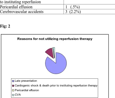
99 (6.7%) patients. 204 (13.8%) did not receive any form of reperfusion for
various reasons. The most common reason for not receiving reperfusion
therapy was late presentation in 175 (85.7%) patients followed by
cardiogenic shock or death in 25 (12.2%). There were 4 patients who were
38
Table 2: Reasons for not utilizing reperfusion therapy
Reasons
Number (n=204)
Late presentation
175(85.7%)
Cardiogenic shock and death prior
to instituting reperfusion
25 (12.2%)
[image:43.612.90.480.172.503.2]Pericardial effusion
1 (.5%)
Cerebrovascular accidents
3 (2.2%)
Fig: 2
Reasons for not utilizing reperfusion therapy
Late presentation
Cardiogenic shock & death prior to instituting reperfusion therapy
Pericardial effusion
CVA
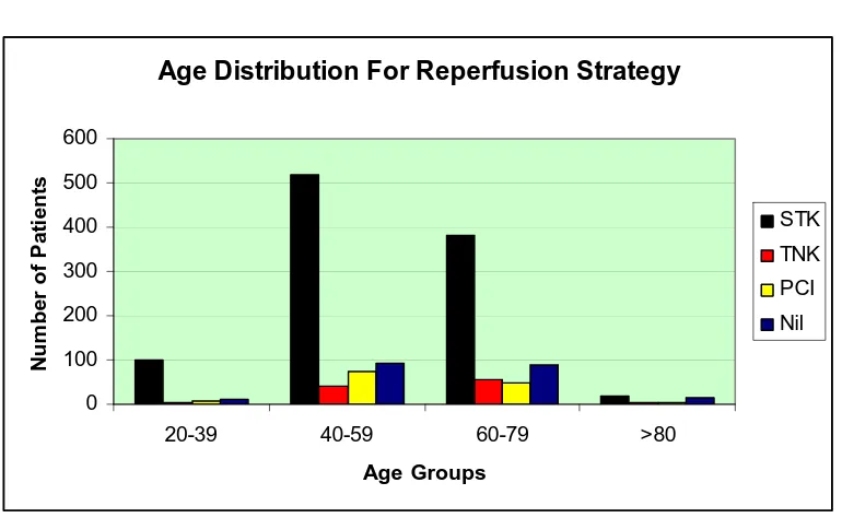
Table 3: Age Distribution of patients receiving different reperfusion
strategies
Age groups
STK
TNK
PCI
Nil
20-39 99
2
9
10
40-59 520 39
75
91
60-79 382 56
48
89
39
Fig 3:
Age Distribution For Reperfusion Strategy
0
100
200
300
400
500
600
20-39
40-59
60-79
>80
Age Groups
N
u
m
b
er
o
f P
a
ti
en
ts
STK
TNK
PCI
Nil
Mean age of patients in the whole study population was 56.3+ 11.8 years.
Maximum patients were in the age groups of 40-59 years with least in >80
years.
Table 4: Trends of revascularization in STEMI
MONTHS
*
Nil (%)
STK (%)
TNK (%)
PCI (%)
Total
1-6
35 (11.8)
216 (72.7) 27 (9.0)
19 (6.4)
297
7-12
55 (16.1) 219 (64.2) 25 (7.3)
42 (12.3)
341
13-18
55 (15.4) 253 (71.1) 21 (5.9)
27 (7.5)
356
19-24
59 (12.7) 331 (71.1) 26 (5.6)
49 (10.5)
465
Pvalue 0.075 0.000 0.840 0.001 0..000
[image:44.612.85.532.482.602.2]40
Fig 4:
0
100
200
300
400
500
Number of
Patients
Aug-Jan
Feb-July
Aug-Jan
Feb-July
Quadrants
Trends of revascularisation in STEMI
STEMI
STK
TNK
PCI
Nil
Patients with STEMI increased progressively in every 6 months assessment.
It was 297 patients to start with in 1
st
half-year which became 465 in the last
half-year.
Fig : 5
Trends of reperfusion in STEMI
0
50
100
150
200
250
300
350
400
450
500
Aug-Jan
Feb-July
Aug-Jan
Feb-July
[image:45.612.89.462.463.701.2]41
STK use as thrombolytic agent also followed the total STEMI trends. It was
216 in the first half-year and became 331 in the last half-year. (Table 5, Fig
5). The percentage of STEMI patients utilizing streptokinase as a
thrombolytic agent remained constant throughout the study period and
corresponded to the rise in patients of STEMI. Patient who did not receive
any form of revascularization too increased from 32 in the first half-year to
59 in the last half-year. The use of tenecteplase remained almost the same
[image:46.612.87.495.342.703.2]throughout the four half-years. It was 27 in the 1
st
half-year and 26 in the last
half-year.
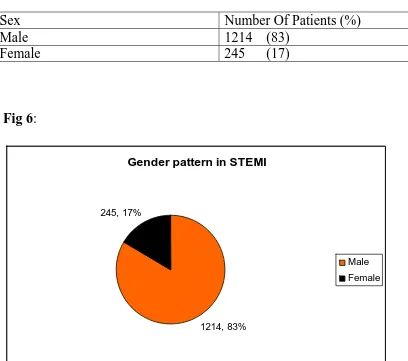
Table 5: Gender Pattern in STEMI
Sex
Number Of Patients (%)
Male
1214 (83)
Female
245 (17)
Fig 6:
Gender pattern in STEMI
1214, 83%
245, 17%
42
Males (1214, 83%) were involved more commonly than females (245, 17%).
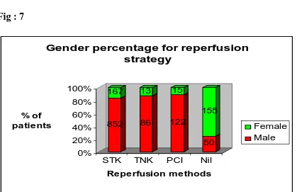
Table 6: Gender Percentage for reperfusion strategy
Sex
STK (%)
TNK (%)
PCI (%)
Nil (%)
[image:47.612.94.513.284.555.2]Male
852 (83.6)
86 (86.8)
122 (89)
50 (24.5)
Female
167 (16.3)
13 (13.2)
15 (11)
155 (75.5)
Fig : 7
852
167
86
13
122
15
50
155
0%
20%
40%
60%
80%
100%
% of
patients
STK
TNK
PCI
Nil
Reperfusion methods
Gender percentage for reperfusion
strategy
Female
Male
In all treatment groups the proportion of males was more than females. In
43
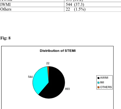
Table 7: Distribution of STEMI
Type of STEMI
Number of Patients (%)
AWMI 893
(61.2)
IWMI
544 (37.3)
[image:48.612.89.498.131.501.2]Others
22 (1.5%)
Fig: 8
Distribution of STEMI
893
544
22
AWMI
IMI
OTHERS
Most common territory involved was anterior wall which constituted 893
(61%) patients. Inferior wall STEMI in various combination was 544 (37%.
Other territories included high lateral, isolated posterior wall etc were 22
(1.5%).49.7% of the patients has type 2 diabetes mellitus. 73% of patients
had some form of lipid abnormalities. 34% of the patients had hypertension.
44
Table 8: Comparison of baseline characterstics and management
strategy outcomes of patients enrolled in earlier study* from the same
centre vs present study
Characterstics Earlier
study
N=1320 (%)
Present study
N= 1459 (%)
Age
56+ 13
56.3+11.8
Males
Females
1106 (83.8)
214 (16.2)
1214 (83%)
245 (17)
Smoking
569 (43.1)
452 (31)
Diabetes Mellitus
531 (40.2)
725 (49.7)
Hypertension
504 (38.2)
500 (34
Location of MI
Anterior wall MI
Inferior wall MI
752 (57)
517 (39)
893
(61.2)
544 (37.2)
Reperfusion therapy
Thrombolytics
STK
TNK
PCI
1093 (82.8)
Nil (0)
Nil (0)
1019 (69.8)
99
(6.7)
137 (9.4)
Data are numbers (%) or mean + SD
•
45
`
DISCUSSION
Mean age of presentation of STEMI in present study was 56.3+11.8 years with
majority of them being males (83%). In the study done by Jose et al, which
included patients from 1999-2003 in the same centre and the CREATE registry
which enrolled patients from 2001-2005, the mean age been 56+13 years and
57.5+12 years. This is suggesting that mean age of patients has not change over a
decade even when comparing data from the same centre. Patients enrolled in this
study presented a decade earlier in comparison to the western data where the mean
age of presentation has increased from 64.1 years to 66.4 years. Early age of
STEMI could lead to tremendous loss of productive years and can have a adverse
outcome on the economy a well as national health.
It is noteworthy that only a small percentage of patients in younger age group of
20-39 years received PCI (6.5%) and tenecteplase (2%) as compared to the 40-59
years age group (PCI 54.7%, tenecteplase 39.4%) age group. This relates to the
social status and financial means of the patients enrolled in the study. Younger
patients who would be benefited the most in terms of productive life-years saved
remain the least benefited by recent advances in reperfusion therapy.
In the present study, women comprised 17% of all patients, which was comparable
to the proportion of women patients in the CREATE study (18%) and the study by
Jose et al (16.2%). The data also highlighted the inferior treatment received by
women in comparison to men, as of the patients who received PCI only 11% were
women. Similarly, women comprised 13% of all STEMI patients who received
46
Trends in STEMI :
There was steady increase in admissions of patients with STEMI over a period of 2
years which was divided into four half-years for analysis. There was almost 20%
increase in the number of STEMI, if we compare the first two half-years with the
last two. This increment raises serious concern that the incidence of STEMI is
likely to be doubled in less than four years in the local population. There was
increase of 34 % of STEMI patients from the first half-year to the last. There was
also parallel increase in the number of patients thrombolysed with streptokinase.
In the first half-year, streptokinase was used in 72% of patients of all STEMI. This
percentage remained almost the same (71%) till the last half-year. So even though
the numbers of patients of STEMI have increased, the role of streptokinase as the
main thrombolytic agent has remained the same over the 2 years period of study.
Streptokinase constituted 91% of all thrombolysed cases. This is in comparison to
data from CREATE study in which thrombolytics (96 % streptokinase) were used
for 58·5% of patients with STEMI and study of Jose et al in which 82.8% of
patients received thrombolysis (all with streptokinase).
A very important result of this study was the analysis of patients who did
not receive any reperfusion therapy because of various reasons. 14% patients of
STEMI did not receive reperfusion therapy over the period of 2 years; there was
41% increase in the number of such patients between the 1
st
and the last half-years,
which is very alarming considering that this number should ideally be expected to
fall. In comparison, in the study by Jose et al 17.2% patient did not receive
reperfusion therapy. So even a decade after the study by Jose et al, the reperfusion
therapy usage in STEMI has only marginally increased. The most important
reasons for no reperfusion therapy usage was late presentation (85.7%) (Table 2,
47
transport facilities and lack of awareness in the general population. Even though,
females formed only 17% of all STEMI patients, they constituted 25 % of patients
not receiving reperfusion therapy. So reperfusion therapy usage rates were lower in
female patients as compared to males.
Cardiogenic shock and death prior to instituting reperfusion therapy (12.2%) was
the second most common cause for not receiving reperfusion therapy. These
patients could have been treated with primary PCI, if they could have afforded it.
High cost of PCI procedures and its non-availability even in some tertiary care
centres remains one of the important draw backs in management of STEMI in
India.
There were 3 patients who had an absolute contraindication to thrombolytic
therapy. Two of them had a recent cerebrovascular accident and one had a
concurrent large ischemic stroke. One patient was not offered thrombolysis in view
of large associated pericardial effusion. PCI would have been the best option in
these of patients, if they could afford it. Tenecteplase was used in 6.7% of
patients, where as in the study by Jose et al study the only thrombolytic used was
streptokinase. The number of patients who were thrombolysed with tenecteplase
remained almost same in the four half-years studied (Table 4, Fig 4).
Tenecteplase was used in 9% of all STEMI patients in the first half-year compared
to only 5.6% in the last (Table 4). The cost of tenecteplase was the reason for
fewer patients being thrombolysed with it. The cost of tenecteplase in our centre is
7-8 times that of streptokinase. Interestingly, it was found that most of the patients
who could afford tenecteplase, could afford PCI as well and so opted for the latter.
So if tenecteplase is to replace streptokinase as a thrombolytic agent in India, its
price has to come down significantly.
48
half-year PCI was used only in 6.3% of all STEMI patients and this percentage
increased to 10.5% in the last half-year. So even though the percentage of primary
PCI between 1
st
and the last half-year has almost doubled, this may not be adequate
considering 34% increase in the rates of STEMI hospitalizations during this period.
This study also analyzed the main risk factors for CAD prevalent in the
Indian population. Diabetes mellitus was found in 49.7% of patients; systemic
hypertension was found in 34%; 31% of patients were either current or ex-smokers
and 73% of patients had some form of dyslipidemia. This data is different from
data from CREATE study where 34% patients had diabetes; 37·7% had
hypertension; and 40·2% were smokers. Study from Jose et al from the same
centre as the present study a decade back showed 40.2% prevalence of diabetes in
STEMI patients. So data the from present study demonstrates an increase in the
proportion of patients with diabetes as compared to the past studies and suggest a
strong correlation of diabetes mellitus with coronary artery disease.
There were many differences between the present study and the study by Jose et al
and the CREATE registry. The study by Jose et al was done a decade ago in the
same centre as the present study. Thrombolysis with streptokinase was the only
mode of reperfusion in patients of STEMI, where as in the present study STK is
considered as a last option for reperfusion therapy after PCI and tenecteplase. Data
on gender and territory of MI has remained almost the same in the two studies.
Prevalence of diabetes mellitus has increased 10% in STEMI patients over a
decade which may be the reason for increased number of patients presenting with
STEMI in the present study. The present study differed from the CREATE registry
as it is a single-center study as compared to the multi-centre CREATE registry.
The present study highlights the current scenario of STEMI and its
49
a day, the rates of primary PCI has been low. Many reasons are responsible for
this. The most important being the financial constraints of the patients; most
patients in India pay for emergency medical expenses themselves and lack
insurance cover.
Lack of awareness in general population and poor referral and transport has
an important role to play. Increasing the numbers of tertiary cardiac centres alone
is unlikely to be sufficient to improve the management of STEMI; the efforts have
50
Conclusions
1.
: Patients with STEMI in this study are decade younger than western
population
2.
: 20% increase in STEMI every year in the studied population.
3.
: Younger patients least likely to get primary PCI or tenecteplase as
reperfusion therapy.
4.
: 86% of patients received some form of reperfusion therapy.
Streptokinase was the most common type of reperfusion therapy.
5.
: Primary PCI rates increasing at 20% per year but still <10% STEMI
patients receive it.
6.
: Tenecteplase is a better replacement for streptokinase was used in
less than 10% due to its high cost.
7.
: 1 in 8 patients of STEMI didn’t receive reperfusion therapy. Most
common reason for not receiving reperfusion therapy was late
presentation.
REFERENCES
1.
Lopaze AD, Mathers CD, Ezzati M, Jamison DT, Murray CJ. Global
and regional burden of disease and risk factors, 2001: systematic
analysis of population health data. Lancet 2006; 367:1747-57.
2.
Yusuf S, Reddy S, Ounpuu S, Anand S. Global burden of
cardiovascular diseases: Part II: variations in cardiovascular disease
by specific ethnic groups and geographic regions and prevention
strategies. Circulation 2001;104: 2855-65.
3.
Gaffar A, Reddy KS, Singhi M. Burden of non-communicable diseases
in South Asia. BMJ 2004;328:807-10.
4.
Fox KA, Goodman SG, Klein W, et al. Management of acute coronary
syndromes. Variations in practice and outcome; findings from the
Global registry of Acute Coronary Events (GRACE). Eur Heart J 2002;
23:1177-89.
5.
Mandelzweig L, Battler A, Boyko V, et al. The second Euro Heart
Survey on acute coronary syndrome: characteristics, treatment, and
outcome of patients with ACS in Europe and Mediterranean Basin in
2004. Eur Heart J 2006; 27:2285-93.
6.
Xavier D, Pais P, Devereaux PJ, Xie C, Prabhakaran D, Reddy KS, et
al; CREATE registry investigators. Treatment and outcomes of acute
coronary syndromes in India (CREATE): a prospective analysis of