Escherichia coli and Mucosal Staphylococci in
Dogs
Thesis submitted in accordance with the requirements of the University of Liverpool
for the degree of Doctor in Philosophy
by
Vanessa Merta Schmidt
Where do you start after four years of blood sweat and tears; and not all mine. First and foremost I need to thank my ever-suffering husband, Stephen Flynn as without his support I would never of even started let alone finished the PhD - death by starvation at the very least! I love you darling (and I promise to do more housework from now on). I would also like to thank my other two boys (Sebbie & Ziggy) for cuddles, walks and for keeping me company (and sane) whilst writing up.
Very importantly, I would like to thank all of my supervisors Dr Nicola J Williams, Dr Gina Pinchbeck, Dr Neil McEwan, Dr Susan Dawson and Dr Tim Nuttall and my advisor, Dr Eithne Comerford, for their support, advice and mentorship. I would especially like to thank Nicola for all of her very hard work, ideas, positivity, support, reviewing, subbing and especially patience – you’re definitely a star!
I would also like to thank Dr Caroline Corless and Erika Tranfield at the Royal Liverpool University Hospital for their amazing and continued help, support, ideas and advice – and especially their valuable time!
Without samples there would be no study – therefore thank you very much to everyone that participated in either sample collection and/or recruitment: Clara MacFarlane, Laura Buckley, Maarja Uri, Ana Martins, Katherine Linney, Helen Yates, Kathleen Gallagher and Atina Unwin. In particular I would like to thank Clara for her co-ordination and organisation in this matter and for accompanying me to dog shows and on home visits when required ☺.
The lab (G10A) has been my second home (sometimes my first) during the last four years. I would like to thank the very many people that work there – THANK YOU ALL for being so nice, understanding and very helpful to this vet!
I would particularly like to thank all the people that have physically helped me at one time or another during the last four years, as without you all I would still be buried under a mountain of plates: Amy Wedley (fountain of all knowledge), Ruth Ryvar, Jenny Fick, Gill Hutchinson, Beatrice Jones, Stevie Snoop, Rebecca Callaby, David Ramsbottom, Mitch Long, Lou
Marriage, Chris Ball, Kirsty Kemmett, Nick Harvey, Jackie Lee and IGH ordering team and Karen Ryan (for keeping me in agar and broth). A special thank you to ‘Aunty’ Ruth for always being there.
Also thank you to Trevor Jones, Dorina Timofte, Andy Watrett, Jan Harris, Tom Maddox, Iuliana Maciuca, Marie McIntyre, Cathy Glover, Cynthia Dare and Anne Forester for your time and kind advice from time to time on all matters microbiological, statistical and grammatical.
Finally thank you to my vet PhD comrades: Dr Camilla Brena and Dr Marco Falchieri, and my friend Dr Steve Shaw. I still can’t believe I have made it!
Staphylococci in Dogs
Vanessa Merta Schmidt
Canine infections with antimicrobial resistant (AMR), particularly multi-drug resistant (MDR) bacteria are increasing, severely limiting therapeutic options, and representing an animal health issue. In addition, with potential transfer of AMR bacteria between dogs, their environment, humans and other animals, there may also be a public health risk. Commensal isolates can be a source of clinical infections and studies reporting the prevalence of AMR and risk factors for such isolates are important. Furthermore, one of the most significant impacts upon commensal bacterial populations is antimicrobial therapy that may select for pre-existing AMR organisms or transmission of resistance determinants. The aim of this work was to investigate AMR amongst canine commensal bacterial populations and the effects of five different antimicrobials, authorised to treat dogs in the UK, on these populations both during and after therapy. Three groups of dogs were enrolled: healthy non-antimicrobial treated, non-vet visiting dogs (n = 28), to investigate longitudinal carriage of faecal E. coli; healthy non-antimicrobial treated, non-vet visiting, dogs (n = 73) and antimicrobial treated, non-hospitalised dogs (n = 127) to investigate longitudinal carriage of mucosal staphylococci and faecal E. coli. Staphylococci and E. coli isolated from swabs (nose/perineum) and faecal samples respectively, were tested for phenotypic AMR and carriage of resistance genes by PCR assay. Staphylococci were assigned to species by PCR assay (nuc gene), MALDI-TOF-MS and sequencing (tuf gene). Healthy dog E. coli underwent phylo-typing, and a selection of longitudinal healthy dog E. coli isolates were genotyped. Questionnaire data were used to formulate independent variables. Statistical analysis included Pearson’s Chi-square, survival analysis and multivariable logistic regression; multilevel for clustered data. The prevalence of meticillin-resistant (MR; 42%) and MDR staphylococci (resistant to ≥ 3 antimicrobial classes; 34%) was high amongst healthy dogs, however MR-coagulase positive staphylococci were not detected. The most common species detected was S. epidermidis (52% of dogs), followed by S. pseudintermedius (44%). S. aureus was only detected in a small number of dogs (8%). Faecal E. coli with AMR to at least one tested drug (63%), MDR (30%) and AmpC-production (16%) were prevalent in healthy dogs, however ESBL-producers (1%) were rare. Healthy dogs carried a predominance of phylogenetic group B1; group B2 E. coli isolates were less likely to have AMR while group D isolates were more likely. Carriage of E. coli
95% CI 95% confidence interval
Amp Ampicillin
AMR Antimicrobial Resistance
BSAC British Society for Antimicrobial Therapy
CAB Columbian Blood Agar
CD Clindamycin
CFX Cefalexin
Chl Chloramphenicol
CLSI Clinical Laboratory Standards Institute
CIP Ciprofloxacin
CoNS Coagulase Negative Staphylococci CoPS Coagulase Positive Staphylococci
CVN Cefovecin
ExPEC Extra-intestinal Pathogenic E. coli
ESBL Extended Spectrum Beta-Lactamases
GNB Gram Negative Bacteria
M Mean
MDR Multidrug resistance
MGEs Mobile Genetic Elements
MIC Minimum Inhibitory Concentration
MLST Multi-Locus-Sequence-Typing
MR Meticillin resistant
MR-CoNS Meticillin Resistant Coagulase Negative Staphylococci
MRS Meticillin Resistant Staphylococci
MRSA Meticillin Resistant S. aureus MRSS Meticillin-resistant S. schleiferi
MSA Mannitol Salt Agar
MSSA Meticillin Susceptible S. aureus
MRSP Meticillin resistant S. pseudintermedius
Nal Nalidixic acid
NOAH National Office of Animal Health
OR Odds Ratio
ORSA Oxacillin Resistance Screening Agar
OX Oxacillin
PBP Penicillin Binding Protein
PCR Polymerase Chain Reaction
PFGE Pulsed Field Gel Electrophoresis
SCCmec Staphylococcal Cassette Chromosome mec
SD Standard Deviation
SDW Sterile distilled water
SIG Staphylococcus intermedius Group
Tet Tetracycline
TM Trimethoprim
TS Cotrimazole
VMD Veterinary Medicines Directorate
General Introduction and Literature Review (Chapter 1) ... 1
Commensal bacteria (1.1) ... 1
Intestinal Microbiome (1.2) ... 1
Escherichia coli (1.2.1) ... 2
Faecal E. coli: an indicator of intestinal health (1.2.1.1) ... 2
Faecal E. coli: commensal and pathogen (1.2.1.2) ... 2
Classification of E. coli strains by phylogenetic group (1.2.1.3) ... 3
Phylogenetic groups: characterisation and distribution (1.2.1.3.1) ... 3
Phylogenetic groups: antimicrobial resistance (1.2.1.3.2) ... 4
Gut resident and transient E. coli strains (1.2.1.4) ... 4
Diversity of E. coli population structure (1.2.1.5) ... 5
Mucosal Microbiome (1.3) ... 5
Coagulase positive staphylococci (CoPS) (1.3.1) ... 6
Coagulase negative staphylococci (CoNS)(1.3.2) ... 7
Antimicrobials (1.4) ... 8
Antimicrobials authorised to treat dogs in the UK (1.4.1) ... 8
Mechanism of action of antimicrobials (1.4.2) ... 8
Beta-lactams (1.4.2.1) ... 8
Fluoroquinolones (1.4.2.2) ... 9
Lincosamides (1.4.2.3) ... 10
Impact of antimicrobials on the gut microbiome (1.4.3) ... 10
Impact of antimicrobials on the mucosal microbiome (1.4.4) ... 11
Antimicrobial resistance (AMR) (1.5) ... 11
Antimicrobial resistance mechanisms (1.5.1) ... 12
Mobile genetic elements (MGEs) (1.5.1.1) ... 13
Transformation, conjugation and transduction (1.5.1.2) ... 13
Beta-lactam resistance (1.5.1.3) ... 14
Beta-lactam resistance: Gram-negative bacteria (1.5.1.3.1) ... 14
Beta-lactam resistance: staphylococci (1.5.1.3.2) ... 15
Fluoroquinolone resistance (1.5.1.4) ... 15
Fluoroquinolone resistance: Gram-negative bacteria (1.5.1.4.1) ... 16
Fluoroquinolone resistance: staphylococci (1.5.1.4.2) ... 16
Fitness costs (1.5.1.5) ... 16
Antimicrobial resistance in Escherichia coli (1.5.2) ... 17
Antimicrobial resistance in staphylococci (1.5.3) ... 20
Prevalence of AMR staphylococciin dogs (1.5.3.1) ... 20
Risk factors of AMR staphylococci in dogs (1.5.3.2) ... 21
Longitudinal carriage of AMR staphylococci in dogs (1.5.3.3) ... 21
Maintenance and spread of AMR by the microbiome (1.5.4) ... 22
Concluding summary and aims (1.6) ... 23
General Materials and Methods (Chapter 2) ... 25
Study populations (2.1) ... 25
Healthy dog cohort study (2.1.1) ... 25
Healthy dog longitudinal study (2.1.2) ... 25
Antimicrobial treated dogs longitudinal study (2.1.3) ... 26
Specimen collection (2.2) ... 26
Processing swab samples: staphylococci (2.3) ... 27
Staphylococcal isolation (2.3.1) ... 27
Staphylococcal isolate selection (2.3.2) ... 27
Antimicrobial susceptibility tests of staphylococci (2.3.3) ... 28
Isolate storage and DNA extraction (2.3.4) ... 29
Processing faecal samples: Escherichia coli (2.4) ... 29
Escherichia coli isolation (2.4.1) ... 29
Escherichia coli isolate selection (2.4.2) ... 29
Antimicrobial susceptibility tests of E. coli (2.4.3) ... 30
Phenotypic detection of ESBL- and AmpC-producing E. coli (2.4.4) ... 31
Conjugation experiments (2.4.5) ... 32
Isolate storage and DNA extraction (2.4.6) ... 32
Polymerase chain reaction (PCR) assay (2.5) ... 32
PCR substrates (2.5.1) ... 32
Visualisation of PCR products (2.5.2) ... 33
MALDI-TOF-MS (2.6) ... 33
Sequencing of the tuf gene (2.7) ... 34
Macro-restriction Pulsed Field Gel Electrophoresis (PFGE) (2.8) ... 34
Preparation of agarose plugs (2.8.1) ... 35
XbaI restriction digest (2.8.2) ... 35
Manuscript 2: (Chapter 4) ... 59
Antimicrobial resistance and characterisation of faecal Escherichia coli isolated from healthy Labrador retrievers in the United Kingdom
Manuscript 3: (Chapter 5) ... 81
Longitudinal study of antimicrobial resistance and characterisation of faecal Escherichia coli isolated from healthy dogs in the United Kingdom
Manuscript 4: (Chapter 6) ... 106
Antimicrobial resistance amongst canine mucosal staphylococci following antimicrobial therapy
Manuscript 5: (Chapter 7) ... 131
Antimicrobial resistance amongst canine faecal Escherichia coli following antimicrobial therapy
General Discussion: (Chapter 8) ... 159
Further work (8.1) ... 166
Conclusions (8.2) ... 167
References ... 169
Appendix I ... 201
Appendix II ... 208
Appendix III ... 213
Appendix IV ... 222
Appendix V ... 245
3GCR Third generation cephalosporin resistance 95% CI 95% confidence interval
Amp Ampicillin
AMR Antimicrobial resistance
BSAC British Society for Antimicrobial Therapy
CAB Columbian blood agar
CD Clindamycin
CFX Cefalexin
Chl Chloramphenicol
CLSI Clinical Laboratory Standards Institute
CIP Ciprofloxacin
CoNS Coagulase negative staphylococci CoPS Coagulase positive staphylococci
CVN Cefovecin
ExPEC Extra-intestinal pathogenic E. coli
ESBL Extended spectrum beta-lactamases
GNB Gram-negative bacteria
M Mean
MDR Multidrug resistance
MGEs Mobile genetic elements
MIC Minimum inhibitory concentration
MLST Multi-locus-sequence-typing
MR Meticillin resistant
MR-CoNS Meticillin resistant coagulase negative staphylococci
MRS Meticillin resistant staphylococci
MRSA Meticillin resistant S. aureus MRSS Meticillin resistant S. schleiferi
MSA Mannitol Salt Agar
MSSA Meticillin Susceptible S. aureus
MRSP Meticillin resistant S. pseudintermedius
Nal Nalidixic acid
NOAH National Office of Animal Health
OR Odds ratio
ORSA Oxacillin resistance screening agar
OX Oxacillin
PBP Penicillin binding protein
PCR Polymerase chain reaction
PFGE Macro-restriction pulsed field gel electrophoresis
SCCmec Staphylococcal cassette chromosome mec
SD Standard deviation
SDW Sterile distilled Water
SIG Staphylococcus intermedius group
Tet Tetracycline
TM Trimethoprim
TS Cotrimazole
VMD Veterinary Medicines Directorate
1. General Introduction and Literature Review
1.1 Commensal bacteria
Commensal microorganisms inhabit ecological niches on the body that are exposed to the environment (Berg, 1996), and the bacteria occupying mucous membranes and the gastro-intestinal tract form part of the normal commensal microbiome. Canine commensal bacteria are generally obtained gradually from the dam during the first week of life (Allaker et al., 1992). Commensal organisms benefit by receiving protection and nutrients from the host and other members of the microbiome, but are usually not detrimental to the host (Tenaillon et al., 2010). The population density, phyla and species composition of the commensal microbiome is generally stable over time for a particular habitat and host (Berg, 1996). A stable
microbiome is crucial for the health and immune status of the host and helps to provide a colonisation barrier against pathogens (Vollaard and Clasener, 1994). If the host is immune-compromised or the microbiome and/or barrier function is disrupted, commensal bacteria may become opportunistic pathogens (Berg, 1996; von Eiff et al., 2002).
1.2 Intestinal microbiome
The large intestinal microbiota of humans consists of approximately 1010 to 1011 bacterial
cells per gram of large intestinal content with more than 500 species, predominately
anaerobes (Backhed et al., 2005; Berg, 1996; Vollaard and Clasener, 1994). Similar findings have been reported in dogs with 108 to 1011 intestinal bacteria per gram of dry faeces (Davis et al., 1977; Mentula et al., 2005; Vanhoutte et al., 2005). Bacterial species compete for
intestinal niches, but may also be mutually beneficial providing nutrients and optimal growth conditions for each other (Jones et al., 2007). A stable intestinal microbiome has metabolic, trophic and protective functions for the host (Guarner and Malagelada, 2003). Together with anatomical and physiological functions of an intact gut, the microbiome provides colonisation resistance (Vollaard and Clasener, 1994). Mechanisms of this resistance are likely to include production of bacteriocins, competition for attachment sites/nutrients or stimulation of the immune system (Hudault et al., 2001; Tenaillon et al., 2010; Vollaard and Clasener, 1994). One of the most common and significant disturbances of the intestinal microbiome is antimicrobial therapy (Berg, 1996; Vollaard and Clasener, 1994), however fluctuations may also occur with disease e.g. acute enteritis or, to a lesser extent, as a result of dietary
1.2.1 Escherichia coli
The most prevalent Gram-negative aerobic bacteria (GNB) in all hosts are Escherichia coli
(Backhed et al., 2005; Berg, 1996; Vollaard and Clasener, 1994). The gastro-intestinal tract of warm-blooded animals and reptiles is the main habitat of E. coli, but they also frequent secondary environmental habitats such as water and sediments (Berg, 1996; Gordon and Cowling, 2003; Savageau, 1983). The prevalence and density of E. coli in the intestine depends on host body size, diet, concurrent microbiota/gut morphology and retention times; human prevalence is > 90% (Penders et al., 2006; Tenaillon et al., 2010). In dogs, E. coli was reported to account for 98% of the faecal aerobic coliforms (Mentula et al., 2005). The intestine of all new-born hosts is colonised shortly after birth by E. coli, likely from maternal microbiota. The bacteria reside in and obtain nutrition from the mucous layer covering the intestinal epithelium, thereafter they and are shed into the lumen and are excreted in faeces (Poulsen et al., 1994). E. coli may contribute to colonisation resistance and inhibit the colonisation of further (exogenous) pathogens (Hudault et al., 2001; Tenaillon et al., 2010; Vollaard and Clasener, 1994).
1.2.1.1 Faecal E. coli: an indicator of intestinal health
E. coli is a well-characterised, widespread gut commensal and potential pathogen that is readily cultured from faeces (Russo and Johnson, 2003; Tenaillon et al., 2010). Faecal culture is an easy, cheap and non-invasive method to study the colonic bacterial microbiota to determine intra or inter-individual differences and investigate antimicrobial selection pressures (Eckburg et al., 2005; Gronvold et al., 2010; Mentula et al., 2005; Wang and Schaffner, 2011). Impairment of the colonisation resistance barrier (mainly composed of anaerobic bacteria) may result in increased detection of antimicrobial resistant E. coli
(Vollaard and Clasener, 1994).
1.2.1.2. Faecal E. coli: commensal and pathogen
E. coli strains have diversified through frequent horizontal gene transfer and recombination events (Rasko et al., 2008; Touchon et al., 2009) and are both gut commensals and adaptable pathogens (Tenaillon et al., 2010). Commensal strains make up the majority of the gut E. coli
survival within the microbiome and possession of certain genes is likely an adaptive strategy to the strain’s environment and only secondarily involved in intra- and extra-intestinal pathogenesis (Tenaillon et al., 2010). Unlike enteric pathogenic strains, ExPEC strains may reside in the microbiome along with commensal E. coli and do not cause gastroenteritis. Gut colonisation is a pre-requisite for extra-intestinal infection, which causes a diverse range of clinical syndromes in humans with considerable morbidity, mortality and associated
healthcare costs (Russo and Johnson, 2003). ExPEC are also a common cause of urinary tract infections in dogs (Johnson et al., 2009).
1.2.1.3 Classification of E. coli strains by phylogenetic group
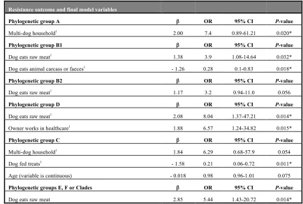
Despite frequent recombination,the population structure of E. coli is predominantly clonal with division into four major phylogenetic groups (A, B1, B2 and D) (Clermont et al., 2000; Doumith et al., 2012). The original PCR assay to assign isolates to these groups was based on the combination of three marker genes: chuA (encoding the outer-membrane hemin receptor gene), yjaA (encoding an uncharacterised protein) and TSPE4.C2 (encoding putative lipase esterase) (Clermont et al., 2000). This PCR assay had 80–85% test specificity compared to multi-locus-sequence-typing (MLST) (Gordon et al., 2008). Doumith et al., (2012) recently updated this method with new primers to amplify conserved regions of the same three markers and glutamate decarboxylase-alpha gene (gadA), an internal control, with further improved specificity (~ 90%). Clermont et al., (2013) also recently updated their method to improve specificity and identify four additional phylo-groups: C, E, F and Escherichia cryptic clade I.
1.2.1.3.1 Phylogenetic groups: characterisation and distribution
Phylo-grouping can help characterise the genetic background, pathogenicity and antimicrobial resistance traits of E. coli strains (Sato et al., 2014; Tenaillon et al., 2010). Phylo-groups B2 and D are more likely to carry virulence genes and cause extra-intestinal infections compared to A or B1 (Clermont et al., 2011; Johnson and Stell, 2000; Picard et al., 1999). Phylo-group C is closely related to group B1 (Clermont et al., 2011; Moissenet et al., 2010), while phylo-group E is related to D (Tenaillon et al., 2010) and phylo-phylo-group F is related to B2 (Jaureguy et al., 2008). Novel Escherichia lineages (cryptic clades) that are genetically distinct, but
phenotypically similar to E. coli have also been identified (Walk et al., 2009).
environmental characteristics such as signalment, body mass, gut morphology, diet, level of hygiene and degree of domestication (Clermont et al., 2011; Escobar-Paramo et al., 2006; Gordon and Cowling, 2003; Gordon et al., 2005; Tenaillon et al., 2010). Generally in humans, phylo-group A predominates followed by groups B2, B1 and D; in animals, group B1
predominates followed by groups A, B2 and D (Tenaillon et al., 2010).
1.2.1.3.2 Phylogenetic groups: antimicrobial resistance
Some genetic backgrounds appear more likely to develop antimicrobial resistance (Tenaillon et al., 2010). This has been reported for the less virulent non-B2 groups in humans, cattle, pigs and dogs (Johnson et al., 2009; Johnson et al., 2003; Moreno et al., 2008). Phylo-group D isolates are more likely to be resistant to fluoroquinolones, third generation cephalosporins (3GCR) or multiple drug classes (MDR) (Deschamps et al., 2009; Platell et al., 2011; Sato et al., 2014; Tamang et al., 2012). Group B2 is more likely to be susceptible (Johnson et al., 2009; Platell et al., 2010; Platell et al., 2011; Sato et al., 2014) than the other groups, however antimicrobial resistance is being reported increasingly amongst B2 (ExPEC) strains (Russo and Johnson, 2003).
1.2.1.4 Gut residents and transient E. coli strains
1.2.1.3 Diversity of E. coli population structure
The E. coli population structure is diverse and dynamic within both the host, the host population, and between host species, for example animals and humans. The aetiology is likely to be multifactorial with host, environmental and bacterial factors all playing a role; possible influences include: host signalment, climate, level of hygiene, diet, health and immune status, bacterial phylogeny, possession of virulence factors, competing bacteria and antimicrobial selection pressure and resistance phenotype (Anderson et al., 2006; Damborg et al., 2009; Schlager et al., 2002; Tenaillon et al., 2010).
1.3 Mucosal microbiome
As with the gastro-intestinal microbiome, the mucosal microbiome consists of a vast number of micro-organisms than inhabit the skin and mucous membranes of humans and other animals; it has been estimated that there are ~ 1 billion bacteria per cm2 of human skin (Grice
et al., 2008). Recent advances in molecular diagnostic methods have used sequencing of bacterial 16S rRNA genes to investigate the microbiome of the mucosa and skin of healthy dogs. This study reported a majority of Staphlyococcaceae, Oxalobacteriaceae, and
Enterobacteriaceae families from the perineum, and Oxalobacteriaceae, Moraxellaceae
followed by Staphlyococcaceae and Corynebacteriaceae from the nasal mucosa (Rodrigues Hoffmann et al., 2014). This is in agreement with the majority of previous culture based studies that have identified staphylococci as important commensals of the mucous membranes and skin of humans and other animals (Kloos, 1980; Saijonmaa-Koulumies and Lloyd, 1996). In humans, the diversity of the microbiome is influenced by the characteristics of the
ecological niche, for example Staphylococcus spp. prefers m oist, humid environments (Grice et al., 2009). Similar findings have been reported for dogs (Saijonmaa-Koulumies and Lloyd, 1996). In humans the most consistent microbiomes are in the ear and nasal cavity (Grice et al., 2009).
The cutaneous barrier is physical, immunological and antimicrobial and together with the microbiome works to prevent adherence, colonisation and infection by pathogens (Elias, 2005; Kong and Segre, 2012; Saijonmaa-Koulumies and Lloyd, 1996). The microbiome may be involved in competitive inhibition for adherence sites and nutrients, production of
become opportunistic pathogens (Pfaller et al., 2007; von Eiff et al., 2002). Atopic dermatitis in humans and dogs is a common cause of reduced barrier function and sufferers have increased carriage of staphylococci and a predilection for pyoderma (Bibel et al., 1977; Fazakerley et al., 2010; Mason and Lloyd, 1989; Olivry and Hill, 2001).
1.3.1 Coagulase Positive Staphylococci (COPS)
The ability to produce free coagulase and clot rabbit plasma differentiates species of
staphylococci. S. pseudintermedius is the main commensal coagulase positive staphylococci (CoPS) of dogs, and the main cause of pyoderma (Berg et al., 1984; Ihrke, 1987; Medleau et al., 1986). S. aureus is the human counterpart (Mainous et al., 2006). Puppies acquire
S. pseudintermedius from their mothers during the first week of life (Saijonmaa-Koulumies and Lloyd, 2002) and the main carriage sites appear to be the mucosa (nose, gingiva, oro-pharnyx, perineum or rectum), where they are considered to be residents; licking by the dog is thought to spread bacteria to the skin and hair (Allaker et al., 1992; Devriese and De
Pelsmaecker, 1987; Mason et al., 1996). These findings have been corroborated by studies reporting homogeneity of strains from mucosa and skin of an individual dog, but
heterogeneity of strains between dogs (Fazakerley et al., 2010; Pinchbeck et al., 2006). In addition, metagenomic analysis identified greater numbers of microbial species and diversity from haired canine skin compared to mucosa (Rodrigues Hoffmann et al., 2014).
The prevalence of mucosal S. pseudintermedius carriage in healthy dogs has been reported to be between 37% to 92% (Devriese and De Pelsmaecker, 1987; Fazakerley et al., 2010; Griffeth et al., 2008; Hanselman et al., 2009; Paul et al., 2012; Rubin et al., 2011), with increased carriage for dogs with certain skin diseases such as atopic dermatitis or pyoderma (Fazakerley et al., 2010; Saijonmaa-Koulumies and Lloyd, 1995). Long-term carriage of
S. pseudintermedius may occur in humans living with dogs (Gomez-Sanz et al., 2013), particularly if owning more than two dogs (Walther et al., 2012).
al., 2008; Yamashita et al., 2005). Prior to re-classification of the S. intermedius group (SIG) in 2007, S. pseudintermedius were referred to as S. intermedius (Bannoehr et al., 2007).
1.3.2 Coagulase Negative Staphylococci (CoNS)
Coagulase negative staphylococci (CoNS) are common cutaneous and mucosal residents of humans and dogs (Kloos and Bannerman, 1994; Saijonmaa-Koulumies and Lloyd, 1996). They were historically considered to be apathogenic (Huebner and Goldmann, 1999), however they are now recognised as a significant cause of nosocomial and community-acquired infections in humans (Barbier et al., 2010; Wertheim et al., 2005), and may also cause infections in dogs and other animals (Beck et al., 2012; Hauschild and Wojcik, 2007; Kern and Perreten, 2013). Certain CoNS species (S. epidermidis, S. saprophyticus, and
S. lugdunensis), like CoPS, may carry virulence factors that increase their pathogenesis and involvement in severe human infections (Dupont et al., 2010).
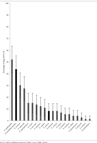
There are at least 40 described species of Staphylococcus with the majority being CoNS.
S. epidermidis is the most common CoNS isolated from the mucosa (nares, perineum and inguinal skin), axillae and interdigital skin of humans (Kloos and Bannerman, 1994; Widerstrom et al., 2012). Numerous CoNS have been detected from dogs including
S. schleiferi subsp schleiferi, S. epidermidis, S. haemolyticus, S. saprophyticus, S. devriesei, S. warneri, S. simulans, S. xylosus, S. capitis, S. caprae and S. sciuri (Bagcigil et al., 2007; Cox et al., 1988; Fazakerley et al., 2010; Kania et al., 2004; May et al., 2005; Medleau et al., 1986; Wedley et al., 2014). The prevalence of nasal CoNS detection in dogs has been reported in one large UK cross sectional study as 38% (Wedley et al., 2014).
1.4 Antimicrobials
1.4.1 Antimicrobials authorised for dogs in the UK
The majority of antimicrobials are usually administered in an oral form. Cefovecin however, is a long-acting subcutaneous preparation of a semi-synthetic third generation cephalosporin. It is authorised in Europe to use every 14 days, for the treatment of skin and urinary tract infections associated with a number of Gram-positive and negative bacteria (NOAH, 2014). It has reported efficacy in the treatment of canine pyoderma (Summers et al., 2012). The majority of cefovecin is excreted unchanged in the urine; however unchanged drug also occurs in the bile (Stegemann et al., 2006). Oral beta-lactam antimicrobials include cefalexin, a first generation cephalosporin that is authorised to treat susceptible bacteria causing skin and urinary tract infections, and clavulanate-amoxicillin, authorised to treat a broad range of aerobic and anaerobic bacteria associated with skin, soft tissue, dental, urinary or respiratory tract infections or enteritis (NOAH, 2014). Both drugs are commonly used to treat canine pyoderma (Damborg et al., 2011; Summers et al., 2012). Veterinary fluoroquinolones, including oral enrofloxacin and marbofloxacin, are broad-spectrum antimicrobials authorised to treat urinary tract, respiratory and skin infections. Fluoroquinolones have been used frequently in dogs to treat pyoderma (Guardabassi et al., 2004; Ihrke et al., 1999). Clindamycin, an oral lincosamide antimicrobial, is also commonly used to treat bacterial pyoderma and dental disease, and while it is not effective against Gram-negative aerobes it is very effective against anaerobes (NOAH, 2014).
1.4.2 Mechanism of action of antimicrobials 1.4.2.1 Beta-lactams
Amoxicillin is a synthetic derivative of penicillin, with slightly less activity against Gram-positive aerobes and anaerobes, but wider activity against Gram-negative aerobes. The addition of clavulanic acid, a potent inhibitor of beta-lactamase enzymes, improves the effectiveness of amoxicillin. Cephalosporins are divided into four classes based on chemical structure and therapeutic activity with decreasing activity against Gram-positive aerobes and anaerobes and increasing activity against Gram-negative aerobes. Cefalexin is a first
generation cephalosporin predominantly active against Gram-positive aerobes and some Gram-negative aerobes including E. coli. It is more effective against anaerobes than
amoxicillin. Third generation cephalosporins are less effective against Gram-positive aerobes and anaerobes but more effective against Gram-negative aerobes than first generation
cephalosporins (Greene and Watson, 2006). Cefovecin is classed as a third generation cephalosporin, but its spectrum of activity is more similar to first- and second-class cephalosporins than third-generation (Hughes et al., 2012).
1.4.2.2 Fluoroquinolones
Fluoroquinolones inhibit DNA synthesis by inhibition of the enzymes DNA topoisomerase II (DNA gyrase) and IV, primarily DNA gyrase in GNB and topoisomerase IV in Gram-positive bacteria. DNA topoisomerases are involved in cutting DNA during replication to remove super-coils and facilitate separation of daughter DNA. The enzymes comprise two pairs of subunits, DNA gyrase consists of GyrA and GyrB and topoisomerase IV consists of ParC and ParE (E. coli) or GrlA and GrlB (staphylococci) (Hooper, 2001). Enrofloxacin and
marbofloxacin are veterinary third generation fluoroquinolones that are mainly active against Gram-negative aerobes and facultative anaerobes and less so against Gram-positive aerobes. Activity against staphylococci is variable. Metabolism is in the liver and excretion of active drug or metabolites is in urine and bile. Fluoroquinolones may cause arthropathy in juvenile dogs and are contra-indicated during the rapid-growth phase; up to one year old in large breed dogs (Greene and Watson, 2006).
1.4.2.3 Lincosamides
resistant. The majority of the drug is excreted in bile (Greene and Watson, 2006; Leigh, 1981).
1.4.3 Impact of antimicrobial therapy on the gut microbiome
Antimicrobials may impact the gut microbiome and disrupt colonisation resistance if they are incompletely absorbed and reach the colon in active form, or if they are excreted in saliva, bile or intestinal mucus (Heimdahl et al., 1985). Suppression of the susceptible flora allows pre-existing, possibly undetectable, resistant isolates (either from mutation and/or from acquired resistant determinants) and/or ingested exogenous antimicrobial resistant bacteria to proliferate. In addition, therapy may trigger the bacterial stress response resulting in
mobilisation and dissemination of resistance determinants (Donskey, 2006; Edlund and Nord, 2000; Sullivan et al., 2001; Wellington et al., 2013).
Early studies in healthy humans reported disturbances of the gut microflora following
administration of amoxicillin, cefotaxime, clindamycin or co-trimoxazole with overgrowth of aerobic bacteria and yeasts and increased concentrations of antimicrobial resistant GNB (Vollaard and Clasener, 1994). Several antimicrobials are more active against anaerobes including clindamycin. Following treatment with this antimicrobial there was reported disturbance of the Bacteroides species composition for up to two years (Jernberg et al., 2007; Lofmark et al., 2006) and increased AMR Enterobacteriaceae for up to nine months (Nyberg et al., 2007). Aminopenicillins, including clavulanate-amoxicillin, were found to suppress some anaerobic bacteria and aerobic Gram-positive cocci and increase AMR
Enterobacteriaceae, but with normalisation within a few weeks to months (Jernberg et al., 2010). Both oral cephalosporins and fluoroquinolones strongly suppressed
Enterobacteriaceae and selected for AMR amongst these bacteria, but had little impact on anaerobes. In addition, cephalosoporins increased aerobic Gram-positive cocci (Edlund and Nord, 2000; Sullivan et al., 2001). Susceptible bacteria may be protected from the
antimicrobial in the intestinal crypts or mucous and proliferate again once the antimicrobial pressure is gone (Jernberg et al., 2010).
1.4.4 Impact of antimicrobial therapy on the mucosal microbiome
pre-therapy may not be as prominent amongst mucosal staphylococci compared with gut GNB. Although staphylococci do commonly possess plasmids, the main mechanism of horizontal gene transfer is by transduction and is species-specific. While SCCmec cassettes, associated with meticillin resistance, can transfer between species, they are generally less mobile than other MGEs (Lindsay and Holden, 2006). Furthermore, multiple chromosomal mutations, such as those required for clinical fluoroquinolone resistance, may take time to accumulate (Hacker and Carniel, 2001).
Fluoroquinolone and beta-lactam therapy may select for fluoroquinolone or beta-lactam resistance amongst staphylococci directly or by co-selection (Westh et al., 2004). MRSA, compared to meticillin susceptible S. aureus (MSSA), are more prone to develop
fluoroquinolone resistance following ciprofloxacin therapy in humans (Isaacs et al., 1988). This may be associated with the up-regulation of adhesion factors, promoting colonisation of resistant strains (Weber et al., 2003). In addition, fluoroquinolone resistant MRSA isolates may demonstrate augmented oxacillin resistance following fluoroquinolone therapy (Venezia et al., 2001). This may be due to a SOS response and increased mutational rates (Mamber et al., 1993), or by repression of mecA regulator genes and upregulation of mecA gene
expression (Venezia et al., 2001). The extent and duration of the effect on the microbiome from antimicrobial therapy depends on the dose, duration, pharmacokinetics and the pharmacodynamics of the antimicrobial, and the level of antimicrobial resistance present before treatment (Edlund and Nord, 2000; Jernberg et al., 2010; Vollaard and Clasener, 1994).
1.5 Antimicrobial resistance
Antimicrobial resistance amongst bacteria is not a new phenonmenom. Even before antimicrobials were used therapeutically, microorganisms had mechanisms to combat
naturally produced antimicrobial substances (Piddock, 2006). However in recent years, under intense antimicrobial selection pressure, there has been the development and global
dissemination of MDR amongst clinical and commensal bacteria of humans and other animals (Ewers et al., 2012; Gould, 2009; Hunter et al., 2010).
1.5.1 Antimicrobial Resistance (AMR) mechanisms
into the cell. Whereas acquired AMR, that is responsible for the development and spread of resistance amongst bacterial populations, arises from either random gene mutation or
horizontal transfer of resistance genes. AMR mechanisms include the prevention of cell entry by porin channel modification, the expulsion of the antimicrobial agents out of the cell via efflux pumps, the inactivation of antimicrobial agents by enzymes and the modification of the antimicrobial target within the bacteria. Multiple mechanisms may occur together and result in MDR (resistance to three or more antimicrobial classes) (Magiorakos et al., 2012) and/or multiple mechanisms may need to occur before there is clinically significant antimicrobial resistance. Efflux pumps in particular are associated with MDR. Some pumps are specific for certain antimicrobials, for example tetracycline in E. coli, while other pumps can remove a variety of structurally diverse antimicrobials including antiseptics, for example
fluoroquinolone and quaternary ammonium compounds in S. aureus (Alekshun and Levy, 2007).
1.5.1.1 Mobile Genetic Elements (MGEs)
Mobile Genetic Elements (MGEs) are segments of DNA that encode factors allowing them to mobilise within or between genomes (Frost et al., 2005). They frequently transfer and often carry AMR and/or virulence genes, and are often responsible for AMR dissemination
between bacteria. MGE’s include plasmids, transposons, staphylococcal cassette chromosome
1.5.1.2 Transformation, conjugation or transduction
Horizontal gene transfer may occur by transformation, conjugation or transduction.
Transformation involves the uptake of free DNA or plasmids into the chromosome by some species of bacteria, conjugation involves the synthesis of pilli to allow passage of plasmids or other elements between cells, and transduction involves the packaging of DNA (chromosomal or MGE) into the bacteriophage head and injection into a recipient cell after the donor is lysed. Conjugation is however, the most common mechanism of acquired AMR in GNB such as E. coli and transduction in staphylococci (Lindsay and Holden, 2006; Tenover, 2006).
1.5.1.3 Beta-lactam resistance
Resistance to beta-lactam antimicrobials occurs via two resistance mechanisms: inactivation of antimicrobial agents by beta-lactamase enzymes and modification of the antimicrobial target site, with the former important for extended spectrum beta-lactamase resistance (ESBL) in Gram-negative bacteria, and the latter for meticillin resistance in staphylococci (MRS).
1.5.1.3.1 Beta-lactam resistance: Gram-negative bacteria
ESBL- and AmpC-producing GNB, including E. coli have emerged (Gould, 2008), and are increasingly detected from clinical and commensal animal samples (Ewers et al., 2012; Li et al., 2007). These beta-lactamase enzymescleave the beta-lactam ring of penicillins and cephalosporins. ESBL enzymes hydrolyse oxyimino-cephalosporins but can be inhibited by clavulanic acid, whereas AmpC enzymes additionally hydrolyse cephamycins and are not inhibited by clavulanic acid (Bradford, 2001; Thomson, 2010). Genes encoding ESBL- (blaSHV, blaTEM, blaCTX-M and blaOXA) and AmpC-type (blaFOX, blaCIT, blaDHA, blaMOX, blaEBCand
blaACC) enzymes are carried on mobile genetic elements so can be readily spread between bacteria by horizontal transmission (Li et al., 2007). CTX-M-type ESBL enzymes and CMY-2 (blaCIT) AmpC- beta-lactamases are the most widespread types. They are prevalent amongst
E. coli isolated from humans and animals, including dogs (Ewers et al., 2012; Jacoby, 2009; Wedley et al., 2011).
AmpC-type genes. MDR may result in co-selection of ESBL- or AmpC-producing E. coli by non-beta-lactam antimicrobials (Jiang et al., 2008; Livermore and Hawkey, 2005). When carried together with ESBLs, AmpC-type enzymes may mask the production of ESBL-type enzymes complicating detection (Thomson, 2010).
1.5.1.3.2 Beta-lactam resistance: Staphylococci
Staphylococci may also produce beta-lactamase enzyme, encoded by the blaZ gene carried on plasmids, resulting in penicillin resistance. However, broad beta-lactam resistance is
associated with an altered target site. Meticillin resistant staphylococci (MRS) carry the mecA gene, which encodes an altered penicillin binding protein (PBP2a) and confers resistance to all beta-lactam antimicrobials (Hartman and Tomasz, 1984). The mecA gene is carried on a large mobile genetic element (MGE), the staphylococcal cassette chromosome mec (SCCmec) that can be transferred horizontally (mechanism unknown) between staphylococci (Black et al., 2009). CoNS may be the original source of the mecA gene (Tsubakishita et al., 2010) and act as reservoirs for CoPS (Barbier et al., 2010; Descloux et al., 2008; Smyth et al., 2011). Integration of SCCmec into the host genome is usually stable and transfer occurs less frequently than other MGEs (Lindsay and Holden, 2006). Several SCCmec types have been described (Descloux et al., 2008; Perreten et al., 2013). They differ by the combination of resistance (mecA) and recombinase (ccr) genes that they possess, but smaller classes, for example SCCmec type IV, may be more easily transferred between strains and have less fitness cost (Lee et al., 2007). Resistance determinants for other antimicrobials may also be carried on SCCmec (Hiramatsu et al., 2001), or other MGEs, and chromosomal mutations may occur also giving rise to resistance. Hence these isolates are commonly MDR, and often fluoroquinolone resistant (Descloux et al., 2008). Co-selection of fluoroquinolone resistant MRS isolates by beta-lactams or fluoroquinolones may lead to the rapid emergence and persistence of these strains (Descloux et al., 2008; Weber et al., 2003; Westh et al., 2004).
1.5.1.4 Fluoroquinolone resistance
Fluoroquinolone resistance is due to altered bacterial target with or without reduced drug uptake. Spontaneous point mutations of DNA gyrase and/or topoisomerase IV genes results in altered antimicrobial target enzymes. Spontaneous mutations conferring various levels of resistance (increased MIC) are thought to occur at a frequency of 106 to 1010 cell divisions.
encoding the DNA gyrase are important for E. coli while mutations in the genes encoding topoisomerase IV are important for staphylococci. Reduced drug uptake via porin channel modification and up-regulation of efflux pumps are conferred by chromosomal mutations. High-level resistance usually involves multiple mutations in DNA topoisomerase/gyrase genes as well as other mechanisms, such as multidrug efflux pumps (Alekshun and Levy, 2007).
1.5.1.4.1 Fluoroquinolone resistance: Gram-negative bacteria
Fluoroquinolone resistance results from mutations of DNA gyrase and/or topoisomerase IV with or without porin modification and/or upregulation of efflux pumps. Multiple mutations of DNA gyrase and/or topoisomerase IV genes, or other concurrent resistance mechanisms are required for clinical resistance. Plasmid-mediated qnr genes encode for a protein that protects DNA gyrase and topoisomerase IV from fluoroquinolones. In addition, plasmid-mediated qepA genes encode for a fluoroquinolone-specific efflux pump. Plasmid-mediated resistance mechanisms result in low-level fluoroquinolone resistance but are additive with other mechanisms for clinical resistance. In addition, strains that solely possess plasmid-mediated mechanisms may also have a selective advantage under low fluoroquinolone concentrations (Alekshun and Levy, 2007; Strahilevitz et al., 2009).
1.5.1.4.2 Fluoroquinolone resistance: staphylococci
Fluoroquinolone resistance in staphylococci is associated with mutations of topoisomerase IV and/or DNA gyrase, which may be augmented by the over-expression of multidrug efflux pumps. Single point mutations in S. aureus strains have been associated with clinical resistance to ciprofloxacin. For newer fluoroquinolones, particularly dual targeting drugs, additional mutations are generally required (Strahilevitz and Hooper, 2005).
1.5.1.5 Fitness costs
In the absence of antimicrobial exposure, susceptible isolates (endogenous or exogenous) may out-compete resistant isolates due to the fitness cost of resistance (Andersson and Hughes, 2010; Lenski, 1998). However, after a period of adaption, compensatory mechanisms may occur negating such costs (Cottell et al., 2012; Karami et al., 2008; Lofmark et al., 2008). Together with co-selection, this can result in long term carriage of antimicrobial resistant isolates and/or genes in the absence of direct antimicrobial pressure (Jernberg et al., 2010).
1.5.2 AMR in Escherichia coli
MDR, ESBL- and AmpC-producing E. coli have been detected in the rectum or faeces of healthy and hospitalised dogs (Gibson et al., 2011a, b; Guo et al., 2013; Wedley, 2012; Wedley et al., 2011), and have been reported to cause opportunistic infections (O'Keefe et al., 2010; Sanchez et al., 2002; Sidjabat et al., 2006). A small number of studies have reported canine carriage prevalence of AMR E. coli in healthy and sick animals. AMR, MDR (resistance to three or more antimicrobial classes), ESBL- or AmpC-producing faecal E. coli
were detected in 29 - 45%, 15 - 18%, 1 - 4% and 3 – 7% of healthy UK dogs (n = 183 - 581)
respectively (Wedley, 2012
;
Wedley et al., 2011). Similarly, a recent Canadian study,investigating dogs frequenting dog-walking parks, reported AMR, MDR (resistance to two or more antimicrobial classes), ceftiofur resistance and cefoxitin resistance in 18%, 10%, 4% and 5% of dogs (n = 251) respectively (Procter et al., 2013). Ampicillin, tetracycline and sulfamethoxazole/trimethoprim resistance was most frequent while 4 - 5% of dogs carried faecal E. coli resistant to clavulanate–amoxicillin and up to 2% of dogs carried
fluoroquinolone resistant isolates (Procter et al., 2013; Wedley et al., 2011).
In sick dogs, including hospitalised dogs and/or dogs under antimicrobial therapy, the reported prevalence of AMR E. coli is higher. Hordijk et al., (2013) detected ESBL- or AmpC-producing Enterobacteriaceae in 55% of diarrhoeic dogs (n = 20) in the Netherlands and Damborg et al., (2011) detected AmpC-producing E. coli in 62% of dogs (n = 13) receiving cephalosporin therapy in Denmark. Guo et al., (2013) detected fluoroquinolone resistant faecal E. coli in 19% (n = 123) of hospitalised dogs in an Australian referral hospital and Moreno et al., (2008) detected ESBL-producing E. coli in 30% of hospitalised dogs (n = 15) treated with enrofloxacin in Chile.
1.5.2.2 Risk factors for AMR E. coli in dogs
Risk factors for the detection of AMR E. coli in dogs have been reported in a small number of studies. Attending a dog day care, where multiple dogs are looked after together, was
associated with the carriage of AMR E. coli, in particular ampicillin resistance (Procter et al., 2013). This may be due to increased sharing of E. coli isolates in multi-dog situations
(Johnson et al., 2008). In addition, consumption of commercial dry food and cooked homemade diets were protective against AMR and MDR faecal E. coli, possibly due to cooking/processing removing bacteria. Whilst large dogs of a mixed breed were more likely to harbour AMR E. coli than smaller mixed or pure breeds, possibly associated with lifestyle differences, and contact with compost was a risk factor for MDR faecal E. coli (Procter et al., 2013), possibly due to ingestion of AMR E. coli in soil/manure/decomposing foodstuffs. Another study examining dogs, dog-owners and a control population of humans found the receipt of antimicrobials in the previous month to be associated with the detection of MDR (resistance to three or more antimicrobials) faecal E. coli in all participants, and drinking from the toilet was a risk factor for ciprofloxacin resistant faecal E. coli in dogs (Stenske et al., 2009).
A number of other studies have also found that antimicrobials, in particular beta-lactams or fluoroquinolones, may select for AMR intestinal E. coli in dogs(Gibson et al., 2011a, b; Gronvold et al., 2010; Lawrence et al., 2013; Moreno et al., 2008; Trott et al., 2004). Cefalexin was a risk factor for rectal carriage of MDR, predominantly AmpC- producing
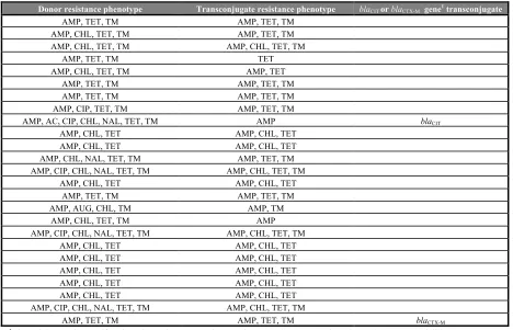
E. coli in hospitalised dogs (Gibson et al., 2011a), and selected for blaCMY-2 (Damborg et al.,
2011). Therapy with cefovecin selected for beta-lactam resistance and carriage of blaCMY-2
risk of acquiring MDR E. coli (Gibson et al., 2011a, b; Hamilton et al., 2013; Ogeer-Gyles et al., 2006). Gibson et al., (2011b) reported that cumulative veterinary admission, greater than or equal to four days was associated with the rectal carriage of MDR rectal E. coli in dogs.
1.5.2.3 Longitudinal carriage of AMR E. coli in animal faeces
There are very few studies that have examined the longitudinal shedding of AMR faecal E. coli in healthy dogs. One study in Denmark, reported the detection of an ESBL-producing E. coli on two occasions from one dog; other antimicrobial resistance profiles were not reported. Genotyping was also performed in this study and while one or two resident clones were present in 69% of dogs (n = 13), the overall E. coli population was highly diverse with multiple clones being detected in the majority of dogs (Damborg et al., 2009). Anderson et al., (2006) reported similar genotypic diversity in humans, horses and cattle in the USA, and in addition, found that a small number of unique AMR phenotypes persisted in some subjects for up to six months. Likewise Maddox, (2010) reported shedding of faecal E. coli with resistance to at least one antimicrobial, in healthy and hospitalised UK horses, for a median of 188 days.
of MDR and ESBL-producing E. coli as a median of 61 and 22 days respectively, in both healthy community and previously hospitalised and antimicrobial-treated horses.
1.5.3 AMR in staphylococci
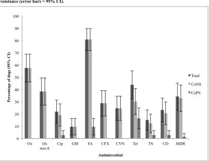
1.5.3.1 Prevalence of AMR staphylococci in dogs
Meticillin resistant S. pseudintermedius (MRSP) has been detected in 0 to 4.5% of healthy dogs (Griffeth et al., 2008; Hanselman et al., 2008; Hanselman et al., 2009; Kania et al., 2004; Murphy et al., 2009; Vengust et al., 2006; Wedley et al., 2014), and from the mucosa and/or skin of between 3.5 - 66% of dogs with skin infections and/or veterinary hospital admission from Germany, USA, or Japan (Beck et al., 2012; Griffeth et al., 2008; Kania et al., 2004; Kawakami et al., 2010; Nienhoff et al., 2011; Onuma et al., 2012; Sasaki et al., 2007). Meticillin resistant S. aureus (MRSA) has been reported to colonise up to 4% of healthy community-dogs in the UK, USA, Slovenia and China and up to 9% of hospitalised dogs in the UK, Canada and Denmark (Weese and van Duijkeren, 2010).
Several studies have reported high prevalence of meticillin-resistant CoNS (MR-CoNS) in humans (Diekema et al., 2001), horses (Bagcigil et al., 2007) and livestock (Huber et al., 2011), but there are few canine reports. Bagcigil et al., (2007) and Vengust et al., (2006) detected MR-CoNS in 13% of healthy dogs (n = 100 and 200, respectively) and Malik et al., (2006) isolated MR–S. haemolyticus from two healthy dogs out of 252 diseased and healthy dogs.
Meticillin-resistant S. schleiferi (MRSS; subsp coagulans or schleiferi) have been detected from the skin and ears of a small number of healthy dogs, and dogs with pyoderma or otitis (Griffeth et al., 2008; Kawakami et al., 2010; May et al., 2005). However, S. schleiferi has not yet been confirmed as a member of canine skin and/or mucosal flora.
1.5.3.2 Risk factors for AMR staphylococci in dogs
antimicrobial courses (greater than three courses in the last six months), treatment with beta-lactams or fluoroquinolones, prolonged hospitalisation, surgical implants, intravenous
catheterisation and contact with hospitalised humans (Faires et al., 2010; Soares Magalhaes et al., 2010). Hamilton et al., (2013) reported extended hospitalisation as a risk factor for MRSA carriage. In addition, recent studies have reported surgery, hospitalisation, frequent veterinary premises contact, and antimicrobial therapy and topical ear medication as risk factors for carriage of, or infection with MRSP in dogs from Germany, Canada and Sweden (Bergstrom et al., 2012; Lehner et al., 2014; Nienhoff et al., 2011; Weese et al., 2012; Windahl et al., 2012). Lehner et al., (2014) also reported an association between glucocorticoid therapy and MRSP infection, but this was not reported in a previous study (Weese et al., 2012). Eckholm et al., (2013) reported antimicrobial therapy and hospitalisation within the last 12 months as risk factors for the detection of MRS in dogs with pyoderma from the USA. Similarly, Huerta et al., (2011) reported the detection of MRS/beta-lactam/ fluoroquinolone resistant
staphylococci to be associated with recurrent pyoderma in dogs that had received long-term antimicrobial therapy and had frequent veterinary premise contact. Furthermore MRS/MDR staphylococci were more likely detected from urban rather than rural dogs and MRS in male dogs from Spain.
The transfer of MRS isolates has been reported to occur between individuals within
households and veterinary clinics, and contaminated environments and clothing may facilitate dissemination (Laarhoven et al., 2011; Paul et al., 2011; Singh et al., 2013; van Duijkeren et al., 2011).
1.5.3.3 Longitudinal carriage of AMR staphylococci in dogs
Longitudinal studies have reported long-term carriage of MRSP in dogs of up to a year following infection (Laarhoven et al., 2011; Windahl et al., 2012) and the duration of
detection was extended by three or more weeks of antimicrobial therapy to which the isolates were resistant (Windahl et al., 2012). However, in the absence of ongoing risk factors, MRSA carriage in dogs is likely to be transient (Weese and van Duijkeren, 2010).
1.5.4 Maintenance & spread of AMR by the microbiome
the development of AMR (Jernberg et al., 2010). Thus the intestinal microbiota of humans and other animals is the main reservoir of AMR GNB (Wellington et al., 2013) and
potentially pathogenic microorganisms including ExPEC strains (Katouli, 2010; Vollaard and Clasener, 1994).
There is evidence that both intestinal E. coli and mucosal staphylococci, including AMR or pathogenic isolates, are shared between in-contact humans and pets in both directions. Transmission is likely to occur within households, but also within veterinary premises (Damborg et al., 2009; Gomez-Sanz et al., 2013; Huebner and Goldmann, 1999; Johnson et al., 2008; Laarhoven et al., 2011; van Duijkeren et al., 2011), where extended veterinary hospitalisation is a risk factor for acquiring MDR E. coli, MRSA or MRSP (Hamilton et al., 2013; Nienhoff et al., 2011). Clinical outbreaks of extra-intestinal infections caused by MDR
E. coli have been reported in veterinary hospitals associated with the carriage of such isolates (Sidjabat et al., 2006). Similarly mucosal microbiomes, particularly after exposure to health-care environments or antimicrobial therapy, may be potential reservoirs of MRS and MDR staphylococci (Eckholm et al., 2013; Guclu et al., 2007; Hamilton et al., 2013; Nienhoff et al., 2011; Salgado et al., 2003) and facilitate the clonal dissemination of strains between patients, staff, the environment and healthy contacts (Eveillard et al., 2004; Huebner and Goldmann, 1999; Miller and Diep, 2008; van Duijkeren et al., 2011).
Very few healthy people carry MRSP (0.4%) (Hanselman et al., 2009). Zoonotic
transmission and infection has been reported (Kempker et al., 2009), but isolation of MRSP from humans tends to be transient (Laarhoven et al., 2011). Carriage may be higher among veterinary staff, owners of infected pets and dogs with concurrent skin disease (Griffeth et al., 2008; Ishihara et al., 2010; Kania et al., 2004; Morris et al., 2010; Paul et al., 2011; van Duijkeren et al., 2011); one study reported two veterinarians being MRSP-positive on two occasions, one month apart (Paul et al., 2011). Additionally, while the source of MRSA carriage in dogs is likely to be an in-contact human (Weese and van Duijkeren, 2010), owners and attending veterinarians of MRSA infected pets were more likely to harbour MRSA
compared to owners and attending veterinarians of MSSA infected pets (Loeffler et al., 2010).
1.6 Concluding summary and aims
suggested that reducing antimicrobial prescriptions will reduce the emergence and spread of AMR bacteria and antimicrobial prescribing guidelines have proven useful in this respect. However, due to reduced fitness cost and co-selection, AMR strains may continue to persist. It is therefore important to characterise the impact of antimicrobial therapy on canine commensal bacterial populations to be able to make informed decisions on preventative strategies. In addition, microbiomes are diverse in healthy individuals and maybe influenced by factors such as signalment, diet, hospitalisation and contact with human health-care. This diversity and associated factors should be taken into account when investigating the effects of antimicrobial selective pressure on microbiomes.
Firstly this work aimed to investigate the mucosal staphylococci and faecal E. coli
2. General Materials and Methods
2.1 Study populations
2.1.1 Healthy dog cohort study
Labrador retriever dogs were recruited on a convenience basis from two dog shows in the North West UK between November 2010 and June 2011; the aim was to recruit 30 dogs per show. Owners were approached during the shows and one healthy dog of any age was enrolled from each household following a clinical examination. Dogs that had received topical or systemic antimicrobial therapy, had been admitted to veterinary premises within the last 12 months, or were determined not to be healthy were excluded. All dog owners gave written informed consent before enrolment in this study and completed a questionnaire regarding potential risk factors for the carriage of antimicrobial resistant bacteria. Swabs (one nasal and one perineal) were taken at the time of enrolment and the owners were asked to collect the very next faecal sample. The University of Liverpool, School of Veterinary Science Ethics-Committee approved the study protocol.
2.1.2 Healthy dog longitudinal study
2.1.3 Antimicrobial treatment longitudinal study
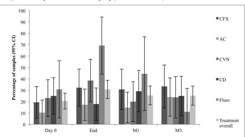
A convenience sample of dogs attending veterinary consultations at three centres including first opinion and referral practice, in the North West of England between June 2011 and September 2012 were recruited for the study if they met the inclusion and exclusion criteria. Inclusion criteria included diagnosis of a bacterial infection requiring systemic antimicrobial therapy with one of five different antimicrobials authorised for use in dogs in the UK (cefalexin [CFX], clavulanate-amoxicillin [AC], cefovecin [CVN], clindamycin [CD], or a fluoroquinolone [enrofloxacin or marbofloxacin; FL]). Exclusion criteria included antimicrobial therapy or veterinary admission within three months of enrolment and dogs aged less than 12 months old. Dogs were excluded during the study if they were prescribed a further course of systemic antimicrobial. The veterinarian in charge of the case determined if the dog required systemic antimicrobials and approached the owner of the dog regarding the study. Before enrolment, all dog owners read the study outline and gave written informed consent and completed a questionnaire regarding potential risk factors for the carriage of antimicrobial resistant bacteria. In addition vets completed a questionnaire regarding diagnosis and previous therapies and/or supplied the clinical history of each case. Samples were collected before starting treatment (D0), at the end of treatment (End) and at one (M1) and three months (M3) after the end of treatment. Swabs (one nasal and one perineal) were collected at the time of consultations and the owners were asked to collect the very next faecal sample. Questionnaires were completed at each sample point. The University of Liverpool, School of Veterinary Science Ethics-Committee approved the study protocol in June 2011.
2.2 Specimen collection
2.3 Processing swab samples 2.3.1 Staphylococcal isolation
On receipt, the swabs and 250 µl of transport media were transferred to 3 ml of nutrient broth with 6.5% sodium chloride and incubated aerobically overnight at 37°C. The broth was vortexed and streaked onto one mannitol salt agar (MSA) plate, one oxacillin resistance screening agar (ORSA) plate, supplemented with 2 µg/ml of oxacillin, and one Columbia 5% horse blood agar (CAB) plate using disposable 5 µl sterile loops; plates were incubated aerobically overnight at 37°C. All media were obtained from LabM Ltd, Bury, UK.
2.3.2 Staphylococcal isolate selection
Where present, isolates typical of staphylococci (small to medium, pink or yellow colonies on MSA, blue colonies on ORSA and white or yellow colonies on CAB) were selected from all plates using a 5 µl sterile loop, sub-cultured onto CAB, and incubated aerobically overnight at 37°C. All fresh cultures on CAB were subject to biochemical tests to identify staphylococci:
1. Gram stain (Sigma-Aldrich Company Ltd., Gillingham, UK). A drop of sterile water was placed onto a clean glass slide. A sterile toothpick was used to touch one colony and emulsify it in the water drop. The slide was air-dried and fixed by passage through the flame of a Bunsen burner. Gram stain was performed according to the standard method. Staphylococci are Gram-positive (dark purple) coccoid bacteria, usually in groups of 2 – 4 cells (X 1000 oil immersion).
1.1 Slide flooded with crystal violet stain for 30 seconds and then rinsed 1.2 Slide flooded with Lugol’s iodine solution for one minute and then rinsed 1.3 Slide washed briefly in acetone and rinsed
1.4 Safranin counter stain flooded over slide for one minute and rinsed
2. Catalase (Sigma-Aldrich Company Ltd., Gillingham, UK) test was performed by placing one to two colonies with a 5 µl loop into a drop of 3% hydrogen peroxide solution in a sterile petri dish. The production of bubbles is a positive reaction and indicates that catalase is causing the formation of hydrogen and oxygen. Staphylococci produce catalase.
2.3.3 Antimicrobial susceptibility testing of staphylococci
Disc diffusion testing was performed on all staphylococcal isolates in accordance with the Clinical and Laboratory Standards Institute (CLSI) (CLSI, 2008). Two Mueller Hinton agar plates with 5% defibrinated horse blood were inoculated with each isolate homogenised in saline (0.5 McFarland standards) for semi-confluent growth using a cotton swab and rotary plating device. Ten antimicrobial discs were then applied to the surface: 1 µg oxacillin (OX), 1 µg ciprofloxacin (CIP), 10 µg gentamicin (GM), 10 µg fusidic acid (FA), 30 µg cefalexin (CFX), 30 µg cefovecin (CVN), 25 µg trimethoprim-sulfamethoxazole (TS), 10 µg tetracycline (Tet), 2 µg clindamycin (CD) and 5 µg vancomycin (Va) (Oxoid, Basingstoke, UK). The plates were incubated aerobically at 35°C for 16 to 18 hours for all discs other than oxacillin and vancomycin, which were incubated for 24 hours. The diameter in millimetres of the zone of inhibition for each antimicrobial disc was recorded. Micro-dilution susceptibility testing1 (Trek Diagnostic Systems, Cleveland, Ohio, USA) was performed on a subset of the
CoNS isolates, using the same antimicrobial panel, other than vancomycin (CLSI, 2008).
Interpretation was based on the CLSI guidelines for animal species-specific zone diameter (mm) interpretive standards and minimal inhibitory concentration (MIC; mg/l) breakpoints for veterinary pathogens or human-derived interpretive standards when available. The European Committee on Antimicrobial Susceptibility Testing (EUCAST) zone diameter interpretive standards and MIC breakpoints were used for CIP and FA (EUCAST, 2013). The breakpoints used for interpretation of OX resistance were a zone of inhibition of ≤ 17 mm and MIC ≥ 0.5 mg/l for S. pseudintermedius and CoNS, and ≤ 10 mm and MIC ≥ 4 mg/l for S. aureus (Bemis et al., 2009; CLSI, 2013). The breakpoints used for interpretation of resistance to CVN were as a zone of inhibition of ≤ 19 mm and MIC ≥ 8 mg/l in accordance with the manufacturer’s recommendations. The reference strain S. aureus ATCC®25923 (LGC Standards, Teddington, UK) was used for quality control for MIC and zone diameter determinations.
2.3.4 Isolate storage and DNA extraction
for 10 minutes and heated at 100°C for 10 minutes before adding 400 µl of SDW. DNA extractions were stored at 4°C before use.
2.4 Processing faecal samples 2.4.1 Escherichia coli isolation
Faecal samples were mixed with an equal volume of brain heart infusion broth containing 5% glycerol (BHI-G) on receipt. Each faecal homogenate was streaked, using a 5 µl sterile loop, onto one eosin methylene blue agar (EMBA) plate without antimicrobials, one EMBA plate impregnated with 1 µg/ml ceftazidime (CZ) and one EMBA plate impregnated with 1 µg/ml cefotaxime (CX) (Sigma-Aldrich Company Ltd, Gillingham, UK) (Liebana et al., 2006), to obtain single colonies. In addition, to detect antimicrobial resistant isolates, one EMBA plate and one MacConkey agar (MAC) plate were inoculated with the faecal homogenate, using a cotton swab and a rotary plating device, for confluent bacterial growth (Bartoloni et al., 2006). Seven antimicrobial discs were applied to the surface: 10 µg ampicillin, 30 µg clavulanate-amoxicillin, 1 µg ciprofloxacin, 30 µg chloramphenicol, 30 µg nalidixic acid, 30 µg tetracycline and 2.5 µg trimethoprim (MAST Group Ltd., Liverpool, UK). A further 500 µl of faecal homogenate was enriched in 4.5 ml of buffered peptone water. All plates and
broths were incubated aerobically for 18 to 20 hours at 37
°
C. If there had been no growth on the EMBA plates impregnated with third generation cephalosporins, the enriched broths were streaked onto EMBA plates impregnated with 1 µg/ml ceftazidime (CZ) and one EMBA plate impregnated with 1 µg/ml cefotaxime (CX) and incubated aerobically for 18 to 20 hours at37
°
C.2.4.2 Escherichia coli isolate selection
Random colonies (3 or 10 depending on the study) were selected from the plain EMBA plate if there morphology resembled E. coli (medium sized metallic green colonies). If present, colonies growing within the zone of inhibition around each antimicrobial disc on the EMBA and MAC plates (medium sized pink colonies) and from the CX and CZ plates were selected. The selected colonies were streaked, using a 5 µl sterile loop, onto nutrient agar and incubated
aerobically for 18 to 20 hours at 37
°
C. Gram stain and biochemical tests to detect E. coli were performed.1. Gram stain was performed as for staphylococci. E. coli are Gram-negative (pink) rod-shaped bacteria (X 1000 oil immersion).