1
Visual processing during short-term memory binding in mild Alzheimer's disease
Gerardo Fernández (a)(*), David Orozco(b), Osvaldo Agamennoni(a), Marcela Schumacher(a), Silvana Sañudo(a), Juan Biondi(a), and Mario A Parra (c,d)
(a) Universidad Nacional del Sur (UNS), Bahía Blanca, Argentina, Instituto de
Investigaciones en Ingeniería Eléctrica (IIIE) (UNS-CONICET), Bahía Blanca, Buenos
Aires, Argentina.
(b) Clínica Privada Bahiense, Bahía Blanca, Buenos Aires, Argentina.
(c) Department of Psychology, School of Social Sciences, Heriot-Watt University,
Edinburgh, UK.
(d) Universidad Autónoma del Caribe, Facultad de Psicología, Barranquilla, Colombia.
(*) Corresponding author: Gerardo Fernández - IIIE – San Andrés 800 - 8000 Bahía
Blanca, Buenos Aires, Argentina – Phone: 54-291-45951801 ext. 3321. e-mail:
2 ABSTRACT
3
Keywords: visual short-term memory binding; Alzheimer’s disease; eye movements; gazing; visual processing
INTRODUCTION
Visual information is thoroughly processed during fixations. Fixations normally last between 150 ms and 300 ms [1] and are driven by saccades which direct the fovea towards a particular element of interest [2]. Fixations are therefore the end result of complex interactions between features of the explored environment (“bottom up”) and the instruction or question to be solved by the explorer (“top down”) [3, 4, 5, 6, 7, 8, 9, 10]. Behind such eye movement
patterns, there are complex cognitive functions such as attention, executive control, and working memory [11, 12, 13, 14, 15, 9, 16, 17]. Hence, the investigation of abnormal patterns of eye movement could provide critical information on the influence that some pathologies causing visual impairments, such as Alzheimer Disease (AD), have on these cognitive abilities, and in doing so unveil the source of capacity loss linked to specific processing levels.
4
motor tasks from the initial course of the disease [25]. It has been suggested that the fixation network is affected in AD patients [24]. Impairments of such a network could result in delayed target visualization, probably due to altered projections from the frontal lobes to the superior colliculus [26]. As suggested by previous research [27, 28, 29, 30], a reduced activity in the fixation pathway could give rise to erroneous gaze shifts in AD patients. It seems that neurons in the rostral pole of the superior colliculus, which are active during visual fixation, become inhibited during gaze shifts in AD patients. This finding suggests that AD may cause a loss of ability to disengage fixation from the stimulus due to a failure of processes responsible for inhibiting fixation neurons. These studies demonstrated that patients with AD show altered visual search strategies and eye movement behaviors, with deficits in smooth pursuit eye movements, an increased number of saccades as well as increased attentional deficits and eye blinks [31, 32, 18, 33]. Porter et al. [34] showed that altered patterns of eye movements in AD patients may account for limitations to organize strategies during visual search tasks that rely on binding functions.
5
6
regions, thus confirming the reliance of such memory function on a dedicated brain network. In none of the above discussed studies was the hippocampus found to contribute to conjunctive binding functions carried out in visual STM. This evidence warrants investigation of the link between eye movements and visual STM binding as the network components which are specific to the latter function seem to be shared with the network supporting the former function (i.e., frontal-parietal network [49, 50]). To date, there is no information available as to whether STM binding deficits and eye movement behaviors may be features of the clinical phenotype of AD patients, which could share pathophysiological mechanisms.
7 2. METHODS
2.1. Participants
8
the principles of the Declaration of Helsinki. All patients and all control subjects signed an informed consent prior to their inclusion in the study.
The mean score of AD patients in the Mini-Mental State Examination (MMSE) [51] was 22.6 (SD = 4.1) thus suggesting mild dementia. The mean score of AD patients in the Adenbrook’s
Cognitive Examination - Revised (ACE-R) [52] was 65.3 (SD = 16.9).
2.2. The STM binding task and eye movement assessment
Stimuli were presented on the center of a 20” LCD Monitor (1024 x 768 pixels resolution). Participants sat at a distance of 60 cm from the monitor. Head movements were minimized using a chin rest. Eye movements were recorded with an EyeLink 1000 Desktop Mount (SR Research) eyetracker, with a sampling rate of 1000 Hz and an eye position resolution of 20-s arc. All recording20-s and calibration were binocular.
Participant’s gaze was calibrated with a standard 13-point grid for both eyes. After validation
of calibration, the STM binding task began. During the task, participants were presented with arrays of object shapes in random positions of a 3x3 virtual grid which sustained 10o of visual angle. Objects were constructed using six different layouts, each defined by a shape and a frame area (see Fig. 1; also see [40]). The shape or frame area of each object (each representing 50% of the surface) was filled with a color. The procedures used to select the colors and the psychophysical features of the colors selected were reported in [40] and can
be found in supplementary materials at
http://www.era.lib.ed.ac.uk/bitstream/1842/2441/1/08-278-MAP.doc. During the task,
9
al., [40] this seems an optimal array size to identify STM binding impairments in AD patients (see also [53]).
Trials began with a fixation screen (i.e., a cross) shown for 250 ms. This was followed by a study display presented for 2000 ms (Fig. 1). After an unfilled interval of 900 ms, the test display was presented until the participant responded. There was then an inter-trial interval of 1000 ms. In half of the trials objects on both displays were the same. In the other half, the objects in the test display showed different colors from those in which they were presented during the study display. Object locations in the test display were always randomly changed to make location an uninformative feature. Participants were requested to detect whether the study and test displays consisted of the “same” or “different” items and to respond verbally
accordingly. Responses were manually typed by a trained instructor.
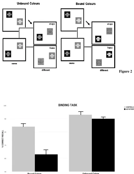
Two experimental conditions were used (Fig. 1). In the Unbound Color condition both the shape and frame areas of each object were shown in different colors. In the “different” trials the color from either the shape (50%) or the frame (50%) area of the two objects was replaced by a new color that had not appeared in the study display. Participants were told to focus on colors and not on their associations as the change would consist of new colors. In the condition assessing memory for Bound Colors both the shape and frame area were also of different colors. However, in the “different” trials the two objects swapped either the color from the shape area (50%) or from the frame area (50%). Participants were told that colors and their associations were both relevant as sometimes colors would be rearranged in different combinations during the test display. For each condition participants performed 15 practice trials followed by 32 test trials. The “same” and “different” trials were fully
10 ---
Insert Fig 1 about here
---
2.3. Statistical analysis
11
factor (i.e., participants’ processing rate). Regression coefficients (bs) standard errors (SEs) and t-values (t=b/SE) are reported for the LMMs. Since there is no clear definition of “degree of freedom” for LMMs, precise p-values cannot be reported. The t-distribution is equivalent
to the normal distribution for all practical purposes (i.e., the contribution of the degrees of freedom to the test statistics is negligible). Our criterion for referring to an effect as significant is t = b/SE >±1.95.
3. RESULTS
3.1 Correct recognition during the STM binding task
12
groups or conditions resulted in significant differences. These results mirror those reported by Parra et al. [40].
3.2. STM binding and gaze duration
In Table 1 we report, (a) effects related to the mean values of gaze duration when averaging over all predictors; (b) interactions of Condition x Group, and (c) those interactions of Condition x Group x Memory Stage (Encoding vs. Retrieval).
Averaging over all predictors. We first evaluated mean log gaze duration as function of %
of correct recognition. As shown in Table 1, % of Correct Recognition as a function of gaze duration was non-significant (t=1.34), but the effect trended toward the expected positive direction: gaze duration increases led to increased correct recognition. Saccade amplitudes exerted a significant effect on gaze duration (t=-3.25) (See Table 1). When we analyzed the effect of Memory Stage (Encoding vs. Retrieval) collapsing across Group and Condition, we noted a significant effect on gaze duration, reflecting longer gazing times during Retrieval than during Encoding (t=2.26). Finally, when we analyzed the effect of Memory Stage and task Condition on gaze duration, we found significantly longer gazing times during Retrieval of BC than during retrieval of UC (t=3.27) (See Table 1).
Interaction of Group x Condition. We then evaluated whether gaze duration was
13
Interactions of Condition x Group x Memory Stage. Then we considered gaze duration as
function of task Condition across Group and Memory Stage. As it is shown in Table 1, only during the BC task there was a significant effect between groups (t=2.38), whereby Controls and AD patients showed well differentiated gaze durations. While controls showed a similar gaze duration between Encoding and Retrieval, AD patients showed significantly shorter gaze duration during Encoding than during Retrieval (See slope for controls and AD patients during Encoding and Retrieval on the BC condition in Figure 3. More details about this striking effect are presented in the Discussion section). In contrast, both groups showed similar gaze durations when performing the UC task (t=1.60) (See Figure 3).
4. DISCUSSION
14
15
succeed in this task, make the visual curiosity hypothesis an unlikely account. Nevertheless, Daffner et al. [57] proposed that visual curiosity impairments in AD patients could be mediated by dysfunction of prefrontal association cortices. Such cortical regions also play a role in STM binding functions. Hence, future studies will need to investigate the extent to which impaired eye movements, altered STM binding functions, and reduced visual curiosity in AD may share neurocognitive mechanisms.
Notwithstanding these knowledge gaps, the results presented here suggest that eye movements may be a sensible approach to collect subjects' performance when analyzing visual information, from the encoding through the retrieval stages of memory. AD patients and controls had longer and similar fixations when encoding UC targets (see Figure 3), being our results compatible with previous evidence about relatively preserved feature processing in mild AD patients [42]. Parra et al. [40] showed that processing BC demanded more cognitive resources than the UC, and that this seems to be true regardless of age [58]. However, previous evidence suggests that AD selectively impairs feature binding [40]. Interestingly, our study shows that gaze duration follows a pattern of impairment similar to that observed through other behavioral measures of STM performance (e.g., percentage of correct recognition), thus suggesting a strong link between these two neurocognitive responses.
16
accuracy of performance in relational visuospatial memory task. Others [64, 65, 66] have argued that the activation of these parietal lobe regions may reinforce and help to produce stronger spatial representations and, consequently, stronger visualization would produce more accurate memory recognitions. The last decade has witnessed a surge of studies documenting a parietal dysfunction in AD patients which seems to be associated to reduced exploration strategies [67, 18]. In our study, controls’ and AD patients’ saccade amplitudes did not show significant differences through the BC and UC conditions of the STM tasks. More research will be needed to understand saccade behaviors during the STM binding task reported here.
In summary, we propose that the analysis of eye movements during STM binding tasks provides a valuable measure to further assess disease mechanisms. Eye movement disorders reflect deficits in attention and working memory processes in AD patients. We suggest that a more comprehensive evaluation of eye movements during binding, incorporating both an in-depth analysis of oculomotor responses and assessment of cognitive processes, may well provide a user-friendly marker of early disease symptoms and future progression. Future longitudinal studies can investigate whether abnormal eye movement responses linked to STM binding impairments can reliably anticipate the diagnosis of AD. Moreover, future studies should refine our understanding of the interplay between eye movements and the cognitive constructs underlying memory binding during STM tasks. Particular emphasis should be placed on the causal relationship between these phenotypic features of the most common form of dementia.
17
This work was supported by Consejo Nacional de Investigaciones Científicas y Técnicas (CONICET) to Dr. Gerardo Fernández and by the Universidad Nacional del Sur and CIC to Dr. O. Agamennoni. Mario A Parra is supported by Alzheimer’s Society (Grant
AS-SF-14-008). We acknowledge the support from the University of Edinburgh‘s Centre for Cognitive Ageing and Cognitive Epidemiology, part of the UK cross council Lifelong Health and Well-being Initiative (MR/L501530/1). Funding from the Biotechnology and Biological Sciences Research Council (BBSRC) and Medical Research Council (MRC) is gratefully acknowledged. We acknowledge the support from Alzheimer’s Scotland Dementia Research
18 6. REFERENCES
[1] Rayner K. (1998). Eye movements in reading and information processing: 20 years of research. Psychological Bulletin. 124:372-422.
[2] Martinez-Conde S, Macknik SL, Hubel DH (2004). The role of fixational eye movements in visual perception. Nature Neuroscience. 5:229-240.
[3] Awh E, Vogel EK, Oh SH. (2006). Interactions between attention and working memory. Neuroscience. 139:201–208.
[4] Cowan N, Morey CC. (2006). Visual working memory depends on attentional filtering. Trends in Cognitive Sciences. 10,139–141.
[5] Gilbert CD, Sigman M. (2007). Brain states: Top down influences in sensory processing. Neuron. 54:677–696.
[6] Khayat PS, Spekreijse H, Roelfsema PR. (2006). Attention lights up new object representations before the old ones fade away. Journal of Neurosciences. 26:138–142. [7] Palmer J. (1998). Attentional limits on the perception and memory of visual information. Journal Experimental Psychology: Human Performance. 16:332–350.
[8] Sigman M, Gilbert CD Learning to find a shape (2000). Nature Neuroscience. 3:264–269.
[9] Yarbus A. (1967). Eye Movements and Vision. Plenum. New York.
19
[11] Hayhoe M, Ballard D. (2005). Eye movements in natural behavior. TRENDS in Cognitive Sciences. Vol.9 No.4.
[12] Hoffman J. (1998). Visual attention and eye movements. In H. Pashler (ed.), Hove, UK: Psychology Press, pp. 119–154.
[13] Itoh N, Fukuda T. (2002). Comparative study of eye movement in extent of central and peripheral vision and use by young and elderly walkers. Perceptual Motor Skills, 94(3 Pt 2), 1283–91.
[14] Miela D, Lobel E, Lehericy S, Pierrot-Deseilligny C, Berthoz A. (2005). Cortical mechanisms of saccadic generation from execution to decision. Annals New York Academy of Sciences. 1039:232-8.
[15] Posner M. (1980). Orienting of attention. Quarterly Journal of Experimental Psychology, 32:3–25.
[16] Fernández G, Schumacher M, Castro L, Orozco D, Agamennoni O. (2015). Patients with Alzheimer disease produced shorter outgoing saccades when reading sentences. Psychiatry Research. 229:470-478.
20
[18] Mosimann UP, Felblinger J, Ballinari P, Hess CW, Müri RM. (2004). Visual exploration behavior during clock reading in Alzheimer’s disease. Brain. 127:431-438.
[19] Fernández G, Manes F, Rotstein N, Colombo O, Mandolesi P, Politi L, Agamennoni O. (2014b). Lack of contextual-word predictability during reading in patients with mild Alzheimer disease. Neuropsychologia. 62:143-151
[20] Levine DN, Lee JM, Fisher CM. (1993). The visual variant of Alzheimer’s disease: a clinicopathologic case study. Neurology. 43:305-313.
[21] Hendrie HC. (1998). Epidemiology of dementia and Alzheimer’s disease. Journal Geriatric Psychiatry. 6:3-18.
[22] Wong AMF. (2008). Eye Movement Disorders. New York: Oxford University Press. p. 165-177.
[23] Cronin-Golomb A, Gilmore GC, Neargarder S, Morrison SR, Laudate TM. (2007) Enhanced signal strength improves visual cognition in aging and Alzheimer’s disease.
Cortex, 43(7), 952 – 966.
[24] Pereira M, Camargo M, Aprahamian I, Forlenza O. (2014) Eye movement analysis and cognitive processing: detecting indicators of conversion to Alzheimer's disease. Neuropsychiatric Disease and Treatment. 10:1273-1285.
[25] Molitor RJ, Ko PC, Ally BA. (2015). Eye movements in Alzheimer's disease. J. Alzheimer's Dis. 44, 1–12. 10.3233/JAD-141173
21
[27] Abel, L. A., Unverzagt, F. & Yee, R. D. (2002). Effects of stimulus predictability and interstimulus gap on saccades in Alzheimer's disease. Dementia Geriatry Cognitive Disorders. 13, 235–243.
[28] Munoz DP, Wurtz RH (1992). Role of the Rostral Superior Colliculus in Active Visual Fixation and Execution of Express Saccades. Journal of Neurophysiology. 67:1000–1002. [29] Munoz DP, Wurtz RH (1993a): Fixation Cells in Monkey Superior Colliculus .1. Characteristics of cell discharge. Journal of Neurophysiology. 70:559 –579.
[30] Munoz DP, Wurtz RH (1993b): Fixation Cells in Monkey Superior Colliculus .2. Reversible Activation and Deactivation. Journal of Neurophysiology. 70:576 –589.
[31] Braak H, Thal DR, Ghebremedhin E, Del TK. (2011). Stages of the pathologic process in Alzheimer disease: age categories from 1 to 100 years. Journal of Neuropathology and Experimental Neurology. 70, 960-969.
[32] Lueck K, Mendez M, Perryman. (2000). Eye movements abnormalities during reading in patients with Alzheimer disease. Neuropsy Neuropsy Be. 13(2):77-82.
[33] Fernández G, Mandolesi P, Rotstein N, Colombo O, Agamennoni O, Politi L. (2013). Eye movement alterations during reading in patients with early Alzheimer Disease. Invest. Ophthalmology and Visual Sciences. doi:10.1167/iovs.13-12877.
[34] Porter G, Leonards U, Wilcock G, Haworth J, Troscianko T, Tales A. (2010) New insights into features and conjunction search: II. Evidence from Alzheimer’s disease. Cortex.
22
[35] Wheeler ME, Treisman AM. (2002). Binding in short-term visual memory. Journal of Experimental Psychology: General. 131(1):48-64.
[36] Olson I, Jiang Y. (2002). Is visual short-term memory object based? Rejection of the “strong-object” hypothesis. Perception & Psychophysics. 64(7):1055-1067.
[37] Smith K, Azami H, Escudero J, Parra MA, Starr JM. (2015). Comparison of Network Analysis Approaches on EEG Connectivity in Beta during Visual Short-Term Memory Binding Tasks. In (pp. 2207-2210).
[38] Ibañez A, Parra MA. (2014). Mapping memory binding onto the connectome's temporal dynamics: toward a combined biomarker for Alzheimer's disease. Frontiers Human Neurosciences. 8:237.
[39] Parra MA, Abrahams S, Logie RH, Mendez LG, Lopera F, Della Sala S. (2010). Visual short-term memory binding deficits in familial Alzheimer's disease. Brain. 133: 2702-2713.
[40] Parra MA, Sala SD, Abrahams S, Logie RH, Mendez LG, Lopera F. (2011). Specific deficit of colour-colour short-term memory binding in sporadic and familial Alzheimer's disease. Neuropsychologia, 49:1943-1952.
[41] Parra MA, Della Sala S, Logie RH, Morcom AM. (2014). Neural correlates of shape-color binding in visual working memory. Neuropsychologia. 52: 27–36.
[42] Parra et al., (2015). Memory binding and white matter integrity in familial Alzheimer's disease. Brain. 138(5):1355-69.
23
for familial Alzheimer's disease. Current Alzheimer Research, 14, 1335-1347 doi: 10.2174/1567205014666170614163316
[44] Olsen RK, Lee Y, Kube J, Rosenbaum RS, Grady CL, Moscovitch M. et al. (2015). The role of relational binding in item memory: evidence from face recognition in a case of developmental amnesia. Journal of Neuroscience, 35:5342-5350.
[45] Smith, K., Ricaud, B., Shahid, N., Rhodes, S., Starr, J. M., Ibañez, A. et al. (2017). Locating Temporal Functional Dynamics of Visual Short-Term Memory Binding using Graph Modular Dirichlet Energy. Scientific Reports, 7, 42013.
[6] Pietto, M., Parra, M. A., Trujillo, N., Flores, F., Garcia, A. M., Bustin, J. et al. (2016). Behavioral and Electrophysiological Correlates of Memory Binding Deficits in Patients at Different Risk Levels for Alzheimer's Disease. Journal Alzheimers Disease. 53, 1325-1340.
[47] Della Sala, S., Kozlova, I., Stamate, A., & Parra Rodriguez, M. (2016). Temporary memory binding: A transcultural cognitive marker of Alzheimers Disease. International Journal of Geriatric Psychiatry.
[48] Petersen, S. E. & Posner, M. I. (2012). The attention system of the human brain: 20 years after. Annual Review of Neuroscience, 35, 73-89.
24
[50] Azuma M, Minamoto T, Yaoi K, Osaka M, Osaka N. (2014). Effect of memory load on eye movement control: A study using the reading span test. Journal of Eye Movement Research. 7(5).
[51] Folstein MF, Folstein SE, McHugh PR. (1975). `Mini-mental state'. A practical method for grading the cognitive state of patients for the clinician. J Psychiatr Res ,12:189-98. [52] Mioshi E, Dawson A, Mitchell J, Arnold R, Hodges JR. (2006). The Addenbrooke's Cognitive Examination Revised (ACE-R): a brief cognitive test battery for dementia screening.Int J Geriatr Psychiatry, 21(11):1078-85.
[53] Della Sala S, Kozlova I, Stamate A, Parra MA. (2017). A transcultural cognitive marker of Alzheimer's Disease. International Journal of Geriatric Psychiatry, n/a.
[54] Bates D, Maechler M. (2013). lme4: Linear mixed-effect models using S4 classes R package versión 0.995-2.
[55] Treisman, A. M. & Gelade, G. (1980). A feature-integration theory of attention. Cognitive Psychology. 12, 97-136.
[56] Festa EK, Insler RZ, Salmon DP, Paxton J, Hamilton JM, Heindel WC. (2005). Neocortical disconnectivity disrupts sensory integration in Alzheimer's disease. Neuropsychology. 19: 728-738.
[57] Daffner, KR., Scinto, LF., Weintraub, S., Guinessey, B.S., Mesulam, M.M. (1992). Diminished curiosity in patients with probable Alzheimer’s disease as measured by
exploratory eye movements. Neurology. 42, 320-328.
25
[59] Xu Y, Chun MM. (2006). Dissociable neural mechanisms supporting visual short-term memory for objects. Nature. 440:91-95.
[60] Shafritz KM, Gore JC, Marois R. (2002). The role of the parietal cortex in visual feature binding. Proc.Natl.Acad.Sci.U.S.A, 99:10917-10922.
[61] Todd JJ, Marois R. (2005). Posterior parietal cortex activity predicts individual differences in visual short-term memory capacity. Cogn Affect.Behav.Neurosci. 5:144-155.
[62] Gazzaley, A. (2013). Top-down Modulation Deficit in the Aging Brain: An Emerging Theory of Cognitive Aging. Stuss, D.T.& Knight, R.T. (Eds.), Principles of Frontal Lobe Function, 2nd ed., p.593-608.
[63] Moskovitch M. (2008). The Hippocampus as a "stupid," domain-specific module: implications for theories of recent and remote memory. Canadian Journal of Experimental Psychology. 62:62-69.
[64] Cummings JL, Houlihan JP, Hill MA. (1986). The pattern or reading deterioration in dementia of the Alzheimer’s type: observations and implications. Brain Lang. 29:315-23.
[65] Kennedy A, Pynte J, Murray W, Paul S. (2012). Frequency and predictability effects in the Dundee corpus. Quarterly Journal of Experimental Psychology. doi.10.1080/17470218.2012.676054.
26
27
Table 1: Parameter estimates for fixed effects of Linear Mixed Models. Threshold of significance is set at t = ±1.95.
Gaze
duration
M SE t-value
Fixed effects
Mean Gaze duration (log) 6.668 0.060 110.02
% of Correct Recall 0.007 0.005 1.34
Saccade amplitude -0.009 0.030 -3.25
Encoding vs. Recognition 0.035 0.015 2.26
UC vs. BC X Encoding vs. Recognition 0.523 0.159 3.27
Group x Task Condition
Control vs. AD X BC -0.467 0.133 -3.51
Control vs. AD X UC 0.027 0.125 0.22
Group x Task Condition x Memory Stage
(Encoding vs. Retrieval)
Control vs. AD X BC X Enc. vs. Rec. 0.271 0.114 2.38 Control vs. AD X UC X Enc. vs. Rec. 0.181 0.113 1.60
Variance components Variance SD
Subject 0.035 0.187
[image:27.612.91.509.136.422.2]28 FIGURE CAPTIONS
Figure 1. Trial design for the two conditions of the STM tasks.
Figure 2. Corrected recognition during the two experimental conditions in both controls and AD patients (error bars = standard errors of the mean).
29 FIGURE 1