0022-538X/91/126597-07$02.00/0
Copyright C) 1991,AmericanSocietyforMicrobiology
N-Terminal
Truncation of the
Scrapie-Associated Form of
PrP
by
Lysosomal Protease(s): Implications Regarding the Site of
Conversion
of PrP
to
the
Protease-Resistant State
BYRON CAUGHEY,* GREGORY J. RAYMOND, DARWIN ERNST, AND RICHARD E. RACE
Laboratory of Persistent Viral Diseases, Rocky Mountain Laboratories, National Institute of Allergy andInfectious Diseases, Hamilton, Montana 59840
Received 24 June1991/Accepted 28 August 1991
Scrapie and related transmissible spongiform encephalopathies result in the accumulation ofa protease-resistant form ofan endogenous brain protein called PrP. As an approach to understanding the scrapie-associated modification of PrP, we have studied the processing and sedimentation properties of
protease-resistant PrP (PrP-res) in scrapie-infected mouse neuroblastoma cells. Like brain-derived PrP-res, the neuroblastoma cellPrP-resaggregated in detergentlysates,providingevidencethat the tendencytoaggregate is anintrinsicproperty of PrP-res andnotmerelyasecondaryconsequenceof degenerativebrain pathology.
ThePrP-res species had lowerapparentmolecularmassesthan the normal,protease-sensitivePrPspecies and
were not affected by moderate treatmentswith proteinase K. This suggested that the PrP-res species were
partially proteolyzed by theneuroblastomacells.Immunoblotanalysisof PrP-res withapanelof monospecific anti-PrP peptideseraconfirmedthat thePrP-res specieswerequantitativelytruncatedatthe Nterminus. The metaboliclabeling of PrP-resinserum-free medium didnot preventtheproteolysis ofPrP-res, showing that the protease(s) involved was cellular rather than serum-derived. ThePrP-res truncation was inhibited in intact cellsbyleupeptinandNH4Cl.Thisprovidedevidence thatalysosomalprotease(s)wasinvolved, andtherefore, thatPrP-reswas translocatedtolysosomes. Whenconsidered with other studies, these results imply that the conversion ofPrPtotheprotease-resistant stateoccursin the plasmamembraneoralonganendocytic pathway beforePrP-resisexposed toendosomal and lysosomalproteases.
The transmissible spongiform encephalopathies form a group ofneurodegenerative diseases which include scrapie and bovine spongiform encephalopathy in animals and
Creutzfeldt-Jakob disease, kuru, and Gerstmann-Straussler
syndrome in humans. A distinguishing feature of these
diseases is the accumulation in the brain of a
disease-specific, protease-resistant form of an endogenous protein,
PrP(6, 10, 18, 23, 36). Unlike the'n'ormal, protease-sensitive PrA (PrP-sen), the protease-resistant PrP (PrP-res) can ag-gregate into amyloidlike fibrils
(19,
21, 23, 37, 43, 56) andplaques (4, 21, 47, 56) and is a major component of brain
fractions enriched forscrapie infectivity (6, 23, 25). Since the
infectious agents ofthe transmissible spongiform
encepha-lopathiesareresistant to treatments harmful to nucleic acids,
it has beenpostulated that these agents are devoid of nucleic acidandcomposedprimarily of protein(1, 26, 41,42).More
recently, PrP-res, orthe fibrilit forms, was proposed to be
thetransmissible agent of these diseases (6, 23, 36, 38) or a
productofavirus-induced amyloidosis(9, 20). However, it
isnotclear whether PrP-res is the transmissible agentitself, a component ofthe agent, or a byproduct of the infection
which happens tocofractionate withscrapie infectivity.
A number of studies have suggested that the differences between PrP-res and PrP-sen arise at the posttranslational level (2, 8, 15, 18, 40). Strong evidence for this has been obtained recently (16). Several posttranslational
modifica-tionsof PrP, including the addition of N-linked glycans and
*
Corresponding
author.aglycophosphatidylinositol moiety, areknown tooccur(7,
14, 15, 35, 50, 51). Although someinvestigatorshave stated that covalent differences between the PrP isoforms must exist (22), no scrapie-specificcovalent modifications of PrP have been identified. Conformational studies have indicated that PrP-res fibrils resembleotheramyloidsinbeing predom-inantly
P-sheet
(17), but it is not known whether this conformation differs from that ofPrP-sen.Thus,the molec-ular basis of the difference between PrP-res and PrP-sen remains unknown.Scrapie-infected (sc+) murine neuroblastoma cells have
become a prototypic in vitro system for investigating PrP
biosynthesis and scrapie (12,44-46). Studies of PrP
biosyn-thesis in both sc+ and uninfected (sc-) neuroblastoma cells have shown that PrP-sen is located on the cell surface in
multipleforms that differ in the amountofN-linkedglycan
they contain (14). All of these forms are linked to the cell surfacebyaphosphatidylinositol moietyandcanbereleased fromintact cells withphosphatidylinositol-specific
phospho-lipase C (PIPLC) (14, 51). PrP-res, which is found in sc+
clones
only
(11, 13), is derived fromaprotease-andPIPLC-sensitive cell surface precursorthat,sofar,is indistinguish-ablefrom PrP-sen ('16). However, mature PrP-res accumu-lates intracellularly and is resistant to release from intact cellsbyPIPLCorproteases(8, 13, 53).Inthepresentstudy, wehavefurtheranalyzed themetabolic fate of PrP-res. Our results show that PrP-res aggregates and is
quantitatively
truncated at the.N terminus by lysosomal, and
possibly
endosomal, proteaseswithinthesecells. These
findings
help
todefine the subcellular siteof theformationof PrP-res and provide evidence thatPrP-res is translocatedtolysosomes.
6597
on November 10, 2019 by guest
http://jvi.asm.org/
MATERIALSANDMETHODS
Neuroblastoma cells. The sc+ and sc- mouse neuroblas-tomaclones were established and maintained as described previously (45, 46).
Antibodies. Synthetic peptides corresponding to amino acid residues 23 to 37, 89to103, and 218 to 232 ofthe full mousePrPsequencewith additional cysteine residuesatthe Nterminiwerekindly made by Michael Buchmeier (peptide 89-103; Scripps Clinic and Research Foundation, La Jolla, Calif.) and John Coligan (peptides 23-37 and 218-232; Na-tional Instituteof Allergy and Infectious Diseases, Bethesda, Md.).Thepeptideswerecoupledvia thecysteinesulfhydryl group to keyhole limpet hemocyanin and inoculated into rabbits aspreviously described (56).
Metaboliclabeling. Flasks(25cm2)ofcellsbetween50and
90% confluent were preincubated for 60 min in 2 ml of methionine-free minimum essential medium (GIBCO,Grand Island, N.Y.) containing 1% dialyzed fetal bovine serum (FBS)exceptin theexperiment (see Fig. 4)inwhich the FBS was purposefully omitted from this and subsequent incu-bation media. Then, 350 to 500 pCi of [35S]methionine (Dupont-NEN) or Tran35S-label (ICN) was added to each flaskfor thedesignatedtimes in thepresence orabsence of lysosomal protease inhibitors (as described in the figure legends).Thecellswerewashed three times with phosphate-buffered balanced salts solution, chase-incubated in com-pleteminimum essential medium with 10%FBS,and washed twice with ice-cold phosphate-buffered balanced salts solu-tion. Thecellswere thenlysedinasolution of ice-cold 0.5% Triton X-100, 0.5% sodium deoxycholate 5 mM Tris-HCl (pH 7.4), 150 mM NaCl, 5 mM EDTA, 0.7 ,ug ofpepstatin per ml, and 0.5 ,ug of leupeptin per ml (1 ml per flask). Phenylmethylsulfonyl fluoride (PMSF; 0.5 mM) was also included in the lysing buffer when the lysates were not
subsequently treated with proteinase K. Nuclei and debris were removed by centrifuging at 1,000 x gfor 5min (4°C). The postnuclear supernatants were treated as described in the figure legends and ultimately either centrifuged at
230,000 x gmax for at least 45 min to pellet aggregates or precipitated by the addition of4 volumesof coldmethanol, cooled at -20°C forat least 30min, and then collected by centrifugationat13,000x g.Intheexperimentshown inFig. 5,the230,000 x gpelletswerewashedonce withasolution containing10%Sarkosyl (N-laurylsarcosinate), 10 mM Tris-HCl, 1 mMEDTA, 133 mMNaCl, and 1 mMdithiothreitol (pH 8.3)toreducenonspecific backgroundinthesubsequent immunoprecipitations.
Immunoprecipitation. The pellets resultingfrom the met-aboliclabeling experimentswereresuspended bysonication indetergent-lipid-protein complex (DLPC)buffer(8) supple-mentedwith 0.5 mMPMSF,0.7 jig ofpepstatinperml,and
0.5 jigofleupeptinperml. Ananti-PrPpeptide89-103serum was diluted 1:250to 1:500 into the PrP-DLPC solutions and incubated at 4°C overnight. Immune complexes were col-lected by binding to protein A-Sepharose (30 ,ul of 10% [wt/vol] protein A-Sepharoseper2
RI1
of undiluted antiserum in sample) for 30 to 60 min at 4°C. The beads were then washed as previously described (8), except that the wash times were reduced to 1 to 3 min. Bound proteins werereleased by boiling the beads in 30 ,u1 of sodium dodecyl sulfate (SDS)-polyacrylamide gel electrophoresis (PAGE) samplebuffercontaining 5% SDS and 4% ,-mercaptoethanol (33).
SDS-PAGE, fluorography, and immunoblotting. Proteins
were separated by SDS-PAGE (12.5% acrylamide) as
previ-ously
described(33).
Gels of radiolabeledsamples
wereprocessed
forfluorography
aspreviously
described(13).
Foranalyses
by
immunoblotting,
proteins
were electroblottedonto Immobilon-P
(Millipore, Bedford, Mass.) by using
astandard buffer (54)
supplemented
with 0.01% SDS. TheImmobilon-Pfilterwasblocked with 5% nonfat dried milk in
10 mM Tris-HCI
(pH
8.0)-150 mM NaCI-0.05% Tween 20(TBST).
Thefilterwas incubated for2hatambienttemper-aturewith the
appropriate
rabbit antiserum diluted1:1,000
to1:2,000 in TBST. After
washing
inTBST,
the filter wasstained with alkaline
phosphatase-conjugated
goat anti-rab-bitimmunoglobulin
(Protoblot; Promega, Madison, Wis.).
Partialpurification of PrP-res.PrP-res was
partially
puri-fied
by
a modified version ofaprocedure
describedprevi-ously
for theextractionoffull-length
PrP-res(except
for thesignal
peptide)
from sc+ brain tissue(29).
Confluent150-cm2 flasks of sc+ neuroblastoma clones were rinsed twice inphosphate-buffered
saline, lysed
inasolutioncontaining
7.5 ml of ice-cold 10%N-laurylsarkosinate
perflask,
1 mMPMSF,
0.7 ,ugofpepstatin
perml,
5jig
ofleupeptin
perml,
and 10 mM sodium
phosphate (pH 7.6),
andhomogenized
with several 10-s bursts ofa
high-speed
tissuehomogenizer
(Tekmar,
Cincinnati,
Ohio).
Although
theprotease inhibitors were notall identicaltothoseusedinapreviousstudy (29),
wehaveshown
by
N-terminalsequencing
thatourinhibitorsare effective in this
procedure
atpreventing
theN-terminal truncation of braintissue-derivedPrP-res(data
notshown).
The
homogenate
wascentrifuged
at22,000
x gfor 30 minat10°C.
The supernatantwas thencentrifuged
at215,000
x g for 150 minat10°C.
Thepellet
wasfrozenat-20°C,
and thenaliquots
were boiled in SDS-PAGEsample
bufferprior
toimmunoblot
analyses.
RESULTS
Aggregation
ofPrP-res.To comparetheaggregation
prop-erties ofPrP-resand PrP-senderived fromsc+tissue culture
cells, detergent
lysates
ofpulse-labeled
sc+ neuroblastoma cells wereultracentrifuged
and theresulting
pellets and supernatants wereanalyzed
forimmunoprecipitable
PrPspecies
thatwereeithersensitiveorresistanttoproteinase
K(Fig. 1).
Thecellswerechasedfor2and24htoallowoptimal
labeling
of PrP-sen andPrP-res,
respectively
(8, 14,
16).
The PrPspeciesinthe230,000
x gsupernatantswereproteinase
Ksensitiveand, thus,
were PrP-senby
definition(Fig. 1A).
After
longer
fluorographic
exposureofthegel
(Fig. 1B),
PrPspecies
werealso observed in the230,000
x gpellets.
Thesespecies
were resistant toproteinase
Kand, thus,
werePrP-res.This result indicated that thedetectablePrP-reswas
aggregated
in thedetergent
lysates
and could beseparated
quantitatively
from thedetergent-soluble
PrP-senby
centrif-ugation.
Comparison
of the relative amounts of labelmaximally
incorporated
into PrP-res and PrP-senby
densitometricscanning
of thefluorograms
inFig. 1indicated that after the 24-hchase,
the label in PrP-res wasonly
approximately
3% of the labelinitially
found in PrP-sen with the 2-h chase.Therefore, only
asmallproportion
ofthetotal PrPsynthe-sized entered the PrP-res
pool.
Proteolytic processingofPrP-res.
Although
theNterminusofmostofthe PrP-res isolated from sc+brain tissue
begins
at the
signal
peptidase cleavage site,
further truncations atthe N terminus can occur in vivo or upon treatment with moderateconcentrations ofproteasesinvitro
(5, 29, 30,
55).
For
instance, proteinase
Ktreatment removes58aminoacid residues from the N terminus ofmouse brain cell-derivedon November 10, 2019 by guest
http://jvi.asm.org/
chase: 2 h 24 h sup. pellet sup. pellet
C PK C PK C PK C PK
preabsorption: - + - + - + - + - + - + - + - +
A M w"
35-41{
1
30 -25-B
35-411{
pp.
W.*$
*ab
-23
-19
FIG. 1. Metabolic labeling and aggregation state of PrP-res. Two flasks of sc+ neuroblastoma cells were labeled for 2.5 h with
L-[35S]methionine,chased with complete growthmedium for 2 or 24
h, and lysed as described in Materials and Methods. Half of the postnuclearsupernatantsfrom the lysates were treated with 20 ,ug of proteinaseK(PK) per ml for 30 min at 37°C, and the control half (C) wasleft on ice. The postnuclearsupernatantswerethen centrifuged at230,000xgfor 45 minat4°C. The 230,000xgsupernatants(sup.) were methanol precipitated. The 230,000 x gpellets were washed oncewith ice-coldwater.The methanolprecipitates and the 230,000 xgpellets were resuspended in DLPC buffer and immunoprecipi-tated asdescribed in Materials and Methods. Bands containing the specific PrP peptide 89-103 epitope were identified as those that were notimmunoprecipitated when the anti-PrP peptide antiserum (R9) waspreabsorbed with the appropriate synthetic PrP peptide (50 ng/,ulof serum) thatwasused tomake the antigen. Panels A and B represent 1-day and 20-day fluorographic exposures of the gel, respectively. The sizes (in kilodaltons) of the PrP bands were estimated by comparison to molecular mass standards and are indicated on the left and right sides of the figure for PrP-sen and PrP-res bands, respectively.
PrP-res and decreases theapparentmolecularmassesofthe PrP-res species by approximately 6 kDa (30). However,
analysis
ofthe sc+ neuroblastoma cellPrP-resin 230,000 xgpelletsasshown inFig.1Bindicated that the proteinaseK
treatmenteliminatedbackground proteins but didnotappear
to alterthe SDS-PAGE mobilities orrelative intensities of
the individual PrP-res bands. This, and the fact that the
PrP-res species, as agroup, had lower apparentmolecular massesthan thePrP-senspecies, provided evidencethatthe PrP-res
species
were already truncated at the N terminuswithin theneuroblastoma cells.
Since background proteins partially obscured the
detec-tion of the 28-to32-kDaPrP-resandperhaps largerPrP-res
species in crude 230,000 x g pellets (Fig. 1B), we also
analyzedtheproteinaseKeffecton PrP-respartiallypurified
by a method shown tobe effective for isolating full-length
PrP-res from sc+ brain tissue (29). This experiment
con-firmed that proteinase K had no effect on the mobility or
relativeintensity ofthe PrP-res bands (Fig. 2). The activity
of the proteinase K was confirmed by the fact that, with
prolonged treatment (50 min), the PrP-res staining was
decreasedas wehavedocumented
previously
(39).Presum-ably the slowdigestion of the epitope (residues 89 to 103),
PK
C 1O' 50'
28-32{ E 20".i 23
-19- _w
-28-32
FIG. 2. Effects of proteinase K (PK) treatment on neuroblas-tomacell PrP-res. Partially purified PrP-res was isolated fromsc+ neuroblastoma cells under conditions that have been shown to be effective in isolating full-length PrP-res from sc+ brain tissue. Aliquots containing less than 1 ,ug ofprotein were suspended in 0.5% Sarkosyl-20mMTris-HCl,pH 7.7, and treated withproteinase
K (2 F±g/ml) for the designated period of time and immunoblotted
with the anti-PrP peptide 89-103 serum, as described in Materials and Methods. Thepositions of the PrP-res speciesaredesignated in kilodaltons.C, control.
which corresponds to the relatively proteinase K-resistant
region of brain tissue-derived PrP-res molecules (30),
ac-countsfor this observation.
Immunoblot analysis of PrP-res with monospecific antisera.
To verify that the neuroblastoma PrP-res species were
truncatedatthe N terminus,weanalyzed PrP-resby
immu-noblot with monospecific antisera to synthetic peptides
correspondingtothe N terminus and two othersequencesof
the PrP polypeptide. Figure 3 shows that all three of the
major PrP-res bands bound antibodies against peptides
89-103 and 218-232which bracketthe sequenceofthe protein-aseK-resistant coreofbraincell-derivedPrP-res(30). How-ever, noneofthe neuroblastoma PrP-resbands reacted with
theantiserum against theN-terminal peptide startingatthe
signal peptidase cleavage site (residues 23 to 37). The
activity ofthe anti-peptide 23-37 was demonstrated bythe
labeling of a subset of bands from a partially proteinase
K-digested preparation ofmouse brain PrP-reswhich
con-tained bothfull-length and truncated forms ofPrP-res. These
results indicated that the PrP-res from the sc+ neuroblas-tomacells was quantitatively truncated at the N terminus. The PrP-res species were 6 to 7 kDa smaller than the
corresponding PrP-sen species (Fig. 1A and B), suggesting
that PrP-res lacked approximately the same number of aminoacidresidues, i.e., 58,asproteinaseK-treated PrP-res from sc+ mouse brain.
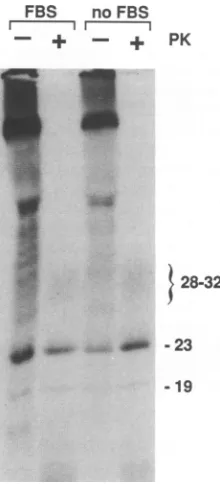
Cellularversus serumoriginofproteolyticactivity.Sinceat leasttheprecursorofPrP-resresides on the cellsurface(16), it waspossiblethat serumproteases in thegrowthmedium, rather than cellular proteases, were responsible for the
proteolysisof PrP-res. Toanalyze this,welabeledPrP-resin
serum-free medium. This had no effect on the SDS-PAGE
migration ofthe PrP-resspeciesobserved,which
comigrated
with proteinase K-treated PrP-res bands(Fig. 4). Thus, the
protease(s)
responsible
fortruncating
PrP-resisnotderivedfromthe FBSand,
by
default, mustbecellular.on November 10, 2019 by guest
http://jvi.asm.org/
[image:3.612.69.309.75.291.2] [image:3.612.399.494.78.260.2]a 23-37 a 89-103 a 218-232
preabsorbed
Br NB Br NB Br
preabsorbed
NB Br NB Br
preabsorbed
NB Br NB
N i , 1 C
23 37 89 103 218 232
Ai
FIG. 3. Analysis of the N-terminaltruncationof PrP-res byimmunoblot withmonospecificantisera.Aliquots of thesamepartially purified neuroblastoma (NB) cell PrP-res used in Fig. 2 (without proteinase K treatment) were immunoblotted usingantisera (a) raisedagainst synthetic peptides correspondingtothedesignated amino acid residuesof the PrPsequence. As apositivecontrol for the activityof the
antisera, samples ofasimilar preparationof sc+mousebraintissue-derivedPrP-res(Br)wereincluded. Thebraintissue-derivedPrP-res had beenpartiallydigestedwithproteinaseKsothatbothfull-lengthandN-terminallytruncated formsof PrP-reswere observed. The relative positions of the peptide antigensin the full PrPamino acidsequence(without theN-terminalsignal peptide correspondingtoresidues1to22) arediagrammed. For comparison,themajor (large triangles)and minor(small triangles)Nterminideterminedfor untreated(filled triangles) andproteinase K digested (open triangle)mousebrain tissue-derivedPrP-resarealso shown(30). The specificity ofthestainingwasshown
by preabsorbingtheantiserawiththecorresponding synthetic PrPpeptidesasdescribed in thelegendtoFig. 1.Thepositionsof themajor neuroblastomacell PrP-res bandsaredesignatedontheright in kilodaltons. The truncated28-to32-kDa PrP-resoftheneuroblastomacells appearsalmostaslarge astheupperfull-lengthbrain PrP-res bandidentified bythea23-37. Thiscanbe attributedtothemoreextensive glycosylation ofPrPin neuroblastoma cells.
Inhibition of PrP-restruncation. To address thequestion of whether PrP-res was truncated bylysosomal orendosomal
proteases and to simultaneously obtain positive evidence that PrP-res was being truncated in the intact cells rather
than in thedetergent lysates,wecompared the SDS-PAGE
mobilities of PrP-res labeled in the presence orabsence of leupeptin and NH4Cl. Leupeptin directly inhibits certain lysosomal and endosomal proteases, and NH4Cl inhibits lysosomalproteolytic activities by raising the internal pH of these organelles (27, 48). Both of these agents inhibit the degradation of plasma membrane proteins thatoccursvia the
endocytic pathway tothe lysosomes (27, 48).
Figure 5 shows that larger PrP-res specieswereobserved
in cells treated with either 10 mM NH4Cl or 200 ,ug of
leupeptinperml.Moreeffective inhibition of truncationwas achieved by using both agents simultaneously. The inhibi-tion of PrP-restruncation byNH4Cl provided evidence that lysosomal proteases were involved in the truncation of
PrP-res. The leupeptin effect suggested the involvement of lysosomal and/or endosomalproteases. The PrP-res species detected in the cells treated with the combined inhibitors
were approximately 3 kDa higher in apparent molecular
massthan thecorresponding proteinase K-treated speciesor theuntreatedspecies from the control cells. However, they remained approximately 3to4 kDa smaller than the corre-sponding PrP-sen species (Fig. 5), suggesting that the inhi-bition ofproteolysis by these compoundswas notcomplete and that theelongated PrP-res species still lacked about 25to
30 amino acid residues that were present in the PrP-sen species. Furthermore, the detection of the PrP-res species of intermediate sizes with the inhibitor treatments indicates that the truncation process involves more than a single
proteolyticstep.
ThetreatmentswithNH4Cl and leupeptin hadnoapparent effecton thecells whenvisualizedby light microscopy. The percentage ofviable cells, i.e., those capable of excluding trypan blue, was slightly decreased by the inhibitor treat-ments but remained greaterthan 90%o in allcases (data not shown).
Since the inhibitors were effective when added to the living cells and not, inthe case ofleupeptin, whenpresent only inthe lysing buffer, theinhibitor-sensitive proteolysis occurred in the intact cells rather than in the lysate. This conclusion was also supported by the observed inhibition withNH4C1, which requires that the lysosomes be intact.
DISCUSSION
In the normal cycle of PrP metabolism, PrP-sen has a
half-life of 3 to6 h (8, 14, 16), which is consistent with the chase-dependent decrease in PrP-sen labeling observed in Fig. 1. Since it appears that only a small proportion of
PrP-sen is released from the cell spontaneously, it is likely thatmostof the PrP-sen is catabolized within the cell(14). PrP-res, onthe otherhand, showsno evidence ofturnover
within the cell (8, 16). However, our present observation
U
}28-32-23 -19
VWw;
on November 10, 2019 by guest
http://jvi.asm.org/
[image:4.612.132.474.74.328.2]FBS noFBS
Ir [1
_
_
_ _:q_
._ ._
}
!_:._
.
....S ::
.4
PK
$ 28-32
1' -23 -19
FIG. 4. PrP-res labeling in the absence of FBS. Four 25-cm2 flasks ofsc+neuroblastoma cellswerelabeled,chasedfor24h, and
lysedasdescribed in Materialsand Methods, except that in two of the flasks the FBS wasomitted from the labelingand chasemedia. The postnuclearsupernatantsfrom the duplicate flasksweremixed andthen divided into equalpartsand either treated with proteinase K (PK) or not and processed for the detection of PrP-res by immunoprecipitation as described in the legend to Fig. 1 and Materials and Methods. The apparent molecular masses of the PrP-resbandsaredesignatedonthe right ofthefigureinkilodaltons.
that PrP-res is truncatedatthe N terminus indicates that it doescome into contactwith cellularproteasesbut is resis-tant tocomplete degradation.
Our inability to detect full-length PrP-res in the sc+
neuroblastoma cellssuggeststhat PrP-res is truncatedatthe N terminus concurrent with, or soon after, its formation. Since PrP-res is made from acell surface precursorthat is similar, if not identical, to the normal PrP-sen (16) and appearstoaccumulateintracellularly (8, 13, 53), it is reason-ableto expectthat PrP-res,oritsprecursor,isendocytosed. Endosomes are commonly translocated to a perinuclear location where they are fused with Golgi-derived vesicles containing proteases and acidified to form lysosomes (32). Protease-sensitive proteins contained within the lysosomes arethendigested. Thispathway is responsible for the normal turnover of most plasma membrane components (27) and thus islikely to account for the degradation of PrP-sen. The fact that PrP-res is truncated by leupeptin- and NH4Cl-sensitive proteases provides evidence that PrP-res follows theendocytic pathway tothe lysosomes. PrP-res maythen accumulate in the lysosomesif it isnotsubsequently trans-located to other organelles. Since PrP-res is not completely degraded in the lysosomes, its conversion to the protease-resistant state must occurprior to its exposure toproteases within endolysosomes and secondary lysosomes. Thus, it is likely that PrP-res is generated at the plasma membrane, where the protease- and phospholipase-sensitive PrP-res precursor is found, or alongthe endocytic pathway before exposure to proteases(16).
A question that remains is why the PrP-res species ob-served in the leupeptin- and NH4Cl-treated cells did not comigrate with the PrP-sen species. The most likely expla-nation would be that we did notachieve complete inhibition oflysosomal and endosomal protease activity. Neither of these inhibitors completely prevents the degradation of
leupeptin
control leupeptin NH4Cl NH4 Cl
I ] 1 1~~~11 1!1
preabsorption: -- + - - + -- + - +
PK: + - - + -.- + - +
-28-32
{
.e
_w
a
23- Q 4116
19-I...
PrP-sen
I35-41
-30 -25
FIG. 5. Inhibition of the truncation ofPrP-res. Flasksofsc+neuroblastoma cellswerelabeled for2h withTran35S-labelandthen chased for 18 h withcomplete medium. Duplicate flaskswereleft untreated(control)orweretreated with200 ,ugofleupeptinperml, 10mMNH4Cl
orbothduring thelabeling and chase periods.Whereastheleupeptinwaspresentduring the entirelabeling period, theNH4Clwasadded1 hafter the initiation oflabeling in ordertoreduceits potential effectsonthelabelingofPrP(49).Postnuclearsupernatantsof theduplicate flaskswereprepared and mixed. One-thirdwastreated withproteinaseK(PK; 20,ug/ml,30min,37°C), andtheremainderwasleft untreated. Bothsampleswerethen treatedwith1mMPMSF.PrP-res was thenpelleted, washedwith10%Sarkosylbuffer, andimmunoprecipitatedwith orwithoutpreabsorption of the antibody (a89-103) withthesyntheticPrPpeptideasdescribed in Materials and Methods and thelegendto
Fig.1. Forcomparison,alanecontainingPrP-senimmunoprecipitated directly fromapostnuclearsupernatantis shown. Thepositionsof the control PrP-resandPrP-senspeciesaredesignated inkilodaltonsontheleft andright sides ofthepanel,respectively.
on November 10, 2019 by guest
http://jvi.asm.org/
[image:5.612.138.248.75.317.2] [image:5.612.145.497.452.640.2]plasma membrane-derived proteins (28, 31, 34). Leupeptin
inhibits onlyasubsetof thelysosomalandendocytic
prote-asesandNH4+ willnotaffectproteasesthatmaybeactive in
a neutral environmentwithinendosomesor anyother
organ-elles playing apartial role in PrP-res truncation. It remains
possiblethat some truncation occurs in thedetergentlysate,
but this is unlikely because there is no evidence for the truncation of PrP-sen in the same lysates. Another
possibil-ity is that the PrP-res precursorissynthesizedde novo in a
partially truncated form, perhaps as a result ofalternative mRNA splicing; however,thefact that full-length PrP-res is found in sc+ brain tissue (5, 29, 30)indicates that this is not generally the case.
The combinedobservations thatthe 19- to 23- and 28- to 32-kDa PrP-res bands each lacked at least the N-terminal
peptide 23-37 epitope (Fig. 5), were insensitive to further
(limited) proteinase K digestion (Fig. 1B and 4), and yet contained epitopes (residues 89 to 103 and 218 to 232) bracketing most of the protease-resistent core of brain
tissue-derived PrP-res, provide evidence that their diverse
SDS-PAGEmobilities were not due primarilytodifferences
inthe length of theirpolypeptidebackbone. Rather, the data support the idea that, like themultiple PrP-senspecies (14), thePrP-res species differ in the amount ofN-linked glyco-sylation they contain and that the 19-kDa PrP-res band lacks N-linked glycans altogether (16, 52). As has been noted previously (12, 52), the fact that the 19-kDa species was a component of PrP-resindicated that N-linkedglycosylation was not essential for protease resistance.
Although the truncation ofPrP-res observed in sc+ neu-roblastoma cells helps to define thesubcellular location and kinetics of PrP-res formation, it is unclear what role the proteolysis ofPrP-res may play in the biogenesis of PrP-res orinscrapie pathogenesis invivo. Little of the brain PrP-res isolated from certain species, e.g., hamsters, is truncated beyond the signal peptidase cleavage site (5, 29). This suggests that much of the PrP-res produced in vivo is not exposed to appropriate proteases, perhaps because of im-pairment of lysosomal function, and that the proteolysis of PrP may have little importance in the scrapie disease pro-cess. Onthe other hand, it is possible that asmallamountof
proteolysed PrP-res could play an important role, for
in-stance, byefficiently nucleatingaconformationalchangeand aggregation of larger amounts of full-length PrPprecursors. Similar processes have been described for the polymeriza-tion of synthetic amyloids (3).
To theextent thatN-terminaltruncation of PrP-res occurs
inscrapie-infected braintissue, the issue arises as to where
the truncation occurs. PrP-res could conceivably be
trun-cated within the cells that produce it, in an extracellular space, or in other cells that scavenge PrP-res from the diseased tissue. Our results have indicated that in tissue culture at least, sc+ neuroblastomacells can quite efficiently truncate their own PrP-res.
Itis tempting to speculate that the aggregation of PrP-res accounts for its protease resistance. Studies showing that PrP-res from sc+ brain tissue is aggregated in detergent extracts (6, 23, 43) raised the question of whether the
tendency to aggregateis anintrinsicproperty ofPrP-res ora
secondary effect of long-termdegenerative brain pathology.
Forinstance, PrP-res might aggregatebecause of
pathology-induced processing or the binding of PrP-res with other
pathology-associated molecules. The observation that sc+
neuroblastoma cell PrP-res aggregates in detergent lysates
(Fig. 1) (52) is consistent with the idea that the tendencyto
aggregateis anintrinsic property of PrP-res, since the cell
cultures shownosigns of scrapie-associated
cytopathology.
Like PrP-res, the scrapie infectivity in detergent extracts ofneuroblastoma cellsisalsorelatively resistantto
protein-ase Kand can be pelleted by ultracentrifugation (39). The
fact that PrP-res and scrapie infectivity share these
proper-ties when derived from tissue culture cells, as wellasfrom
brain tissue (6, 23, 24, 43), provides further suggestive evidence that the protein is physically associated with the transmissible agent, ifnotthe agentitself.
Thus, the subcellular location of PrP-res formation may
also be the site of scrapie agent replication. However, it
remains possible that the replication of the agent and its
apparent association with PrP-resoccur as separate events. Inany case,thetruncationofPrP-res thatwehaveobserved within sc+ neuroblastoma cells provides a kinetic and or-ganelle-specific marker that helps define the metabolic fate of PrP-res. Our future studies will focus on the plasma membrane and endocytic pathway as likely sites for the
formation of PrP-res and perhaps, therefore, the scrapie
agent.
ACKNOWLEDGMENTS
We thank Bruce Chesebrofor helpfuldiscussions and Bob Evans and Gary Hettrick forgraphics assistance.
REFERENCES
1. Alper, T., W. A. Cramp, D. A. Haig, and M. C. Clarke. 1967. Does theagentof scrapie replicate without nucleic acid?Nature (London) 214:764-766.
2. Basler, K., B. Oesch, M. Scott, D.Westaway, M. Walchii,D.F. Groth,M. P. McKinley,S. B. Prusiner, andC. Weissman. 1986. Scrapie and cellular PrP isoforms are encoded by the same chromosomal gene. Cell 46:417-428.
3. Beaven, G. H., W. B. Gratzer, and H. G. Davies. 1969. Forma-tion and structure of gels and fibrils from glucagon. Eur. J. Biochem. 11:37-42.
4. Bendheim, P. E., R. A. Barry, S. J.DeArmond,D. P.Stites,and S. B. Prusiner. 1984. Antibodies to a scrapie prion protein. Nature(London) 310:418-421.
5. Bolton, D. C., P. E. Bendheim, A. D. Marmostein, and A. Potempska. 1987. Isolation and structural studies of the intact scrapie agentprotein. Arch. Biochem. Biophys. 258:579-590. 6. Bolton, D. C., M. P. McKinley, and S. B. Prusiner. 1982.
Identification of a protein that purifies with the scrapie prion. Science 218:1309-1311.
7. Bolton, D. C., R. K. Meyer, and S. B. Prusiner. 1985. Scrapie PrP 27-30 is asialoglycoprotein. J. Virol. 53:596-606. 8. Borchelt, D. R., M. Scott, A. Taraboulos, N. Stahl, and S. B.
Prusiner. 1990. Scrapie and cellular prion proteins differ in the kinetics ofsynthesis and topology in culturedcells. J. Cell Biol. 110:743-752.
9. Braig, H. R., and H. Diringer. 1985. Scrapie: concept of a virus-induced amyloidosis of the brain. EMBO J. 4:2309-2312. 10. Brown, P., M. Coker-Vann, K. Pomeroy, M. Franko, D. M. Asher, C. J. Gibbs, Jr., and D. C. Gajdusek. 1986. Diagnosis of Creutzfeldt-Jakob disease by Western blot identification of marker protein in human brain tissue. N. Engl. J. Med. 314:547-551.
11. Butler, D. A., M. R. D. Scott, J. M. Bockman, D. R. Borchelt,A. Taraboulos, K. K. Hsiao, D. T. Kingsbury, and S. B. Prusiner. 1988.Scrapie-infected murine neuroblastoma cells produce pro-tease-resistant prion proteins. J. Virol. 62:1558-1564.
12. Caughey, B. 1991. In vitro expression andbiosynthesis of prion protein. Curr. Top. Microbiol. Immunol. 172:93-107.
13. Caughey, B., K. Neary, R. Buller, D. Ernst, L. Perry, B. Chesebro, and R. Race. 1990. Normal and scrapie-associated forms of prion protein differ in their sensitivities to phospholi-pase and proteases in intact neuroblastoma cells. J. Virol.
on November 10, 2019 by guest
http://jvi.asm.org/
64:1093-1101.
14. Caughey, B., R. E. Race, D. Ernst, M. J. Buchmeier, and B. Chesebro. 1989. Prion protein (PrP) biosynthesis in scrapie-infected and uninfected neuroblastoma cells. J. Virol. 63:175-181.
15. Caughey, B., R. E. Race, M. Vogel, M. J. Buchmeier, and B. Chesebro. 1988. In vitro expression in eukaryotic cells of the prion protein gene cloned from scrapie-infected mouse brain. Proc. Natl.Acad. Sci. USA 85:4657-4661.
16. Caughey, B., and G. J. Raymond. 1991. The scrapie-associated form of PrP is made fromacell surface precursor that is both protease- and phospholipase-sensitive. J. Biol. Chem. 266: 18217-18223.
17. Caughey,B.W.,A.Dong,K.S. Bhat,D.Ernst,S. F.Hayes,and W. S. Caughey. 1991. Secondary structure analysis of the scrapie-associated protein PrP 27-30 inwaterbyinfrared spec-troscopy. Biochemistry 30:7672-7680.
18. Chesebro, B., R. Race, K. Wehrly, J. Nishio, M. Bloom, D. Lechner, S.Bergstrom,K. Robbins,L.Mayer, J.M. Keith, C. Garon, and A. Haase. 1985. Identification of scrapie prion protein-specific mRNA in scrapie-infected and uninfected brain. Nature(London) 315:331-333.
19. Cho, H. J. 1986. Antibodyto scrapie-associated fibril protein identifiesacellularantigen. J.Gen. Virol. 67:243-253. 20. Czub, M., H. Braig, and H. Diringer. 1986. Pathogenesis of
scrapie: study of the temporal development of clinical symp-toms,of infectivity titers and scrapie-associated fibrils in brains of hamsters infectedintraperitoneally. J. Gen. Virol. 67:2005-2009.
21. DeArmond, S. J.,M. P.McKinley,R. A.Barry,M. B.Braunfeld, J.R.McColloch,and S. B. Prusiner.1985. Identification ofprion amyloidfilaments inscrapie-infectedbrain. Cell 41:221-235. 22. Diringer, H.,H. Blode,andU. Oberdieck.1991. Virus-induced
amyloidosis inscrapieinvolvesachange in covalentlinkagesin thepreamyloid. Arch. Virol. 118:127-131.
23. Diringer, H.,H.Gelderblom,H.Hilmert,M.Ozel,C.Edelbluth, andR. H. Kimberlin. 1983. Scrapieinfectivity, fibrils and low molecularweight protein. Nature(London) 306:476-478. 24. Diringer, H.,H. Hilmert,D. Simon,E. Werner,and B. Ehlers.
1983. Towards purification of the scrapie agent. Eur. J. Bio-chem. 134:555-560.
25. Gabizon, R., M. P. McKinley, D. Groth, and S. B. Prusiner. 1988. Immunoaffinity purification andneutralization ofscrapie prion infectivity. Proc. Natl.Acad. Sci. USA 85:6617-6621. 26. Griffith, J. S. 1967. Self-replication andscrapie. Nature
(Lon-don) 215:1043-1044.
27. Hare, J. F. 1990. Mechanisms of membrane protein turnover. Biochim.Biophys. Acta 1031:71-90.
28. Hare, J. F., and M. Huston. 1984. Degradation of surface-labeledhepatoma membranepolypeptides: effect of inhibitors. Arch.Biochem. Biophys. 233:547-555.
29. Hope, J., L. J. D. Morton, C. F. Farquhar, G. Multhaup, K. Beyreuther, and R. H. Kimberlin.1986. The major polypeptide of scrapie-associated fibrils (SAF) has the same size, charge distribution and N-terminal protein sequence as predicted for the normalbrainprotein(PrP). EMBO J. 5:2591-2597. 30. Hope, J.,G.Multhaup,L.J.D.Reekie,R. H.Kimberlin,and K.
Beyreuther. 1988. Molecular pathology of scrapie-associated fibrilprotein (PrP)inmousebrainaffectedbythe ME7 strainof scrapie. Eur. J.Biochem. 172:271-277.
31. Hyman, C.,and S. C. Froehner. 1983. Degradation of acetyl-choline receptors in muscle cells:effect ofleupeptinonturnover rate, intracellular pool sizes, and receptorproperties. J. Cell Biol.96:1316-1324.
32. Kelly, R. B. 1990. Microtubules, membrane traffic, and cell organization. Cell 61:5-7.
33. Laemmli,U. K. 1970.Cleavageof structuralproteins duringthe assembly of the head ofbacteriophage T4. Nature (London) 227:680-685.
34. Libby, P.,S.Bursztajn,and A. L. Goldberg. 1980.Degradation oftheacetylcholinereceptorinculturedmusclecells: selective inhibitors and the fate ofundegradedreceptors.Cell19:481-491. 35. Manuelidis, L.,S.Valley,and E. E. Manuelidis.1985. Specific
proteins associated withCreutzfeldt-Jakob disease andscrapie share antigenic and carbohydrate determinants. Proc. Natl. Acad.Sci. USA82:4263-4267.
36. McKinley, M. P., D. C. Bolton, and S. B. Prusiner. 1983. A
protease-resistant protein is a structural component of the scrapie prion.Cell 35:57-62.
37. Merz,P.A.,R.J. Kascsak,R.Rubenstein,R.I.Carp,andH. M. Wisniewski. 1987. Antisera to scrapie-associated fibril protein and prion protein decorate scrapie-associated fibrils. J. Virol. 61:42-49.
38. Merz,P.A.,R.A.Somerville,H.M.Wisniewski,L.Manuelidis,
and E. E. Manuelidis. 1983. Scrapie associated fibrils in Creutzfeldt-Jakob disease. Nature(London)306:474-476. 39. Neary, K.,B. Caughey,D.Ernst,R. E.Race,and B.Chesebro.
1991.Proteasesensitivityand nuclease resistance of thescrapie agent propagated in vitro in neuroblastoma cells. J. Virol. 65:1031-1034.
40. Oesch, B.,D.Westaway,M.Walchli,M.P.McKinley,S. B. H.
Kent,R.Aebersold,R. A.Barry,P.Tempst,D. B.Teplow,L. E.
Hood,S. B. Prusiner,andC. Weissmann. 1985. Acellular gene encodesscrapiePrP27-30protein.Cell 40:735-746.
41. Pattison,I.H.,andK. M.Jones. 1967. Thepossiblenatureof the transmissible agent ofscrapie. Vet. Rec. 80:2-9.
42. Prusiner,S. B. 1982. Novelproteinaceous infectious particles
causescrapie. Science216:136-144.
43. Prusiner, S. B., M. P.
McKinley,
K. A.Bowman, P. E.Bend-heim, D. C. Bolton, D. F. Groth, and G. G. Glenner. 1983.
Scrapie prionsaggregatetoformamyloid-like birefringentrods.
Cell 35:349-358.
44. Race,R.1991.Thescrapieagent invitro. Curr.Top.Microbiol. Immunol. 172:181-193.
45. Race, R. E.,B.Caughey, K.Graham, D. Ernst,and B. Chese-bro.1988.Analysesoffrequencyofinfection,
specific
infectiv-ity, and prion protein biosynthesis in scrapie-infected
neuro-blastoma cell clones. J.Virol. 62:2845-2849.
46. Race, R.E.,L. H.Fadness,andB.Chesebro. 1987. Character-ization ofscrapie infection in mouse neuroblastoma cells. J. Gen. Virol. 68:1391-1399.
47. Roberts,G.W.,R.Lofthouse,R.Brown,T.J.Crow,R. A.Barry,
and S. B. Prusiner. 1986. Prion-protein
immunoreactivity
in humantransmissibledementias.N.Engi.
J. Med. 315:1231-1232. 48. Seglen, P. 0. 1983. Inhibitors oflysosomal
function. MethodsEnzymol. 96:737-764.
49. Seglen,P.O.,B.Grinde,and A.E. Solheim. 1979.Inhibitionof
the
lysosomal
pathway ofprotein degradation in isolated rathepatocytes by ammonia, methylamine, chloroquine and leu-peptin. Eur. J. Biochem. 95:215-225.
50. Stahl, N., M. A.Baldwin,A.L.Burlingame,andS.B.Prusiner. 1990. Identification of glycoinositol
phospholipid
linked and truncated forms of the scrapie prionprotein. Biochemistry
29:8879-8884.
51. Stahl, N., D. R. Borchelt, K. Hsiao, and S. B. Prusiner. 1987.
Scrapieprion proteincontainsa
phosphatidylinositol
glycolipid.
Cell 51:229-240.
52. Taraboulos, A.,M. Rogers,D. R.Borchelt,M. P.McKinley,M.
Scott, D. Serban, and S. B. Prusiner. 1990.
Acquisition
ofprotease resistance by prion proteins in
scrapie-infected
cellsdoes not require asparagine-linked
glycosylation.
Proc. Natl.Acad. Sci. USA 87:8262-8266.
53. Taraboulos, A., D. Serban, and S. B. Prusiner. 1990.
Scrapie
prion proteins accumulate in the cytoplasm of
persistently
infected cultured cells. J. Cell Biol. 110:2117-2132.
54. Towbin, H.,T.Staehelin,andJ. Gordon.1979.
Electrophoretic
transferofproteins from
polyacrylamide gels
tonitrocellulose sheets:procedureandsomeapplications.
Proc.Natl.Acad. Sci. USA 76:4350-4354.55. Turk, E., D. B.Teplow,L. E.Hood,and S. B. Prusiner.1988. Purification andpropertiesofthecellular and
scrapie
hamsterprion proteins. Eur. J.Biochem. 176:21-30.
56. Wiley,C.A.,P. G.Burrola,M.J.Buchmeier,M. K.Wooddell,
R. A.Barry,S. B.Prusiner,and P. W.Lampert.1987.
Immuno-gold localization of
![FIG.1.flaskswasatwerepostnuclearproteinaserespectively.onceL-[35S]methionine,h,were(R9)tatedng/,ulestimatedPrP-resrepresentindicatedspecificx g and 230,000 Metabolic labeling and aggregation state of PrP-res](https://thumb-us.123doks.com/thumbv2/123dok_us/1311714.84508/3.612.69.309.75.291/flaskswasatwerepostnuclearproteinaserespectively-methionine-tatedng-ulestimatedprp-resrepresentindicatedspecificx-metabolic-labeling-aggregation.webp)