0022-538X/92/031622-13$02.00/0
CopyrightX 1992, AmericanSocietyforMicrobiology
Human CD8+ Herpes Simplex Virus-Specific Cytotoxic
T-Lymphocyte Clones
Recognize
Diverse Virion Protein
Antigens
MICHAELA.
TIGGES,1*
DAVID KOELLE,2tKARINHARTOG,1
ROSEE.SEKULOVICH,l
LAWRENCE COREY,2AND RAE LYNBURKE'Chiron Corporation, Emeryville, California 94608-2916,' andDepartments of LaboratoryMedicine, Microbiology, andMedicine, University of Washington, Seattle, Washington981052
Received 3September 1991/Accepted 9 December1991
The role of the HLA classI-restricted, CD8+, herpes simplexvirus(HSV)-specific cytotoxicTlymphocytes (CTL) in the control of human HSV infections is controversial because previous reports suggest that a
substantialportion of theantigen-specific lyticresponseis mediatedby CD4+ cells. To address this question
directly,weisolatedHSV-specific CD8+CTL clones fromapatientwithrecurrentgenital herpes.TheseCTL
wereclonedby coculturing responder peripheralblood mononuclear cells (PBMC)with phytohemagglutinin-stimulated PBMC that had been infected with live HSV-2 and then irradiatedpriortothe addition ofresponder cells. After 1week,CTLwereclonedby limitingdilutionusing phytohemagglutininstimulation andallogeneic
feeder PBMC. Seven clones were isolated; all sevencloneswere CD8+ CD4- CD3+
DRbeght,
six lysedonlyHSV-2-infected targets, andone lysed both HSV-1- and HSV-2-infected targets. Antigen presentation was
restricted bytwo tothree differentHLAclassIloci. To determine theantigens recognized by theseHSV-specific CTL,targetcellswereinfected with HSV in thepresenceofacyclovir, 5,6-dichloro-1-f-D-ribofuranosylbenz-imidazole, or cycloheximide in a series ofdrug block/release protocols to limit the repertoire of viral gene
expression to select transcriptional classes. Five of the clones exhibited a different pattern ofcytotoxicity, suggestingthat eachrecognized adistinct HSVantigen. One of the clonesappears tobedirectedagainstan
immediate-early antigen; six of the clonesrecognizevirionproteins. Five of these clones recognized internal
virion proteins that could be introduced into target cells by HSV infection in the absence of virus gene
expression. Antigenspecificitywasfurthertestedby usingvaccinia virusvectorsthatexpressglycoproteins gD2 andgB2orthe tegumentprotein VP16.One clonelysedvacciniavirus/gD2-infected target cells;theremaining clones did notrecognizeanyof thesegeneproducts.ThediversityoftheCD8+responsefromasingleindividual indicated that several differentantigensarerecognizedwhenpresentedin the context ofavarietyof class I HLA alleles,apattern thatmarkedlydiffers from that described for anotherhumanherpesvirus, cytomegalovirus.
While several lines of evidence suggest that the cell-mediatedcytolyticcomponentof the immuneresponse may
be crucial in controlling herpes simplexvirus(HSV) infec-tions in humans, the exact role of these cells is poorly understood. Inmice, CD8+ cytotoxicTlymphocytes (CTL) have been showntofacilitate clearance of infectedcells, as
demonstrated by immunodepletion experiments (25, 42). CD8+ CTL have also been shown to be responsible for reduction of virus atthe site of infectionbyusing adoptive transfer of this subset oflymphocytes from immune donors (6, 48). Inhumans, however, the existence ofa substantial CD8+ HSV-specific response has beensurprisingly difficult
todemonstrate. Previousworkers have usedUV-inactivated virus to stimulate HSV-specific memory CTL in vitro. In short-term bulkcultures,themajorityof theHLA-restricted HSV-specific lytic activitywasshowntobemediated by the CD4+ T-cellsubset (47, 54). Usinga similar invitro
stimu-lation protocolwith UV-inactivated virusor soluble
glyco-proteins, Zarling and coworkers isolated bifunctional HSV-specific CTL clones that were cytolytic, secreted
interleukin-2 (IL-2), and were, with one exception, CD4+
(63-65). Recently, Yasukawaetal. (62)reported thatCD8+, HSV-specificCTLcanbe demonstrated in
HSV-1-seropos-itive individuals when peripheral blood mononuclear cells
* Corresponding author.
tPresent address: Virology Department, Children's Hospital, Seattle,WA98105.
(PBMC) are stimulated with cell-associated viral antigens. This study emphasizes the critical role of the in vitro stimulation conditions for the activation ofantigen-specific, HLA-restrictedCTLresponses.
By using HSV-infected antigen-presenting cells (APCs) for in vitro stimulation, we have detected HSV-specific
CD8+CTL inanindividual withfrequently recurringtype2 herpes and succeeded in isolating seven CD8+,
HSV-spe-cific CTL clones from a second individual with recurrent genital herpes. One of the clones and a significant level of
activity by CTL in bulk cultures is directed against glyco-protein gD2 as expressed by a recombinant vaccinia virus vector in infected target cells. The other clones exhibit a
diversityofantigen recognition, including geneproducts of
thea, I1, andpossibly y transcription classes. It is also of
interest that most of the CD8+ CTL clones were HSV-2
specific. Thisdiversityofresponsesfromasingle individual appearstodiffer fromthat described forother herpesviruses suchas cytomegalovirus (CMV). However,as with human
CMV, manyof the targetproteins for CD8+ HSV-specific CTLmaybe internalvirionproteinswhichcanbepresented
inthecontextof HLAclassIinthe absence ofendogenous
protein synthesis,thusrenderinganinfected cell susceptible
to lysisvery earlyin theprocessof virusinfection (46).
MATERIALS ANDMETHODS
Cell linesand viruses. Virus stocks ofHSV-1 strain Patton and HSV-2 strains MS and333werepreparedfromVeroor
1622
on November 10, 2019 by guest
http://jvi.asm.org/
human embryo tonsil cells infected at low multiplicity of infection (MOI), usually 0.01. Cell-associated virus was released from cells by freezing and thawing or by sonication. Low-titer preparations of HSV-2 were concentrated by centrifugation. Vaccinia virus stocks were propagated in
BSC-40 cells and purified from cytoplasmic extracts as
described elsewhere (31). Epstein-Barr virus
(EBV)-trans-formed lymphoblastoid cell lines (B-LCL)were obtainedby
coculture of PBMC with supernatants of B95.8 cells
persis-tently infected with EBV in the presence of 1 ,ug of
cyclo-sporin A (Sandoz Research Institute, East Hanover, N.J.)
per ml (45). The lines were maintained in RPMI-F (RPMI
1640 [J. R. H. Biosciences, Lenexa, Kans.] containing2mM
glutamine supplemented with 1 mM sodium pyruvate and
10% fetal bovine serum [FBS; HyClone, Logan, Utah]).
Human dermal fibroblast lines were initiated from punch
biopsies of the skin in the deltoid area. The minced tissue
was cultured in Waymouth's medium (GIBCO, Grand
Is-land, N.Y.) supplemented with 20% FBS and 10 ng of
recombinant human acidic fibroblast growth factor (Chiron Corp., Emeryville, Calif.) per ml. Once established, the lines
were cultured in Waymouth's medium supplemented with
10% FBS and 10 ng of fibroblast growth factor.
Donors. Blood was collected by venipuncture into
hep-arinized blood collectiontubes fromvolunteers with clinical
recurrent genital herpes. The donors were seropositive for
HSV-2 andseronegativefor HSV-1, asdeterminedby
West-ern immunoblot assay (2). Their clinical status was
con-firmed by isolation ofHSV-2 from genital lesions (28). The
donors participated in a protocol approved by the Human
Subjects Review Committee of the University of
Washing-ton.
Antibodies and surface marker analysis. The hybridomas
W6/32 and 2.06 were obtained from the American Type
Culture Collection, Rockville, Md. Antibodies were
pre-pared from ascites fluid (W6/32) or conditioned culture
medium (2.06). The anti-CD8 monoclonal antibody (MAb)
Leu 2 used for panning was provided by Chris Walker, Chiron. Goat anti-mouseimmunoglobulin wasobtained from
Protos (South San Francisco, Calif.). Flowcytometry
anal-ysis wasperformed by theClinical Immunology Laboratory
atthe UniversityofCalifornia, San Francisco.
Construction ofrecombinant vaccinia viruses. The recom-binantviruses vac/gB2-tk- and
vac/gD2-tk-,
whichcontain the gB and gD genes of HSV-2, respectively, under thecontrol of the early/late vaccinia virus promoter p7.5, were
obtained from Ian Ramshaw of the John Curtin School of
Medical Research, Australian National University,
Can-berra. The virus
vac/VP16-tk-,
containing the VP16 gene ofHSV-2 also under the control of the vaccinia virus p7.5 promoter, was constructed as described by Sekulovich (47a). To preparevacciniavirus-infected target cells,B-LCL
were infected with the vaccinia virus recombinants at an
MOI of 10 18 to 20 h prior to51Cr loading and CTL assay. Generation ofHSV-specific CTL. (i) Preparation of stimu-lators.PBMCwere purified fromheparinized whole bloodby
centrifugation onto a Ficoll (Pharmacia, Piscataway, N.J.)
cushion. ThewashedPBMC(5 x
106)
were seededinto T-25flasks in 5 ml of RPMI-CF (RPMI 1640 containing 2 mM
L-glutamine supplemented with 25 mM
N-2-hydroxyeth-ylpiperazine-N'-2-ethanesulfonic acid [HEPES; pH 7.2], 1%
minimal essentialmediumwith nonessential aminoacids, 1%
minimal essential medium with vitamins, 1 mM sodium
pyruvate [GIBCO], 20 ,ug ofasparagine per ml 5 x 10⁵ M
2-mercaptoethanol, 100 U of penicillin per ml, 100 U of
streptomycin per ml, and 10% fetal calf serum) and
stimu-lated by incubation with 200 ng of phytohemagglutinin-P
(PHA-P; Burroughs-Welcome, Research Triangle Park, N.C.) per ml for 48 h in upright flasks. Thecells were then
washed once with serum-free RPMI, returnedto the flaskin
0.5 mlof serum-free RPMI, and infected with HSV-2 strain
MS at an MOI of 4. After 1 h, 5 ml of RPMI-F containing
serumwas added, and the infection was continued for 16h.
The cells were then washed free of virus, resuspended in 5
ml of RPMI-CH (RPMI-CF except that fetal calf serum was
replaced with 10% heat-inactivated, pooled human serum),
and returned to the flask. Autologous B-LCL stimulators
were also infected at an MOI of 4 in serum-free RPMI and
then incubated overnight in RPMI-F. Autologous fibroblast stimulator cultures were initiated by seeding T-25 flasks with fibroblasts in Waymouth's medium lacking fibroblast growth
factor and cultured until 90% confluent. The monolayers
were washed with serum-freemedium, infected withHSV-2
at an MOI of 5, and incubated overnight in 5 ml of
Way-mouth's medium containing 10% FBS. The infected
fibro-blasts were washed with phosphate-buffered saline (PBS)
and treated for 1 h with 1
jig
ofmitomycin perml (17). Theother infected stimulator cultures wereexposed to 7,500 R of
-y radiation and placed at 4°C for 1 h. For the PBMC, the
nonadherent cells wereremoved from the flasks andwashed
with RPMI-CM. The adherent cells remaining in the flasks
were also washed, and then the nonadherent irradiatedcells
were returned to the starting flask in 5 ml of medium. The
responder cells were then added to these same flasks for
stimulation.
(ii) Preparation ofresponders. PBMC were purified from
fresh whole blood or recovered from cyropreservation,
washed, and resuspendedin 5 ml of RPMI-CH. Between 1 x
107
and 3 x 10⁷ responders were seeded per flask ofstimulator cells, and the flasks were incubated standing
upright for 1 week. The cells were then recovered from the
stimulation flasks, washed, and assayed or further purified
by panning (56, 60). Briefly, the responders were incubated with 1 jig of anti-CD8 MAb per 106 cells for 20 min at 4°C.
The unbound antibody wasremoved by washing the cellsin
PBS containing 1% FBS, and then the cells were added to
petri dishes precoatedwith goatanti-mouse immunoglobulin
antibodies and incubated at 4°C for 2 h. Nonadherent cells
were removed by washing with PBS, and then CD8+ cells
wereremovedfrom theplates with ahigh-pressure streamof
buffer. The cells were concentrated by centrifugation and
then added to T-25 flasks for bulk culture. Approximately 10% of the stimulated responder cells were recovered as CD8+ cells from the panning plates. The recovered T cells
were stimulated with PHA-P and sustained by cocultivation
with allogeneic feeder PBMC (37). The allogeneic feeder
PBMC were prepared from fresh blood or buffy coats
obtainedfrom a blood bank. After Ficollseparation,thecells
wereexposed to3,000 R ofyradiationand added to the T-25
flasks at a final concentration of 106/ml in RPMI-CH
con-taining 200 ngofPHA-P per ml and purified human IL-2
(32
BRMPunits/ml, finalconcentration; Pharmacia). After48
h,
cells were removed from the T-25
flasks,
washed,
and returned to the same flask in mediumcontaining
IL-2. The cultures were maintained for 10 to 14 days before assay.Establishment of HSV-specific CTL lines.
CD8+
T cellsstimulated by infected PHA blasts and isolated
by panning
were seeded into Terasaki wells at an average oftwo cells per well in 10
,Il
of RPMI-CH.Allogeneic
feeder cells(104)
were added in 10 jil of RPMI-CHcontaining
200 ng of PHA-P per ml and 32 U ofIL-2 per ml for each well of the Terasaki plates. Theplateswere fed at 4- to5-day
intervalson November 10, 2019 by guest
http://jvi.asm.org/
byadding mediumcontaining IL-2 until day 14, when T-cell growthwasapparent. Cellswereremovedfrom the positive
wells and expanded bycultivation in 96-well plates with 105
allogeneic feeders per wellin medium containing PHA and
IL-2. After 48 h, the medium was replaced with medium
containingIL-2, and theplateswererefedondays6and 10.
On day 14,the cultures were split into threereplicates; two
wereusedfor screeningassays,and thethird waspreserved
for expansion. The replicates were tested in a
51Cr
release assayusing uninfectedorHSV-2-infected autologousB-LCL.A candidate was scored as positive ifthe counts perminute
released from infected B-LCL was greater than 3 standard
deviations above themean spontaneousrelease and twofold
greater than the value for uninfected B-LCL target cells. Positive cultures were expanded by cultivation in 24-well
and then 6-well plates and retested in a
51Cr
release assaywith graded effector/targetcell(E/T)ratios,usingautologous and HLA-mismatched targets. The resulting HSV-specific cultures were then cultured in RPMI-F containing 32 U of
IL-2 per ml and restimulated with PHA at 2- to 3-week intervals.
SubcloningofCD8+ CTL. The CTLlines were subcloned
in U-bottom 96-well plates by seeding 0.3 cells per well in
RPMI-CHcontaining
105
irradiated feeder PBMC and 200ngof PHA-Pperml. After48 h, themediumwasreplacedwith
RPMI-CH containing 48 U of IL-2 per ml, and the plates wererefedat4-day intervals with mediumcontaining 32 U of IL-2 per ml. After 2 weeks, the plates were stimulated a
second time with fresh feeders andPHA. T-cell growthwas
generally apparent 7 to 9 days later. The resulting clones
were expandedand testedforHSV-specificlysis. The initial clone is indicatedhereby the designation 1H6, for example, and the subclone derived from theparentclone is designated 1H6.5
Chromium release assays. Autologous or
HLA-mis-matched EBV-transformed
P-LCL
infected with HSV-2 atanMOI of2to4were used as targetcells. After incubation
overnight, the cells were washed, concentrated, and
resus-pended in0.2to0.4 ml of medium containing 50to 100
,uCi
of 51Cr for90 min. The target cells were then washed threetimes and resuspended at0.5 x
105
or 1 x10⁵
cells per ml,and 0.1-ml aliquots were added to the effector cells. For screening of clones, effector cells were removed from their
96-well plates and seeded into two identical replicates in
96-well U-bottomplates. Targetcellswere either uninfected
orinfected autologous B-LCL. For assays ofbulk cultures
or isolated clones, the effector cells were recovered from
culture flasks, washed, and seeded into triplicate wells of U-bottom microtiter plates to achieve E/T ratios of 100:1 (bulk cultures) or 10:1 (clones). The effectors were then
diluted seriallytwice intwo-orfourfoldsteps. Control wells
containing target cells alone to determine the spontaneous
51Cr counts per minute released or target cells plus 0.5%
Nonidet P-40 todetermine the total 51Cr counts per minute releasedwereincludedin allassays. The plates were
centri-fuged briefly at 800 x g and incubated for 4 h at
37°C
in a humidified CO2 incubator. The assays were completed by removing 100 ,ul of the supernatants for counting. Percentspecific lysis was calculated as [(average cpm released
average spontaneous cpm)/(average total cpm released
average spontaneous cpm)] x 100.
Preparation of drug-treated targets. To prepare target
B-LCL treated with IL-2 and acyclovir, the cells were
infected with HSV-2 in serum-free RPMI for 1 h, and then RPMI-Fcontaining32 UofIL-2permlor150
,uM
acyclovir (Burroughs-Wellcome) wasaddedtothe cultures. The cellswere cultured with the added drugs present continuously during infection,
51Cr
loading, and the CTL assay. To prepare targets treated with the RNA synthesis inhibitor5,6-dichloro-l-,3-D-ribofuranosylbenzimidazole
(DRB; Cal-biochem, San Diego, Calif.), B-LCL were treated with 100,uM
DRB for 1 h and then the cells were infected at anMOI
of 10 in serum-free RPMI containing DRB. After 1 h, RPMI-F containing DRB was added, and the cells were cultured for 0.5 to 2 h before being washed,concentrated,
and loaded with51Cr
for 2 h. The inhibitor was present during the loading, washes, and assay. The DRB treatment inhibited transcription by greater than95%, as measured by incorporation of[3H]uridine
into acid-precipitable material. The preparation of targets treated by sequential cyclohexi-mide block and release asdescribed by Del Val et al. (12) is outlined in Fig. 3. Briefly, the B-LCL were pretreated for1 h with cycloheximide (Sigma, St. Louis, Mo.) at a concen-tration of 25,ug/ml,
infected in serum-free RPMI, and cultured for 4 h after dilution in the presence of cyclohexi-mide. The cells were then washed twice in medium free of drugs or medium containing5,g
ofactinomycin D (ActD; Calbiochem) per ml and then loaded with51Cr
for 2 h in 0.2 ml of medium. The cultures were washed three times in drug-free or ActD-containing medium and then added to the effector cells without additional drugs orin medium contain-ing 20,ug
of ActD per ml. ActD was included during loading at a concentration of 10,ug/ml,
during subsequent washes at SpLg/ml,
and during culture with the CTL at 10,ug/ml.
The cells were kept in the dark as much aspossible bywrapping tubes and assay plates with aluminum foil during incuba-tions.Preparation of RNA and Northern (RNA) blot analysis. RNA was isolated from 3 x
107
infected B-LCL, which were washed once with PBS containing 1% FBS and immediately frozen in a dry ice-ethanol bath prior to storage at-70°C.
Total RNA was prepared from the culture samples by a modification of the method of Han and Rutter (18) as described previously (9). To prepare the Northern blots, the RNA was denatured by incubation with 1.1 M formalde-hyde-50% formamide in R buffer (5 mM EDTA, 5 mM sodium acetate, 20 mM morpholinepropanesulfonic acid [MOPS], pH 7) for 5min
at65°C.
The RNA was electro-phoresed through a 1% agarose gel formed in 1.1 M formal-dehyde plus R buffer. The gel was stained for 30min
with 1,ug
of ethidium bromide per ml, photographed, and destained for 60min
in R buffer, followed by blot transfer to Gene-Screen Plus (Dupont Co., Boston, Mass.) as directed by the manufacturer. The filters were prehybridized for 4 h at42°C
in 60% formamide-10% dextran sulfate-1% sodium dodecyl sulfate(SDS)-1
M NaCl.32P-labeled
probes (specific activ-ity,>109
cpm/,g)
were prepared by using a random-priming kit from Amersham (Chicago, Ill.). For hybridization,32P_
labeled probe(106
cpm/ml), total yeast RNA (50,ug/ml;
Calbiochem), denatured salmon sperm DNA (10jig/ml),
and poly(A) (1,ug/ml;
Pharmacia) were added to the prehybrid-ization mixture and hybridized for 16 h at42°C.
For detec-tion ofICPO, the HSV-2 3.9-kbBamHI
fragmentpG-BamS
ofpGR90
was used as a probe (35). A 1.2-kbBamHI
fragment derived from a plasmid containing the ICP4 gene in a 2.2-kbHindIII-PstI
fragment was used for detection of ICP4 mRNA (49). The VP16 probe was derived from a plasmid carrying the HSV-2 VP16 gene (47a) as a 1.2-kbBglI
fragment. The probe for gD2 consisted of a 845-bpSmaI
fragment obtained from a plasmid containing the HSV-2 DNAHindIII
L restriction fragment that encodes residues 27 to 313 of the mature gD2 glycoprotein (57). Followingon November 10, 2019 by guest
http://jvi.asm.org/
100
U'
-i 0
0
Cl)
80
60
40
20
100
E:TRatio
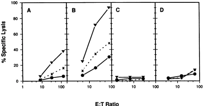
FIG. 1. Comparison of stimulation conditions for HSV-2-specific CTL. PBMC were purified from fresh whole blood (A and C) or recovered fromcryopreservation (B and D). Stimulator cells consisted of HSV-2-infected autologous PHA blasts (A and B), infected autologous EBV-transformed B-LCL (C), or infected autologous fibroblasts (D). The infected lymphoblasts were irradiated, and the fibroblasts treated with Mitomycin before responders were added. After 1 week, responder cells were recovered from the stimulation flasks andwashed, and graded numbers were added in 100 ,ul of medium to the assay plates to achieve the E/T ratios indicated. Autologous(V) or completely mismatched(x)EBV-transformed B-LCL were infectedovernightwith HSV-2 at an MOI of 4.Uninfected(0)autologous B-LCL and theinfected B-LCL were loaded with5"Crfor 90min, and104targets were added in 100,ul per well to the assay plate. After 4 h, 100p. wasremoved for counting.
hybridization,thefilterswerewashed in 0.1 x SSC(lx SSC
is 0.15 M NaCl plus0.015 M sodiumcitrate)-0.1% SDS at
65°C
for 3 h. The dried filterswere exposedto XAR-5film(Eastman Kodak Co., Rochester, N.Y.) at -70°C with an
intensifyingscreen (Dupont)for 16h.
RESULTS
Conditions of stimulation influence HSV-specific CTL re-sponses. Inpreliminary experiments,wetested various
stim-ulation conditions to optimize the amplification of
HSV-specific
CTL. PBMC from an individual with frequentlyrecurring genital herpes were stimulated in vitro by using
threedifferent potential APCs, autologous PBMC stimulated
with PHA(PHAblasts) torender themsusceptible to HSV
infection (17), autologous B-LCL, and autologous
fibro-blasts.Tosimplify the timing of subsequent experiments,we
also compared the cytolytic effector function of
cryopre-served versus freshly prepared PBMC. The stimulator cell
populations
were infected with HSV for 16 h and thenirradiated to attenuate viral infectivity prior to addition of
theresponder cells. The cellswereculturedtogether without
added
lymphokines
for 1 week at aresponder/stimulator
ratioof approximately 5:1. Thecultures were then assayed
forthe ability to lyse HSV-infected and uninfected
autolo-gous B-LCL as monitored by 51Cr release
(Fig.
1). Acompletely
HLA-mismatchedallogeneic
B-LCL wasin-cluded as a control target cell to determine the extent of
HLA-unrestricted
(natural killer) lytic activity
generated
under theseconditions.Theinfected PHA blasts werepotentstimulatorsof CTL for bothfreshly
prepared (Fig.
1A) andpreviously frozen (Fig. 1B)
responder
cells, while neithertheB-LCL(Fig. 1C) northefibroblasts
(Fig.
1D) wereefficientAPCs under these conditions. The cultures stimulatedwith
the infected PHA blastsalso exhibiteda
significant
level ofunrestricted lytic activity, whereasthe infected B-LCL- or
fibroblast-stimulated cultureswere
uniformly
unresponsive.
Thedifferences observed in the induction of
lytic activity
intheresponder cell
populations
werenotduetodifferences inviability, since all four cultures were similar inappearance
andthe numberof respondersrecovered from each culture
and theirviabilitiesas determinedbytrypanblueexclusion
were comparable.
This experiment did not distinguish between lysis by
CD4+orCD8+ CTL.Todetermine whetherHLA-restricted
lysis
mediated byCD8+ CTL could be detected, PBMC froma secondindividual with frequently recurrent genital herpes
were stimulated for 7 days by cocultivation with infected
autologous PHA blasts, and then CD8+ cells were isolated
by panning(56, 60). The recovered CD8+ cellswere
stimu-latedwithPHAand thencultured for14daysinthepresence
of irradiated allogeneic feeder cells and human IL-2 (37).
Lytic activity was assayed against several types oftarget
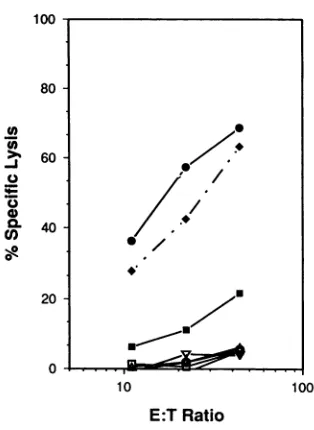
cells (Fig. 2). This CD8+ cell population efficiently lysed
autologous HSV-infected B-LCL as well as HSV-infected,
partially HLA-matched B-LCL but not uninfected targets.
These bulk cultures had no measurable natural killer
activ-ity, asindicated by the absence oflysis of K-562 cells. To
determinewhether thecytolyticactivitywasdirectedagainst
certain abundant virion proteins, recombinant vaccinia
vi-rusesexpressingtheHSV-2
glycoproteins
gD andgBorthetegument-associated
transactivatorprotein VP16 wereusedto infect autologous B-LCL targets. The bulk CD8+ CTL
population
efficientlylysed
vac/gD2-infected
targetcells and didnotlyse
targetcells infected withthewild-type
parental
strain. In contrast, gB- or
VP16-specific
lysis
was notdetected
despite
higher levels ofexpression
of thesegenesthan of the
gD2
gene in infectedB-LCL,
as measuredby
Western blot assay. Moreover, mouse fibroblasts infected
with this vac/gB2 vector are
recognized by
HSV-immunemousesplenocytes
(data
notshown).Cloning of
HSV-specific
CTL. Tobegin
todetermine therepertoire
of HSVantigens
recognized
by
theCD8+
CTL,
clones were isolated from apatient
whowas HSV-2solely
seropositive
and who hadculture-proven
recurrentgenital
HSV-2 disease. To obtaintheseclones,
PBMCwere stimu-lated by cocultivation withHSV-infected,
PHA-activatedlymphoblasts
for 1 week.CD8+
cells were isolatedby
on November 10, 2019 by guest
http://jvi.asm.org/
[image:4.612.150.480.79.250.2]100
-80
-U)
0
-J 0
4)
U,)
*
-60
40
2:1
10 100
E:TRatio
FIG. 2. Lysis by CD8+ CTLexpandedin bulkcultures. PBMC
werestimulated with HSV-infected PHA blasts for 7days,andthen
CD8+ cells were isolated by panning. The recovered cells were
cultured in RPMI-Hcontaining200ngofPHA-Pperml,32 UofIL-2 perml,and107 irradiatedallogeneicPBMC for 48h;then the cells
werewashed and returnedtotheflask inRPMI-Hcontaining32Uof IL-2 per mlfor 12days. Forpreparationof targets,autologous
(0)
andpartiallyHLA-matched
(*)
B-LCLwereinfectedat anMOI of4withHSV-2or at anMOIof 10 withwild-typevacciniavirus strain WR
(l)
orrecombinant virusesexpressingHSV-2glycoproteingD2(U),
gB2 (A), or VP16 (V). After a 16-h infection, the B-LCL,uninfectedautologousB-LCL
(0),
and the natural killer target line K-562(K)
werewashed and loaded with51Cr,
and104cells in100,ul ofmediumwere added tothe assay plates. After4h, 100 ,ulwasremoved forcounting.TheallogeneicB-LCL sharedHLAB7,Cw7,
and DR2 with the CTL.
panning
andwereseededintothe wellsof Terasakiplates
at an averageinput
oftwo cells per well. The cultures weresupplemented by
the addition ofallogeneic
feedercells,
PHA,
and IL-2(37).
After 14days,
206of282(73%)
ofthe wellscontainedgrowing
cells. The candidateswerescreened forHSV-specific cytolytic activity,
and seven clones were identified.Because greater than 66.7% of the wells in the initial
cloning
stepgenerated
viablecultures,
the isolates couldnotbe considered
truly
clonal.Nevertheless,
the HSVantigen
specificity
of the isolated lines isprobably
conferredby
asingle
CTL clone in thepotentially
oligoclonal populations,
since the
frequency
ofHSV-specific
CTLamong thecandi-date lines was 0.035. To avoid
awkwardness,
theHSV-specific
candidates will be referredto asclones. Four of the seven clones were subclonedby
limiting
dilutionand havebeen characterized. The subclones retain the
specificity
oftheir parent
clones,
so it islikely
that the results of theanalysis
of the clones are characteristic of the CTL sub-clones isolated from thatpopulation.
One of the clones lostspecificity
for HSVandwasnotsuccessfully
subcloned. Theremaining
two clones have been characterized to a morelimited extent.
Characterization of
HSV-specific
CTL. The clones wereanalyzed
indetailtodeterminevirustypespecificity,
surfacephenotype,
andHLArestriction.AsshowninTable1, six of the sevenHSV-2-specific
clones did not recognize target [image:5.612.95.252.75.290.2]cells infected with HSV-1. The surface
phenotype
of theTABLE 1. Type specificity and HLA class restriction of seven HSV-2-specific CTL clones
%Specific lysisof
virus-infectedtargetsa %Inhibition by
Clone _____________W6/32b
HSV-2 HSV-1
1H6 23 4 30±9
3B3 59 48 60 ±9
2H12 30 3 75± 13
2E7 21 Oc 88± 1
2C1 34 0 100
2F11 38d 2 NDe
2D12 51d 1 ND
a Autologous B-LCLwereinfected with HSV-2 strain MSorHSV-1strain
Pattonat anMOIof 4 and culturedfor16hpriortoCTLassay.The E/T ratio
was 5:1.
bHSV-2-infected B-LCLwereloadedwith51Crand thenresuspendedin
medium containinga1/50or1/80 dilution of W6/32 ascitesfluid and incubated
for 20min beforeaddition of CTL. Inhibition was calculatedby comparison
with lysisof target cells treated withthe same dilutionofanascites fluid
containing an isotype-matched control antibody. The E/T ratio was 5:1.
Values are means and standarderrorsoftwo tofiveexperiments.
IA valueof0 wasassignedwhenthecountsperminute released in thetest
wells werefewer than in the spontaneous release wells.
dE/Tratio of10:1.
eND,notdone.
sevenCTL cloneswasCD8+ CD4- CD3+ HLA
DRbfight,
ascharacterized by flowcytometry analysis (data notshown).
The class ofthe HLArestriction elementrecognized bythe
clones and their subclones was testedby using MAbW6/32,
specific for a monomorphic class I determinant, to block
lysis (3). The antibody was added to infected, 51Cr-loaded
B-LCL targets for 20 min before addition of effector cells
(Table 1).Thelytic capacity ofallclones testedwasinhibited
by preincubation of target cells with MAb W6/32, although
the1H6.5 subclonewas moreresistanttoblocking thanwere
the other clones. In a similar experiment using MAb 2.06,
which recognizes monomorphic determinants on HLA DR
(11), lysiswas notinhibited (data notshown).
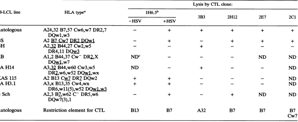
The class I HLAelement restricting recognition of HSV
epitopesfor five clones was determined by using apanel of
B-LCL that matched thedonor HLA type at one or more
loci as target cells. The pattern ofrecognition of partially
HLA-matched target cells and the deduced restriction
ele-ment for each of the five HSV-specific CTL clones are
shown in Table 2. Three of the five clones recognize their cognate HSV-2antigen in the context ofHLA B7, and one clone does so in the context ofeither B7 orCw7. The fifth
clone, 3B3, recognizesanHSVtype-commonepitope in the
context of HLA A32. In addition to recognizing an HSV
antigen, the 1H6.5 subclone exhibited alloreactivity to
B-LCLbearingthe HLA B13 classI molecule.
Identification ofantigen specificity with recombinant vac-cinia virus vectors. Vaccinia virus recombinants expressing
the HSV-2 gB, gD, or VP16 gene were used to determine
whether any of the clones recognized one of these virion
proteins. As shown in Table 3, none of the clones lysed
vaccinia virus-infected targets expressing gB2 or VP16; one
clone, 1H6, recognized
vac/gD2-infected
targets.Identification ofantigen specificity with drug-treated target cells. AsHSVencodes atleast 70proteins(33), reagents to
identifyallofthem are not available. To narrow the focus for
furthercharacterization oftheantigen-specific CTL clones,
infectedtarget cells were treatedwithselective drugs to limit
therepertoireof viral geneexpression to specific
transcrip-tionalsubclassesasoutlinedinFig. 3. Fourbroad classesof
9 /
.
on November 10, 2019 by guest
http://jvi.asm.org/
[image:5.612.313.552.102.209.2]TABLE 2. LysispatternofHSV-specific CTL clones from patient JC for partially or fullyHLA-matchedB-LCL
Lysis byCTLclone:
B-LCLline HLAtypea 1H6.5b
3B3 2H12 2E7 2C1
-HSV +HSV
Autologous A24,32 B7,57 Cw6,w7 DR2,7 - + + + + +
DQw1,w3
GS A2B7Cw7DR2DOwl - + - + + +
GH A2,32 B44,27Cw2,w5 - - + - -
-DR4,11
DQwf
RB A1,2 B44,37Cw-DR2,X NDC - - - ND ND
DQwA,w7
SAH14 A3,32B44,w60Cw3,w5 ND - + - - ND
DR2,w6,w52
DQwA,wx
KAS 115 A2B13 Cw DR7DQw2 + + - - - ND
SAH3.1 A3,x B13,35Cw4,wx + + - - - ND
DR6,w11(5),w52DQw1,w3
BSch A2,3 B7,w62 C- DR5,w6 - + - + ND ND
DQw7(3),1
Autologous Restriction element for
CTJL
B13 B7 A32 B7 B7 B7Cw7
a
HLk
matches with theeffectors areunderlined.bSubclone of 1H6.
cND,notdone.
genes are transcribed sequentially during an infection (re-viewed in reference 55). The first class ofimmediate-early
(IE) (a) transcripts is expressed in the absence of viral
protein synthesis. Early
(O)
transcriptsaretransactivatedbyaproteinsandthus require viral protein synthesis for their
expression. As ,Btranscription begins, mostatranscription
ceases and the corresponding a gene products rapidly dis-appear (50, 58). Early-late transcripts
(-yl),
are normallyexpressedasviral DNAreplicationcommences,buttheyare
also expressed when replication is blocked (55, 58). The
glycoproteinsgDandgB are members of the
-Yl
transcriptionclass (55). The true late
(Y2)
class, which encodes manystructural virionproteins,isnotexpressed inthe absence of viral DNAsynthesis.
Todetermine whether any of theremainingsixclones also
recognizevirionproteinsasdoes thegD2-specificCTL clone
1H6.5,targetcellsweretreated withacyclovir after infection
toblockHSVDNAsynthesisandthusinhibit_Y2expression. Parallel cultures ofB-LCLwereinfectedwith HSV-2 for 1 h,
and
thenacyclovirwasaddedto oneof the culturesat afinalTABLE 3. AntigenspecificityofsevenHSV-2-specific CTL clones
CTL % Specificlysisof vaccinia virus-infectedtargetsa
clone vac/WR vac/gD2 vac/gB2 vac/VP16-2
1H6 8 64 6 5
3B3 14 10 3 7
2H12 3 2 2 1
2E7 1 ob 0 0
2C1 1 2 3 0
2F11 3 0 0 2
2D12 1 1 0 2
aAutologousB-LCLwereinfected with
wild-type
vaccinia virus(WR)orvacciniavirusrecombinantsat anMOIof 10 for 16 hpriortoCTFLassay. The
EiTratio was 10:1.
bThe counts per minute released in the effector-containing wellswere
fewer thaninthespontaneousrelease control wells.
concentration of 150 ,uM.The target cells were incubated for
anadditional16 to 18h and then loaded with51Crand mixed
withthe CTL. As shown in Table 4, two of the subclones, 3B3.4 and2H12.5, lysed acyclovir-treated target cells with
efficiencysimilar to that of untreated target cells. In a second
experiment with the 3B3.4 subclone, specific 51Cr release
was 64.4% from acyclovir-treated cells, compared with
69.8% from untreated cells, at a 10:1 E/T ratio (data not
shown). The lytic capacity of the 2C1 clone, on the other
hand, was enhanced substantially when _Y2 gene expression was suppressed by acyclovir addition compared with un-treated cells. In contrast,the1H6.5subclone and 2E7 clone
exhibitedreduced lysisof theacyclovir-treated target cells.
Theremainingtwoclones, 2F11 and 2D12, also lysed HSV-2
infected cells with comparable efficiency, with and without
acyclovir treatment (data not shown). Thus, three distinct
patternsofacyclovirsensitivityof thecytolytic activitywere
observed for the seven clones: decreased (two clones), enhanced(one clone), andunalteredcytolysis(four clones).
Duringthe initial characterization experiments, the CTL
weremaintained in mediumcontaining IL-2 evenduringthe incubation with target cells. Lysis by the 2C1 clone was
quite variable under these conditions. Because B cells
ex-press IL-2 receptorsandcan be inducedtodifferentiate by
thelymphokine (5, 40, 41,53),itwaspossiblethat thecourse
of an HSV infection
might
also be affected by IL-2 treat-ment. Therefore, the B-LCL targets were infected withHSV-2,IL-2wasaddedto32U/ml(finalconcentration), and
the cellswereincubatedovernightinparallelwith untreated
or acyclovir-treated cells (Table
4).
Lysis by four of fiveCITL
clones tested was notsignificantly
affectedby
this treatment, butlysis by2C1was abolished.As analternateapproachtolimiting
presentation
tovirionproteins, cells were
exposed
to DRBprior
to infection toprevent anyviral RNA
synthesis.
Under theseconditions,
onlyvirionproteinsreleased into thecytoplasm
after fusion of theenvelopewith theplasma
membranewould be proc-essed and presented. DRB was chosen instead of ActD because itcauses lessdamage
tothe cellswhilepreventing
on November 10, 2019 by guest
http://jvi.asm.org/
1
"Cr CTL
| 16-20 hr
I
]
wash | 2hr wash | 4hr A] wash LIZX wash
I
l BHSV-2
wash wash C
eDRB uDRB
eDRB
HSV-2
lb
CX
wash |X ]wash D
E
ActD
wash wash F
ActD ActD
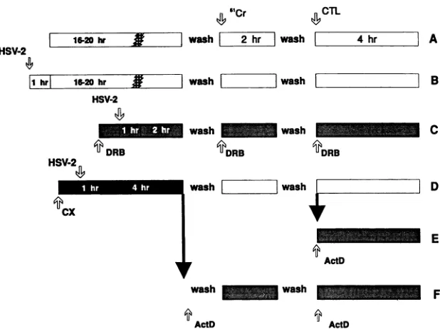
FIG. 3. Schematicfor thepreparationofdrug-blocked/releasedtargets.The inhibitorscycloheximide (CX)and DRBwereaddedtoB-LCL 1hpriortoinfectionwith HSV-2. Infectionswereinitiatedbyincubation of cells and virus in 0.5 ml of serum-free medium for 1 hpriorto
dilution with mediumcontainingserumanddrugs. Drugswereincludedduring washes whereappropriate. (A)Uninfectedtargets;(B)HSV-2 infectedtargetsuntreatedortreatedwith150,uMacycloviror32 U of IL-2perml; (C)DRB-treatedtargets;(D)cycloheximide-treatedand infectedtargetsreleased fromdrug block; (E)cycloheximide-treated targetsreleased fromdrugblock for 2 hand thenblocked withActD; (F)cycloheximide-treatedtargets shiftedtoanActDblock.
RNA synthesis as effectively (20). Target B-LCL were
pretreated with DRB for 1 h, infected in the presence of
DRB, and incubated for 1.5to3 hpriortoloading with
5"Cr
and exposure to the CTL. A parallel culture of untreated cells wasinfected and incubatedforthe same time periods
were as the DRB-treated target cells. A third culture was
infected andincubated in the absence of drugs for 16to18 h. Assummarized in Table 5, three clones, 3B3.4, 2H12.5, and 2F11,wereabletolyseDRB-treated cellsasefficientlyasdid
target cells infected for the same amount of time in the absence ofdrug. Another clone, 2D12, lysed DRB-blocked cells with half the efficiency of untreated target cells. In contrast, the 2C1 clone lysed untreated target cells very
poorly. However, cells treated with DRB were lysed
effi-ciently. The 2E7 clone and 1H6.5 subclone did not lyse
targetcellstreated with DRB. Thesesameclonesalsolysed
cells infected for less than 6 h with substantially lower efficiency thandid targets that hadbeen infectedovernight. The targetantigen specificity of the 3B3 and 2H12 clones
wasfurther examinedby comparing the lysissusceptibilities
oftarget cells infected with HSV-2 and exposed to one of
threedrug treatment protocols: (i) infection with HSV-2 in thepresenceof theprotein synthesis inhibitor cycloheximide followed by release from drug arrest, (ii) treatment with
cycloheximide followed by drug removal and addition of
ActD,or(iii)treatmentwithcycloheximidefollowedby drug removal and a 2-h delay prior to the addition of ActD as
diagramed in Fig. 3D toF. For the firsttreatmentprotocol,
thecycloheximide wasremoved 5 h afterinfection; the cells
wereloaded with 51Cr and then mixed with effector cells for a4-h incubation in the absence ofdrugs (Fig. 3D). Under
these conditions, the time course of viral gene expression
TABLE 4. Effect ofacyclovirand IL-2treatmentofHSV-infected targetcellsonlysisby CTL clones
E/T %Specificlysis %
Treatment ratio. 1H6.5 2B3.4 2H12.5 2E7 2C1 Spontaneousrelease
Uninfected 10:1 Oa 3 8 0 7 11
2.5:1 0 0 8 0 5 11
Noneb 10:1 55 66 35 37 15 9
2.5:1 40 29 40 32 12 9
Acyclovirc 10:1 12 82 40 5 44 10
2.5:1 12 58 33 6 42 10
IL-2C 10:1 45 54 23 34 1 10
2.5:1 46 21 15 37 1 10
aAvalue of0wasassigned when the mean countsper minute released were fewer than in the spontaneous release control wells.
bThe targetB-LCLwereinfected with HSV-2 at an MOI of 2, incubated overnight (18 h), and then loaded with
51Cr
for 90min prior to assay.cTargetcellsweretreated with 150 ,uMacyclovir or32 UofIL-2per ml from 1 h after addition of virus. The cells were maintained with acyclovir or IL-2 throughout 5"Crloadingandincubationwith theeffectorcells.
HSV-2
|1 hr| 16-20 hr ,
on November 10, 2019 by guest
http://jvi.asm.org/
[image:7.612.151.460.80.311.2] [image:7.612.62.555.593.702.2]TABLE 5. Effect ofDRB treatmentof HSV-infected target cells on lysis by CTL clones
Treatment E/T % Specific lysis % Spontaneous
ratio 1H6.5 3B3.4 2H12.5 2E7 2C1 2F11 2D12 release
Uninfected 10:1 1 0 10 2 2 oa 0 12
2.5:1 0 0 8 0 0 0 0 12
None 10:1 71 82 62 28 2 37 53 13
18 hb 2.5:1 21 55 48 18 0 25 42 13
3_5 hc 10:1 14 42 29 11 NDd 18 30 13
2.5:1 10 48 28 10 ND 13 29 13
DRBe 10:1 4 48 31 0 31 14 15 24
2.5:1 2 45 26 2 6 5 11 24
aAvalueof 0 was assigned when the counts per minute released in the test wells were fewer than in the spontaneous release wells.
bTarget cells were infected with an MOIof 5 and then incubatedovernight(18 h) prior to
51Cr
loadingand assay.cTarget cells were infected with an MOI of 10 and incubated in parallel with the DRB-treated cells but in the absence of drug.
dND, not done.
eTarget cells were treated with 100p.MDRB for 30min before infection with HSV-2 at an MOI of 10. DRB was maintained continuously for the 1.5- to 3-h
incubation period after virus addition and during the subsequent loading of the cells with51Cr,washing,and addition of effector cells for the cytolytic assay.
was delayed by5hours such thata, 13, and
-yl
proteinswereexpressed during 51Cr loading andexposuretothe CTL. The effects of thedrugtreatmentprotocolsonthepatternof HSV
genetranscriptionwereconfirmedby Northern blot analysis as described below. The second set of target cells was
treated with cycloheximide for 5 h, followed by the imme-diate addition and continuouspresence of ActD during the
washes, 51Cr labeling, and lysis (Fig. 3F). Under these conditions, a genetranscripts accumulate during the cyclo-heximide block, and these are expressed rapidly after
re-moval of the drug. Because further transcription was
blocked, only aantigenswereexpressed in these cells. The
third set of cells was treated with cycloheximide for 5 h,
followed by drug removal and 51Cr labeling for 2 h in the absence ofdrugs and then the addition of ActD and effector cells simultaneously (Fig. 3E). Under these conditions, predominantly gene products should have been present, since only a transcripts accumulate during the cyclohexi-mide block andare translatedimmediately after removal of
thedrug. These proteins activate geneexpression with the
concomitant decline ofaproteins, ashas beendescribedby
Del Valetal. (12) formouse CMV undera similarregimen.
As can beseen inTable 6, clone 2H12 lyses virus infected
targetstreated withcycloheximide followed by drug removal
or immediate replacement with ActD but not cells treated with cycloheximide followed by delayed addition of ActD. Incontrast, the 3B3 clonelyses all three drug-treatedtarget cells with equal efficiency and at approximately the same
levelas dotargetcells infected with virus in the absence of drug treatment. From these results and those of DRB treatment, itwould appearthat the 2H12 clone may
recog-nize an a antigen that is also packaged in the virion and
whose synthesis declines upon the onsetof gene
expres-sion. Theproducts ofagenessuchasICP4orICPO(61)are
likely candidates. The 3B3 clone may recognize a virion
protein of the transcriptional class introducedas aresult of
viral infectionoranageneproduct whose synthesis doesnot declineasrapidly during the period of geneexpression.
It was notpossible toapply the samedrug block/release
strategy tofurtheranalyze the other three clones. In thecase
of the 1H6.5 and 2E7clones,cognateantigensareexpressed
at levelstoolowto sufficiently sensitize thetargetcells ina
short-term infectiontodistinguish effects of the application and removal of the cycloheximide block. Treatment of the B-LCL withcycloheximide for longer time periods orwith higher concentrations of drug rendered them resistant to lysis by the 1H6.5, 2E7, and 3B3.4 clones in addition to causing unacceptably high levels of spontaneously released 51Cr. The 2C1 clonewasnotanalyzed further because of loss ofantigen specificity.
Expression of HSV RNA in drug-treated B-LCL target cells. Toensurethat thedrugtreatmentswereeffective in limiting
HSVgene expressionas predicted, RNAwas purified from
HSV-infected, drug-treated cells and assayed in Northern blots. IE a gene RNAwas detected by using DNA probes
that were generated from restriction fragments containing
the ICPO and ICP4 gene sequences. -Yl transcripts were
detected with a gD probe, and Y2 transcripts wereassayed
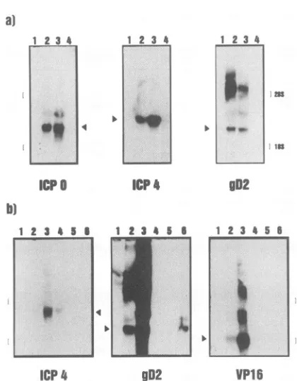
withaVP16probe. DRB treatmentprevented expression of ICPO or ICP4 a transcripts or the more abundant gD2 -Yl transcripts (Fig. 4a, lane 4), while all three were readily
[image:8.612.64.564.94.213.2]detected in untreated B-LCL infected for 6 h (lane 2)orin
TABLE 6. Lysis ofcellsexpressing differenttranscriptionalclasses of HSV-2bytwoCTL clones
%Specificlysisb
Target Treatment' Transcription %Spontaneous
classexpressed 3B3 2H12 release
(E/T, 8:1) (E/T, 13:1)
Uninfected CXfolloweddirectly byActD None 7 8 35
HSV-2 infected None a,
PI,
'Yl'Y2 39 15 21CX followedby6hin the absence of CX
ax,
1, i 49 24 17CX followeddirectlytoActD aX 52 23 29
CX followedby2-hdelayand then ActD 1, (oa) 35 2 22
a B-LCL targetsweretreatedwithcycloheximide(CX)for5 hbefore
51Cr
loadingasoutlined inthelegendtoFig.3and detailedinMaterialsandMethods.b The CTLwereaddedtoU-bottommicrotiterplatesin100
±d
toachievetheE/Tratios indicated;104target cellswereadded,andlysis
wasallowedtoproceedfor4h.
on November 10, 2019 by guest
http://jvi.asm.org/
[image:8.612.70.566.621.708.2]a)
1 23 4 1 2 34
1
Icp
1 2 3 4 5 I 1 2 3 5
4
1 2 34
I.
FF,
2,t
gD2
1 2 34 56
I....
ICP4
902
VP16
FIG. 4. HSV-2RNAsin drug-treatedB-LCL. RNAwaspurified
from3 x10' B-LCL, and10jig of RNAwasaddedperlaneof1.1 Mformaldehyde-1%agarosegels.The positionsofthe28S and 18S rRNAs were determined by ethidium bromide staining prior to
blotting and are indicated by brackets. Replicate blots were
pre-paredand hybridized with probestothegenesindicated below the
lanes. The lanes in panel acontained RNA from uninfected cells
(lane 1), cellsinfected for6h in the absenceof drugs (lane 2), cells
infectedfor5hin thepresenceofcycloheximide and then 2 h in the
absenceof drugs(lane 3),orcellsinfected for3hin thepresenceof
DRB(lane4). The lanes in panel b contained RNAfrom uninfected cells (lane 1), cells infectedfor 6 h (lane 2) or24 h(lane 3) inthe
absenceof drugs, cellsinfectedfor5hinthepresenceof
cyclohex-imide(lane4),cells infected for5h in thepresenceof cycloheximide
and thentreatedwith DRB for 2 h (lane 5),orcells treatedfor23of
24hwithacyclovir(lane 6).
cells treated with cycloheximide for5 h and then released from the block for an additional 2 h before the cells were
harvestedfor RNA isolation(lane 3).OverexposedNorthern blots did not yield signals above background in the lanes
containingRNA from DRB-treated cells (data not shown).
Cells maintainedin cycloheximidefor 5 h before isolation of RNA containedICP4 transcripts butweredevoid ofgD2
transcripts(Fig. 4b, lane 4). Treatmentof infected cellswith 150puMacyclovirfor 23 h blocked VP16 expression (lane6). The inhibitor also significantly reduced gD2 RNA levels compared withuntreatedcells (lanes6and 3,respectively), characteristic ofthe Yl transcription class. This depression of gD2 expressionwas reflectedas areduction in sensitivity
to lysis by 1H6.5 CTL in acyclovir-treated cells (Table 4).
Treating infected cells with cycloheximide and then shifting
the cells to DRB for 3 h led to a loss of the ICP4 RNA
present atthe end of the cycloheximide treatment (lanes 4
and 5).No gD2orVP16 transcriptsweredetected in the cells treated with cycloheximide or cycloheximide followed by DRB (lanes 4 and 5). As expected, this treatment limited
HSVtranscription tothe agene class.
DISCUSSION
We have investigated the nature of the HSV-specific
CD8+
CTL response in humans with culture-proven and serologically provenrecurrent
HSV-2 infection. Although HSV-specificCD4+
CTL have been repeatedly observed and such clones have been isolated (64, 65), the isolation of HLA class I-restricted, HSV-specificCD8+
CTL clones from virus-infected humans has proven considerably more difficult. Other investigators have detected HSV-1 specific, HLA class I-restricted CTL in bulk assays, but the activity represented a minor fraction of the cytolytic activity in the culture (54). A likely explanation for previous failures to cloneCD8+
CTL from humans could be that theAPCs in the in vitro stimulation phase processed the soluble viral anti-gens or the noninfectious UV-inactivated virus through the HLA class II pathway in a manner similar to that described for influenza virus antigens (8, 38). In the experiments reported here,CD8+
HSV-specific CTL clones were iso-lated by stimulating memory CTL in vitro with HSV-in-fected PBMC rather than using solubleglycoprotein antigens or UV-inactivated virus. In addition,CD8+
T cells were then isolated by panning prior to expansion and cloning. Thus, it is clear that the cellular immune response to HSV infection, like the response to other viruses, includes a broad class I-restricted CTL response. [image:9.612.72.282.76.345.2]Five of the seven HSV-specific clones clearly recognize different viral protein antigens. Because two clones, 2F11 and 2D12, were not characterized as extensively, it is not possible to assert that they recognize an HSV antigen distinct from that recognized by either the 3B3 or 2H12 clone. Additional vaccinia virus recombinants expressing specific HSV-2 a and
P
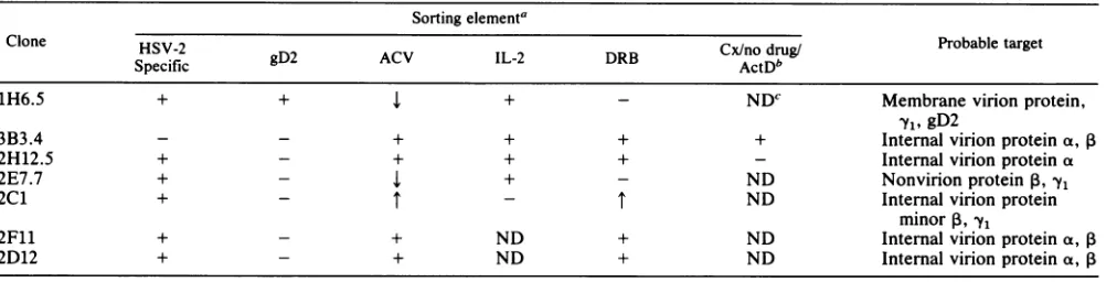
gene products will be needed to differentiate these clones. The minimum criteria that enable the clones to be distinguished from one another are shown in Table 7. These sorting elements include the type specificity of HSV recognition, recognition of a specific antigen (gD2), andrecognition
of infected target cells treated continuously with acyclovir, DRB, or IL-2 or treated with cycloheximide followed by drug removal and delayed addition of ActD. All of these sorting elements discriminate between different viral proteins with the sole exception of type specificity. This sorting element could distinguish between different proteins or different epitopes on the same polypeptide chain. On the basis of the pattern of recognition of drug-blocked target cells, a probable target antigen class has been assigned for each clone.The
1H6.5
subclone recognizes a virion protein, gD2, an abundant glycoprotein inserted in the viral envelope. How-ever, DRB-blocked target cells were not lysed by this gD2-specific clone. Thus, the gD2 epitope was presented on the target cells only as a result of de novo synthesis. Acyclovir treatment reduced gD2 mRNA expression in the target cells. In concordance with the reduced gD2 expres-sion, the ability of the clone to lyse acyclovir-treated target cells was diminished (Fig. 4b, lane 6; Table 4). A similar effect was noted in cells infected for 3 to 5 h compared with target cells infected for longer time periods (Fig. 4b, lanes 2 and 3; Table 5). These data and those of Zarling et al. (64, 65) indicate that bothCD4+
andCD8+
CTL specific for gD2 are present in HSV-2 infected patients. Thus, these data suggest that gD2 may be an important target for control of HSV infections.It is interesting that in this limited sample, five of seven clones recognize exogenous virion proteins introduced into the target cell and presented to the immune system via the
ICP
4
on November 10, 2019 by guest
http://jvi.asm.org/
TABLE 7. Assignment of probable CTL target antigens or transcription classes Sortingelementa
Clone HSV-2
Cx/no
drug/
ProbabletargetSpecific gD2 ACV IL-2 DRB ActDb
1H6.5 + + 4 + - NDC Membrane virion protein,
-yl,gD2
3B3.4 - - + + + + Internal virion protein a, 1B
2H12.5 + - + + + - Internal virion protein a
2E7.7 + - l + _ ND Nonvirion protein 13,
Yl
2C1 + - - T ND Internal virion protein
minor1, -yl
2F11 + - + ND + ND Internal virion protein a, ,B
2D12 + - + ND + ND Internalvirion proteina, ,B
aCondition that distinguishes one clone from the others. Symbols indicate enhancement oflysis(t),noeffect(+), reduction oflysis
(t),
resistance tolysis(-).
bInfection in the presence of cycloheximide (CX) followed by 2 h without drugs followed byActD.
cND, not done.
classI pathway inthe absenceof viral protein synthesis. In
most instances, exogenously acquired antigens are
proc-essed through endocytic vesicles and presented to the
im-mune system in the context ofa class II HLA restriction
element, although it is possibletointroduce proteins directly
into the cytoplasm for classIpresentationby osmotic shock
(36). Byfusion with the plasma membrane(15, 22, 51),HSV
virions bypasstheendocyticpathway of cellular entry, thus
providing a ready source of viral proteins for processing
throughapathway that normally handles proteins produced
by de novo synthesis. Recently, Riddell et al. (46) have
shown for HCMV, the majority ofthe CMV-specific CTL
from seven seropositive individuals also recognize virion
protein(s) introduced into the target cell in the absence of
any viralgene expression. The classIpresentation ofthese
antigenswasbrefeldinAsensitive, demonstratingthat
intra-cellularprocessing of the antigens wasrequired. Moreover,
like the case with HSV gD glycoprotein, the CMV gB
homologtothe HSVgBglycoproteinwas notrecognized in
the absence of viral gene expression. This result suggests
that the topology of the viral proteins is preserved upon
virus-hostcellmembranefusionand that viral glycoproteins
arenotefficiently injected intothecytoplasm. The parallels
of these results in CMV and oursinHSVare striking. That
the CTLresponsein this individual appears to be biasedto
a seemingly minor source of HSV antigens may reflect an
adaptation to one way that the virus uses to evade the
immunesystem; anextremely efficienthostshutoff function
(14,21, 27). The effects of this shutoff functionarereflected
in theinability of primary fibroblast lines infected with live
virus to serve as APCs in vitro (Fig. 1) and our failure,
despite repeated attempts, to use these sameprimary lines
infected with live virus astarget cells forthree ofthe CTL
clones (26).
For the case of murine CMV, Reddehase and
Koszi-nowski have shown that a
single antigen
issurprisingly
immunodominant;40 to50%of all memory CTLaredirected
against a single
protein,
themajor
IE transactivator (IE1,pp89) (44). Similarly, in two CMV-immune humans, itwas
suggested that the 72-kDa major IE
protein
is asignificant
target antigen, since 46% of the CTL
lysed
target cellsinfected with a vaccinia virus recombinant
expressing
theCMV major IE
protein,
whereas less than 6%lysed
vac/CMVgB-infected
cells(7).
In contrast to theseprevious
results, the diversity ofourclones suggest that there is no
such extreme immunodominance for the case of HSV.
However,inagreementwiththeresults ofRiddellet al. (46)
forCMV, internal virion proteinsappear tobepreferentially
recognized.
In the mouse, in which HSV-specific CTL are easier to
demonstrate, manyresearchers have focusedonthe
poten-tialof particular glycoproteins toelicit CTL. These efforts
haveyielded conflicting results.Three groupshavedetected
gB-specific CTL in some butnot allhaplotypes(4, 34,59),
andHankeet al. (19) have recentlylocalizedaCTLepitope
in gB to residues 489 to 515 for the H-2b haplotype. In contrast, Martin et al. reported that gC but not gB or gD serves as a class I-restricted target antigen (31). Although several researchers have been unable to detect gD-specific
CTL (4, 31, 59), recently Johnson et al. (23) reported the
isolation ofa gD-specific CTL clone from an
H_2k
mouse.These contradicting results may arisefrom methodological
differences or, more likely, from the possibility the
glyco-protein-specific memory CTL are rare. In support of this
view, Martin et al. (29, 30, 32) suggested that a gene
products may represent as much as 30% of the target
antigens and that virion components serve as minortarget
antigens. Inthe
H-2k
haplotype, thea generecognition maybeentirely directedtoICP4; ICP0was notrecognized (59).
The differences between the results in the mouse and our
studies in the human, even with a small sample, i.e., the
demonstration ofgD-specific CTL in two individuals, the
lack ofagB-specific response, and the predominant
recog-nition ofinternal virion proteins, underscorethenecessityto
continue to do such studies to more fully characterize the
human immuneresponse toHSV.
It is notablethat six of thesevenCTL clonesare HSV-2
type specific. The overall DNA sequence
homology
ofHSV-1 and HSV-2 as measuredby the kineticsof solution
hybridization is 50% (24). For
specific
essential genes suchasthose
encoding
gB, gD, and VP16,theidentity
increases up to86%(47a,52, 57).Thus,
manyHSV-specific
antibodiesarecross-reactive(1, 39).Theresults
presented
heresuggestthat the type
specificity
ofthecellular immune response may be greater than thatoftheantibody
response. Whether this pattern will be maintainedin alarger panel
of clonesfrom this and other individualsremainstobedetermined.Inmice,
the
HSV-specific
CTL response is alsopredominantly
typespecific (16, 43), although type-common
recognition
hasbeen
reported
(10,13).
These observations on the typeselectivity
of the immune response areimportant
for thedesign
of vaccinesto preventorlimit HSV disease.on November 10, 2019 by guest
http://jvi.asm.org/
[image:10.612.66.572.92.220.2]Efficient in vitro stimulation was achieved only with the useof HSV-infectedlymphoblastsasstimulator cells. Autol-ogous B-LCL orfibroblasts didnot function asAPCs. The
major difference between the PHA blast cultures and the B-LCL or fibroblast line cultures was the presence of accessory cells, including adherent cells, during the blast
transformation, virus infection, and stimulation phases of the cultures. Thus, the failure of both the B-LCL and
fibroblaststostimulateeffector cellsmaybetheir inabilityto
provideaccessoryfunctions suchaslymphokine secretionin
additiontopresenting viral antigens. Inaddition, the failure
of HSV-infected fibroblasts toserve as stimulators may be
duetothe effect of the efficient HSV hostshutoffmechanism
onantigen presentation in these cells (14, 21, 27). Consistent
with this hypothesis, we haveobserved that HSV-infected
primary fibroblast lines also do notserve astarget cells for these CTL clones (26).
In summary, we have, using a novel in vitro stimulation
system,been able todemonstrate HSV-specific
CD8+
CTLactivity in two individuals. In the one individual in whom
extensive characterization of
CD8+
CTL clones wasper-formed, we found extensive antigenic diversity to
HSV-2-specific proteins. While we were able to demonstrate
cy-tolytic activity specific to gD2, we were not able to detect
CTL specifictogB2, amoreabundant HSV-2 glycoprotein.
Mostof the cloned CTLwere specific to exogenous virion
proteinsthatwereprocessed andpresentedpriortodenovo
virion protein synthesis. Quantitative characterization of CTL responses in humans to evaluate their importance in
controllingHSV infections in bothimmunizedand naturally
infected patients can nowbe undertaken.
ACKNOWLEDGMENTS
This work was supported in part by NIH grants A120381 and
A129681 and by Chiron Corp. D.K. is a research fellow of the
AmericanSocial Health Association.
We thank John Hansen and Brenda Nesperos, Department of
Medicine, Universityof Washington, forHLAtyping severalofthe
B-LCLlines andfor providing others usedtoidentify HLA
restric-tion. We also thank Sergio Abrignoni, Ciba-Geigy, and Susan
Masecicz, MLC Laboratory, Fred Hutchinson Cancer Research Center, for additionalB-LCL lines. We acknowledge the assistance
ofJeffreyNeidleman in preparing the manuscript. Finally, we are
especiallygrateful to Chris Walker and Phil Greenberg for many
helpfuldiscussions.
REFERENCES
1. Ashley, R.L., L. Corey, andW.E. Rawls.1989. Herpes simplex
virusesand herpes B virus, p. 263-317. In N. J. Schmidt and
R. W. Emmons (ed.), Diagnostic procedures for viral,
rickett-sialandchlamydialinfections. AmericanPublic Health
Associ-ation,Washington, D.C.
2. Ashley, R. L., J. Milton, F. Lee, A. Nahmias, and L. Corey.
1988. Comparison ofWestern blot (immunoblot) and
glycopro-tein G-specific immunoblot for detecting antibodies to herpes
simplex types 1 and 2 in human sera. J. Clin. Microbiol.
26:662-667.
3. Barnstable, C. J., W. F. Bodmer, G. Brown, G. Galfre, C.
Milstein, A. F. Williams, and A. Ziegler. 1978. Production of
monoclonalantibodiestogroupAerythrocytes, HLA and other
human cell surface antigens-new tools for genetic analysis.
Cell 14:9-20.
4. Blacklaws,B. A.,A. A. Nash, and G. Darby. 1987.Specificityof
the immuneresponseof micetoherpessimplex virus
glycopro-tein BandD constitutively expressedonLcell lines. J. Gen.
Virol.68:1103.
5. Blackman, M. A., M. A. Tigges, M. E. Minie, and M. E.
Koshland.1986. A modelsystemfor peptidehormoneactionin
differentiation: interleukin 2 induces a B lymphoma to
tran-scribe theJchain gene. Cell 47:609-617.
6. Bonneau, R. H., and S. R. Jennings. 1988. Modulationof acute and latent herpes simplex virus infection in
C57BL/6
mice by adoptive transfer of immune lymphocytes with cytolytic activ-ity. J. Virol. 63:1480-1484.7. Borysiewicz, L. K., S. Graham, J. K. Hickling, P. D. Mason, and J. G. P. Sissons. 1988. Human cytomegalovirus-specific cyto-toxic T cells: their precursor frequency and stage specificity. Eur.J. Immunol. 18:269-275.
8. Braciale, T. J., L. A. Morrison, M. T. Sweetser, J. Sambrook, M.-J. Gething, and V. L. Braciale. 1987. Antigen presentation pathways to class I and class II MHC-restrictedTlymphocytes. Immunol. Rev. 98:95-114.
9. Burke, R. L., K. Hartog, K. D. Croen, and J. M. Ostrove. 1991. Detection and characterization of latent HSV RNA by in situ and Northern blot hybridization in guinea pigs. Virology 181: 793-797.
10. Carter, V. C., P. L. Rice, and S. S. Tevethia. 1982. Intratypic and intertypic specificity of lymphocytes involved in the recog-nition of herpes simplex virus glycoproteins. Infect. Immun. 37:116-126.
11. Charron, D. J., and H. 0.McDevitt. 1979. Analysis of HLA-D region-associated molecules with monoclonal antibody. Proc. Natl. Acad. Sci. USA76:6567-6571.
12. Del Val, M., K. Munch, M. J. Reddehase, and U. H. Koszi-nowski. 1989. Presentation of CMV immediate-early antigen to cytolytic T lymphocytes is selectively prevented by viral genes expressed in early phase. Cell 58:305-315.
13. Eberle, R., R. G. Russell, and B. T. Rouse. 1981. Cell-mediated immunity to herpes simplex virus: recognition of type-specific and type-common surface antigens by cytotoxic T-cell popula-tions. Infect. Immun. 34:795-803.
14. Fenwick, M. L., and R. D. Everett. 1990. Inactivation of the shutoff gene (UL41) of herpes simplex virus types 1 and 2. J. Gen. Virol. 71:2961-2967.
15. Fuller, A. O., and P. G. Spear. 1987. Anti-glycoprotein D antibodies that permit adsorption but block infection by herpes simplex virus 1 prevent virion-cell fusion at the cell surface. Proc. Natl. Acad. Sci. USA 84:5454-5458.
16. Glorioso, J., U. Kees, G. Kumel, H. Kirchner, and P. H. Krammer. 1985. Identification of herpes simplex virus type 1 (HSV-1) glycoprotein gC as the immunodominant antigen for HSV-1 specific memory cytotoxic Tlymphocytes. J. Immunol. 135:575-582.
17. Grammer, S. F., G. Y. Ishioka, and R. W. Chesnut. 1988. Studies on the capacity of B cells as well as T cells to serve as accessory cells for the activation of herpes simplex virus-specific cytolytic T cells. J. Immunol. 140:2016-2022.
18. Han, J. H., and W. J. Rutter. 1988. Isolation of intact mRNA and construction of full length cDNA libraries: use of a new vector,,ugt22, and primer-adapters for directional cDNA clon-ing. Genet. Eng. 10:195-219.
19. Hanke, T., F. L. Graham, K. L. Rosenthal, and D. C. Johnson. 1991. Identification of an immunodominant cytotoxic T
lympho-cyte recognition site in glycoprotein B of herpes simplex virus using recombinant adenovirus vectors and synthetic peptides. J. Virol. 65:1177-1186.
20. Jack, H.-M., and M. Wabl. 1988. Immunoglobulin mRNA stability varies during B-lymphocyte differentiation. EMBO J. 7:1041-1046.
21. Jennings, S. R., P. L. Rice, E. D. Kloszewski, R. W. Anderson, D. L. Thompson, and S. S. Tevethia. 1985. Effect of herpes simplex virus types 1 and 2 on surface expression of class I major histocompatibility complex antigens on infected cells. J. Virol. 56:757-766.
22. Johnson, D. C., M. Wittels, and P. G. Spear. 1984. Binding to cells of virosomes containing herpes simplex virus type 1 glycoproteins and evidence for fusion. J. Virol. 52:238-247. 23. Johnson, R. M., D. W. Lancki, F. W. Fitch, and P. G. Spear.
1990. Herpes simplex virus glycoprotein D is recognized as antigen byCD4+and