Vol. 52, No. 3 JOURNALOF VIROLOGY, Dec. 1984,p. 750-760
0022-538X1841120750-11$02.00/0
Copyright C) 1984, American Society for Microbiology
Multiple Binding Sites for Polyomavirus
Large
T
Antigen Within
Regulatory Sequences of Polyomavirus
DNA
ALISON COWIEANDROBERT KAMEN* GeneticsInstitute, Boston, Massachusetts 02115
Received 6June 1984/Accepted 24 August 1984
Polyomavirus large T antigen binds specifically tomultiplesites within the regulatory region of the viral
genome.Experiments done with crudeextractsfrom wild-type virus-infectedmousecells and immunoprecipi-tation of protein-DNA complexes localizedtwo high-affinity binding siteson the early regionsideofthe DNA replication origin. Purification of the large T antigen by immunoaffinitychromatography made it possibleto refinethe analysis through application of DNase I footprinting. The high-affinity interactionswereresolved into
three closely spaced, but distinct, binding regions. These begin ata site only slightly overlapping the early
boundary of thecorereplication origin,alocation highly homologoustothatof simian virus 40 largeT antigen-binding siteI, but then extendawayfrom the origin toward the early codingsequenceand thusspanthe early
region transcriptional initiation sites. Each tight-binding region contains from two to four copies of the
sequence5'-(A=T)G(A>G)GGC-3' repeatedat9-to11-base-pair spacing. At high protein concentrationsand at low ionic strength, additional sites within the core replication origin and in the enhancer region were
protected from DNase I digestion. These minor binding sites also includedrepeatsofsequencesrelatedtothe
consensus, butatdifferent spacings. Our resultssuggestthat, unlike simian virus 40 DNA, the polyomavirus genome may have distinct regions of interaction with its large T antigen which separately are involved in
initiation of DNA replication and the regulation of viral transcription. The large T antigen of polyomavirus is oneofthree T(for
tumor) antigens encoded by the early region of the viral genome. Activities of this 100-kilodalton nuclear phospho-protein have been implicated in several processes that occur
after virus infection. These include the stimulation ofhost
cell DNAsynthesis, the initiation of viral DNA replication,
the repression of early gene expression, the switch from
early to late transcription, and the events that lead to integration, excision, and amplification ofthe virus DNA within thehost chromosome (for a review, seereference46).
Expression ofthelarge Tantigen fromDNAintegrated into
the cellular genome results in remarkablephenotypic
alter-ations,but does not causetumorigenic transformation.Itcan
reduce the serum growth factor requirement of established
rodent cell lines and complement the serum-dependent
transformed stateinduced insuch cellsbytheexpression of
the viral middle T antigen (42). Transfection of rodent
embryo fibroblasts with DNAencodingthelargeTantigen,
orencodingitsamino-terminal 40kilodaltons, results in the
establishment of permanent cell lines without passage
throughcrisis.Thecelllinesthusgeneratedhave thegrowth
properties of nontransformed fibroblastswithrelatively low
serum requirements; they are dependent on continued
expressionof the large Tantigen forpropagationinculture
(43). This establishment function ofthelargeT antigencan
complement theactivityof cellularorviral oncogenes in the
tumorgenic conversion ofmortalrodent cells(29, 45).
Polyo-mavirus largeTantigenis thusoneofaninteresting class of
oncogene products, also including adenovirus ElAand the
mycprotein, which reside in the nucleus, induce
establish-ment (but not tumorgenic transformation) of cells, and
functionastranscriptional regulators (15, 21, 22, 29, 39, 45,
48).
Two biochemical activities of the polyomavirus large T
antigen have been demonstrated in vitro: ATP hydrolysis
and DNAbinding.Wehaveundertaken the detailedanalysis
*
Corresponding
author.ofDNAbinding withtheview that thisfunctionis
fundamen-talto theinitiation of viralDNA replication, therepression
of earlytranscription, andpresumablytheactivationof late
transcription. In addition, specific bindingto cellular DNA
sequences may be involved in cellular immortalization.
Previous investigation has shown thatlargeTantigen binds
with high selectivity and affinity to sequences within the
noncoding region of the polyomavirus genome (6, 13, 41).
Thisportionof the viralDNAandits situation ina
recombi-nant plasmid containing full-length strain A2 DNA is
illus-trateddiagrammatically in Fig. 1.Within the 0.5kilobase of
DNA shown are a number of important sequences that regulate viral gene expression and DNA replication. The
central featureistheorigin ofDNAreplication (ORI), which
has beenmapped by deletion analysis (26, 32)to a
minimal
coreof65to70basepairs (bp). The ORIcomprisesan
AT-rich tract (15 of 16 base pairs) adjacentto a sequencewith
dyad symmetry followed by a 14-bp inverted repeat. The ORI sequences shareapproximately 80%
homology
with thecorresponding region of simian virus 40
(SV40)
DNA(53).
To the late side ofORI (leftward in Fig. 1) lies the 244 bp
demonstrated to contain the
polyomavirus
transcriptional
enhancer(5, 31,63).Theprecise
positions
ofwhat appeartobe severalsequenceelements within this
region
involved inenhancer function have yet to be well
defined,
but recentevidence (64) suggested that the
principal
element lies be-tween auniqueBclI site and thefirstPvuII site(nucleotide
[nt] 5128). Sequenceswithin the enhancer are
essential
notonly for viral transcription, but also in cis for viral DNA
replication (32, 36, 63). Multiple initiation sites for late
transcription occurwithin this
region
ofthe genome(3). Ontheearlyregionside of ORI(rightward in
Fig.
1) liesome80bp of DNA, spanning a unique
BgII site,
to which noessential function has yet been
assigned.
This isthen
fol-lowedbyaTATA boxand the cap sites of the
early
region
transcripts (2). The pentanucleotide
5'-(G/T)(A/G)GGC-3'
occurs15timesthroughout this
region:
9of the copiesontheearlyside ofORI,4copieswithin the ORI core,and 2
copies
750
on November 10, 2019 by guest
http://jvi.asm.org/
Bcltl I
BcII PVUII P.], BgII HphI
FIG. 1. Diagram of theregulatory regionofpolyomavirus. The regulatory regionof thepolyomavirusgenomeis shown withrespect
to itspositionintherecombinantplasmid p37.3.A2,which contains
the entire viral DNA sequences cleaved withBamHI and inserted
into the homologous siteofplasmid pAT153. The nucleotide num-bering systemisfrom Soeda et al.(54).Thestippledareashows the
enhancersequences.The structuralfeatures of the ORIare
indicat-ed as follows: A/T, AT-rich region; DYAD, dyad symmetry; IR,
inverted repeat. The crosshatched boxes representthe margin of
errorin the mappingof the minimal ORI coresequencessufficient
for DNA replication (26, 32). The region of close homology with
SV40is shown(53).Theearlyandlatetranscriptioninitiation sites
are marked by black boxes with arrowsto show the direction of transcription. The small arrows above and below the diagram represent copies of the pentanucleotide 5'-(G/T)(A/G)GGC-3' and indicate onwhich strand of the DNAthey occur. Certain relevant
restriction endonucleasecleavagesitesareindicated.
onthe late side. This maybehighly significantfor
polyoma-viruslarge T antigen binding, because the analogous SV40 protein has been shown to interact directly with repeats of thesamepentanucleotidesequenceinSV40DNA(4, 24,
55-59).
DNA binding studies on polyomavirus large T antigen
have used, in almost all reports thus far, crude or slightly enrichedpreparations andindirectassaytechniquessuchas
DNA immunoprecipitation (41) or measurement of DNA-bound ATPase activity (13). By contrast, SV40 large T
antigen has been extensively purified, and arather compre-hensiveelucidationof its DNAinteractions andof itsrole in
transcriptional autoregulation (reviewedin references59; 4, 17, 30, 37, 44, 55-58) has been achieved. The absence of
genetically engineered recombinant viruses that
overpro-duce the polyomavirus protein and ofan efficient
purifica-tion scheme hassignificantly impeded comparabledefinition of its DNA-binding properties.
Recently, Dilworth et al. (6) reported the purification of polyomavirus large T antigen utilizing a monoclonal
anti-body coupled to Sepharose. By a simple one-step method,
theprotein waspurified toapproximately50% homogeneity
from mouse 3T6 cells infected with wild-type virus. The large T antigen retained its specific DNA-binding activity,
andpreliminary experiments suggestedthatitcould beused for DNase I footprinting. This work thus opened the way
toward detailed characterization of polyomavirus large T
antigen-DNA interaction by the application of methods possible only with purified protein.
In thispaperwe describethe interactions between
polyo-maviruslargeTantigenandregulatorysequencesof the viral genome. We first used the indirect DNA
immunoprecipita-tion approach to localize two high-affinity binding sites for
the large T antigen within the noncoding region of viral DNA. These results were obtained with crude extracts
derived from mouse cells infected with wild-type virus. Fractionation of the extracts by a modified version of the immunoaffinity purification described by Dilworth et al. (6)
generated useful quantities of large T antigen which, as
judged by the DNA immunoprecipitation method, retained the specific DNA-binding activity of the native molecule present in crude preparations. DNase I footprint
experi-ments with the purified protein revealed multisite interac-tions between the Tantigen and a series of closely situated binding regions in the viral DNA. Our results suggest that polyomavirus T antigen-bindingregions functional in DNA
replicationmaybe separate from those involved in
transcrip-tionalregulation.
MATERIALS AND METHODS
Cells and viruses. Mouse 3T6 fibroblasts andmonkey CV1 cells were routinely grown in Dulbecco modified Eagle mediumsupplemented with 5% fetal calf serum and antibiot-ics. Polyomavirus (A2 and A3 strain) was propagated in
secondary mouse embryo cells at low multiplicity. SV40
(SV-S strain) waspropagated in CV1 cells.
Recombinant DNAs. Plasmid p37.3.A2 (63) comprises BamHI-cleaved polyomavirus DNA strain A2 cloned into the homologous site of plasmid pAT153 (62). Plasmids
p43.25.67andp43.34.70areidentical,except thePvuII sites
at nt 5128 and nt 5265, respectively (Fig. 1), have been
replaced by an inserted XhoI linker (63). These plasmids
wereusedtogeneratethepd12000seriesof deletionmutants from either PvuII site(63). Thepdl3000 plasmidsarefrom a series of mutants with deletions from the single polyoma
BglI
site (nt93; Fig. 1) that weredescribed previously(23).PlasmidspA68 andpA81 also containmutantsdeleted at the
BglI site and were provided by M. Katinka (27). Plasmid
p32.2contains thefragmentofpolyomavirusDNA from the
BclI site(nt5021)totheBglI site (nt 93), spanning the origin
ofreplication, cloned between the PvuII and EcoRI site of
plasmid
pBR328 (52). Details ofall recombinants used arepresented in Table 1.
Preparationofradiolabeled DNAfragments. Polyomavirus
andSV40 viral DNAswereisolated from 3T6 and CV1cells,
respectively, by selective sodiumdodecylsulfate extraction
(20) andCsCl-ethidiumbromidedensity gradient
centrifuga-tion. Plasmid DNAs were prepared from Escherichia coli strainHB101bythealkaline sodiumdodecylsulfate method
(1) and similar density gradient banding. All restriction
endonucleases were purchased from New England Biolabs and used asrecommended. Labeled DNAfragmentsfor the
immunoprecipitation
assaywereprepared byT4DNApoly-merase-catalyzed replacement synthesis (40). After
diges-tion with the
appropriate
restriction endonucleases thedi-gestion mixes were heated at 70°C for 5 to 15 min to
inactivate the enzymes. The resultant DNAfragmentswere labeled
by incubating
100 ngofadigestin 66 mMTrisacetate(pH 7.9)-33
mMpotassiumacetate-10 mMmagnesiumace-tate)-1mM dithiothreitol with 1 U ofT4 DNA polymerase
(PL
Biochemicals)
for5minat37°C; 10 ,uCi of [a-32P]dCTP(3,000
Ci/mmol;
Amersham Inteinational) was then added,together
with dATP, dGTP, and dTTP to 0.2 mM, andincubationwascontinuedfor 5 minat37°C. Unlabeled dCTP
wasthen addedto 10 ,uM, and theincubation was continued forafurther 10min;thefinalreactionvolumewas 10 ,ul. The reaction was stopped with 90
pul
of10 mMTris-hydrochlo-ride
(pH 7.5)-i
mM EDTA and heat inactivation of theenzymeat
70°C
for 10 min.on November 10, 2019 by guest
http://jvi.asm.org/
[image:2.612.58.297.76.226.2]752 COWIE AND KAMEN
For the DNase I footprint experiments, DNA fragments werelabeledatthe 5' endswith [-y-32P]ATP(3,000Ci/mmol; New England Nuclear Corp.) andT4polynucleotide kinase (PLBiochemicals)asdescribed byKamenetal.(25). Forthe DNA E-strand studies either strain A2 viral DNAorplasmid p37.3.A2 was cleaved with DdeI, labeled at the 5' phos-phate, and thencutwith Hinfltoyieldafragment of 410 bp. For the DNA L-strand studies either p43.25.67orp43.34.70 was cleaved with XhoI, labeled at the 5' end, and thencut
with Hinfltogive fragments of 556 and 413 bp, respectively. The resultant digests were resolved on 8% polyacrylamide gels in the presenceof 8.3 Murea, but under nondenaturing conditions. Therequired fragmentswereelutedfrom the gel by the method of Maxam and Gilbert (33).
Preparation of crude extracts from infected cells. Mouse 3T6cellsorCV1 monkey cellswereinfected with polyoma-virus A2 strainorSV40, respectively,at50to100PFU/cell. After 40 to 42 h at 37°C the cells were washed twice with Tris-buffered saline and lysed with 1 ml of lysis buffer (50 mMTris-hydrochloride [pH 8.0], 150 mM NaCl, 2% Nonidet P-40, 1% Aprotinin [Sigma Chemical Co.]) per90-mm
cul-turedish. After 30minonice the lysateswerecentrifuged for 5 min in an Eppendorf microfuge. The supernatants were stored in liquid nitrogen.
Purification of polyomavirus largeT antigen. We used the purification method described by Dilworth et al. (6), modi-fied to reduce the level of contaminating actin in the final preparation. This provednecessaryfor theuseof the partial-ly purified protein in DNase I protection experiments be-causeof the DNase-binding activity of actin. Typically, 1 x
109to2 x
109
mouse3T6fibroblastswereinfected withwild-type polyomavirus strain A2 at50 PFU/cell. The cells were
grown at 32°C in Dulbecco modified Eagle medium contain-ing 5% fetal calfserumandharvestedat40 hpostinfection by lysis with 1 ml of 100 mM Tris-hydrochloride (pH 8.0)-100 mMNaCl-1% NonidetP-40-1% Aprotininper2 x 107 cells on ice. After centrifugation at 5,000 rpm for 10 min, the supernatant was made 25 mM with potassium phosphate buffer (pH 7.2) and passed over a 10-g Bio-Gel HTP (Bio-Rad Laboratories) column. The HTP was washed with 10 volumes of 30 mM potassium phosphate buffer (pH 7.2) (actin was one of the proteins eluted at this phosphate concentration), and the remaining proteins, including the
large T antigen, were then eluted with 5 volumes of 0.3 M potassium phosphate buffer (pH 7.2). To this fraction was addedanequal volume of1% Nonidet P-40, and itwasthen mixed with the cxPyLT1 monoclonal antibody Sepharoseas described by Dilworth et al. (6). After elution from the monoclonalantibody Sepharose with3.5 MMgCl2, thefree antigenwas dialyzed against 20mMTris-hydrochloride (pH 7.0-0.1 M NaCl-1 mM EDTA-2 mM dithiothreitol-20% glycerol; bovineserumalbuminwasaddedto0.01%, and the protein was stored in liquid nitrogen. The purity of each preparation was assayed by sodium dodecyl sulfate-poly-acrylamide gel electrophoresis and silver staining before the addition of bovineserumalbumin. The concentration of the large T antigenwasapproximated by visual comparison with known quantities of proteins run asmarkers on the gels; it wasusually 10to 50
jig/ml.
Immunoprecipitation of 32P-labeled DNA fragment. The method used for immunoprecipitation of 32P-labeled DNA was essentially a variation of the McKayprocedure (35)as describedby Haydayetal. (19),further modifiedasfollows. 32P-labeled DNA fragments (5 to10ng; ca.
10-3
pmol)were incubated with 100 ,ul of infected cell lysate in 1 ml of MB buffer(10 mMsodium phosphate [pH 7.0], 2 mM dithiothrei-tol, 0.01% [wt/vol] bovineserumalbumin [BoehringerMann-heim], 1 mM EDTA, 0.05% Nonidet P-40, 3% dimethyl sulfoxide, 0.1 M NaCl, 5 ,.g of sheared salmon spermDNA
perml) for1h atroomtemperature. Samples (5 ,.Jeach) of
tissue culture supernatant, containing the monoclonal anti-bodiescxPyLT1 and aPyLT4 (7) in thecaseof
polyomavirus-infected cell lysate or 10 ,ul of tissue culture supernatant
containing PabL416 (18) in the case ofSV40-infected cell
lysate,werethenaddedfor1hatroomtemperaturefollowed by 20 ,ul offixed Staphylococcusaureus cellsto precipitate theDNA-protein complex. The immunosorbentwaswashed
with 1 ml of MW buffer(20 mM Tris-hydrochloride [pH 8.0], 2 mM dithiothreitol, 0.01% bovine serum albumin, 0.5%
Nonidet P-40, 1 mM EDTA, 0.1 M NaCl, 10 p.g of sheared
salmon spermDNAperml). The DNAwas eluted fromthe
[image:3.612.67.558.532.726.2]immune complex with 1% sodium dodecyl sulfate-10 mM Tris-hydrochloride (pH 7.5)-i mM EDTA, extracted twice with TE (10 mM Tris-hydrochloride [pH 7.5], 1 mM EDTA)-saturatedphenol and precipitatedwith 2.5 volumes ethanol. The DNA fragments immunoprecipitated in this manner
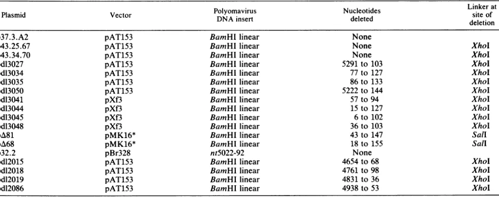
TABLE 1. Structuresof recombinant plasmidsused in large T-Antigen bindingstudies
Polyomavirus Nucleotides
~~~~~~~~Linker
atPlasmid Vector DNAinsert Nucleotides siteof
deletion
p37.3.A2 pAT153 BamHI linear None
p43.25.67 pAT153 BamHI linear None XhoI
p43.34.70 pAT153 BamHI linear None Xhol
pd13027 pAT153 BamHIlinear 5291to103 Xhol
pdl3034 pAT153 BamHllinear 77to127 XhoI
pdl3035 pAT153 BamHIlinear 86to 133 XhoI
pdl3050 pAT153 BamHI linear 5222to 144 XhoI
pdl3041 pXf3 BamHIlinear 57to94 XhoI
pdl3044 pXf3 BamHIlinear 15to 127 XhoI
pd13045 pXf3 BamHIlinear 6 to102 XhoI
pdl3048 pXf3 BamHI linear 36to 103 XhoI
pA81 pMK16* BamHIlinear 43to 147 Sall
pA68 pMK16* BamHIlinear 18to 155 Sall
p32.2 pBr328 nt5022-92 None
pdl2015 pAT153 BamHIlinear 4654to68 XhoI
pdl2018 pAT153 BamHllinear 4761to98 XhoI
pdl2019 pAT153 BamHI linear 4831to36 XhoI
pdl2086 pAT153 BamHI linear 4938to53 Xhol
J. VIROL.
on November 10, 2019 by guest
http://jvi.asm.org/
wereidentified by agarose gel electrophoresis and
quantitat-edbyexcision ofthegelbands and Cerenkovcounting.The
radioactivity recovered in corresponding fragments excised from marker lanes servedas normalization standards.
DNase I footprinting. Protection from partial DNase I
digestionof labeled DNAfragmentsboundbypolyomavirus
large T antigen was examined by a modification of the method of Galas and Schmitz (12). Known amounts of 5' end-labeled DNAfragment (1 x
10-3
to2x10-3
pmol) wereincubated with between 1 and 20 pmol (based on the
monomeric molecular weight) of partially purified large T
antigen in a final volume of 100 ,ul with DB buffer (20 mM
Tris-hydrochloride [pH 7.0],0.1 M NaCl,2 mM dithiothrei-tol, 1 mM EDTA, 0.01% [wt/vol] bovine serum albumin) at
20°C per30 min.PartialDNase I digestionwasdone bythe
A. p37.3.A2 p37.3.A2 PyA3 PyA3
Bam +
Bams-Bgll+Sac HinfI Bgll+Sac Hinfl
12
3
41
51
6'M Pabs aT M PabsoT M Pabs M Pabs
ban'
U...
-41
3
ar-b
A2
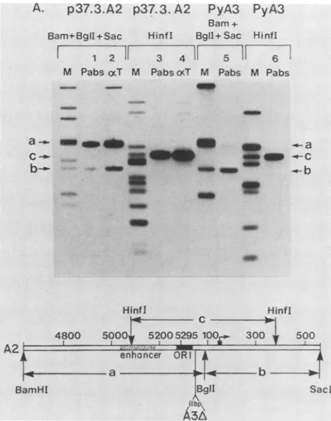
FIG. 2. Specific immunoprecipitation ofpolyomavirus ORI re-gionDNA. 32P-labeledrestriction endonucleasedigests (5to10ng,
ca10-3pmol)wereincubated withcell extract from 2 x10⁶infected
3T6cellsasdescribed in thetext. Lanes 1 and 2show immunopreci-pitation froma combined BamHI-BgI-SacI digest of theplasmid p37.3.A2 witheither cxPyLT1 plus aPyLT4monoclonal antibodies
orwithanti-tumorserum.Lane 5shows thesamedigestwithstrain
A3 DNA after immunoprecipitation with cxPyLT1 and aPyLT4.
Lanes 3, 4, and 6 show the results ofimmunoprecipitation froma
Hinfldigestof eitherp37.3.A2orA3DNA,respectively,witheither
aPyLT1 plus aPyLT4 orantitumor serum. The diagramindicates
the positions of the Hinfl fragment c and the BamHI-BglI-SacI fragmentsaandb withrespectto eachother and thepolyomarvirus regulatory region. The position ofthe 11-bpdeletion in A3 strain
DNAismarked(53).Thelanes labeled Mcomprise1/50 of theinput
DNA used as size markers and to aid quantitation. Note that the
fragmentaband in themarker lanes of theBamHI-BglI-Sacldigest comigrates with another fragment and therefore appears to be disproportionatelyintense.
addition ofMgCl2 to 5 mM, CaCl2 to 1 mM , and DNase I (Miles-Yeda) to a final concentration of 5 ng/ml and incuba-tion for a further 10min at 20°C. The reaction was terminat-ed by the addition ofan equal volume of 2 M ammonium acetate-100 mM EDTA-100 ,ug of sheared salmon sperm DNA per ml. Thesampleswereextracted with TE-saturated phenol and precipitated from ethanol. They were then dis-solved in formamidedye mix (33) and run on 8% polyacryl-amide-urea denaturing gels. The gels were dried before autoradiography.
RESULTS
Specific immunoprecipitation ofpolyomavirus largeT
anti-genbound to regulatory region DNAdemonstrates twotight
bindingsites.Our initialexperimentsused as a sourceof the
viral largeTantigenextractsprepared from 3T6 mouse cells infected with wild-type virus. These studies demonstrated highly specific DNA interactions, approximated the loca-tions of two high-affinity binding sites, and provided an assay to assess the activity of the protein through the
purification used for subsequent experimentation.
The
immunoprecipitation
method developed by McKay(35) allowed us to examine the DNA-binding activity in unfractionatedextracts. Inourapplication of this procedure mixtures of radiolabeled DNA fragments were incubated with crude lysates of infected cells. The assay was made
highlyselective forspecific DNA-protein interactions by the
addition of nonradioactive carrier DNA (5 ,ug of salmon sperm DNA per 5 to 10 ng of radiolabeled restriction
fragments) and 100 mM NaClto the bindingbufferused by
McKay (35). Thosefragments bound tothe largeT antigen
were
immunoprecipitated
by reaction with either polyclonalantitumorserumor amixture oftwodifferentratanti-largeT
antigen
monoclonalantibodies(7), followed by adsorptiontofixed S. aureus bacteria. The labeled DNA eluted from the washed immunocomplexes was identified by agarose gel
electrophoresis andautoradiography.
Figure
2 shows the results obtained with two differentrestriction endonuclease digests of recombinant plasmid
p37.3.A2 (Fig. 1), which contains fulllength viral A2 strain
DNA cloned at the BamHI site into plasmid pAT153. The
unique
DNAfragment
immunoprecipitated from the Hinfldigestion (band
cinFig.
2)byeitherantitumorserum(lane4)ormonoclonal antibodies(lane 3)spanstheenhancer region,
ORI,and the early regionpromoter(Fig. 2).Noother DNA
fragments
weredetectably immunoprecipitated
(the adjacentlane M shows theinputDNAdigest). We therefore conclud-ed that the
large
Tantigen
binds withhigh
specificity
toone or more sequences within the regulatory region of thepolyomavirus
genome. Thesingle high-affinity binding
sitelocalizedby Gaudrayetal. (13), withadifferentprocedure,
isincludedwithin DNAfragment c.
Totestthehypothesis proposed by Gaudrayetal.(13) and
Kamen et al. (25) that
polyomavirus
large T antigen bindsseparately
tosequencesatthetranscriptional
initiation sitesas well as to those in the ORI region, we digested plasmid
p37.3.A2
with theendonuclease BgIl, which cleavespolyo-mavirus DNAat asingle site between the ORIand theearly
region
capsites,
plusBamHIandSacl. Twofragmentswereimmunoprecipitated
fromthis digest (Fig. 2,lanes 1and2):fragment
a, which includes ORI, and fragment b, whichspansthe TATA box and the cap sites.Fragmenta wasmore
efficiently immunoprecipitated
thanfragmentb. By excisionof the
gel
bands and Cerenkov counting the molar ratio betweenfragments
a and b was determined to beapproxi-mately
4:1.The regulatory regionthus contains atleasttwoon November 10, 2019 by guest
http://jvi.asm.org/
[image:4.612.52.291.247.551.2]754 COWIE AND KAMEN
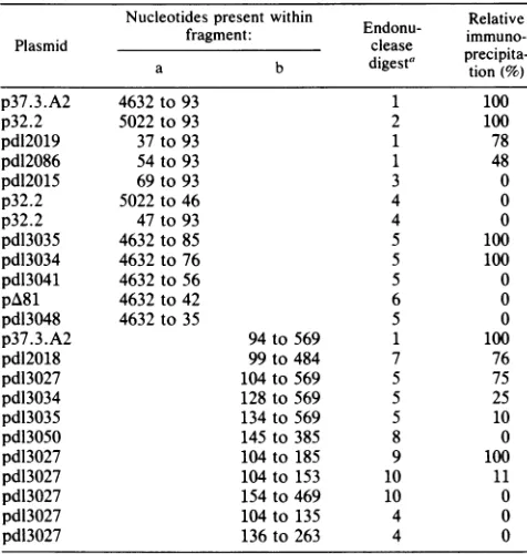
TABLE 2. Mapping of the large T antigen high affinity binding regions with deletion mutants and endonuclease cleavage
Nucleotidespresentwithin Relative
Plasmid fragment: Endonu-
immuno-a b digesta precipita-tion(%)
p37.3.A2 4632 to 93 1 100
p32.2 5022 to 93 2 100
pdl2019 37to 93 1 78
pdl2086 54 to 93 1 48
pdl2015 69 to 93 3 0
p32.2 5022 to 46 4 0
p32.2 47 to 93 4 0
pdl3035 4632 to 85 5 100
pdl3034 4632 to 76 5 100
pdl3041 4632 to 56 5 0
pA81 4632 to 42 6 0
pdl3048 4632 to 35 5 0
p37.3.A2 94 to 569 1 100
pdl2018 99 to 484 7 76
pdl3027 104 to 569 5 75
pdl3034 128 to 569 5 25
pdl3035 134 to 569 5 10
pdl3050 145 to 385 8 0
pdl3027 104 to 185 9 100
pdl3027 104 to 153 10 11
pdl3027 154 to 469 10 0
pdl3027 104 to 135 4 0
pdl3027 136 to 263 4 0
"Key to the endonuclease digestions. 1, BamHl 1-BglI-SacI; 2, Rsal-EcoRI; 3,BglI-PstI;4,MbolI;5,BamHI-XhoI-SacI;6,BamHI-Sall-SacI;7,
XhoI-PstI;8,HinI;9,DdeI;10,HphI.
independent large T antigen-binding sites, with apparently
different affinities, located on either side of the BgIl site.
From the experiment shown in Fig. 2 the binding region
withinfragment b, on the early side of the BgIl site, maps
between this position (nt 93) and the Hinfl site at nt 385.
Further analysis with a number of different restriction en-zymes anddeletion mutants(Tables 1 and 2) demonstrated
that all of the sequences important for binding within
frag-ment b are located between nt 103 and nt 188. This DNA
spans the early region TATA box and cap sites (2, 25).
Truncation ofthe DNAfragmentattheprincipalcapsite(nt
153)by cleavage with endonucleaseHphI reduced the
bind-ing by 10-fold (Table 2).
Results were also obtained with polyomavirus strain A3
DNA. This strain differs from strain A2 DNA by an 11-bp
deletion (nt 44 through 55) (53), removing one of the three
AGAGGC repeats adjacenttotheORI (Fig. 1and2). Hinfl
fragment c was efficiently immunoprecipitated from the
digest of strain A3 DNA (Fig. 2, lane 6), but in the
BgII-BamHI-SacI digest, fragment a binding was only 8%
effi-cientrelativetothestrain A2fragment. BindingtoA3 strain
fragment b was at the same efficiency as observed for A2
DNA (Fig. 2, lane 5). The difference between A2 and A3
DNAsindicatedto usthat thebinding regionwithinfragment
a involves the deleted sequences and suggested that the
number of AGAGGC hexanucleotide sequences within the
binding region (two in strain A3 DNA versus three in A2)
influencesthe binding affinity when measured bythe
selec-tive immunoprecipitation assay. Our data further implied
thatthe strongbindingsite located withinfragmenta was not
importantfor viability because strains A2 and A3 are both
wild type and grow equally well in culture.
Origin-proximal binding sitemapsoutside theORI region.
The data presented in Fig. 2 suggest that the high-affinity
binding site within fragment a might not include the ORI
sequence. We define the ORI as one of the two elements necessary in cis for viral DNA replication (32, 36, 63). It has been mapped by deletion analysis to a minimal core se-quence of ca. 70 bp between a late boundary at nt 5265
through5269 (32) and anearly boundaryat nt36through 42
(26). The DNA sequencefrom nt 42 to 64 plays a quantitative role (its removal reduces replicatoryefficiencyto 10 to20%
ofwild-type level),but it is not essential for DNA replication
(26; our unpublished observations). The second element
essential for DNA replication is noncontiguous and occurs
within the enhancer element (32, 36, 63).
Tobetterlocalizethebinding regionwithinfragmentaand
to ask more explicity whether large T antigen binds to the
ORI, we tested a series of restriction endonuclease frag-ments extending from the BamHI site (nt 4632) to various
endpointswithinfragment a.These werederived from a set
of deletionmutants(Table1) with XhoI orSalllinkers at the
site ofdeletion.Theresults(Fig.3) demonstrate the absence of detectablebindinguntil thefragment extends well past the ORI to nt 76. Thelarge Tantigen did not bind to the DNA
fragments, which includecompetentreplicationorigins such
asthosewithendpointsat nt 42 or nt56.Thiseffectis not an
artifact causedby the use of restriction endonuclease
frag-ments with ends near the essential region. A
deletion-insertionmutantin whichpolyomavirus DNAfromnt43 to
147 wasreplaced withaportion ofthe mouse
metallothion-ine gene (G. M. Veldman, unpublished results) did not contain any large T antigen-binding sites detectable by
immunoprecipitation (data not shown), yet thisDNA
repli-cated at 10 to
20%
ofthe wild-type level after transfectioninto permissive mousecells.
The experimentsto locate thebinding regionwithin
frag-ment a were extended to include a number of additional
restriction fragments derived from wild-type or deletion
mutant DNAs (Table 2). We concluded that there are no sequencesessential orsufficient forhigh-affinityDNA
bind-ing before nt 36 or after nt 76. This would imply that the
DNA ip2,i3027indiMi;)5pdi;3044P|dt3048p37 A2 end poi nit52891 nt5 nt 15 nt35 Al 92
M T M T M T M T M T
b _-4 _ _
am - _ _
4632 5100 5200
~~~~~
Barnr
- --
-'--P&8. Dd304id 304.diO35 77 At
nt42 nt56 nt76 nl86 Pt92 M TM TI|M TISM T,M T
m-* * *@*i..-_ _a.
_ - _ _ tll;- 3 t
5289 55 354256 768692
[image:5.612.327.566.472.617.2]g1A II
...
. ..-?Rt~
e~~~gI
L._ -_
FIG. 3. Mappingthe regionoffragmenta towhichthelarge T
antigenbinds. Deletionmutantswith endpointswithinfragment a
(Table 1)were cleaved withBamHI, Sacl, andeitherBglI inthe case of p37.3.A2, XhoI for pdI3027, pdI3045, pdI3044, pdI3048,
pd13041,pdI3034,andpdI3035,orSallforpA81tocleave the linkers
atthe deletionsites. After incubationwith infectedcellextractand
immunoprecipitation with polyclonal antibodies atPyLT1 and aPyLT4, the resultant DNA fragments were separated on 2% agarose gelswith 1/50 ofthe radiolabeled DNAdigests as marker tracks.Thediagramshowsthe deletionendpointsinfragmentawith respect to ORI. The repeated pentanucleotide sequence
(G/T)(AIG)GGCisindicatedby the half-arrows.
J. VIROL.
on November 10, 2019 by guest
http://jvi.asm.org/
Hif I Pvull Pvufl Dde I
1
E strand
L strandl
L strand2
5295 BglI
5000 51Ot 5200 3 1?0 200 300 400
major , ORI L_earlycops
late cops
FIG. 4. DNAfragments usedintheDNase Ifootprinting studies. TheDNAfragments usedinFig. 5, 6, and 7 are shownaligned with various restriction endonuclease sitesandthe following structural features oftheviralDNA:ORI, earlyRNA capsites,and late RNA capsites. Viral DNA or p37.3.A2recombinant DNAwasused for the E-strand fragment. Arecombinantplasmid (p43.34.70) that has anXhoIlinker inserted into thePvuII siteat nt5265wasused forthe L-strand'fragment.Asimilarplasmid (p43.25.67)has thePvulI site atnt 5128 replaced with an XhoI linker and was used for the
L-strand2 fragment. Theposition ofthe5' 32p label is markedby an asterisk.
binding region is contained within these limitsandtherefore overlapstheearly boundaryof the ORI corebyno more than afew base pairs.
Purification of polyomavirus large T antigen. DNA
frag-mentimmunoprecipitation isa sensitive technique for study
ofa DNA-binding protein present in crude cell extracts.
When combined with a sufficiently extensive set ofDNA
deletion mutants, it can be used, as illustrated above, to
approximate the positions of specific, independent
DNA-binding regions.Weconsideredtheseassignmentsas
prelim-inary, however,because the method does notdirectly
identi-fythe DNA sequences contacted bytheproteininwild-type
DNA. Wewerealsounableto study potentially cooperative
binding that could extend into the ORI. A satisfactory
understanding ofthelarge Tantigen-DNA interactionwould
only be possible iftheproteinwerepurified. Itwas ourgood
fortune at this time to enter into a collaboration with S.
Dilworth to evaluate an immunoaffinity purification
proce-dure he had developed by using monoclonal antibodies
against polyomavirus large T antigen (7). We demonstrate
elsewhere (6) that hissingle-step immunoaffinity purification
yields, from wild-type virus-infected 3T6cells, protein30 to
50% pure which retains its DNA-binding specificity. We have now modified the procedure to reduce frequent
con-tamination with a protein, presumed to be actin, that
inter-fered with the DNase I footprint analysis. Details of the
purification steps aredescribed above.
Detection ofthree high-affinity binding regions byDNaseI
footprinting. With the availability ofpurified protein, albeit
in small quantities, we were able to apply the footprinting
procedure of Galas and Schmitz (12) to study the binding
interactions more definitively. The
32P-labeled
DNAfrag-ments usedforthefootprint studies areillustrated in Fig.4.
Weincubated the DNA withincreasing amounts ofpurified
large T antigen in a fixed final reaction volume. A typical
saturationpattern isshown inFig. 5. Three distinctregions
of the DNA, designated A, B, and C, were protected from DNase I digestionwith as little as 1 pmol (5 ,ulofasolution
ofca. 20
jig/ml)
oflarge Tantigen monomer.Alignmentof the protected regions withthe marker lanes
and the Aand Gsequencing track allowed precise localiza-tion with respecttothe DNA sequence (see Fig. 8). Region
Ais adjacent to the replication origin, overlapping slightly
the ORIearly boundary (Fig. 1). Region B spans thesingle
BglIsiteat nt93 andends justbeforethe TATAbox,where
there are a few base pairs unprotected. Region C extends from the TATAsequence to halfwaybetween theearlycap sites (nt 148 through 153 [32]) and the ATG of the coding region (nt 173).
The positions of binding regions A and C agree verywell with the conclusions derived from DNA immunoprecipita-tion mapping experiment. Binding of large T antigen to region B, however, could not be demonstrated by the indirect assay method. In repeated efforts, we were unable to immunoprecipitate an endonuclease MboII fragment (nt 48through 133), which includesall ofregionB. The signifi-cance of thisdiscrepancybetweenfootprintand immunopre-cipitation results is considered below.
The prominent regions of DNaseI protection (A through C) manifest in Fig. 5 confirmed that the high-affinity binding sites for polyomavirus large Tantigen occur outside of the ORI core sequence.Nevertheless, inspectionof data such as thatpresented for the DNA E strand in Fig.5suggestedthat the T antigen was also perturbing the DNase I digestion pattern in the ORI region, but that this required higher
E ST RAN D L STRAND
A ..L_T ....A
+
G02 5 020 50M 02 5IO0 205OG
3Z
2C
IE
AL
BL
C
b.
a
s~~~~~~~~Z_
qw
_~~~~~~~~~~~~~~~~~~~"I
_~~~~~~~~~~~e c
8
LA
FIG. 5. Binding of polyomaviruslarge Tantigentothe noncod-ing region of the genome. To localize the binding sites of largeT
antigentoboth E- and L-DNA strands,approximately lo-3 pmol of 5'-32P-labeled restriction endonuclease fragments (E strand and L strand' from Fig. 4) were incubated with increasing amounts of partiallypurifiedlarge T antigen in a total volumeof100,ul of DB bufferasdescribed in the text. After DNase Idigestionthesamples weresubjected to denaturing gel electrophoresis in an8% polyacryl-amide-urea sequencinggel. The A+G sequencing reaction (33) of each fragment was used as a marker track together with a size marker.
I
-.4- -- ---I
---Am
I
.T.0 wlwn.go-- . ...ft"M.-M.
.1-aft 11"- ..
on November 10, 2019 by guest
http://jvi.asm.org/
[image:6.612.55.292.75.162.2] [image:6.612.323.543.298.624.2]756 COWIE AND KAMEN
",t-_
L:4
A
_w
_wop.
i_ _
a
.
4i-L
STRAND
A
T C M O T C G
]
IB
B
A
2
.7. :
--W*l
_~~~~~~~~~I E
[image:7.612.86.284.75.506.2]~~~~~~~~~.48t:14
...
__
binding reaction to the maximum level technically feasible
(Fig. 6). We also used the longer5'
32P-labeled
fragment for the DNA L-strand probe(Fig. 4). To show that the protec-tion was specific for the presence of T antigen, control DNase Idigestions were done in parallel in thebinding bufferand thelarge Tantigenstorage buffer (see abovefor details).
As aresult,Tantigen-specific effects, in addition to
protec-tion ofregionsA, B,andC,wererevealed(Fig.6).
Consider-ing first the E-strand DNA, we note above region A an enhanced cleavage siteand then aregion ofprotection. As this protected region is interrupted by a site of enhanced digestion, we tentatively define twoadjacent binding sites, 1 and 2. Above region 2, some 50 bp removed, we note a
further region of DNase Iprotection, indicated in Fig. 6 as
region3. Protection ofregions 1 and 2was lessobvious on
the DNA L strand, but region 3 was apparent, as was a marked region of enhanced DNase I digestion between regions 2 and 3 (denoted by E in Fig.6). Theinteractions at
regions 1 and 2 map around nt 5295/1, withintheORI core
sequence(cf. Fig. 1and8).Region3 isonthelatesideofthe
ORI(around nt5220; see Fig. 8).
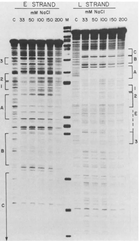
We next tried varying the NaCl concentration in the
binding reaction.Allprevious experiments had been carried
out at0.1 M NaCl. Figure 7 shows the results obtained by
usingthe same amount of large T antigen in each reaction,
butincreasing the NaClconcentration from33 mMto0.2 M. At33 and 50 mMNaCltheprotectionatsites 1, 2,and 3was moreclearly visible on both DNAstrandsthanat100mM,as wastheregion of enhanceddigestion(E)between 2 and 3. At 150 mMNaClnoprotection of sites 1, 2,and 3wasdetected,
but regions A, B and C were unchanged. The regions
protectedatlowionicstrength map tothe same
positions
atthose shown at high protein concentration, i.e., on either
sideofthent5295/1andaroundnt5220. Thedependence of
theprotection of sites 1,2,and 3onhighprotein
concentra-tion or low ionic strength may indicate that the large T
antigenhas asignificantly loweraffinity forthese sites than
forregionsA, B, andC,which couldexplain why theywere
not detected in the immunoprecipitation assay. At present, however, wecannotruleoutthealternative
explanation
thataminorsubset ofT-antigen moleculesbindsto
regions
1,2,
and 3.
FIG. 6. Binding of large T antigen at high concentration. To localize the binding sites of large T antigen, approximately 10-3 pmol of 5'-32P-labeled DNA fragments (E strand and L strand2 in Fig. 4)wereincubated with 100,ul (ca.20pmolofmonomer) largeT antigenatroomtemperaturefor 30min.After DNase Idigestionthe resultant fragments were resolved on an 8% urea-polyacrylamide denaturinggel. Size markers and theA+Gcleavage reaction(33)of each fragment were run as marker lanes. Lane 0 represents the
reaction inthepresence of100 ,u of DB buffer, laneT showsthe reaction with 100 pLI of large T antigen, and lane C shows the
reaction with100,ul of largeT storage buffer(seethetextfor buffer compositions).
concentrations ofprotein. The putative additional binding sitesareindicatedas1, 2,and 3 inFig.5forconsistencywith conclusions derived from the subsequent experiments de-scribed below.
Detection of additional binding regions with ORI and the enhancer sequences by DNase I footprinting. To further
investigatethe interaction between large T antigen and the ORI region, we increased the amount of protein in the
DISCUSSION
We have presented results that define multiple interac-tions between polyomavirus large T antigen and the viral genome.Theproteinis knowntobe involved in theinitiation of eachsuccessive round of viral DNAreplicationand in the
repression of early transcription (46) and, by analogy to
SV40 large T antigen, could also play a direct role in the
augmentation of late transcription (28). Specific DNA
bind-ing perhapsmediates each of these activities. Ouraimwasto
identifythe DNA sequencesrecognized bytheproteinwith theanticipationthatlocalization ofbindingsiteswithrespect tootherregulatory signalswould suggest their function.
Indirect mapping experiments withcrudeprotein
prepara-tions. The study ofpolyomavirus large T antigen has been
impeded bytechnical difficulties encounteredin its purifica-tion. It was nevertheless possible to examine its
DNA-bindingproperties bytheapplicationoftwodifferent indirect
approaches.The earliestreport(13) usedanovelmethod in
which theTantigenpresentin rather crudepreparationswas
absorbed to double-stranded calf thymus DNA-cellulose.
PolyomavirusTantigenATPaseactivitycouldbe specifical-ly elutedby incubation of the DNA-cellulosewithplasmids
E
STRAND
A GO0
80
L
C
J.VIROL.
.I...
111:1Tl..,
m
on November 10, 2019 by guest
http://jvi.asm.org/
BINDING SITES FOR POLYOMAVIRUS LARGE T ANTIGEN 757
E
STRAN D
mM NaCI
L
STRAND
mM NaCI
C 33 50 100 150 200 M C 33 50 100 150 200
_
__*11#s
_iaIME--r9~_ _ mm
21- _ _ ...~~~ino
r *
o *
_~~~~~~~~ow
_C
]
B]A
13
method, but itwasfound thatsequences onbothsidesof the high-affinity site contributed to the binding s-trength (P.
Gaudray and F. Cuzin, personal communication).
Several laboratories subsequently used the McKay (35)
DNA immunoprecipitation technique to study the DNA-binding properties of large T antigen in unfractionated ex-tracts. Our results, reported above, localized two
indepen-dent binding sites for the large T antigen (Table 2; Fig.2and 3). A higher-affinity site in strain A2 DNA was included between nt 37 and nt 76. It did not overlap the replication
origin by more than a few nucleotides. A lower-affinity site, located between nt 104 and nt 188 (Fig.1and8),spannedthe TATA box and theinitiation sites for early region transcrip-tion andcontained three TGAGGChexanucleotides andone AGAGGChexanucleotide. This site isanobvious candidate
3 2
A. a 2
6209 5119 6229 5259 5249 529 SN 52?19
L AA9AG59A9C AAAAAOCCTC TCCACCCA9O CCTAGAATQTTTCCACCCAATCATTACTATrACAAC5ACT9TTTTTrrTA E TTCTCCTTCOrTTTTCGASAGO T6O7TCCSATCTTACAAA9OTO9O9T A9TAATOATACTgTIr9COACAAMAAT
1 A
5269 I10 CT o 50 40 , OQ_ 60
L STATrAAGCA GAUCC Go9o cCCT9GOCtCCTTA cTcT99AGAAAAAGAAUAAGOCATTITAGA9GC'rrCCA9
E CATAATTCOT
5/
CTCC9O~~~~~~GA
CCCCCGO9OACC COAAT9A6AOTCTTTTTCTTCTCT CtGTAACATC TCCOAA2TC70 so 90 100 II0 120 1J0 140
L ASOCAACTTOTCAAAACAGG ACTGGCOCCTTOG29COCT GTSGOCCACCCAAATT9AT ATAATTAC CCCAACCOCC
E TOCTTOAACAGrTT6OTCCTSACCoCoGAACCTCCGCO CACCCCGSTG GeTTrAACTATATTAATTC6 OOGTTOOCOG
C
100 160 170 I80 190 200
L TCTTCCCOCC TCATrTCAOCCTCACCACCATCATOGATAGAOTrCTGA2C AGAICTOACA E AG6A9OCO9 AGTAGTCOGA6TGTGOGTAGTACCTATCTCAAGACTCGTCTCGACTGT
m
B. PvuE Pou Hpal BglI POLYOMA
3 2 H A B C
WJ. .-En hancer- - - - .TEiymN
[image:8.612.53.296.75.492.2]fiHomology
FIG. 7. Effect ofNaCl concentration on large T antigen DNA binding. The effect of NaCl concentrationonlargeTantigen binding was determined byincubating 1 x 10-' to2 x 10' pmol of 5'-32P-labeledDNAfragments (Fig. 4) with 40,ul(ca. 8pmol of monomer) ofpartially purified large T antigen ina total volumeof 100 ,ulat differentNaCIconcentrationsasshown. After DNase Idigestion the sampleswere run on an8%polyacrylamide-urea denaturing gel. The control lanes, marked C, werecarried out at 100 mMNaCl in the absenceoflarge T antigen. Because the change in NaCl
concentra-tion also affected the rate of DNase I digestion, the amount of
enzyme wasvaried between 2.5ng/ml and 33 mM and 10 ng/ml at 200 mMtoobtainequivalent degress of digestion.
containing the viral regulatory region. Guadray et al. (13) wereabletomeasurethe affinityof the protein for its binding site andtoapproximate the genomic location of this site by use of several different restriction endonucleases and dele-tionmutants.Inthis waytheyidentifiedasingle high-affinity binding region immediately adjacent tothe core replication originontheearly side. They suggested thattwoof the three copies of the hexanucleotide AGAGGC repeated within the binding region may be required for high-affinity interaction. No further independent binding sites were detected bythis
m E I
72 72 21 21 r
-Ennhancer -- A
BglI Hind m
[image:8.612.312.552.241.516.2]SV40
FIG. 8. Diagrammatic representation of the polyomavirus large Tantigen-binding sites. (A) The DNAsequenceof the polyomavirus regulatory region fromnt5200to200(numberingsystemof Soedaet al.[54] asmodified by Tyndalletal. [63]). The publishedsequence of strainA2polyomavirus DNA (53) contains threeerrorsin theORI region. The Aat nt5isactuallyaG, andtwobasepairs (CT)mustbe inserted between nt 13 and nt 14. These changes eliminate the differences between the A2 and A3 ORI sequence (9). To avoid confusion between the numbering system employed in previous
papers and the work presented here, we have refrained from
adjusting the numbering of the sequence to include the additional dinucleotide. Instead we represent the additional basesabove and below the sequenceshown in thisfigure. Theareas protected from DNase I digestion by the large T antigen are bracketed. The pentanucleotide repeats 5'-(G/T)(A/G)GGC-3' aremarked with
ar-rows.TheORIcoreextends fromnt5269tobetweennt36andnt42
(26, 32). (B) The top line is a diagram of the binding sites of
polyomavirus large T antigen shown with respect tothe functional elementsof theregulatory region: the earlypromoter.The ORIcore
sequencesand the enhancer. Thebottom line isasimilardiagram of
SV40aligned withpolyomavirusattheORI homology. The position oftheearly RNAstartsitesand the enhancersequencesareshown,
with arrows indicating the 72-bp and 21-bp repeats (taken from
reference 37). C
.=19
VOL.52, 1984
on November 10, 2019 by guest
http://jvi.asm.org/
758 COWIE AND KAMEN
for the interaction mediating repression of transcription from the majorearly cap sites.
In strain A2 DNA, the higher-affinity site was the origin-proximal site. It was immunoprecipitated about four times moreefficiently than the origin-distal site. Strain A3, a wild-type variant of polyomavirus has an 11-bp deletion within the origin-proximal T antigen-binding site which removes oneof the three AGAGGC sequences(53). This reducedthe apparent binding affinity by more than 10-fold and led us to speculate that at least three repeats must be present in a
binding site for efficient immunoprecipitation under the
highly selective conditions used.
The DNA immunprecipitation results we describe confirm and extend related reports (19, 41). Pomerantz et al. (41),
usinganA3-typestrain, found two binding sites and reached
similar conclusions for the location of the origin-distal site,
although they failed to note the 10-fold reduction in binding efficiency caused by HphI cleavage at nt 153. They assumed that the origin-proximal site included the replication origin, but did not present supporting evidence. It is clear from Fig. 3 and Table 2 that the ORI core sequence neither has high affinity for polyomavirus large T nor contributes significant-ly to the binding strength we measure for the adjacent site.
One could postulate that binding of large T antigen to the
originis dependent on simultaneous occupation of the
origin-proximal high-affinity site, but because recombinant
plas-mids lacking both of these T antigen binding sites replicate (26; our unpublished results), such cooperative binding, if it occurs, cannot be essential.
DNase I footprinting experiments. The availability of monoclonalantibodies directed against polyomavirus large T
antigenallowed us to purify enough of the protein from
wild-type virus-infected 3T6 cells for direct analysis of DNA
binding by DNase I footprinting. The antibody used for
immunoaffinityfractionation, cxPyLT1, reacts with all of the
largeTantigen detectable in polyomavirus-infected cells (7). It is not known whether polyomavirus large T antigen
oligomerizes in vivo or whether the forms of the protein that
have been previously identified (7, 61) have different DNA
binding activities. By using the cxPyLT1 monoclonal
anti-body, the risk of enriching for particular subspecies was
minimized.
Our initial footprint analyses (Fig. 5) demonstrated three
closely spaced, but noncontiguous, regions of DNase I
protection (designated A, B, and C in Fig. 8). The lengths of
theprotected regions were approximately 43, 35, and 38 bp, respectively. The locations of regions A and C agree with the deductions made from the DNA immunoprecipitation
ex-periments.Region B, which spans theBglI site at nt 93, was
not detectable in the indirect assay. We also note that
footprint
saturation experiments such as that shown in Fig. 5indicated nearly equivalent protection of all three sites,
whereas irnmunoprecipitation data implied a much greater affinity of T antigen for region A than for region C. These
discrepancies suggest that the footprint experiments were
relatively insensitive to differences in binding affinity
be-causeof the high reactant concentrations employed. In addition we detected protection of three other regions, identified as 1, 2, and 3 in Fig. 8. Regions 1 and 2 together span most of the ORI core sequence. These studies have thusafforded the first evidence for an interaction between T antigen and the sequence necessary for DNA replication. Region 3 is near sequences known to have enhancer func-tion. Approximately 10 times more T antigen was required to protectregions 1, 2, and 3 than to saturate regions A, B, and
C (Fig. 5), and this protection was highly sensitive to ionic
strength in the 33 to 150 mM range (Fig. 7). This may suggest that a minor fraction of the T antigen molecules bind to regions 1, 2, and 3, that the affinity for these sites is very low, or that a combination of both hypotheses pretains.
The division of the protected region between approximate-ly nt 5260 and nt 10 into two subregions (1 and 2 in Fig. 8) is based on the hypersensitivity of the sequence around the nt
5295/1junction to DNase I when the rest of the region was
protected (Fig. 6, E-DNA strand). Region 1 is far smaller than the other protected regions and therefore we suspect that region 1 plus 2 together comprise a single domain of
interaction. Further work, using more concentrated T anti-gen preparations and methods such as dimethyl sulfate
protection for footprinting should resolve this issue. Consensus recognition sequences. Repeated
pentanucleo-tides of the consensus family 5'-(G>T)(A>G)GGC-3' are
distributed in the three regions ofSV40 DNA contacted by SV40 large T antigen (4, 56-58) and are thought to represent recognition sequences. Soeda et al. (53) originally noted that the related hexanucleotide, AGAGGC, is repeated
numer-ous times near the polyomavirus origin. These have since
been implicated in polyomavirus large T antigen binding (13, 41). As noted in Fig. 1 and shown more clearly in Fig. 8, there are in fact 15 copies of the SV40 consensus pentanu-cleotide sequence arranged in five clusters of two to four tandem repeats within the 250 bp spanning thepolyomavirus replication origin. The regions protected from DNase I digestion by large T antigen coincide with these clusters.
The three strong binding regions A, B, and C contain, respectively, 3, 2, and 4 pentanucleotides. The homologies in these regions extend to six bases, the consensus being 5'-(A=T)G(A>G)GGC. The repeat intervals are all between 9 and 11 bp, indicating that the recognition sequences are accessible on the same side of the double helix. Although it is likely that sequences outside the putative recognition hexanucleotide are also important in T-antigen binding, the frequency with which the repeat occurs at similar spacingis most remarkable.
A consensus polyomavirus repeat of 5-'G(A>G)GGC can be deduced from the comparison of all binding regions. It is apparent that the strong binding regions differ from the weaker ones in both the number and the length of the conserved sequence and in the spacing of the repeats. All of the results we have obtained by DNA immunoprecipitation can be understood under the hypothesis that a threefold repeat spaced at one turn of the helix is optimal for high-affinity binding. The different arrangement of therecognition sequences within regions 1 plus 2 and in region 3 may imply a different mode of interaction between the protein and the DNA.
Implications for papovavirus large T antigen function.One of the several significant differences between the genome organization of murine polyomaviruses and their simian counterpart, SV40, is the displacement of the polyomavirus early region promoterfrom the core ORI(Fig. 8). Theearly region cap sites and TATA box are included within theSV40 ORI sequence (14), andDNA essential foreffective invitro transcription lies immediately to the late side of the ORI (16, 37, 44). Bycontrast, theprincipal polyomavirus earlyregion
TATA box and cap sites are some 80 bp on the early sideof the ORI, withinDNA sequence not homologously represent-ed in the SV40 genome. The sequences at the polyomavirus ORI, although very similar to the SV40 sequence, are not transcriptionally active in vitro, and indeed no specific
sequence upstream of the TATA box is important for polyomavirus early transcription in vitro(23).
J. VIROL.
on November 10, 2019 by guest
http://jvi.asm.org/
The large T antigens of polyomavirus and SV40 both repress early region transcription and activate DNA replica-tion. SV40 large T antigen recognizes three adjacentregions
spanning its ORI, called I, II, and III (Fig. 8), which bind
noncooperativelyand with different affinities in the order of
I > II > III (55, 57, 58, 60). In vitro experiments have
suggested that SV40 large T antigen-binding regions I and
possiblyII areinvolved in the repression of early
transcrip-tion, which initiates within region II (17, 44). Considerable
genetic evidence demonstrates that the DNA within SV40
region II is essential for viral DNA replication, whereas
sequenceswithin regionI has a quantitative, if nonessential,
role (8, 34, 35, 38, 49-51).
Coincident with the displacement of the polyomavirus
early promoterfromthe ORI is a major shift in the location
of high-affinity large Tantigen-bindingregions. Region A of
polyomavirus occurs at a position precisely analogous to
SV40region I and also appears to be the highest-affinity site, at least in strain A2 DNA. Sequences within polyomavirus
region A, like that comprising SV40 region I, influence the
quantitative efficiency of DNA replication, but are not
essential. Regions B and C, unlike SV40 II and III, extend
toward and across the early transcriptional regulatory
se-quencesrather than in the replication ORI direction (Fig. 8).
The location ofthemajorbinding regions strongly suggests
that these are primarily involved in transcriptional
regula-tion.
We have detectedpolyomavirus T antigen binding to the
ORI (region 1 and 2), at a position very similar to that of
SV40 region II, but this interaction is either much weaker
thanthattoregions A, B, and C or is the property of a small
subset of large T-antigen molecules. Of the polyomavirus
largeTantigen-binding regions,only 1 and 2 occur within the
sequence essential forDNA replication. We therefore sus-pect that the minor interaction we have detected is that
which is principally involved in the replication initiation
function of the protein. This topological segregation of
functional binding sites distinguishes polyomavirus from
SV40 and should prove useful in the further study of the
multifunctional interaction between the viralprotein and its
genome.
The bindingatregion3occurs outside the ORI core within
theviral enhancerregion, but within asequencethat can be
deleted with no phenotypic consequence (32). Region 3,
however, includes the sequence mutated and duplicated in
polyomavirus variants able to grow in theembryonal
carci-nomacell lineF9 (10, 27, 47). Furthermorethissequence has
been shown to be necessary in cis for both viral DNA
replicationandearly gene expressionin embryonal
carcino-macells(11, 31). Future experimentswill test whether large
T antigen binding at region 3 is involved in enhancer
function,and whether region 3 plays a role in the activation
oflatergeneexpression.
ACKNOWLEDGMENTS
This work was initiated in the Transcription Laboratory of the ImperialCancerResearch Fund, London.
We aregratefultoSteve Dilworth and Furzana Chaudry for their advice and to Steve Dilworthfor themonoclonal antibodies used. Steve Lupton, Parmjit Jat, and Chiara Tyndall kindly provided deletionmutants. We would also like tothank PeterTegtmeyerand Angelo DeLucia, whose interest and comments stimulated our search for an ORIinteraction.
LITERATURE CITED
1. Birnboim, H. C., and J.Doly. 1979. A rapidalkaline extraction procedure for screening recombinant plasmid DNA. Nucleic
Acids Res. 7:1513-1523.
2. Cowie, A., P. Jat, and R. Kamen. 1982. Determination of sequences at the capped 5' ends of polyomavirus early region transcripts synthesized in vivo and in vitro demonstrates an unusual microheterogeneity. J. Mol. Biol. 159:225-255. 3. Cowie, A., C. Tyndall, and R. Kamen. 1981. Sequences at the
capped 5' ends of polyomavirus late region mRNAs: an example ofextreme terminal heterogeneity. Nucleic Acids Res. 9:6305-6322.
4. DeLucia, A. L., B. A. Lewton, R. Tjian, and P. Tegtmeyer. 1983. Topography of simian virus 40 A protein-DNA complexes: arrangement of pentanucleotide interaction sites at the origin of replication. J. Virol. 46:143-150.
5. deVillers, J., and W. Schaffner. 1981. A small segment of polyomavirus DNA enhances the expression of the clone ,B-globin gene over a distance of 1400 base pairs. Nucleic Acids Res.9:6251-6264.
6. Dilworth, S. M., A. Cowie, R. Kamen, and B. E. Griffin. 1984. DNA binding activity of polyomavirus large T antigen. Proc. Natl. Acad. Sci. U.S.A. 81:1941-1945.
7. Dilworth, S. M., and B. E. Griffin. 1982. Monoclonal antibodies against polyomavirus tumor antigens Proc. Natl. Acad. Sci. U.S.A.79:1959-1963.
8. DiMaio, D., and D. Nathans. 1982. Regulatory mutants of SV40. Effect ofmutations at aTantigen binding site on DNA replica-tion and expression of viral genes. J. Mol. Biol. 156:531-548. 9. Friedman, T., A. Esty, P. LaPorte, and D. Deininger. 1979. The
nucleotide sequence and genome organization of the polyoma early region: extensive nucleotide and amino homology with SV40. Cell 17:715-724.
10. Fujimura, F. K., P. L. Deininger, T. Friedmann, and E. Linney. 1981. Mutation near the polyoma DNA replication origin per-mitsproductive infection of F9 embryonal carcinoma cells. Cell 23:809-814.
11. Fujimura, F. K., and E. Linney. 1982. Polyoma mutants that productively infect F9 embryonal carcinoma cells do not rescue wildtype polyoma in F9 cells. Proc. Natl. Acad. Sci. U.S.A. 79:1479-1483.
12. Galas, D. J., and A. Schmitz. 1978. DNase footprinting: a simple method for detection ofprotein-DNA binding specificity. Nu-cleic Acids Res. 5:3157-3710.
13. Gaudray, P., C. Tyndall, R. Kamen, and F. Cuzin. 1981. The high affinity binding site on polyomavirus DNA for the viral large T antigen. Nucleic Acids Res. 21:5697-5710.
14. Ghosh, P. K., and P. Lebowitz. 1981. Simian virus 40 early mRNAs contain multiple 5' termini upstream and downstream from aHogness-Goldberg sequence; a shift in 5' termini during the lyticcycle ismediated by largeTantigen. J. Virol. 40:224-240.
15. Green, M. R., R. Treisman, and T. Maniatis. 1983. Transcrip-tional activation of cloned human ,B-globin genes by viral immediate-early gene products. Cell35:137-148.
16. Hansen, U., and P. Sharp. 1983. Sequences controlling in vitro transcription ofSV40promoters. EMBO J. 2:2293-2303. 17. Hansen, U., D. G. Tenen, D. M. Livingston, and P. A. Sharp.
1981. Tantigenrepression ofSV40 early transcription from two promoters. Cell 27:603-612.
18. Harlow, E., L. V. Crawford, D. C. Pim, and N. M. Williamson. 1981. Monoclonal antibodies specific for simian virus 40 tumor antigens. J. Virol. 39:861-869.
19. Hayday, A. C., F. Chaudry, and M. Fried. 1983. Loss of polyomavirus infectivity as a result of a single amino acid change ina region ofpolyomavirus large T antigen which has extensive amino acid homology with simian virus 40 large T antigen. J. Virol.45:693-699.
20. Hirt, B. 1967. Selective extraction of polyoma DNA from infected mouse cell cultures. J. Mol. Biol. 26:365-369. 21. Houweling, A., P. J. van denElsen, and A. J. van der Eb. 1980.
Partialtransformation of primary rat cells by the leftmost 4.5% fragment of adenovirus5DNA. Virology 105:537-550. 22. Imperiale, M. J., L. T. Feldman, and J. R. Nevins. 1983.
Activation of gene expression by adenovirus and herpes virus regulatory genes acting in trans and by acis-acting adenovirus enhancer element. Cell 35:127-136.
on November 10, 2019 by guest
http://jvi.asm.org/
760 COWIE AND KAMEN
23. Jat, P., U. Novak, A. Cowie, C. Tyndall, and R. Kamen. 1982. DNA sequences required for specific and efficient initiation of transcription at the polyomavirus early promoter. Mol. Cell. Biol. 2:737-751.
24. Jones, K. A., and T. Tjian. 1984. Essential contact residues within SV40large Tantigen binding sites I andI1 identified by
alkylation-interference. Cell 36:155-162.
25. Kamen, R., P.Jat, R. Treisman, J. Favaloro, and W. R. Folk. 1982. 5' termini ofpolyomavirus early region transcripts synthe-sized in viivo by wild type andviable deletion mutants. J. Mol. Biol. 159:189-224.
26. Katinka, M., and M. Yaniv. 1983. DNA replication origin of
polyomavirus: early proximal boundary. J. Virol.47:244-248. 27. Katinka, M., M. Yaniv, M. Vasseur, and D. Biangy. 1980.
Expression of polyoma early functions in mouse embryonal carcinoma cells depends upon sequence rearrangements in the
beginningofthe late region. Cell 20:393-399.
28. Keller, J. M., andJ. C. Alwine. 1984. Activation ofthe simian virus 40late promoter: direct effect ofTantigen in the absence ofviral DNAreplication. Cell36:381-389.
29. Land, H.,L. F. Parada, and R.A.Weinberg.1983.Tumorigenic conversion ofprimary embryo fibroblasts requires atleast two
cooperatingoncogenes. Nature (London) 304:596-602. 30. Lewton, B.A., A. L.DeLucia, and P. Tegtmeyer. 1984. Binding
of simianvirusAproteintoDNAwithdeletionsat theorigin of
replication. J.Virol. 49:9-13.
31. Linney, E.,andS.Donerly. 1983. DNAfragmentsfrom F9 PyEC mutantsincreaseexpression ofheterologous genesin transfect-ed F9cells. Cell 35:693-699.
32. Luthman, H.,M.G.Nilsson, and G. Magnusson. 1982. Noncon-tinuous segments ofthe polyoma genome required in cis for DNAreplication. J.Mol. Biol. 161:533-550.
33. Maxam, A., and W. Gilbert. 1980. Sequencing end-labelled DNAwith basespecificchemicalcleavages.MethodsEnzymol. 65:499-560.
34. McKay, R.,and D.DiMaio.1981. Binding ofanSV40T antigen-related proteintothe DNAofSV40regulatorymutants.Nature
(London)289:810-813.
35. McKay, R. D. G. 1981. Binding ofan SV40T antigen related
proteintoDNA. J. Mol. Biol. 145:471-488.
36. Muller,W.J.,C. R.Mueller,A.-M.Mes,andJ.A.Hassel. 1983.
Polyoma virus origin for DNA replication comprises multiple geneticelements. J. Virol. 47:586-599.
37. Myers, R. M., D. C. Rio, A. K. Robbins, and R. Tjian. 1981. SV40gene expressionis modulatedbythecooperative binding ofT-antigen toDNA. Cell 25:373-384.
38. Myers,R.M.,and R.Tjian.1980. Construction andanalysisof SV40originsdefectiveintumor bindingand DNA replication. Proc. Natl. Acad. Sci. U.S.A. 77:6491-6495.
39. Nevins, J. R. 1981. Mechanism of activation of early viral
transcriptionbytheadenovirusElA geneproduct. Cell 26:213-220.
40. O'Farrell, P. 1981. Replacement synthesis method of labelling DNAfragments. Bethesda Research LaboratoriesFocus3:1. 41. Pomerantz, B. J., C. R. Mueller, and J. A. Hassel. 1983.
PolyomaviruslargeTantigen binds independentlytomultiple.
unique regionson the viralgenome. J. Virol. 47:600-610. 42. Rassoulzadegan, M., A. Cowie, A. Carr, N. Glaichenhaus, R.
Kamen, and F. Cuzin. 1982. The roles ofindividual polyoma-virusearlyproteinsinoncogenic transformation. Nature
(Lon-don)300:713-718.
43. Rassoulzadegan, M., Z. Naghashfar, A. Cowie, M. Grisoni, R. Kamen,and F.Cuzin. 1983. Expressionof thelargeTantigenof
polyomavirus promotes the establishment in culture of "nor-mal rodentfibroblast cell lines. Proc. Natl. Acad.Sci. U.S.A. 80:4354-4358.
44. Rio, D. C., and R. Tjian. 1983. SV40 T antigen binding site mutations thataffect autoregulation. Cell 32:1227-1240. 45. Ruley,H. E. 1983.Adenovirusearlyregion1A enables viral and
cellular transforming genes to transform primary cells in cul-ture. Nature (London) 304:602-606.
46. Schaffhausen,B.1982. Transforminggenesand geneproducts of polyomaand SV40. Crit. Rev. Biochem. 13:215-286.
47. Sekikawa, K., and A. J. Levine. 1981. Isolation and character-ization of polyoma hostrange mutants that replicate in
nullipo-tential embryonal carcinoma cells. Proc. Natl. Acad. Sci. U.S.A. 78:1100-1104.
48. Shiroki, K., H. Handa, H. Shimojo, S. Yano, S.Ojima, and K. Fukinaga. 1977. Establishment and characterization ofrat cell lines transformed by restriction endonuclease fragments of adenovirus 12 DNA. Virology 82:462-471.
49. Shortle, D., and D. Nathans. 1978. Local mutagenesis; a method forgeneratingviralmutantswith basesubstitutions in
preselect-ed regions of the viral genome. Proc. Natl. Acad. Sci. U.S.A. 75:2170-2174.
50. Shortle,D., and D. Nathans. 1979. Regulatory mutantsofsimian virus 40 constructed mutants with base substitutions at the origin ofDNA replication. J. Mol. Biol. 131:801-817.
51. Shortle,D., and D. Nathans. 1979. MutantsofSV40with base substitutions at the origin of DNA replication. Cold Spring
HarborSymp. Quant. Biol. 43:663-668.
52. Soberon, X., L. Covarrubias, and F. Bolivar. 1980.Construction and characterization ofnewcloning vehicles. Gene 9:287-305. 53. Soeda, E., J. R. Arrand, N. Smolar, and B. E. Griffin. 1979.
Sequence from early region ofpolyomavirus DNAcontaining viral replication origin and encoding small, middle and (partof) large T antigens.Cell 17:357-370.
54. Soeda, E., J. R. Arrand, N. Smolar, J. E. Walsh, and B. E. Griffin. 1980. Coding potential and regulatory signals of the polyomavirus genome. Nature (London) 283:445-453. 55. Tegtmeyer, P., B. Anderson, S. B. Shaw, and V. G. Wilson.1981.
Alternative interactions of the SV40 A protein with DNA. Virology 115:75-87.
56. Tegtmeyer, P., B. A. Lewton, A. L. DeLucia, V. G. Wilson, and K.Ryder. 1983.TopographyofSV40Aprotein-DNA complex-es: arrangementof protein bound totheorigin ofreplication. J. Virol. 46:151-161.
57. Tenen, D. G., L. L. Haines, and D. M. Livingston.1982.Binding ofananalogueofthesimian virus40 Tantigentowild-typeand mutantviral replicationorigins. J. Mol. Biol. 157:473-492. 58. Tenen, D. G., T. S. Taylor, L. L. Haines, M. K. Bradley,R.G.
Martin, and D. M. Livingston. 1983. Binding of simianvirus40 large T antigen from virus infected monkey cells to wild-type and mutantviral replicationorigins. J. Mol. Biol. 168:791-808. 59. Tjian, R. 1978. The bindingsite ofSV40DNAfor aT
antigen-related protein.Cell 13:165-179.
60. Tjian, R. 1981. Regulation of viral transcription and DNA replication by theSV40large T antigen. Curr.Top. Microbiol. Immunol. 93:5-24.
61. Turler, H., and C. Solomon. 1977. Characterization ofpolyoma Tantigens. INSERM 69:131-144.
62. Twigg, A. J., and D. Sherrat. 1980. Trans-complementable copy-number mutants of plasmid ColEl. Nature (London)
283:216-218.
63. Tyndall, C., G. Lamantia, C. M. Thacker, J. Favaloro,and R. Kamen. 1981. A region of the polyomavirus genome between the replication origin and late protein coding sequences is required in cis for both early gene expression and viral DNA replication. NucleicAcids Res. 9:6231-6250.
64. Weber, F., J. deVilliers, and W. Schaffner. 1984. An SV40 "enhancer trap" incorporates exogenous enhancers or gener-ates enhancersfromits own sequences. Cell 36:983-992.
J. VIROL.