JOURNAL OFVIROLOGY,
JUlY
1991, Vol.65, No. 0022-538X/91/073906-05$02.00/0Copyright C)1991,American Society forMicrobiology
The Sequence
of
Human
Immunodeficiency Virus Type 2
Circle
Junction
Suggests That
Integration
Protein Cleaves the Ends of
Linear DNA Asymmetrically
JEANNETTE M. WHITCOMB AND STEPHEN H. HUGHES*
ABL-BasicResearch Program, National Cancer Institute-Frederick Cancer Research andDevelopmentCenter, P.O. BoxB, Frederick, Maryland 21702-1201
Received 28 January1991/Accepted2April1991
The sequence of the human immunodeficiency virus type 2 circle junction was determined. The most commonsequencefoundbetween theconserved CA and TGdinucleotides at the ends of theintegrated provirus wasfive bases long(GGTAC).Thissuggeststhat theintegration of humanimmunodeficiencyvirustype2 DNA is accompanied by theasymmetric loss oftwoandthreebases, respectively, from the U3 and U5 ends of the linear double-stranded DNApriortointegration.
Akeystep inthe early stage oftheretrovirallifecycle is
the reverse transcription ofthe viral RNA genome into a
linear double-stranded DNA (dsDNA) molecule and the
integration of this linear dsDNA into the host genome (30, 33).These two events, unique toretrovirusesand retrotrans-posons, should be excellent targets for interrupting the retroviral life cycle. Understanding the molecular basis of
these steps in the replication of human immunodeficiency
virus (HIV) may be of great importance in the design of successfultherapies againstAIDS.
The generation ofthe linear dsDNA moleculeby reverse
transcription involves a complex series of events (30, 33). The structuresof the viral RNA, the linear dsDNA, and the
integratedproviral DNA are shown diagrammatically in Fig. 1. The reverse transcription reaction generates terminally redundant sequences known as long terminal repeats (LTRs). Theinitiation of plus- and minus-strand DNA syn-thesis atdiscrete sites within the RNA genome defines the
boundariesof the LTRs (Fig. 1). Minus-strand synthesis is
initiatedfrom a cellular tRNA primer that is annealed to a
specific complementary primer-binding site (PBS) located
immediately adjacenttothe U5 sequence. Plus-strand DNA synthesis is initiated from an RNA primer that is generated
byaspecific RNase H cleavage of the RNA genome within a polypurine tract (PPT) adjacent to U3. The most widely accepted model of reverse transcription dictates that the
sites of initiation of both plus- and minus-strand synthesis
precisely define the ends of the unintegrated linear dsDNA
molecule.Morespecifically, the first DNA base added to the tRNA primer defines the end of the U5 region in the right LTR and the subsequent removal of this tRNA primer by RNase H exactly defines the right end of the linear dsDNA
molecule. The left end of the linear dsDNA molecule is
defined bytwoRNase H events; the first generates the RNA
primer that initiates plus-strand synthesis, and the second removes the RNA primer from the left end of the DNA
molecule.
Following reverse transcription, the linear dsDNA
mole-cule, which is bounded by LTRs, is transported to the
nucleus,where itis the precursor to the integrated provirus.
*
Corresponding
author.The linear molecule also gives rise in the nucleus to two circular forms of DNA that contain eitherone or twoLTRs. Thebasicmechanismof retroviral integrationisemerging
fromin vivo and invitrostudies of several retroviruses(1-4, 7-12, 16, 26). It is now clear that the linear viral DNA
molecule, and not one ofthe circular forms, is the direct precursor tothe integrated provirus(1, 2, 8, 19). A virally
encoded product, integration protein (IN), recognizes se-quences atthe ends of the linear molecule and removes a
specific numberofbases, usually two, from each of the 3' endsofthelinear viralDNA(Fig. 1) (7,10, 16, 17, 31).The IN thenjoins the recessed viral 3' ends to the 5' ends ofa double-stranded staggered cutin the host DNA. This inter-mediate is repaired, presumably by host enzymes. The
resulting integrated provirus is shorter than the linear dsDNA molecule by the number of bases removed by IN
priorto integration (Fig. 1).
Studies with various mutant forms of retroviruses have shown that the terminal 15 to 20 bases ateach end ofthe
linearDNAareimportant forcorrectintegration to occur (3, 6,17, 21, 26). However,thespecificity ofthecleavageevent
byINisnotcompletelyunderstood. Tofullyunderstand the
integration reaction, it is important to define the exact substrate for IN (i.e., the ends ofthe linear dsDNA mole-cule).
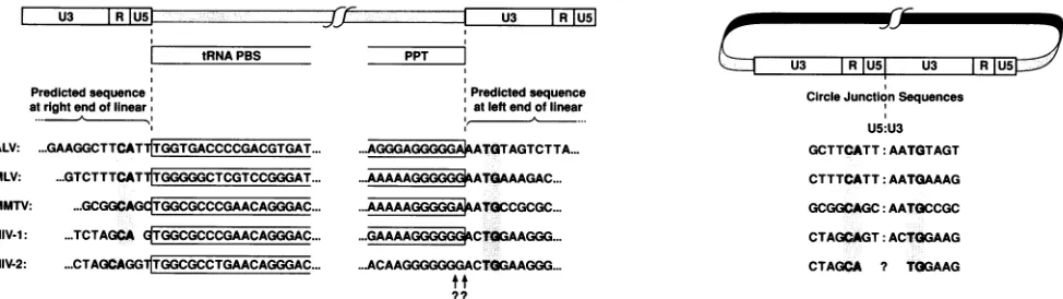
Figure 2 compares the sequences of the genome/LTR boundaries of a number of retroviruses. Examination of
these sequences should allow us to predict the sequences presentatthe endsofthelinearsubstratefor integration.For
example,theright end of the linear dsDNA molecule should
contain the U5 sequences adjacent to the PBS(Fig. 2, box). It shouldalso bepossibletopredictthe bases removed by IN
prior to integration. All known integrated proviruses are boundedontheleft(U3) endbyaTG dinucleotide andonthe
right(U5) endby aCAdinucleotide(Fig. 2, shaded boxes). The bases presentbetween the tRNA primer (boxed) and the CAdinucleotide(shaded)should be thebases removed from
the right (U5) end prior to integration. A similar analysis should be possible for the left (U3) end of the molecule.
However, the precise site of RNase H cleavage which defines the plus-strand primeris not knownfor all viruses. Where the site has been determined, it is always found within the PPT,twobases from the TGdinucleotide (Fig. 2,
box) (20, 23, 25).
3906
on November 10, 2019 by guest
http://jvi.asm.org/
GpppR
UPT I
U3
RlAn
PBS~~
;")PPT
4RT
LTR LTR
U3 R
|U5|U3
JR
I
U51
C
/(U3
IRU5H
tj3
U3
I
RIU5|
U3
I
RIU5Ei-'
D
I
U3I
R
U5:
U3 RIU51
IN
LI
U3 IR U5zzzlz
(,iz U3 IRIU5LH
IN
F
[image:2.612.96.512.73.584.2]A U3 IR
U5U3
R
|U5
FIG. 1. Outlineof the reverse transcription andintegration of the retroviralgenome. The single-stranded RNA (ssRNA)genome (A) resemblesacellular mRNAin thatit hasa3'poly(A)tail
(A.)
anda5'cap(Gppp).Thegenomecontainsshortrepeatsequences(R)andunique3'and 5' sequencesateach end(U3andU5). The PPT and thetRNAPBS,whereplus-and minus-strand DNAsynthesisisinitiated,are
shown.Followingreversetranscription,thedsDNA(BandD)isboundedbyLTRsconsistingofU3,R, and U5sequences. In the nucleus
thelineardsDNAmoleculegivesriseto twocircular forms of dsDNA(C)containingoneandtwoLTRs. IN cleavesaspecificnumber of
bases,usuallytwo,fromeach 3' end of the linear dsDNA(E).The recessed 3'endsarecovalentlyattachedtothe5'ends ofadouble-stranded
staggeredcutinthe host DNA. After DNArepair,theintegrated provirus (F)isshorterthan itsdouble-strandedlinearprecursorand is flanked
byashortdirectrepeatofhost DNA. Additional detailsaregiveninthetext(seealsoreferences 30and33).
Additionalinformationthatcanbeusedtopredictthe ends of the linear DNA comes from analysis of the junction
sequences in the two LTR circles found in the nucleus. Circles with two LTRs appear to be formed by blunt-end ligationof the lineardsDNA molecule(28, 30, 33).Whether
this ligation is catalyzed by cellular or viral enzymes is unclear. However, the bases thatare lostfromthe ends of the linear viral DNA during integration are present at the
circlejunctioninthecircles with two LTRs.
We, and others, have reported previously on the
se-A
B
E
on November 10, 2019 by guest
http://jvi.asm.org/
U3 R
tRNAPBS
Predicted sequence'
atrightend oflinear
ALV: ...GAAGGCTTCAT TGGTGACCCCGACGTGAT... MLV: ...
GTCTTTCATfTGGGGGCTCGTCCGGGAT
MMTV: ...GCGGCAG TGGCGCCCGAACAGGGAC
HIV-1: ...TCTAGGA
qTGGCGCCCGAACAGGGAC
HIV-2: ...CTAGCAGGlTGGCGCCTGAACAGGGAC
:7L U3 RJU5
PPT
Predictedsequence
atleftendoflinear
...AGGGAGGGGG*ATGTAGTCTTA...
...AAAAAGGGGG kATGAAAGAC...
..AAAAAGGGGG*A*tCCGCGC...
...GAAAAG=*C!TGAAGGG...
..ACAAGGGGGGGACTGGAAGGG...
?t
-~~~~~~~~~~
U3
IRIU5I
U3 IRCircle JunctionSequences U5:U3 GCTTCATT:AATGTAGT CTTTCATT:AATGIAAAG GCGGCAGC:AATGCCGC CTAGCAGT:ACTGGAAG CTAGCA ? TGGAAG
FIG. 2. Comparison of the viralgenome/LTR boundariesandpredictedcirclejunctionsequencesof several retroviruses. Roussarcoma virus (Pr-Cstrain) (ALV),Moloney murine leukemia virus(MLV),andmousemammarytumorvirus(MMTV)sequencesarefromreference
33. HIV-1sequenceis fromreferences24,27, and32.HIV-2 sequenceis from reference 13.The tRNAPBS andthePPTareshowninopen boxes. TheconservedCA/TG dinucleotides foundattheboundaries of theintegrated provirusareshown inshaded boxes. The site ofRNase
Hcleavage of thePPTthatproducestheplus-strandprimer isnot known forHIV-2. Thetwo mostlikelysitesaredesignatedbyquestion
marks. Ourdata(seeFig. 3)suggest thatthecleavage site will be betweentheG andtheA.
quencesfound at the circlejunctions of human
immunode-ficiency virus type1 (HIV-1)circles with twoLTRs(15, 18, 22, 29, 34). These data suggest thatintegration is
accompa-nied by the loss oftwo bases from each end, as for other
retroviruses. This was contrary to whatwaspredictedfrom cloned HIV-1proviral sequences (Fig. 2) (24, 27, 32). Most retroviruses have twobasesbetween the tRNA PBS(which
ends GGT) and the CAdinucleotide found atthe end of the
integrated provirus. HIV-1 has only one base at this posi-tion. However, as we and others have shown, two bases
fromthe U5 sideare presentin HIV-1 circlejunctions (15, 18, 22, 29, 34), suggesting that the information containedin
terminalAof the tRNAprimer is found at therightend of the
linearDNA molecule. We also presented evidence
suggest-ingthat the terminal ribo-A remains attached to the dsDNA
molecule because the RNase H of HIV-1 reverse
tran-scriptase does not remove the last A ofthe tRNA primer (34). These results led us to postulate that there was selec-tive pressure that caused HIV-1, and perhaps all retrovi-ruses, tomaintainasubstrate forintegrationfrom which two bases could be removedsymmetrically fromeach endbyIN. However, the sequences of HIV-2 proviruses (13) suggest that retroviral INs may not always act symmetrically. As shown inFig.2, there are threebases between the end of the PBS and the conserved CA found at the right U5 boundary of the HIV-2provirus. Even if HIV-2 RNase H removes the
entiretRNAprimer,three bases would be removed from the U5 endbyINprior tointegration.Thereis no simple rule to
predict how many bases will be lost from the U3 end. By
analogy with other retroviruses, it could be predicted that twobases should be removed from the U3 side in
prepara-tion forintegration (Fig. 2). Our results with HIV-1 suggest that HIV-1 IN acts symmetrically on both ends of the linear molecule. If this rule of symmetry is also true for HIV-2, three bases might be removed from the U3 side as well. The
primerused forinitiation of plus-strand synthesis in HIV-2 has not been precisely defined, and, depending on the precise site of RNase H cleavage, it is possible that either twoorthreebases (AC or GAC) would be removed from the U3 side of the linear molecule prior to integration. This wouldpredict that five or six extra bases could be present at the circle junction of the circles containing two LTRs in
HIV-2-infected cells.
HIV-2 (strain NIH-Z)-infected CEM cells harvested 24
and 48 h postinfection were kindly provided by David Waters. DNAwaspreparedbythe method of Hirt(14), and thecirclejunctionsequences were amplifiedby using
prim-ers in U3 and U5 (Fig. 3). Primer sequences containing
EcoRI sites are as follows: U5, GCCGAATTCGGTCAT TCGGTGTTCACCTG; U3, GCCGAATTCGTGTAGTTCT GCCAATCTGC. The polymerase chain reaction (PCR)was performed asfollows. The final reaction mixture contained 50mM Tris chloride(pH 8.3); 50 mM KCl; 7 mM MgCI2; 170 ,ug of bovine serum albumin per ml; 1 mM each dATP,
dCTP, dGTP, and dTTP; 1 ,uM each primers U3 and U5;4% dimethyl sulfoxide; 1
RI
of Hirt lysate; and 2 U of Taq polymerase (United States Biochemical Corp., Cleveland, Ohio) in a total volume of 25 pI. Thirty cycles were per-formedby usingaPerkin-Elmer Cetus DNA thermalcyclerwith astep-cycle file programmed for 89°C for 40 s and 59°C for 80 s. Theproducts of the amplification werefractionated on a2% low-melting-temperature agarose gel and visualized bystaining with ethidium bromide. TheHIV-2-specific band (190 bp) was excised, extracted from the gel, anddigested with EcoRI. The EcoRIfragmentcontaining the circle junc-tion was ligated intoM13mpl8 and used totransform com-petent Escherichia coliDH5a F' (Bethesda Research
Labo-ratories). M13 clones with inserts were processed and sequenced byfollowing the Sequenase DNASequencing Kit protocol (United States Biochemical Corp.).
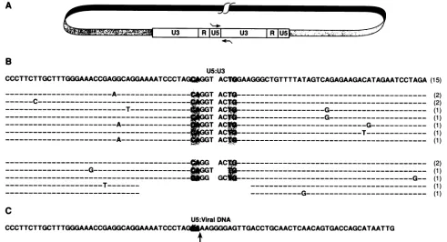
We sequenced 30 circlejunction clones; their sequences aresummarized inFig. 3B. Themajority of the clones (24 of 30) contain five extra bases (GGTAC), three bases from U5 and two bases from U3. Wealso foundseveral variant circle junction clones. One type of clone lacked the terminal T from the U5 side, andanotherlacked the terminal two bases (AC)fromthe U3side. This type of aberrant circle junction has beendescribedpreviously in reports on the sequence of HIV-1circlejunctions (18, 29). In two clones, seven nucle-otides from U3 and 15 nucleotides from U5 were missing
fromthe circlejunction. We alsofound one clone in which the terminal base was lostfrom each end concomitant with the insertion of one extra base. It is not clear from the sequenceexactlyhow this circlejunctionwasformed. None of thecloneswe sequenced contained sixextrabasesatthe circle junction, as would be expected if three bases were removed symmetrically from each end of the linear dsDNA prior to integration of HIV-2. The data also allow us to
on November 10, 2019 by guest
http://jvi.asm.org/
[image:3.612.71.558.72.209.2]U3 RU5 U3 RU5K
CCCTTCTTGCTTTGGGAAACCGAGGCAGGAAAATCCCTAG*G
---____-______----A---C
---
-
---C-____ __ __
ACT
C
U5:U3
GT ACTGAAGGGCTGTTTTATAGTCAGAGAAGACATAGAATCCTAGA(15) GGT
ACTQ---3GT
ACTG---
TACN7A-G---T A
---G---3GT AC --- G ________
GT AC* ---T---~GT AC
---(2) (2)
(1) (1) (1) (1) (1)
(2)
(1) (1) (1) (1)
CCCTTCTTGCTTTGGGAAACCGAGGCAGGA
U5:ViralDNA
JAATCCCTAG&AAGGGGAGTTGACCTGCAACTCAACAGTGACCAGCATAATTG
tFIG. 3. (A) Positions of primers (indicated bythe arrows) in U3 and U5 usedtoamplify the circle junction sequencesof HIV-2. (B)
Sequences of HIV-2 circle junction clones. TheconservedCA and TG dinucleotides foundat the end of integrated HIV-2provirusesare
shown inshaded boxes. Thesequencefoundmostoften isshowninthetopline. The number in parenthesestothe right of each clone is the
number of clonesseenwith eachsequence.Sequence identity is indicatedby dashed lines. Point mutations in individual clonesareindicated.
(C) Sequence ofaclone representing the right end ofanautointegrationevent.The U5 boundary is indicated byan arrow.
predict the site of RNase H cleavage in the PPT of HIV-2. Thecleavagemusttake place between the G and theA,two
bases from theconserved TG foundatthe U3 boundary of theintegrated HIV-2 provirus.
The 30 HIV-2 circle junction clones reported here were
isolated following a single round of PCR amplification (30 cycles) with Hirt supernatant DNA as the substrate. We
believe that most ofthe sequence diversity seen in these
clones reflects differences in the population of circular HIV-2 DNAs used as a substrate for the amplification reaction. Thisdiversity probably aroseduringreverse
tran-scription of the retroviral DNA, althoughwecannotruleout the possibility that some of the sequence differences were
introduced during PCR amplification. Therearetwoclones thathaveanidentical deletion in U3 and U5, and it islikely
that these clones derive from the amplification ofa single
circlejunction.
In addition to the circle junction clones, we report the
sequences of clones derived from the amplification ofone
end ofan autointegration event (Fig. 3C). When we
orig-inally attemptedtoclone the HIV-2 circlejunction,wechose
PCRprimersthat didnotinclude EcoRI sites. Our attempts to clone the blunt-end fragments generated by PCR were
unsuccessful (only one clone was isolated that contained HIV-2 circlejunction sequences). However, whenasample
of this material was used as asubstrate for a second PCR with primers containing EcoRI sites, the amplified product
was easily cloned intoM13. Many of theclones, however, did not contain HIV-2 circle junctions but instead were
found to represent one boundary from an autointegration
event (Fig. 3C). These clones contain U5 sequences up to the CA residues found at the boundary of a normal
inte-grated provirus. Examination of the adjacent sequences
shows that the siteofintegration isintheenvregionatbase 7468. We were able to clone the PCR products from this integration event because a downstream sequence (bases
7557to7577)shareshomologywiththe U3primer chosenfor PCR (an 18- of21-base match). PCR with the U3 primer annealed in this position gives rise to a 210-bp fragment
which was cloned along with the 190-bp circle
junction-specific fragment.
These clones represent the right end ofaprovirus
inte-grated into a target DNA, showing that integration in this particularstrain isaccompanied byaloss of three bases from US. Ten clones were sequenced, all of which we assume were amplified from a single autointegrant. There are no sequencedifferences in these 10 clones in the regionshown in Fig. 3C. However, two of the clones have independent point mutations outside this region. These clones were
isolatedfollowingatotal of 60cyclesofPCR,and it islikely thatthese mutationswereintroducedbytheTaq polymerase during amplification. Therelative lack ofsequencediversity
amongthese clonesarguesthat the conditionswehave used
for PCR donotgenerate sufficient mutations to account for thediversityof circlejunction sequenceswehave isolated.
We know of no other naturally occurring retrovirus in
which the INremoves adifferent number of bases fromthe 3' and 5' ends priortointegrationof theprovirus. Goff and coworkers have constructed and analyzed a number of
mutant Moloney murine leukemia viruses with changes at
therightend of US(5, 6, 26).Some ofthese mutationsresult
inU5 ends thatare similartothe endsof HIV-1and HIV-2
with respectto spacing and basecontent. Moloney murine
leukemia virus mutants containing only one base between
the PBS and the end oftheprovirusand mutantscontaining four basesatthisposition replicate reasonablywell. It is still
A
C
B
:;
NWAlk
'A
\-40.
on November 10, 2019 by guest
http://jvi.asm.org/
[image:4.612.69.560.63.329.2]3910 NOTES
unclear why HIV-1 uses a specific RNase H cleavage to generate a linear DNA substrate forintegration with sym-metric ends, whereas HIV-2 apparentlygenerates an asym-metric substrate for integration. AlthoughHIV-1 andHIV-2 are related viruses, the specificity of the RNase H and IN activities of these two viruses may differ. We are now attempting to use the specific substrates and enzymes in experiments to discover preciselywhat determines the spec-ificity of these two reactions in HIV-1and HIV-2.
We aregratefultoDavidWatersandLarryArthurfor thegift of HIV-2-infected CEM cells, toMarilynPowersfor the oligonucleo-tides, andtoHildaMarusiodisfortyping themanuscript.
ResearchwassponsoredbytheNationalCancerInstitute, under contractNO1-CO-74101 with ABL.
REFERENCES
1. Brown, P. O., B. Bowerman, H. E. Varmus, and J. M.Bishop. 1987. Correct integration of retroviral DNA in vitro. Cell 49:347-356.
2. Brown, P.O., B. Bowerman, H. E. Varmus, and J. M. Bishop. 1989. Retroviral integration: structure of the initial covalent productandits precursor, and a role for the viral INprotein. Proc. Natl. Acad. Sci. USA86:2525-2529.
3. Bushman, F. D., and R. Craigie. 1990. Sequence requirements forintegration of Moloney murine leukemia virusDNAin vitro. J. Virol. 64:5645-5648.
4. Bushman, F. D., T. Fujiwara, and R. Craigie. 1990. Retroviral DNAintegration directed by HIV integration protein in vitro. Science249:1555-1558.
5. Colicelli, J., and S. P. Goff. 1985. Mutantsandpseudorevertants of Moloney murine leukemia virus with alterations at the integration site. Cell42:573-580.
6. Colicelli,J., and S. P. Goff.1988.Sequenceandspacing require-mentsofaretrovirus integration site. J.Mol. Biol. 199:47-59. 7. Craigie,R., T.Fujiwara, and F. Bushman. 1990. The INprotein
of Moloney murine leukemia virus processes the viral DNA ends and accomplishes their integrationin vitro. Cell 62:829-837.
8. Ellis, J., and A. Bernstein. 1989. Retrovirus vectors containing aninternal attachment site: evidence that circlesare not inter-mediates to murine retrovirus integration. J. Virol.
63:2844-2846.
9. Ellison, V., H. Abrams, T. Roe, J. Lifson, and P. Brown. 1990. Human immunodeficiency virus integration ina cell-free sys-tem.J. Virol. 64:2711-2715.
10. Fujiwara, T., and R. Craigie. 1989.Integration of mini-retroviral DNA: acell-free reaction for biochemical analysis of retroviral integration. Proc. Natl. Acad. Sci. USA 86:3065-3069. 11. Fujiwara, T., and K. Mizuuchi. 1988. Retroviral DNA
integra-tion: structure of anintegration intermediate. Cell 54:497-504. 12. Goff,S.P. 1990. Integration of retroviral DNA into the genome
of theinfected cell. CancerCells2:172-178.
13. Guyader, M., M. Emerman, P. Sonigo, F. Clavel, L. Montagnier, andM. Alizon. 1987. Genome organization and transactivation of the humanimmunodeficiencyvirustype 2. Nature (London) 326:662-669.
14. Hirt, B. 1967. Selective extraction of polyoma DNA from infectedmousecellcultures. J. Mol. Biol. 26:365-371. 15. Hong, T., K.Drlica, A.Pinter, and E.Murphy. 1991. Circular
DNA of human immunodeficiency virus: analysis of circle junction nucleotide sequences. J. Virol.65:551-555.
16. Katz, R.A.,G.Merkel, J. Kulkosky, J.Leis,and A. M. Skalka. 1990. The avian retroviral IN protein is both necessary and sufficient forintegrative recombination in vitro. Cell 63:87-95.
17. Katzman,M., R. A. Katz, A. M. Skalka, and J. Leis. 1989. The avian retroviral integration protein cleaves the terminal
se-quencesof linear viral DNAatthe in vivo sites ofintegration.J. Virol. 63:5319-5327.
18. Kulkosky, J., R. A. Katz, and A. M. Skalka. 1990. Terminal nucleotides of the pre-integrative linear form of HIV-1 DNA deduced from the sequence of circular DNA junctions. J. Acquired Immune Defic. Syndr. 3:852-858.
19. Lobel, L. I., J. E.Murphy, and S. P. Goff. 1989. The palindro-mic LTR-LTRjunctionofMoloneymurine leukemia virus is not anefficient substrate forproviral integration. J. Virol. 63:2629-2637.
20. Luo, G. X., L. Sharmeen, and J. Taylor. 1990. Specificities involvedin theinitiationofretroviral plus-strand DNA.J. Virol. 64:592-597.
21. Panganiban, A. T., and H. M. Temin. 1983. The terminal nucleotides of retrovirus DNA arerequired for integration but notvirusproduction. Nature (London) 306:155-160.
22. Pauza, C. D. 1990. Two basesare deleted from the terminiof HIV-1 linearDNA during integrative recombination. Virology 179:886-889.
23. Pullen, K. A., and J. J. Champoux. 1990. Plus-strand origin for humanimmunodeficiency virus type 1: implications for integra-tion.J. Virol. 64:6274-6277.
24. Ratner, L., W. Haseltine, R.Patarca, K.J. Livak, B. Starich, S. F.Josephs, E. R. Doran, J. A. Rafalski, E. A. Whitehorn, K. Baumeister, L. Ivanoff, S. R. Petteway, Jr., M. L. Pearson, J. A. Lautenberger, T. S. Papas, J. Ghrayeb, N. T. Chang, R. C. Gallo,and F. Wong-Staal. 1985. Completenucleotidesequence of the AIDSvirus, HTLV-III. Nature (London) 313:277-294. 25. Rattray, A. J., and J. J. Champoux. 1989. Plus-strandpriming
by Moloney murine leukemia virus: the sequence features importantforcleavage byRNase H. J.Mol. Biol.208:445-456. 26. Roth, M. J., P. L.Schwartzberg,andS. P.Goff. 1989. Structure of the termini of DNA intermediates in the integration of retroviral DNA:dependenceonINfunctionandterminal DNA sequence. Cell 58:47-54.
27. Sanchez-Pescador, R., M. D. Power, P. J. Barr, K. S. Steimer, M. M.Stempien,S. L.Brown-Shimer, W. W. Gee, A. Renard, A. Randolph, J. A. Levy, D. Dina, and P. A. Luciw. 1985. Nucle-otide sequence andexpressionofanAIDS-associatedretrovirus (ARV-2). Science227:484-492.
28. Shank, P. R., and H. E. Varmus. 1978. Virus-specificDNA in the cytoplasmof aviansarcomavirus-infected cells isa precur-sor tocovalently closed circular viral DNA in the nucleus. J. Virol. 25:104-114.
29. Smith, J. S., S. Kim, and M. J. Roth. 1990. Analysis oflong terminal repeat circlejunctions of human immunodeficiency virustype 1. J. Virol.64:6286-6290.
30. Varmus, H., and P. Brown. 1989. Retroviruses, p. 53-108. In D. E. Berg and M. M. Howe (ed.), Mobile DNA. American Society forMicrobiology, Washington, D.C.
31. Vora, A. C., M. L. Fitzgerald, and D. P. Grandgenett. 1990. Removalof 3'-OH-terminalnucleotides from blunt-ended long terminal repeat termini by the avian retrovirus integration protein.J. Virol. 64:5656-5659.
32. Wain-Hobson, S., P. Sonigo,0.Danos, S.Cole, and M. Alizon. 1985. Nucleotide sequence of the AIDS virus, LAV. Cell 40:9-17.
33. Weiss,R., N. Teich,H. E. Varmus, and J. Coffin (ed.). 1985. RNA tumor viruses. Cold Spring Harbor Laboratory, Cold Spring Harbor,N.Y.
34. Whitcomb, J. M., R. Kumar, and S. H. Hughes. 1990. Sequence ofthecirclejunction of human immunodeficiency virus type 1: implications forreverse transcription andintegration. J. Virol. 64:4903-4906.