0022-538X/90/094152-10$02.00/0
Copyright C) 1990,American SocietyforMicrobiology
Involvement
of
Nuclear
Factor
I-Binding
Sites in Control of Akv
Virus Gene
Expression
HENRIKSTEEN OLSEN,t STEEN LOVMAND,JETTE LOVMAND, POUL J0RGENSEN, NIELSOLE KJELDGAARD, AND FINN SKOU PEDERSEN*
Departmentof Molecular Biology and Plant Physiology, University of
Aarhus,
DK-8000 Aarhus C, DenmarkReceived30 January1990/Accepted 14 May1990
The U3 region of Akv murine leukemia virus carries a 99-base-pair repeat that is associated with transcriptional enhancementinmurineNIH3T3cells.Deletionanalysis pointsto acritical function ofaregion within the repeat unitrelatedtothe recognition sequences for nuclear factor Iproteins but distinct from the sitespreviously analyzed in relatedviruses. Nuclearproteinsbindingtothecritical siteweredetected in NIH 3T3 cells and in mouse livers. A protein fraction binding to this site was purified from mouse livers by ion-exchange and DNAaffinity chromatography and showntohavenuclear factor Iproperties. Mutations that causedapartialorcomplete reduction ofthe invitrobindingwereintroduced intoanAkvlongterminalrepeat with one99-base-pair repeat copydrivingareporter gene,and theexpression activities of the mutants in NIH 3T3 cells were foundtocorrespondtotheir invitrobinding activities. This correlationstronglysupports the roleof nuclear factor I proteinsin Akv expression. Residual expression activity was, however, detected in mutants devoid ofin vitrobinding. Thisresidual activity may relatetothe presence of additional sequences
withhomology to nuclearfactorIbinding sites bothwithinand outside the repeatregion. The ability of these sitestobind crude andpurified protein fractions with nuclear factorIactivitywasanalyzed,andthe role of the sites within and outside the repeatregion for control of geneexpressionofAkvand related virusesis discussed.
The promoter-enhancer regionin U3 ofmurine leukemia viruses (MuLVs) contains multiple sequence elements that
interact with DNA-bindingtranscription factors of the host
cell (33, 36, 48). Variations in the U3 structure among
isolates of murine leukemia viruses can determine differ-encesintheir tissuespecificity of transcription and
oncogen-esis (1, 3, 4, 17, 29, 31, 38, 46, 50, 53). In the cases investigated (15, 17, 25, 29, 32, 44), the structures within U3 that are critical for these differences have been located to tandem repeatsabout 200 to 400 base pairs upstreamof the cap site at the U3-R border. In a previous study (32), we
analyzedthe overall functionalorganizationof the enhancer region in a prototype virus, Akv MuLV, with a U3 region characterized by a 99-base-pair tandem repeat. In mouse
fibroblast cells we found that sequences within the repeats were critical for enhancer function. Sequences outside the
tandemrepeats, however, werefound to contribute
cooper-atively to enhancer function, a contribution that may be essential whenone ofthe 99-base-pairtandem repeatunits has beendeleted (32).
Inthis studyweanalyzed asite in the 99-base-pair repeat
of crucial significance for function of the Akv U3 in
tran-scription. We presentbiochemical and genetic evidence that this site binds proteins of the nuclear factor I(NF-I) family and thatthe binding plays a role for expression in NIH 3T3
fibroblasts. The NF-I proteins represent a family of
DNA-bindingproteins (12, 39, 43), with roles in transcription and DNA replication that have been identified in birds and mammals (5, 20, 26, 30, 35, 41, 42). The consensus DNA recognition sequence is characterized by two symmetrical
half-siteswith a TGG motif separated by less critical spacer
nucleotides(9, 16, 20, 26, 36, 37), but elements with only one
*Correspondingauthor.
tPresentaddress: Department of Molecular Oncologyand Virol-ogy, RocheInstitute ofMolecularBiology, Nutley, NJ 07110.
half-sitehave also been showntobe functional(2, 11, 12, 30,
39).Differentsitesmayshow functionaldifferences,
depend-ingonthestrengthandexactspecificity for bindingof NF-I
proteins (36) and on the possibility for interactions with
otherDNA-binding proteins (12).
Thetandemrepeatregions ofdifferent MuLVs differ with respecttothenumberof sites that may beimplicatedin NF-I
bindingandwithrespecttotheexact structureand sequence environment of thesesites(33,36,48),andsites correspond-ingto that studied here havenotbeenpreviously analyzed. On the basis ofresults of biochemical and genetic studies,
we discuss the possibility that expression is regulated by
multipleNF-I-binding siteslocated both within andoutside
the repeatregion.
MATERIALS ANDMETHODS
Expressionvector plasmids. The transcription unit in the
expression vector plasmids contains anAkv long terminal
repeat with a stretch of viral 5' untranslated sequences linkedto achloramphenicol acetyltransferasereporter gene
andsimian virus 40polyadenylation signals. Thenucleotide
sequence ofthe plasmids in the relevant portion of U3 is shown in Fig. 1. Plasmid pl-99 was derived from the standard Akv expression vector plasmid pAkv6-cat (8, 32,
38) by digestion with ApaI and self-ligation to generate an
Akv U3 region carrying only one copy of the 99-base-pair tandem repeat. Plasmids pDD3, pD10,pDll, pDD4,pDD5,
pD12, pD13, pDD7, pDD6, pD15, pD16, and pD17 are derivatives of pl-99 harboring previously described
dele-tions in U3 (32), and pD19is a parallel derivative described in Fig. 1. The plasmids pAkv8.1 (enhancer negative,
pro-moter positive) (32) and pL6-cat (enhancer negative,
pro-moter negative) (40) are pAkv6-cat derivatives with long
terminal repeat deletions covering base pairs -441 to -89 and base pairs -441 to +27, respectively. Plasmids pMl,
pM2, pM3, and pM4werederived from pl-99 bysite-specific
4152
on November 10, 2019 by guest
http://jvi.asm.org/
NF-I-BINDING SITES IN U3 OF Akv VIRUS 4153
mutagenesis around base pairs -240 to -260 in U3. The mutagenesis was carried out byinsertion of double-stranded DNA fragments harboring the mutations by using the unique PstI andApaI sites in the U3 region. The inserted fragments were generated by ligation of a chemically synthesized fragment for each mutation (MspI-ApaI orRsaI-ApaI) and a nonmutated, plasmid-derived fragment (PstI-MspI or PstI-RsaI). The structure of the plasmids in the relevant regions was confirmed by nucleotide sequence analysis.
Transfections and assays for transient gene expression. Transfections were performed by the calcium phosphate precipitation method (14) as previously described (32). The transientexpression activity of theplasmidswasdetermined
as previously described (32). The procedure measures the
activity of chloramphenicol acetyltransferase (13) under conditions that ensure substrate conversions within the time-linear range and corrects forvariations in transfection
efficiencyby measurement of the activity of
3-galactosidase
expressed from a cotransfected
plasmid
(18). The results were corrected for background values from mocktransfec-tions, and for each transfection series the transient expres-sion activities were calculated relative to themean
activity
obtainedfor parallel pAkv6-cat
plasmid transfections,
arbi-trarily set at 100.
DNA probes. The nucleotide sequences of the DNA
probes are summarized in Fig. 1 and 6. DNA
probes
forelectrophoretic mobility shift assays, DNase I
footprinting,
andmethylation interferenceexperiments
were made in thefollowing way. Chemically
synthesized
oligonucleotides (5
to10pmol)wereradiolabeled withT4
polynucleotide
kinase and[y-32P]ATP
(carrier free, crude; ICNRadiochemicals),
heated to 90°C for 5 min,hybridized
tothecomplementary
oligonucleotide by slow
cooling
to room temperature, andpurified by gel
electrophoresis.
The DNA probes wt,
Ml,
M2, M3, and M4(Fig. 1)
weremade in several steps as follows. The
oligonucleotides
5'-CCTCTAGACAGAGAGGCTGGAAAGTACCGGGAC
TAGGGCCAAACAGGATATCTGTGG-3'
(wt),
5'-CCTCTAGACAGAGAGGCTGGAAAGTACCGCCACTAGG
GCCAAACAGGATATCTGTGG-3'
(Ml),
5'-CCTCTAGACAGAGAGGCTGGAAAGTACCGGGACTAGGGGGAAA
CAGGATATCTGTGG-3'
(M2),
5'-CCTCTAGACAGAGAGGCTGGAAAGTACCGGGTTTAGGGCCAAACAGGA
TATCTGTGG-3' (M3), and
5'-CCTCTAGACAGAGAGGC
TGGAAAGTACCGCCACTAGGGGGAAACAGGATATC
TGTGG-3' (M4)(mutations
underlined)
were end labeled with T4polynucleotidekinase,
hybridized
to theoligonucle-otide
5'-CCTCTAGATTTCTGGGGACCATCTGTTCTTG
GCCCTGGGCCGGGGCCCTAGTGCTTGACCACAGAT
ATCCTGT-3'
(the
complementary
nucleotides are under-lined), incubated with reversetranscriptase
and the fourdeoxynucleotide
triphosphates
for 1h at37°C
togenerate
acompletely
double-strandedoligonucleotide,
digested
withEcoRV, and
purified by
gel
electrophoresis.
The structuresofthe
resulting
completely
double-strandedprobes
wt,Ml,
M2, M3, and M4 are shown in
Fig.
1.Proteinpurification. A nuclearextractwas
prepared
fromapproximately
25 fresh mouse livers(kindly
donatedby
J.T0nnesNielsen)asdescribed
previously (2),
exceptthat thefollowing
proteinase
inhibitors wereadded:soybean
trypsin
inhibitor (50,ug/ml),
aprotinin
(30
,ug/ml),
benzamidine(1
mM),leupeptin
(20,ug/ml),
andphenylmethylsulfonyl
fluo-ride (0.1 mM).The crude nuclear extract
(approximately
15ml)
wasdiluted to 100 mM KCl with buffer A
(20
mM HEPES[N-2-hydroxyethylpiperazine-N'-2-ethanesulfonic acid]
[pH
8.0],
2 mMMgCl2,
10%glycerol,
0.2 mMEDTA,
1 mMdithiothreitol)
andapplied
ontoaheparin-Sepharose
column(1.5
by
10cm).
Theproteins
were eluted from theheparin
column with a linear
gradient
ofKCl(100
to 500mM)
in bufferA,
and the fractionscontaining DNA-binding
activi-ties were identified in this and in allsubsequent
stepsby
electrophoretic mobility
shift assays withprobe
82(Fig.
1).
The active fractions werepooled,
diluted to 100 mM KCl with buffer Acontaining
0.1 mMphenylmethylsulfonyl
fluoride,
applied
ontoanFPLC MonoQ
Column(Pharmacia
Biotechnology,
Uppsala,
Sweden),
and eluted with alineargradient
ofKCl(100
to1,000 mM)
in buffer A.Theactivefractionswere
again
dilutedto100 mMKClby
using
buffer Acontaining
0.1 mMphenylmethylsulfonyl
fluoride,
0.1%NonidetP-40,
and 10 mMMgCl2
and loadedonto an
affinity
column with matrix-bounddouble-strandedDNA
(21).
Thecolumncarried double-stranded DNAcorre-sponding
toprobe
82(Fig.
1),
and the boundprotein
waseluted with buffer A
containing
600 mMKCl,
0.1% NonidetP-40,
and 10 mMMgCl2.
The active fractions werepooled
and diluted
again
forsubsequent
chromatography cycles
onthe same
affinity
column.Thefinal
yield
from25liverswasin therangeof0.1to1 ,ug ofprotein,
and we estimate that thepurification
scheme results in ayield
of about 20% and apurification
of thespecific binding activity
of about104-fold
from the crude nuclearextract.Sodium
dodecyl
sulfate-polyacrylamide
gel
electrophore-sis and silver
staining analysis
ofpurified protein
fractionswere
performed by
standardtechniques
(24,
49).
Size exclu-sionchromatography
on aSuperose
12column(Pharmacia)
under
nondenaturing
conditionswas carried outin bufferAwith0.1% NonidetP-40 and 10 mM
MgC12,
and theprotein
fractions withbinding activity
wereidentifiedby
theelectro-phoretic
mobility
shiftassay withprobe
82.Crude nuclear
protein
extractsfrom NIH 3T3 cellsgrownin Dulbecco modified
Eagle
medium with 10%newborn calfserum weremadeasdescribed
by Dignam
etal.(10),
except
that proteaseinhibitors were included asdescribed above.
Electrophoretic
mobility
shift assay.TheradiolabeledDNAprobe
(5,000
to10,000
cpm)
was mixed withpoly(dI)-poly(dC),
or withspecific
nonradioactivecompetitor
DNAas
specified,
in 25,ul
of 50 mMKCl-20
mM HEPES(pH
7.5)-5%
glycerol-10
mMMgCI2-1
mMdithiothreitol-1 mMEDTA. A
protein
sample
in 0.5to 1,ul
wasadded,
and themixturewasincubated for 10to20minatroom
temperature.
Then5
RI
of50%glycerol
with0.01%bromophenol
bluewasadded,
and the mixture was loaded onto a 5%polyacryl-amide
gel.
Theelectrophoresis
buffer formobility
shiftassays was 0.19 M
glycine-25
mM Tris-0.2 mM EDTA. Afterelectrophoresis,
thegel
was dried andautoradio-graphed by using intensifying
screens. Thebands were cut out of the driedgel
after exposure, and the amount ofradioactivity
was determinedby
Cerenkovcounting.
DNase
footprinting
andmethylation
interference. DNase Ifootprinting
wasperformed
as apreparative
mobility
shiftassay
by
using
fivefoldlarger
quantities
ofallcomponents.
Immediately
beforeloading
onto the gel, the mixture wasdigested
with DNase I(0.05
U;
Sigma
ChemicalCo.,
St.Louis, Mo.)
for1min. Aftergel
electrophoresis,
the wetgel
wasexposed
for 1 to 2h,
and the DNA in the retarded andnonretarded bands was extracted and run on a 12%
poly-acrylamide sequencing gel.
For the
methylation
interferenceexperiment,
aprepara-tive
mobility
shiftassaywasperformed
with100,000
cpmofa
methylated
probe
(47),
and DNAbandsin thewetgel
wereVOL.64, 1990
on November 10, 2019 by guest
http://jvi.asm.org/
NF-I site 2 II
H I 99
-bprepeat
-230 -220 -210
1
-200 -190 -180 -170
AACAAGGAAGTACAGAGAGGCTGGAAAGTACCGGGACTAGGGCCAAACAGGATATCTGTGGTCAAGCACTAGGGCCCCGGCCCAGGGCCAAGAhr.AGATrGGTCCCCAGAAATAGC-T
---GAGGCTGGAAAGTACCGGGACTAGGGCCAAACAGGATATCTGTGGTCAAGCACTAGGGCCCCGGCCCAGGGCCAAGAACAGATGGTCCCCAGAAATAGCT ---CCGGGACTAGGGCCAAACAGGATATCTGTGGTCAAGCACTAGGGCCCCGGCCCAGGGCCAAGAACAGATGGTCCCCAGAAATAGCT ---TAGGGCCAAACAGGATATCTGTGGTCAAGCACTAGGGCCCCGGCCCAGGGCCAAGAACAGATGGTCCCCAGAAATAGCT
-- -- --- -- -- -- -- -- -- -- -- -- -- -- ----TAGGGCCAAACAGGATATCTGTGGTCAAGCACTAGGGCCCCGGCCCAGGGCCAAGAACAGATGGTCCCCAGAAATAGCT --- GTGGTCAAGCACTAGGGCCCCGGCCCAGGGCCAAGAACAGATGGTCCCCAGAAATAGCT
---AGGGCCCCGGCCCAGGGCCAAGAACAGATGGTCCCCAGAAATAGCT
---GGGCCCCGGCCCAGGGCCAAGAACAGATGGTCCCCAGAAATAGCT
---GGGCCCCGGCCCAGGGCCAAGAACAGATGGTCCCCAGAAATAGCT
AACAAGGAAGTACAGAGAGGCTGGAAAGT---CCGGCCCAGGGCCAAGAACAGATGGTCCCCAGAAATAGCT
AACAAGGAAGTACAGAGAGGCTGGAAAGTA---CCGGCCCAGGGCCAAGAACAGATGGTCCCCAGAAATAGCT
AACAAGGAAGTACAGAGAGGCTGGAAAGTAC---TATCTGTGGTCAAGCACTAGGGCCCCGGCCCAGGGCCAAGAACAGATGGTCCCCAGAAATAGCT
AACAAGGAAGTACAGAGAGGCTGGAAAGTACCGGGACTAGGGCCAAACAGGATATCTGTGGTCAAGCAC---AACAAGGAAGTACAGAGAGGCTGGAAAGTACCGCCACTAGGGCCAAACAGGATATCTGTGGTCAAGCACTAGGGCCCCGGCCCAGGGCCAAGAACAGATGGTCCCCAGAAATAGCT AACAAGGAAGTACAGAGAGGCTGGAAAGTACCGGGACTAGGGGGAAACAGGATATCTGTGGTCAAGCACTAGGGCCCCGGCCCAGGGCCAAGAACAGATGGTCCCCAGAAATAGCT AACAAGGAAGTACAGAGAGGCTGGAAAGTACCGGG=TAGGGCCAAACAGGATATCTGTGGTCAAGCACTAGGGCCCCGGCCCAGGGCCAAGAACAGATGGTCCCCAGAAATAGCT AACAAGGAAGTACAGAGAGGCTGGAAAGTACCGeCACTAGGGGGAAACAGGATATCTGTGGTCAAGCACTAGGGCCCCGGCCCAGGGCCAAGAACAGATGGTCCCCAGAAATAGCTr
CAT
ACTIVITY
Mean S.D. 40 9 26 2 14 6 3.5 0.7 2.9 1.1 3.2 1.4 2.1 0.7 1.1 0.6 3.1 0.2 3.1 0.3 2.6 0.2 3.3 0.5 1.7 0.5 54 5
8.9 2.0 6.8 0.4 14 4
4.8 1.6
CCGGGACTAGGGCCAAACAGGATATCTGTGG
CCGGGACTAGGGCCAAAC
cctctag4CAGAGAGGCTGGAAAGTACCGGGACTAGGGCCAAACAGGAT
cctctagACAGAGAGGCTGGAAAGTACCGGGACTAGGG§AAACAGGAT
cctctagACAGAGAGGCTGGAAAGTACCGCACTAGGGXAAACAGGAT
AAGTACCGGGACTAGGGGAAACAGG
[image:3.612.68.556.69.431.2]AAGTACCGGGflTAGGGCCAAACAGG
FIG. 1. Structureofplasmids and DNA probes.Allplasmidsarederivativesof theexpressionvectorplasmid pAkv6-cat (32),inwhicha
completeAkvlongterminalrepeatdrives theexpressionofachloramphenicolacetyltransferase (CAT)gene.The basepairnumbersreferto thecapsiteofpAkv6-cat.Only thestructuresinthepartofU3 where theplasmidsdifferfrompAkv6-catareshown. Plasmidpl-99differs frompAkv6-catbydeletion ofone copyof the99-base-pair tandem repeatsequences. PlasmidspDD3, pD10, pDll, pDD4, pDD5, pD12, pD13, pDD7, andpDD6all differ frompl-99 by furtherdeletionofasinglestretch ofnucleotides shownbythebrokenlines. Intheseplasmids,
thedownstreamdeletion bordersarelocatedatvariouspointsin the99-base-pairrepeatsequenceasindicated, andthedeletions extend all
through theupstream partofU3intovarious points of thevectorsequences(32).Plasmids pD15, pD16, andpD17differ from pl-99byinternal
deletionsinthe99-base-pairrepeatsequence asshown.Plasmid pD19differs from pl-99 bydeletionofsequencesfromthemiddle of the 99-base-pair repeat unit as shown and downstream through base pair -119. Plasmids pMl, pM2, pM3 differ from pl-99 by a single
dinucleotidesubstitution aroundbasepairs-250to-240asshown by theunderlinedbases.PlasmidpM4carries thecombined substitutions ofpMlandpM2.Thechloramphenicol acetyltransferase activitymeasurementsgivetheactivitiesas meansandstandard deviations forat leastsixindependenttransfectionsinto NIH 3T3fibroblasts. The valuesaregiven relativetothemeanactivityforparalleltransfections with
thepAkv6-catplasmid, arbitrarilyset at100.PlasmidspAkv8.1 (deletion from base pairs-441to-89)and pL6-cat(deletionfrombase pairs
-441to +27)givechloramphenicol acetyltransferase activitiesof 0.2 + 0.2and 0.0±0.0,respectively. The perfectly double-strandedDNA
probesaregivenasthesequenceofonestrand listed below the homologous Akvsequence. Eightbasepairsatoneend of thewt,Ml, M2,
M3, and M4 probes withnohomologytoAkvareshownaslowercase letters.Themutations inMl,M2, M3, and M4 and in 1102, 1103, 1104,
and1105,correspondingtopMl, pM2, pM3, and pM4, respectively, areunderlined.
localized as described above. The DNA was electroeluted
and treated with piperidine for 1 h at 95°C and
electro-phoresedona 12% sequencing gel with size markers
gener-atedfromthesameprobes by Maxam-Gilbert guanosineand
adenosine cleavagereactions(34).
RESULTS
Deletion analysis of the 99-base-pair repeat unit. We
wanted to identify cis-acting elements in the U3 region of AkvMuLVthatareofcritical significance fortranscription
in mouse fibroblasts. In our previous study of the overall
functional organization of this U3 region, the effect of
deletions in U3upongeneexpressionwasanalyzed (32).To
identify important cis-acting elements, we inspected the
nucleotidesequencesofanumber of critical deletionmutant
plasmids, all affectingnucleotide sequences of the
99-base-pairrepeatunit. Figure1showsthenucleotidesequencesof
the repeatunitandadjacent DNA,given in theupperline for
plasmid pl-99, in which exactly one copy of the repeat
sequence has been deleted, and belowfor 13 deletion
mu-tantscarryingless thanonecompletecopyof therepeatunit.
Thetransientexpression activity of the linked
chloramphen-icol acetyltransferase gene is given relative to that of an
PLASMIDS
-380 (-372)
NF-I site 1
Il
-260 -250 -240
pl-99
pDD3
pDlo pDll
pDD4 pDD5 pD12
pD13
pDD7 pDD6
pD15 pD16 pD17
pDl9
pMl
pM2
pM3
pM4
PROBES 794 933 136 94
wt
Ml
M2 M3 M4 1102 (Ml)
1103 (M2)
1104 (M3)
1105 (M4)
CAGAGAGGCTGGAAAGTACCGGGACTAGGGCCAAACAGGATATCTGTGGTCAAGCACTAGGGCCCCGGCCCAGGGCCAAGAACAGATGGTCCCCAGAAAT
on November 10, 2019 by guest
http://jvi.asm.org/
NF-1-BINDING SITES IN U3 OF Akv VIRUS 4155
I 3R 4 5 A9 1() 11 12 13 14 15 16t 17 18 19 B
eu-- -± + t
-FIG. 2. Analysis of protein-DNA complexes formed with an NF-Isite 1 probe. NIH 3T3 crude nuclear extract (lanes 1 and 3), liver crude nuclear extract (lanes 2 and 4), and affinity-purified extract from liver(lanes 5 and 6) were analyzed for binding to DNA probe 82 in the presence of 0.5 ,ug of poly(dI)-poly(dC) by the electrophoretic mobility shift assay. The assays represented by lanes 1, 2, and 6 were performed as described in Materials and Methods. The protein fractions used for the assays representedby lanes 3, 4, and 5 were treated with trypsin (final concentration, 8 ,ug/ml) for 10 min at roomtemperature; then trypsin activity was inhibitedbyadditionof trasylol (finalconcentration,250,ug/ml),and theprotein samples were used in the electrophoretic mobility shift assay.
intact U3 with two copies of the tandem repeat unit,
arbi-trarily set to100. Byinspection of the series of 5' deletions, we note a sequential reduction in activity by deletion of sequencesthrough base pairs -270 to -250, whereas further deletions through the 99-base-pair sequence have no
signif-icanteffect upon expression activity. The critical sequences around base pair -250 are related to the recognition se-quencesfor NF-I proteins (20). This site and a more
down-streamsequence that hasalso been proposed to represent a NF-I recognition site in related viruses (33, 36, 48) are marked in Fig. 1 as NF-I site 1 and NF-I site 2, respectively. DNA-binding proteins with NF-I properties. To identify
possible sequence-specific DNA-binding proteins of
impor-tanceforthecriticalfunctionofthenucleotidesaround base
pair -250, double-stranded DNA probes covering this
re-gion (DNA probes 82, 794, and 933; Fig. 1) were used in
mobility shiftassayswith nuclear extractsof NIH 3T3 cells. A prominent complex was formed for nucleotide probes containingsequencesbetween basepairs-240and-260,as
shown inFig. 2for probe82. Topurify proteinsable toform this complex in sufficient quantities, we started from a mouse liver nuclear extract. In most cases the DNA
com-plexes formed with the liver proteins showed a somewhat faster electrophoretic mobility than did the NIH 3T3
com-plexes (Fig. 2). Different relative amounts ofthe different
complexes were, however, formed with separate liver ex-tracts. After mild proteolysis by incubation with trypsin, complexeswithsimilar mobilitieswereobservedin allcases
(Fig.2). Thedifferencesincomplexmobilitiesmaytherefore
relate tovariable proteolytic degradationof the proteins in the crude nuclearextracts orpossibly alsoto differences in accessoryproteins associatedwith thecomplexes.We
there-fore believethat similarDNA-bindingactivities arepresent in the nuclearextractsfrom thetwosources,an
assumption
that is further supported by analysis of binding activitiestoward differentprobesandby
footprinting
studies(datanotshown). Also, ourbinding studies with mutated
competitor
DNAs(see below andFig. 6) didnotreveal any difference inbindingspecificitybetweentheNIH 3T3and liveractivities.
4 k,I) i El)
I
I
169 kI) 44 kl) 14ki) ' r.1)
FIG. 3. Analysis of the purified protein fraction. The DNA affinity-purified fraction of mouse nuclear proteins with binding activity towardNF-I site1 wasobtained as described in Materials andMethods.(A)Fractionationof thepurified protein fraction on a Superose 12column undernondenaturing conditions. The binding activityofthefractionswasassayed in the electrophoretic mobility shift assay withprobe 82. Anautoradiogram of thepart of the gel containingthecomplexesis shown,and the positions ofproteinsof known molecular weights on the same column are marked. (B) Sodium dodecylsulfate-polyacrylamide gel electrophoresis analysis of thepurifiedprotein fraction. The positions of marker proteins of known molecularmasses(kilodaltons) in parallel lanes are marked byarrows.
Aprotein fractionthatshowedbinding to DNA probes from this region (Fig. 2) was obtained from mouse liver nuclear extracts by using heparin-Sepharose, cation-exchange, and DNA affinity chromatography asdescribed in Materials and Methods. Theprotein fraction subjected to the most exten-sivecharacterization (Fig. 3, 4, and 5) wasderived froman
extract withapparentlylittle proteolytic degradation.
A rough estimate of the size of the protein with DNA-bindingactivity wasobtained by size-exclusion chromatog-raphy on a Superose 12 column (Pharmacia) under
nonde-naturing conditions (Fig. 3A) in the presence of protein
markers of known molecular weight. The specific
DNA-bindingactivitywasdetectedbytheelectrophoretic mobility shift assaywithprobe 82. We notebinding activities in the size range between 44 and 67 kilodaltons. The protein species forming the two characteristic protein-DNA
com-plex bands observed in the mobility shift assay can be
separated on the size-exclusion column, indicating that
proteins with different molecular weight interact with the
binding site in vitro. The polypeptide composition of the
purified protein fraction was analyzed by using sodium dodecyl sulfate-polyacrylamide gel
electrophoresis
(Fig.
3B). Thepurified fraction containedatleastfourmajor bandsin the 50- to 70-kilodalton range, a molecular mass
pattern
somewhat similar to that reported for NF-I isolated from HeLacells (20, 41).Toelucidatethe
precise
bindingspecificity
oftheaffinity-purified
protein
fraction,
DNase Iprotection
and methyla-tion interference experiments were performed. For the DNase I protection assay, a DNA-protein complex wasformed anddigested
briefly
with DNase Ibeforeseparation
of thecomplexedandnoncomplexed
DNAby
electrophore-sisonnondenaturing gels.The DNAbandswereisolated andrerun on a denaturing
gel
for identification of DNase Icleavage sites
(Fig.
4B). Withprobe
136spanning
one full99-base-pair repeat segment, two
protected
areas can befound, each
containing
a match of 7 of 8 basepairs
to theconsensus sequence for
binding
of NF-Iproteins
5'TGGA/
VOL. 64,1990
on November 10, 2019 by guest
http://jvi.asm.org/
[image:4.612.128.224.75.203.2] [image:4.612.323.553.77.212.2]4156 OLSEN ET AL.
A Ni N12 \13
2 3 4 5 6 2 3 4 5 6 2 3
M313 Nl14 %It
4 5 6 2 3 4 _ 6 2 3 4
.
l ._
o .a
4,
[image:5.612.342.535.72.326.2]O ..
FIG. 4. DNase Ifootprintingandmethylationinterference
anal-ysis. The analysis was carried out with samples of the affinity-purifiedprotein fraction analyzed inFig. 3. (A) DNase Ifootprint analysis with probe 136 (Fig. 1), spanningafull99-base-pairrepeat
sequence. Lanes: 1, free DNA probe; 2, probe-protein complex. Only the coding strandwasradioactively labeled.Thenucleotides in
theprotected regionsare indicated. Theuppersitecorrespondsto NF-Isite 2 of Fig. 1, and the lower site correspondstoNF-I site1.
Theextentof the protectionattheupstreamborder of NF-I site 1 is
not clearly defined and may notinclude the first four nucleotides indicatedatthebottom. (B and C) Methylation interference analy-sis. The analysis was conducted as described in Materials and
Methods with probe 94. In experiment B the coding strand was
radioactively labeled, and in experiment C the noncoding strandwas
radioactively labeled. Lanes: 1 and 4, noncomplexed probe; 2, probe from the complex with the faster migration; 3,probe from the complex with slower migration. Guanosine residues that show methylation interference are marked by filled circles (0) and one
residuethatshows weaker interference is marked byan opencircle (0).
CN5GCCAA
3'(16).
Likewise,
for themethylation
interfer-ence experiment, a preparative mobility shift experiment was setup, this time with methylated probe 94. The
nonre-tardedband and thetworetarded bands wereisolated after
nondenaturing gel electrophoresis, and the DNA was
ana-lyzed on a denaturing gel after treatment with piperidine (Fig. 4C). On the codingDNAstrand, the interfering
meth-ylations wereobservedatallthree G residues that matched
theconsensus sequence(base pairs-243, -250, and -251in
Fig. 1). On the noncoding strand the GG sequence (base
pairs-241and 242 in Fig. 1) interacted withprotein binding, andaweaker interactionwasfoundinsomeexperiments for
the G atbase pair -248, representing anonhomology with
the consensus sequence. Identical interference patterns were observed forboth the faster-and slower-moving
com-plexes. Thepatternsof methylation interferenceand DNase Iprotectionarein accordance withresultsobtained for NF-I
proteins analyzed in other circumstances (6, 9, 20, 41). Effect of mutations in NF-I-binding site. We wanted to introducemutationsinthesequenceofthe NF-Irecognition
B-2
A-A
-~O
C.: |~~~~~~~~~~~~~~~~~~~~~~~~~~C
FIG. 5. Electrophoretic mobilityshiftassayswithwild-typeand mutatedprobes.Approximately50pgofradioactivelylabeled DNA
probes(Ml, M2, M3, M4,and wt;Fig. 1)weremixedwith various
amounts ofnonradioactiveprobe136. The mixtureswereanalyzed by the electrophoretic mobility shift assay with samples of the
affinity-purified protein fraction analyzed in Fig. 3. (A)
Autoradio-gramsof thepartof thegelcontainingtheprotein-DNA complexes.
Lanes 1 through 6represent assays with0, 50, 100, 200, 500, and
1,000 pg of nonradioactive probe 136, respectively. (B) Graphic representation of the percentage of the radioactivity in the probe
retained in thecomplexes. DNAprobes: wt(LI), Ml (0), M2(0),
M3(A), M4(A).
sequence around base pair -250to test the effect of such mutations both on protein binding in vitro and on gene
expression in cultured cells. Thefour mutations chosen for this study represent three dinucleotide substitutions (pMl, pM2, and pM3) and one substitution oftwo dinucleotides
(pM4) (Fig.1;seeMaterials andMethods).Thesubstitutions ofpMl, pM2, andpM4involve nucleotides thatrepresenta
perfect match with the consensus sequence, whereas the
pM3 change was introduced at a position suspected to be lesscritical.
Five different synthetic DNA probes (Fig. 1) carrying
either the wild-type sequence (wt probe) or the four
muta-tions(probesMl, M2, M3, andM4)wereusedforanalysisof
binding of the purified protein fraction. To obtain further informationonthequantitativeaspects of DNAbindingand
toconfirm thatproteinsboundtothe mutatedprobeswould also bindtothewild-type sequence, assayswereperformed
withvarious amountsofnonradioactive competitorDNAof the wild-type sequence (probe 136). The complexes were
autoradiographed (Fig. 5A), and the probe retardation
effi-ciency was determined (Fig. SB). The mutation in the downstream half-site of the consensus NF-I binding
se-quence, as found in pM2 and pM4, completely abolished
protein binding, indicating that this half-site has a critical function. The mutation in theupstreamhalf-site (asinpMl)
led to some reduction in binding, whereas the mutation in
pM3hadonlyaminor effect. The addition ofexcessDNAof the wild-type sequence led to competition in complex
for-fi
_ fN
(i
-io \ r;
\+
J. VIROL.
k A 'k c, 0,,
t-N *11
40,
'Mir. ,a 4m 40-1 ftk..
I;
I
.a so
on November 10, 2019 by guest
http://jvi.asm.org/
[image:5.612.78.279.75.330.2]NF-I-BINDING SITES IN U3 OF Akv VIRUS 4157
NiX
31'3crudie
itckleair extractN1I (I102) M2
1110s3
-NM13f1104
1 2 3 4 5 6 1 2 3 4 5 6 2 3
N13
.14
1105i. wti933)4 ^5 6 2 3 4 6 1 2 3 4 5 6
Afflinity
1)lrified
extract frorn liverMNI1 1102) M2 1103) NB 1104
1 2 3 4 .5 6 1 2 3 4 5 6 1 2 3
_ib W
~~M-
-* JS
NI4
stlno5)
wVt(933)4 5 6 1 2 3 4 .5 6 2 3 4 5 6
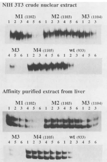
FIG. 6. Effect of mutations in DNA-binding competition assays. Approximately 0.06 pmol of radioactively labeled probe of the wild-type sequence (probe 933) was mixed with various amounts of the five nonradioactive probes, 1102 (Ml), 1103 (M2), 1104 (M3), 1105 (M4), and 933 (wt). The mixtures were analyzed by the electrophoretic mobility shift assay as described in Materials and Methods with NIH 3T3 crude nuclear extract or affinity-purified extract from mouse livers. Each assay with the NIH 3T3 extract contained 0.5,ug of poly(dI)-poly(dC). Lanes: 1, assays without specific competitor DNA; 2 to 6, assays witha 10-, 50-, 100-,500-,
and 1,000-fold molar excess of specific competitor DNA,
respec-tively.
mation for theMland M3 probes as well as for the wild-type probe. For the two mutated probespMland pM3, however, we noted an increase in complexed radioactive DNA upon the addition of smaller amounts of competitor DNA, a reproducible phenomenon for which we have no explanation (Fig. 5).
To study the effect of the same mutations on the binding activity under different experimental conditions, we per-formed electrophoretic mobility shift assays with a probe of wild-type sequence (probe 933, Fig. 1) in the presence of various concentrations of competitor DNAs of mutated or wild-type sequence (probes 1102, 1103, 1104, 1105, and 933; Fig. 1). This type of experiment was performed both with the affinity-purified liver fraction and with a crude nuclear extract from NIH 3T3 cells.
The mutations had the same effect in the assays with the purified liver fraction and in the assays with the crude nuclear extract from NIH 3T3 cells (Fig. 6). The competitor DNAs with the M2 or M4 mutations did not affect protein binding to the wild-type probe, even in a 1,000-fold molar excess, whereas strong competition was observed with a competitor of the wild-type sequence (Fig. 6). The probes of
Ml or M3 sequence showed intermediate competitor activ-ity. These conclusions, derived from inspection of the auto-radiograms, are supported by quantitative analysis of the amount of radioactivity in the bands representing complexed
probeandfreeprobe in two independent experiments (data notshown).
The results of the competition experiments therefore indicate that the different relative effects of the mutations observed in Fig. 5 are valid for NIH 3T3 cells as well as for liver cells. The difference between theMl and M3 sequences was not resolved by this competition assay.
The effect of the mutations in the plasmids pMl, pM2, pM3, and pM4 were studied by comparing their activity with that ofp99-1 in a transient expression assay. All mutations caused a reduction in expression of chloramphenicol acetyl-transferase activity (Fig. 1). The activity of thepM3 plasmid was about 30% of the pl-99 value, whereas the activities of theremaining three plasmids ranged from 20 to 10% of the
pl-99 activity. A statistical analysis with Student's t test showed that all five values (for pl-99, pM3, pMl, pM2, and pM4) were significantly different (P < 0.01). The relative effect of the mutations on the expression level corresponded to theireffect on the in vitro binding of the purified protein fraction with NF-I-like properties, with the exception that the difference between the M2 dinucleotide substitution mutant and the M4 double dinucleotide substitution mutant could not be resolved in the protein binding assay. These results therefore strongly support the notion that binding of NF-I proteins to this site is important forexpression in the cells.
The values observed for thepMl, pM2, and pM4 plasmids were significantly higher than the values for the deletion mutants that lacked the complete NF-I site 1 (plasmids pDD5, pD12, pD13, pDD7, pDD6, pD15, pD16, and pD17; Fig. 1). This may indicate thatsequences immediately down-stream of NF-I site 1 areimportant or that exactpositioning
of sequences upstream of the NF-I site plays arole. The second NF-I site in the99-base-pair repeat unit. The nucleotide sequence termed NF-I site 2 in Fig. 1 has
previ-ously been proposed to represent an NF-I-binding site in murine leukemia viruses (33, 36, 48). Even though this site was intact in pMl, pM2, pM3, and pM4, these plasmids showed a severe reduction in expression activity. More than one NF-I-binding site may therefore be required, or a
functional NF-I site in the natural sequence environmentof
NF-I site 1 may be essential. These possibilities are sup-ported by the low activity ofplasmids pD15 andpD16 (Fig.
1). The deletions of thesetwoplasmidsbring NF-Isite2into the upstream sequence environment of NF-I site 1, except for the additional removal of one(pDl6) or two (pDl5)base pairs. That NF-I site 2 was less important than NF-I site 1 for expression in our assay was supported by results ob-tained with plasmid pD19 (Fig. 1), which showedan
activity
similar to that ofpl-99. In pD19, NF-I site 2 and adjacentsequences in the99-base-pairunit were deletedtogetherwith 55 base pairs downstream of the repeat unit.
Although
the activity of this plasmid may be affected by alterations ofpromoter-enhancer distances, the result clearly demon-strates that NF-I site 2 is notabsolutely required for
expres-sion.
Potential NF-I-binding sites outside the repeat unit. In a
previous study (32) we found that the function of one
99-base-pair repeat sequence alone is dependent upon its normal sequence environment, whereas thefunction oftwo
99-base-pair sequences in tandem is
independent
ofse-quenceenvironment. Sincewehavefoundacriticalfunction of binding sites for NF-I proteins, and since two repeats
contain four NF-I sites and one contains
only
two, wespeculated that the sequences in the Akv U3 outside the tandem repeats may contain NF-I sites that are of
impor-VOL.64, 1990on November 10, 2019 by guest
http://jvi.asm.org/
[image:6.612.92.271.69.339.2]ACTGCAGTAACGCCATTT 790 CCGGGACTAGGGCCAAAC 794
TGACGTCATTGCGGTAAA GGCCCTGATCCCGGTTT GCCCCTAGTTGGGGTTCCGGGGATCAACCCCAAGC 798
site 4 site 3 = site 1 site 2 > site 5
-440 /-415 \ -246 /-201\ -105
CATGGGAAAATACCAGA 792 GTACCCTTTTATGGTCTC
-345 -300
CCCCGGCCCAGGGCCAAG 796 GGGGCCGGGTCCCGGTTC
FIG. 7. Probes forsequences in AkvU3withpartialhomologytoNF-I-bindingsites. Themapcorrespondstothe U3sequenceof Akv. Onlyonecopyof the99-base-pair tandemrepeatisshown(arrow). Thenucleotide numbers refertothecapsiteasin Fig.1. The five sites were identifiedby partial homologytothe NF-I-bindingsite consensus sequenceTGGA/cN5GCCAA (16). Thedots above the sequences indicate the consensus matches.Sites1and2arealsomarked inFig.1.Thedouble-strandedsequencesgiven correspondtothe DNAprobes
790,792, 794,796,and798.
tance when only one 99-base-pair sequence is present. We
therefore searched for possible NF-I-binding sites in U3
upstream or downstream of the repeats. The NF-I-binding
sites aretypically characterized by an inverted repeat, but sequencescontaining onlyonehalf-site ofarepeathave also
beenimplicatedinbinding(2, 11, 12, 30, 39).Thepublished NF-I consensus recognition sequences include TGGA/
CN5GCCAA
(16),
PyTGGA/cN A/TGCCA
(37),
TGGA/CA/
TN3
A/TGCCAA
(26),
TTGGCTN3AGCCAA(20),
and TTGGAN5GCCAAT
(9)and theideal recognition sequencePyPy-PyTGGCACAGTGCCAPuPuPu (36). We found two
se-quences,upstream
and one sequence downstream of the99-base-pair repeat that exhibited good homology to the
NF-I consensus (Fig. 7).
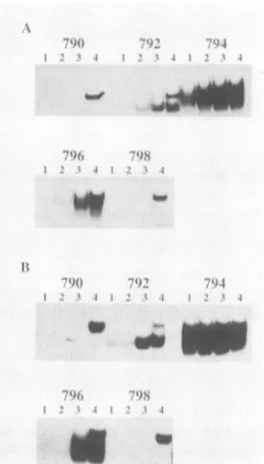
Synthetic double-stranded probes (probes 790, 792, 794, 796, and 798)correspondingtothese sequences(Fig. 7)were
used toassessproteinbinding. The probeswere madeshort
(18 base pairs) to minimize thepossibility ofcomplexesdue to interactions with non-NF-I proteins. A series ofparallel band shift assays was first carried out with the affinity-purified fraction. A weakbinding activity was detectedfor
probe 792, andno orlittle bindingwasobserved forthetwo
remainingprobes (probes 790 and 798) (data not shown). As
expected, theparallel experiments demonstrated clear
bind-ingtothe sequencesof NF-I sites 1 and 2 within the repeats
(probes 794 and 796, respectively).
Tofurtherinvestigatethe large differences in the
protein-binding abilities of the five probes with NF-I binding site
homology, weperformed binding studies with crude nuclear
extractsorwithsemipurified NF-I preparations.Theprotein
fractionsused wereliver crude nuclear extract (Fig. 8A) and
pooledtopfractions fromaheparin column chromatography
(Fig. 8B). Thebinding capacity was assessed without or with
two different concentrations ofpoly(dI)-poly(dC). Protein-DNA complexes could be detected with all probes in the
absence ofpoly(dI)-poly(dC). Complexes of the same
mo-bilities occurred with the probes within the repeats (probes 794 and796),althoughless complex was formed with probe 796. The complexes formed with probe 792 were less effi-ciently competed with by poly(dI)-poly(dC) than were the complexesformedwith the other two probes (790 and 798). Furthermore, the positions of shifted bands for probe 792
weremorelike the bands occurring for probes 794 and 796,
representing repeat recognition sites, although the same
degreeof complexity did not show up.
Thecomplexes observed for probes 790 and 798 are most
likely due to unspecific protein binding, since the bands
disappeared after competition with low concentrations of
poly(dI)-poly(dC)(0.1
pLg
forprobe 798 and 0.5 ,ug for probe790). Further studies will be requiredtodetermine whether the poly(dI)-poly(dC)-resistant complexes formed with probe792representspecific protein bindingand whetherthe proteins belongto theNF-I family.
DISCUSSION
In thepresentstudyweanalyzedasequencein thetandem
repeat ofthe U3 region of Akv murine leukemia with the properties ofaNF-I-bindingsite andprovidedevidence that
binding of NF-I-like proteins to this site plays a role for
transcriptional activity ofthis U3 in mousefibroblasts. Homology to NF-I recognition sites can be found inthe
repeatmodules of a number of other MuLVs. The viruses withinthisgroupdiffer withrespect totheorganization and
)f
h)l#' ...
[image:7.612.96.528.76.177.2]*
-Imp
FIG. 8. Analysis of protein binding to probes representing pos-sible NF-I-bindingsites in the U3region of Akv. A crude nuclear proteinextractfrommouse livers (A) and pooled top fractions from
asubsequent heparincolumnfractionation step (B) weretested in the electrophoretic mobility-shift assay with the five double-strandedDNA probesshown inFig.6. The assays wereperformed asdescribed inMaterials andMethods with thefollowing additions (lanes): 1, 1p.gofpoly(dI)-poly(dC);2, 0.5 ,ugofpoly(dI)-poly(dC);
3,0.1,ugpoly(dI)-poly(dC);4,noaddition.
on November 10, 2019 by guest
http://jvi.asm.org/
[image:7.612.373.505.407.639.2]NF-I-BINDING SITES IN U3 OF Akv VIRUS 4159
the primary nucleotide sequences of repeat modules, and
theirrepeat regions showdifferences in the numberof sites with homologies to the NF-I consensus recognition se-quence and in the nucleotide sequences of these sites. Different sites in one virus or sites in different viruses are
therefore notnecessarily functionally equivalent.
The Akv repeatregion contains four elements with homol-ogy tothe NF-I consensusrecognition sequence, two in each tandem repeat unit represented by the site 1 and site 2 sequencesinFig. 1. Inthis study we focused on the function
of site 1. The site 1 sequence is also found in the repeat regions of the Akv-related viruses SL3-2 (8), Gross passage A(52),OA-1 (27), FBJ MuLV (51), and Soule MuLV (7), in allcasesinahomologous position and in only one copy. This
sitehas not previously been subjected to functional studies in any ofthe viruses. The site 2 sequence is found in the repeatregionof all viruses that are closely related to Akv. In
SL3-3virus the ability of this sequence and mutants thereof
to bind proteins in nuclear extracts suggests that it
repre-sents aNF-I-bindingsite (36). Three copies of this sequence
are found inSL3-3 (28)and in FBJ MuLV (51), two copies
arefound in OA-1 (27) and SL3-2 (8), and one copy is found in Gross passage A virus (52). The Moloney (45) and Friend
(23)MuLVsboth containanelementnearlyidenticalto site 2 in their tandem repeat sequences (23, 33, 45, 48). The
function of these sequences in the Moloney and Friend MuLVsas NF-I-bindingsites has been subject to
biochem-ical and genetic studies (33, 48). In addition, the Moloney MuLV tandem repeat contains anotherpotential NF-I site
(5'-TGAATATGGGCCAA-3') that is more different from the sites in Akv (45). This site is located in the
promoter-distalpartof the repeat unit in apositioncorresponding to
theNF-Isite 1 of Akv. It has beenfound(44) that thisregion
canbedeletedfromaMoloney MuLVU3withonetandem repeat copy without significant effecton its ability to drive
transient expression in fibroblasts. Potential NF-I sites of different structures are also found in the repeat region of
MCF viruseslikeMCF247(5'-TGGGACAGGGGCCAA-3') (22)andofaradiationleukemiavirusisolate (5'-TAGACAT AGGCCAA-3') (19).
This comparison stresses thecomplex differences among theindividual viruseswith respecttothe number and
organ-ization of sites that may be
implicated
in NF-Ibinding.
Interestingly, all the sites discussedabove contain a
down-streamhalf-site with the sequence 5'-GGCCAA-3' but differ
substantially in their upstream half-sites. We also note the presence oftwo site 1 elements in Akv, whereas the other
virusescontain one or no copies ofthis sequence.
Our firstindication forarole ofNF-Isite1in
transcription
directed by the U3 ofAkv came from deletion analysis,whichpointedtoacriticalfunction of sequences around base
pair
-250 within the99-base-pair
repeat sequence. Thenucleotidesequencein thisregionmatches the NF-I consen-sus recognition sequence
5'-TGGA/cN5GCCAA-3'
(16),ex-ceptforadifference in the first base
pair.
Frommouselivernuclei we purified a protein fraction with
specific binding
activity toward the Akv sequence in this
region.
The purifi-cationprocedure includedDNAaffinity
chromatography
onmatrix-bound double-stranded DNA of the Akv sequence in thisregion. NF-I proteins purified fromHeLacell fractions
were shown to consist of a
family
ofpolypeptides
with molecular weight rangefrom 52,000to 66,000(20, 41).
Thepurified fraction of mouse nuclear
proteins
used in the presentstudyshows asimilar molecularweight
pattern,andour DNase I
footprint
andmethylation
interference resultsare in accordance with
published
results withpurified
NF-Ipreparations (6, 9, 20, 41). We therefore believe that our
purified proteins belongto the NF-Ifamily.
To test whether the critical function of the sequences around basepair -250 isin fact due totheirabilityto bind
NF-I proteins, we used site-specific mutagenesis. Three
dinucleotide substitutionmutants(pM1,pM2,andpM3)and
onedouble dinucleotide substitutionmutant (pM4) with the combined mutations ofpMlandpM2werefirstanalyzedfor their
ability
tobind NF-Iproteins
in vitro in theelectropho-retic mobility shift assay and then for their transcriptional
function inmousefibroblasts. ThepMl mutationrepresents
a
change
in thebasepairs
of thetwomethylation-sensitive
G residues of the upstream half-site from 5'-CGGGAC-3' to5'-CGCCAC-3' and the pM2 mutationa changein the base
pairs
of the twomethylation
sensitive G residues of the downstream half-site site from 5'-GGCCAA-3' to 5'-GGGGAA-3'. The pM3 mutation represents the
change
ofpre-sumably lesscriticalbase
pairs
between the half-sites. ThepM2 mutation in the downstream half-site was found to
abolish NF-I
binding
invitro,
whereasthe pMlmutationinthe upstream half-site allowed some binding activity. The downstream
half-site,
which representsaperfectmatch with the consensussequence andis conserved in all of the NF-I sites discussedabove,
therefore seems critical forbinding.
ThepM3 mutation ledto abinding
withanactivity
betweenthose of the
wild-type
sequenceand thepMl sequence. The mutatedplasmids
pMl, pM2, pM3, and pM4 all showed reducedactivity
in the transientexpression assay, whencompared with that oftheir parentplasmid, pl-99.The relative order ofexpression activities waspl-99 > pM3 >pMl >pM2> pM4,in agreement with the in vitro
binding
activities. We take the covariation between in vitrobinding
and invivoactivity
as astrongindication ofafunctional role of the NF-Ibinding
in thisregion.
However,complete
impairment
of NF-I site1,asinourpM2andpM4mutations,
still allowstranscriptional activity
abovebackground
levels. This residualactivity
in the absenceofafunctional NF-I site 1 maydepend
upon thebinding
of NF-I to other sequences in the U3region
of Akv and/oruponthebinding
ofother
transcription
factors. Asexpected
fromtheprevious
studies ofotherviruses,
ourpurified
fraction ofNF-Ipro-teinsalso boundtothe sequencesinAkvtermed NF-I site2
(Fig. 1),
and theobserved DNase Iprotection (Fig. 4)
andmethylation
interferencepatterns(data
notshown)
fit thoseexpected
forNF-Iinteractions. NF-I site 2alone, however,
is not sufficient for the residualexpression
activity
oftheplasmids
with animpaired
NF-I site1,
since deletionsaffecting
site1all ledtoabolishment ofactivity. Specifically,
we note the low
expression activity
of mutants pD15 andpD16, with deletions that
bring
NF-I site 2 into the site 1position,
andofmutantpD17,
withadeletion that eliminates the site 1 sequences. Mostlikely
additionalprotein-DNA
interactions in this
region
arerequired,
and theinterpreta-tion may be
quite complex.
Theinability
of site 2 toreplace
site 1functionin these deletedconstructs mayrelatetothe differentstructuresofthetwo
sites,
orit may reflectarole ofinteractions with elements
immediately
downstream of site 1 thataredeleted inpD15
andpD16
orwith elementsupstreamof site1 that maynotinteract
properly
with site 2 inpD17.
Apparently,
NF-Isite2isnotasimportant
asNF-Isite1forexpression
in NIH 3T3cells,
assuggested by
the resultsobtainedwith the
pD19
deletionmutant.Thisplasmid
showshigh-level
expression,
although
it lacks NF-I site 2 andadjacent
sequences.Obviously,
our deletionanalysis
is too crude toclearly
identify
specific
contributions from individualbinding
sitesVOL.64, 1990
on November 10, 2019 by guest
http://jvi.asm.org/
outsideof NF-Isite 1.
Site-specific mutagenesis
of NF-Isite2 and of other
potential binding
sites may berequired
infurther
analyses.
We note,however,
that three differentlevels of disturbance of the functional
organization
may bereflected in the levelsofactivityofpM2andpM4 (6.8and 4.8
relative
units,
respectively),
inwhichNF-I site 1isimpairedby
substitution,
ofpD15
andpD16 (2.6
and 3.3units),
inwhichNF-Isite 2 withdownstreamsequencesistransposed
to the
position
of NF-I site1,
and ofpD17 (1.7 units),
inwhich NF-I site 1 has been deleted.
Our
previous
results (32) have shown that the transcrip-tionalactivity
ofone99-base-pair
repeatunit isdependent
uponitssequenceenvironment, indicating
arequirement
forcooperativity
between one repeat unit and itsflanking
se-quences in U3. To
investigate
thepossible
role of NF-Iinteractionsfor this
cooperativity,
we searchedthe Akv U3sequences outside the repeats for
homologies
tothe recog-nition sequence for NF-I. Two sites(sites
3 and4)
werefoundupstream of base
pair -385,
andone site(site 5)
wasfound downstream of the repeats. These three sites each
containoneTGGsequenceandatleasttwomatches with the TGG sequences of the other half-site. The formation of
complexes
with DNAprobes corresponding
to these sites wasanalyzed
withaffinity-purified,
semipurified,
and crudenuclear
protein
fractions. Whereas sites4and 5 gaverisetolow-specificity complexes,
site 3produced complexes
ofapparently higher
specificity. Furthermore,
site 3 gave riseto
complex
mobilities almost similartothose obtained withsite 1and site2
probes, although
thebinding activity
of thesite 3
probes
was somewhat lower.Interestingly,
theonly
differencesoutside the repeat
region
between the U3s of Akv and the related viruses SL3-2(8),
SL3-3(28),
and Gross passageAvirus(52)
arelocatedin thepotential NF-I-binding
site 3. The Akv and Gross passage sequences both have a
6-base-pair
spacerbetween the TGG andCCAhalf-sites but differ inoneposition
ofthe spacer. The SL3-2 and SL3-3 sequences carry anadditionalbasepair
atthisposition
andhence contain 7 base
pairs
between two half-sites. TheMoloney
MuLV sequence(45)
also differs from the Akv sequence in thisregion.
Additionalexperiments
will berequired
to elucidatethepossible regulatory
function of thesite 3 sequence in Akv and the variant viruses.
In summary,wehave identifiedtwosequenceelementsin the tandem repeats of Akv MuLVinvolved in NF-I
binding
andpotential NF-I-binding
sites located in U3 outside the tandem repeats andpointed
to identical and divergent sitesin the U3
regions
of other MuLVs. A number of features mayinfluencetheexactfunction ofeachofthesesites. Theexistence of different subclasses ofNF-I
proteins
presentinvariousamountsand
possibly
with slightly different bindingspecificities
may affect the pattern ofbinding
to multipleNF-I sites. Molecular
cloning
ofthe genes encoding NF-Iproteins
from humans (43), rats (39), and hamsters (12)confirmsthe existence of different subclasses of NF-I pro-teins that all
recognize
the 5'-TGG-3' sequence. Theanaly-ses
predict proteins
with nearly identical amino acidse-quences in their NH2-terminal half, which contains the
DNA-binding
domain and with divergence in theCOOH-terminal
regions.
ProteinsbindingtoDNAflanking the NF-I sites may also contribute to their functional diversitythrough
protein-protein
interactions affectingthe DNAbind-ing
of NF-I and other proteins. Also, an NF-I functionalmonomer couldbindto half therecognition site,
5'-NNTG-GNN-3' ina
complex
with anotherproteinwithaffinity foraneighboring
sequence, asproposed previously(12). Finally,the exact location of an NF-I site in U3 may affect its
possibility to participate in formation of a functional
tran-scriptioncomplex.
Studiesemployingpermutations of NF-I sites withinone
virusorbetween viruses, together with specificmutagenesis
of individual sites,mayprovide insight into the role of these different regulatory principles. These viruses may thereby
provide interesting material for studies of the role of NF-I-binding sites in regulation of gene expression.
ACKNOWLEDGMENTS
WethankPeterV.Mathiasen,LoneH0jgaard, and Bente Christ-ensen for technical assistance and J. T0nnes Nielsen for mouse livers.
This workwas supported by grants from the Carlsberg Founda-tion, the Danish Cancer Society, Euratom (BI6-086-DK), andthe Danish Biotechnology Programme.
LITERATURE CITED
1. Boral, A. L., S. A. Okenquist, and J. Lenz. 1989. Identification of the SL3-3virus enhancercoreas aT-lymphoma cell-specific element. J.Virol. 63:76-84.
2. Borgmeyer,U., J. Nowock,and A. E.Sippel.1984. The TGGCA-binding protein: a eukaryotic nuclear protein recognizing a symmetricalsequence ondouble-stranded linear DNA. Nucleic Acids Res. 12:4295-4311.
3. Bosze, Z., H.-J. Thiesen, and P. Charnay. 1986. A transcrip-tional enhancer withspecificity for erythroid cells is located in thelong terminal repeatof the Friend murine leukemia virus. EMBOJ. 7:1615-1623.
4. Celander, D., andW.A. Haseltine. 1984. Tissue-specific tran-scription preferenceas adeterminant ofcell tropismand leu-kaemogenic potential of murine retroviruses. Nature(London) 312:159-162.
5. Cereghini, S.,M. Raymondjean,A. G.Carranca,P. Herbomel, and M. Yaniv. 1987. Factors involved in control of tissue-specific expression of albumin gene. Cell 50:627-638.
6. Chodosh,L.A.,A.S.Baldwin,R. W.Carthew,and P. A.Sharp. 1988.HumanCCAAT-binding proteinshaveheterologous sub-units. Cell 53:11-24.
7. Corcoran,L.M., J.M.Adams,A. R.Dunn,and S.Cory.1984. MurineTlymphomas in which the cellularmyc oncogene has been activatedby retroviral insertion. Cell37:113-122. 8. Dai, H. Y., M. Etzerodt, A. J. Bekgaard, S. Lovmand, P.
J0rgensen,N.0.Kjeldgaard,and F. S. Pedersen. 1990.Multiple
sequenceelements inthe U3region of the leukemogenic murine retrovirusSL3-2 contributetocell-dependentgeneexpression. Virology 175:581-585.
9. DeVries, E., W.vanDriel, S. J. L. van den Heuvel, and P. C. van der Viet. 1987. Contact point analysis of the HeLa nuclear factor Irecognitionsiterevealssymmetrical bindingat one side of the DNA helix. EMBO J.6:161-168.
10. Dignam, J. D., R. M. Lebovitz, and R. G. Roeder. 1983. Accurate transcription initiation by RNA polymerase II in a solubleextractfrom isolatedmammalian nuclei. Nucleic Acids Res. 11:1475-1489.
11. Gil, G.,T. F.Osborne,J. L. Goldstein, and M. S. Brown. 1988. Purification of a protein doublet that binds to six TGG-con-taining sequences in the promoter for hamster 3-hydroxy-3-methylglutaryl-coenzyme A reductase. J. Biol. Chem. 263: 19009-19019.
12. Gil, G.,J. R.Smith,J. L.Goldstein, C. A. Slaughter, K. Orth, M.S.Brown,and T. F.Osborne. 1988. Multiplegenes encode nuclear factor 1-like proteins that bind to the promoter for 3-hydroxy-3-methylglutaryl-coenzymeA reductase. Proc.Natl. Acad. Sci. USA 85:8963-8967.
13. Gorman, C. M., L. F. Moffat, and B. H. Howard. 1982. Recombinant genomeswhichexpress chloramphenicol acetyl-transferasein mammaliancells.Mol.Cell. Biol. 2:1044-1051. 14. Graham,F.L., and A. J. van der Eb. 1973. A newtechnique for
the assayofinfectivity ofhumanadenovirus5 DNA.Virology 52:456-467.
on November 10, 2019 by guest
http://jvi.asm.org/
NF-I-BINDING SITES IN U3 OF Akv VIRUS 4161 15. Graves, B. J., R. N. Eisenman, and S. L. McKnight. 1985.
Delineation of transcriptional control signals within the Molo-neymurinesarcoma virus long terminal repeat. Mol. Cell. Biol. 5:1948-1958.
16. Gronostajski, R. M., S. Adhya, K. Nagata, R. A. Guggenheimer, and J. Hurwitz. 1985. Site-specific DNA binding of nuclear factor I: analysis of cellular binding sites. Mol. Cell. Biol. 5:964-971.
17. Hallberg, B., and T. Grundstrom. 1988. Tissue specific se-quence motifs in the enhancer of the leukaemogenic mouse retrovirus SL3-3. Nucleic Acids Res. 16:5927-5944.
18. Herbomel, P., B. Bourachot, and M. Yaniv. 1984. Two distinct enhancers with different cell specificities coexist in the regula-tory region of polyoma. Cell39:653-662.
19. Janowski, M., J. Merregaert, J. Bonvier, and J. R. Maisin. 1985. Proviral genome of radiation leukemia virus: molecular cloning of biologically active proviral DNA and nucleotide sequence of its long terminal repeat. J. Virol. 55:251-255.
20. Jones, K. A., J. T. Kadonaga, P. J. Rosenfeld, T. J. Kelly, and R. Tjian. 1987. A cellular DNA-binding protein that activates eukaryotic transcription and DNA replication. Cell 48:79-89. 21. Kadonaga, J. T., and R. Tjian. 1986. Affinity purification of
sequence-specific DNA binding proteins. Proc. Natl. Acad. Sci. USA 83:5889-5893.
22. Kelly, M., C. A. Holland, M. L. Lung, S. K. Chattopadhyay, D. R. Lowy, and N. H. Hopkins. 1983. Nucleotide sequence of the 3' end of MCF 247 murine leukemia virus. J. Virol. 45:291-298.
23. Koch, W., W. Zimmerman, A. Oliff, and R. Friedrich. 1984. Molecular analysis of the envelope gene and long terminal repeat of friend mink cell focus-inducing virus: implications for the functions of these sequences. J. Virol. 49:828-840. 24. Laemmli, U. K. 1970. Cleavage of structural proteinsduringthe
assembly of the head of bacteriophage T4. Nature (London) 227:680-685.
25. Laimins, L. A., P. Gruss, R. Pozzatti, and G. Khoury. 1984. Characterization of enhancer elements in the long terminal repeat of Moloney murine sarcoma virus. J. Virol. 49:183-189. 26. Leegwater, P. A. J., P. C. van der Vliet, R. A. W. Rupp, J. Nowock, and A. E. Sippel. 1986. Functional homology between the sequence-specific DNA-binding proteins nuclear factor I from HeLa cells and the TGGCA protein from chicken liver. EMBO J. 5:381-386.
27. Leib-Mosch, C., J. Schmidt, M. Etzerodt, F. S. Pedersen, R. Hehlmann,and V.Erfle. 1986. Oncogenic retrovirus from spon-taneous murine osteomas. II. Molecular cloning and genomic characterization. Virology 150:96-105.
28. Lenz, J., D. Celander, R. L. Crowther, R. Patarca, D. W. Perkins, and W. A. Haseltine. 1984. Determination of the leukaemogenicity of a murine retrovirus by sequences within the long terminal repeat. Nature (London) 308:467-470. 29. Li, Y., E. Golemis, J. W. Hartley, andN.Hopkins.1987.Disease
specificity of nondefective Friend and Moloney murine leuke-mia viruses is controlled by a small number ofnucleotides. J. Virol. 61:693-700.
30. Lichtsteiner, S., J. Wuarin, and U. Schibler. 1987.Theinterplay of DNA-binding proteins on thepromoterof themouse albumin gene. Cell 51:963-973.
31. LoSardo, J. E., L. A. Cupelli, M. K. Short,J. W. Berman, and J. Lenz. 1989. Differences in activities of murine retrovirallong terminal repeats in cytotoxic T lymphocytes andT-lymphoma cells. J. Virol. 63:1087-1094.
32. Lovmand, S., N. 0. Kjeldgaard, P.J0rgensen, and F. S. Peder-sen. 1990. Enhancer functions in U3 ofAkv virus: a role for cooperativity of a tandem repeat unit and its flanking DNA sequences. J. Virol. 64:3185-3191.
33. Manley, N. R., M. A. O'Connell, P. A.Sharp,and N. Hopkins. 1989. Nuclearfactorsthatbindto theenhancerregion of nonde-fective Friendmurineleukemiavirus. J. Virol. 63:4210-4223. 34. Maxam, A. M., andW. Gilbert. 1980. Sequencingend-labeled
DNA with base-specificchemicalcleavages.MethodsEnzymol. 65:499-560.
35. Nagata,K., R.A.Guggenheimer,T.Enomoto,J. H.Lichy, and J. Hurwitz. 1982. Adenovirus DNAreplication in vitro: identi-fication of a host factor thatstimulatessynthesis of the preter-minal protein-dCMP complex. Proc. Natl. Acad. Sci. USA 79:6438-6442.
36. Nilsson,P., B.Hallberg,A.Thornell, and T. Grundstrom. 1989. Mutant analysis ofprotein interactions with anuclear factorI bindingsite in the SL3-3 virus enhancer. Nucleic Acids Res. 17:4061-4075.
37. Nowock, J., U.Borgmeyer,A. W.Piischel, R. A. W.Rupp,and A.E. Sippel. 1985. TheTGGCAprotein binds tothe MMTV-LTR, the adenovirus origin ofreplication, and the BK virus enhancer. Nucleic Acids Res. 13:2045-2061.
38. Paludan, K., H. Y. Dai, M. Duch, P.J0rgensen, N.0.
Kjeld-gaard, and F. S. Pedersen. 1989. Different relativeexpression
from two murineleukemia viruslongterminalrepeatsin unin-tegrated transfected DNA and in integrated retroviral vector
proviruses. J. Virol. 63:5201-5207.
39. Paonessa, G., F. Gounari, R. Frank, and R. Cortese. 1988. Purification ofa NFl-likeDNA-binding protein fromrat liver andcloning of thecorrespondingcDNA.EMBO J. 7:3115-3123. 40. Pedersen, F. S., M. Etzerodt, S. Lovmand, H. Y. Dai, A. J. Baekgaard, J. S0rensen, P. J0rgensen, N. 0. Kjeldgaard, J. Schmidt, C.Leib-Mosch,A.Luz, and V. Erfle.1987.
Transcrip-tional control andoncogenicityof murineleukemiaviruses,p. 17-35. In N. 0. Kjeldgaard and J. Forchhammer (ed.), Viral carcinogenesis, Alfred Benzon Symposium 24. Munksgaard,
Copenhagen.
41. Rosenfeld, P. J., and T. J. Kelly. 1986. Purification of nuclear factor I by DNA recognition site affinity chromatography. J. Biol. Chem. 261:1398-1408.
42. Rossi, P., G.Karsenty,A. B.Roberts,N.S.Roche,M. B.Sporn,
and B. deCrombrugghe. 1988. ANuclearFactor Ibindingsite mediates the transcriptional activation of a type I collagen
promoter bytransforminggrowth
factor-P.
Cell52:405-414. 43. Santoro, C., N.Mermod,P.C.Andrews, and R.Tjian. 1988. Afamily of human CCAAT-box-binding proteins active in
tran-scription andDNAreplication: cloningand expressionof mul-tiplecDNAs. Nature (London) 334:218-224.
44. Schulze, F., E. Boehnlein, and P. Gruss. 1985. Mutational analyses oftheMaloney murinesarcomavirusenhancer. DNA 4:193-202.
45. Shinnick, T., R. A. Lerner, andJ. G. Sutcliffe. 1981. Nucleotide sequenceofMoloneymurineleukemia virus. Nature(London)
293:543-548.
46. Short, M. K., S. A.Okenquist,andJ.Lenz.1987. Correlation of leukemogenic potential of murine retroviruses with
transcrip-tional tissue preference of the viral long terminal repeats. J. Virol. 61:1067-1072.
47. Siebenlist, U., and W. Gilbert. 1980. Contacts between Esche-richia coliRNApolymeraseandanearlypromoterofphageT7. Proc. Natl. Acad. Sci. USA 77:122-126.
48. Speck, N. A., and D. Baltimore. 1987. Six distinct nuclear factors interact with the 75-base-pair repeat of the Moloney
murineleukemia virus enhancer. Mol. Cell. Biol. 7:1101-1110. 49. Switzer, R.C.,III,C. R.Merril,andS. Shifrin. 1979. A
highly
sensitive silver stain for detecting proteins and peptides in polyacrylamidegels. Anal. Biochem. 98:231-237.
50. Thiesen, H.-J., Z. Bosze, L. Henry, and P. Charnay. 1988. A DNAelementresponsibleforthe differenttissue
specificities
of Friend andMoloney retroviralenhancers. J. Virol. 62:614-618. 51. Van Beveren, C., F. van Straaten, T. Curran, R. Muller, and I. M. Verma. 1983. Analysis ofFBJ-MuSVprovirus
and c-fos (mouse) gene reveals that viral and cellular fos geneproducts
havedifferent corboxy termini. Cell 32:1241-1255.
52. Villemur, R., E. Rassart, L. DesGroseillers, and P. Jolicoeur. 1983. Molecularcloningof viral DNA fromleukemogenicGross passage Amurine leukemiavirus and nucleotide sequence of its long terminalrepeat. J. Virol. 45:539-546.
53. Yoshimura, F. K., B. Davison, and K. Chaffin. 1985. Murine leukemiaviruslongterminalrepeat sequencescanenhance gene activity inacell-type-specific manner. Mol. Cell. Biol. 5:2832-2835.
VOL.64, 1990