JOURNAL OFVIROLOGY,JUlY1990, p. 3310-3318 Vol. 64, No. 7 0022-538X/90/073310-09$02.00/0
Copyright © 1990,American SocietyforMicrobiology
Infectious Cycle
of Human
Papillomavirus Type
11 in Human
Foreskin Xenografts in Nude Mice
MARK H. STOLER,
4t
APRILWHITBECK,1
STEVEN M.WOLINSKY,2t
THOMAS R.BROKER,3'4
LOUISET. CHOW,3.4* MARY K. HOWETT,5 ANDJOHN W. KREIDER6Department of Pathology andLaboratory
Medicine,'
DepartmentofMedicinelInfectious
Diseases Unit,2DepartmentofBiochemistry,3 and The CancerCenter,4 The University
of
Rochester Schoolof Medicine, Rochester, New York 14642, and DepartmentofMicrobiology andImmunology'
andDepartment of Pathology,6 ThePennsylvania State UniversityCollegeof Medicine, Hershey, Pennsylvania 17033 Received 2February1990/Accepted 16 April 1990
We have performed the first molecular analysis ofa time course of infection by a papillomavirus. The Hersheyisolate ofthe human papillomavirus type 11wasused to infect human foreskin tissues,which were thenimplanted under therenal capsulesof nude mice. The xenograftswererecoveredevery 2 weeks for 14 weeks, fixedinformalin, and embeddedinparaffin.Four-micrometer serial sectionswereexamined by light microscopyformorphological changes,byimmunocytochemistryfor virionantigen production,andbyin situ hybridization with 3H-labeled RNA probes for viral DNA replication and expression of the major mRNA species. Aftera lagperiod,probes spanningthe E4 andE5 open reading frames, whicharepresentin all E region viralmRNAs, generated the first detectablesignals atweek 4. Signalsof other Eregion probes were minimally detected atweek 6. Between weeks6 and 8,there was anabrupt changein theimplantsuch that cellularproliferation, viralDNAreplication,and E and LregionmRNAtranscriptionwererobust and reached a plateau. By weeks 10 to 12, the experimental condylomata were morphologically and histologically indistinguishable from naturally occurring condylomata acuminata. These findings suggest that cellular hyperproliferation and the morphologic features ofcondylomata aredirect results ofviral genetic activities. Unlike other DNA viruses, the E region transcripts increased with cell age and cellular differentiation and persisted throughout theentire experiment. In particular, the mRNAencoding the Eli^E4 and perhaps E5 proteins remainedoverwhelmingly abundant. In contrast, viralDNAreplication, LregionmRNAsynthesis, and virionantigen productionwererestricted tothe mostdifferentiated, superficialcells.
Papillomavirusesare afamilyof small DNA viruses which containacircular DNA chromosome ofapproximately 7,900 basepairs. Thehuman papillomaviruses (HPVs)arespecies specificandarerestrictedin their tissuetropism. Infection of target cutaneous ormucosal epithelium results in hyperpro-liferation. The closely related human papillomaviruses type 6(HPV-6) andtype11(HPV-11)areusuallyassociated with
benign genital warts (condylomata acuminata). One major
obstacle to research on human papillomaviruses is the
in-abilitytopropagatethemin cultured cellsor toinfect tissues
of laboratory animals. Nevertheless, it has been demon-strated thatinfection byaparticular HPV-11 isolate, called Hershey, alone is sufficientto generatecondylomatouscysts in chips of human foreskin or uterine cervix following
implantation under the renal capsule of athymic mice (11, 13). These experimental condylomata have morphologic
features identical to those of human lesions. The HPV-11 Hershey isolate has been seriallypassaged in this system and produces largeamountsof virions(10).The tissue specificity of the Hersheyisolate has been examined byusing skin from differentbodysites from the sameindividual, with foreskin and urethral epithelium being most supportive of cellular transformationand viral transcription(12). HPV-11 Hershey has been molecularly cloned. Physical and functional
anal-* Correspondingauthor.
tPresentaddress: DepartmentofPathology L25,The Cleveland ClinicFoundation, Cleveland, OH 44195.
tPresent address: Department ofMedicine/Infectious Diseases Unit, Northwestern University School ofMedicine, Chicago, IL 60611.
yses of this isolate have not uncovered any significant differences (8) from the HPV-11 prototype clone (7). The
seemingly unique properties of HPV-11 Hershey have been attributedto an unusually high virus titer in theextracts of thepatient lesions usedtogenerate the initialexperimental
condyloma.
HPV-6 and HPV-11 mRNAs from human lesions aswell asfrom theexperimental condylomatahavebeenmapped by electron microscopic examination ofR-loops (6, 16). The twovirusesgenerate completely analogous families of over-lapping mRNAs that are transcribed from several distinct promotersandpolyadenylatedatoneoftwosites locatedat the 3' ends of the E or L genetic regions (Fig. 1). The structures and the coding potentials of the alternatively
processed mRNAs have been determined by various meth-ods (6, 14-17, 21). We have prepared whole genomic and
subgenomic RNA probes specific forDNA orfor different viralmRNAspecies and performedin situ hybridization with serial thin sections of biopsies of patientcondylomata asso-ciated with HPV-6or HPV-11 (18, 19). These studies show that both viral DNA replication and mRNA transcription increase with cellular differentiation. DifferentmRNA spe-cies are present in dramatically different relative abun-dances. ThemRNAspeciesaencodinganEli^E4(^denotes a splice orfusion) protein (see the legend to Fig. 1 for the change in nomenclature) is by far the most abundant viral RNA,presentin nearly all the cells,whereas other messages areexpressedat0.1to0.01times thatamount orless, mostly in themoredifferentiated cells. Theseresults provide snap-shotsof the viral activityatthetimeswhen the biopsies were taken. They do not, however, reveal the time course of 3310
on November 10, 2019 by guest
http://jvi.asm.org/
"-in zV 0 0
VIt- Ve lo *# a) I *)
I I II
to t
-fn OCO ES
'-I' I0CD
I I I I
0. _ b. C. d.I e. f.I o h. i. -k. :7 0 EI!AE4 El-M, E2-C
El, Pro e Pro
m E6, E7, ELAE4
E6, E7, E2
E2 r E2-C
m
n3~~~~~~~~~
r
L2
1
2] L
El |
t
E22 3 kb 4
[Eb
5 6 I Pre EI('E4, LI r L2 3 7FIG. 1. Genetic organization and message-specificprobesofHPV-11. The circulargenomeof 7,933 base pairs is represented inalinear
fashion fromaBstI site in the upstreamregulatory region. The ORFs deducedfromthe DNA sequence are representedby open boxes.
Verticaldotted lines mark the locations of the first AUGcodonineach ORF. Viralmessages aredepictedas arrowsinthe 5'to3'direction,
withgapsindicating introns(6, 14, 16). The dotsatthe 5'ends of the mRNAsrepresentproven orputativepromoters.Thepresumptive L2
mRNA(species k) hasnotbeen observed frequently enoughtopredictitspromoter. Coding potentials,asdeduced from thecDNAsequences
(16),arelisted, with the exception of the E5 protein(s), which could potentiallybetranslatedfromanyorallof theEregion mRNAsorfrom
anmRNAyettobedefined. Shaded boxes indicate regions spanned byexon-specificsubgenomic clones in pGEMorBRL19vectors(19).
Pre,Putativeprecursor.TheEli^E4 protein has previously been designatedEl^E4protein.Wenowintroduce thisnewterminology, with "i"
signifying translationinitiation, todistinguish this protein, which derives only the initiation methionine and fouradditional amino acids from
the ElORF, from other fusion proteins with substantial El domains (C.-M. Chiang,T. R. Broker, and L. T. Chow, unpublished results).
infectionandtherelationship of viralgeneexpressiontothe development of a condyloma. Taking advantage of the availability ofinfectious HPV-11 Hershey virions and the reproducibilitywithwhich they induceexperimental condy-lomata, we systematically surveyed viral DNA replication
and the synthesis of the major mRNA species at different times after the implantation of infected human xenografts. Thisreportprovides thefirstmolecular analysis ofthetime
courseofaproductiveinfectionbyapapillomavirus. Atime
courseofcystdevelopmentinskingrafts infected bybovine papillomavirushas beenanalyzedformorphological changes (9).
MATERIALS AND METHODS
Infection of human foreskin chipsand subrenal implanta-tion. Humanforeskin wasobtained fromaroutine neonatal circumcision at an areahospital. Split-thickness skinchips (0.5 by2.0by2.0mm)werecut withascalpeland incubated either with HPV-11Hersheyextracted fromprevious
exper-imentalcondylomata orwithcontrol saline for 1 h at 37°C. Theyweretheninserted beneath the renalcapsules of nude
mice, as previously described (11, 13). Female athymic mice,NIHstrain,4 to6 weeksold,wereusedashosts.Two
mice were used for each time point. Each mouse received
one infectedimplantandone controlimplant.
In situ hybridization with HPV-l1 message-specific
ribo-probes. Two mice were sacrificed every 2 weeks for 14
weeks after implantation. The kidneys were removed, and
the xenografts werefixed in 10% neutral-buffered formalin andthen embeddedin paraffin. Serial sections (4 ,Lm)were
mounted onto polylysine-coated microscope slides. The controlspecimenswereexaminedhistologically. Sections of each of thetwo setsoftheinfectedxenograftswere
hybrid-izedto 3H-labeled,asymmetric wholegenomicor
message-specific riboprobes (Fig. 1) in two separate experiments, each withaset ofprobesof thesame specific activity. The probeshavebeen describedpreviously (18, 19). Aprobefor E5 open reading frames (ORFs) (nucleotides 3901 to 4557) which does not react with mRNA j wasalso used (Fig. 1).
Probe concentrations were normalized according to their lengths. Autoradiograms were developed after a 4-week
exposure. Therefore, the intensities ofthesignalsgenerated
were proportional to relative copy numbers of the target molecules. Negative controls included hybridization of whole genomicand subgenomic sense-strand riboprobesto specimenswithoutpriorheatdenaturation ofthe viral DNA. We define sense-strand probes to be those of the same
polarityasthe viralmRNAs. Thesecontrolswereuniformly
negative (data not shown). All slides from one of the
experimental groups were photographed with bright-field illumination for histological examination and immunocyto-chemistry and with dark-field illumination for optimal 3H
signaldetection.
RESULTS
Kidneys from two mice, each withone infected and one
control foreskinimplant,wereharvestedevery 2 weeks after
cIe
Is
s. ,;,,,..., .., .. , ...-.-.---r..:;..;.
4 = uLOD
on November 10, 2019 by guest
http://jvi.asm.org/
[image:2.612.121.494.83.328.2]3312 STOLER ET AL.
implantation. Uninfected human foreskin xenograftsformed squamous epithelial cysts and have nevershown koilocyto-sis or viral antigen. Serial thin sections of HPV-infected xenografts from both sets of animals were hybridized with whole genomic or subgenomic 3H-labeled riboprobes in separate experiments, as described in Materials and Meth-ods. Because the amounts ofprobe used were normalized against the probe size, the intensity ofthe3Hsignalsreflects the relative abundances of individual RNA species. Essen-tially identical results were obtained with the two parallel
experiments. Only one set of sectionswasphotographedand presented in this report. Due to the necessity of taking individual time points from different animals, there were occasional small deviations in the trend of viral activities overthe 14-week period. However, at any one time point, consistent assessments were obtained with regardto histol-ogy, viral DNA replication, and transcription ofindividual mRNA species. We point out that it takes perhaps 1 or 2 weeks for the keratinocytes to migrate from the parabasal
layer to the surface. Accordingly, the topography of the epithelium itselfpresents an ongoing time course. The sa-lient features of the time course of infectionaresummarized below.
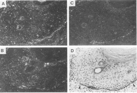
Replication of viralDNA.WholegenomicRNAprobewith the same polarity as the viral messages was used todetect viral DNA replicationfollowingheatdenaturation ofDNAin thespecimens. Viral DNA was not detectableforthefirst4 weeks, and the signals were barely detected in the week 6
sample (Fig.2A andB). By week 8, mostof the cells in the foreskinepitheliumcellswerepositive,includingcells inthe lower stratum spinosum (Fig. 2C and D). The strongest
signals were in the upper stratum spinosum and stratum granulosum. Thereafter, DNAsignalswererestrictedtothe moredifferentiatedcells(Fig. 2E to H). Relative to week 8or week 12 specimens, the week 10 xenograft appeared less active in both DNA replication and in mRNAtranscription
(data not shown). We attributed these lower activities toa somewhat slower rate with which the 10-week graft was established in the particular animal.
Expression of individual mRNA species. The locations of the message-specific subgenomic probes (19) are presented in Fig. 1. With the exception of the E4-E5 and E5 probes, each probe is complementary to a region (E6-E7, El, E2, L2, and
Li)
unique for the target mRNA. The E4-E5 probeoverlaps the carboxyl-terminal half of the E2 ORF, which encodes the E2-C protein, and also hybridizes to the
Li
mRNAspecies
j
(Fig. 1). TheE4-E5and the E5probes also hybridize to all the other E region mRNAs. The E2-C mRNA isextremely rare in patient lesions and in the experimentalcondylomata(6, 16) andtherefore accounts for a very small fraction of theabundant signals detected, except perhaps in asubpopulation ofthe cells (see Discussion). The amount of the Eli^E4 mRNA species a can be deduced from the difference between the cytoplasmic signals generated from the E4-E5 probe and those specific for the other E region messages and for the
Li
mRNA. This adjustment makesonly a very small difference as, all together, these other
transcripts amount to but a small fraction of the Eli^E4
mRNA(see Fig. 4). By using these message-specific RNA probes, we examined adjacentthin sections for the expres-sion ofeach mRNA for each of the timepoint specimens.
The two probes that span the E4-E5 and E5 region first generated marginally detectable signals at week 4 in the relatively undifferentiated, basallike cells (data not shown), and the signals clearly increased in strength in the week 6 specimen (Fig. 3B and D). From week 8 on, when cellular proliferation and condylomatous differentiation were evi-dent, E4 and E5signals weredetected in the basal cells and increased dramatically with cellular differentiation (Fig. 4D and E).They were overwhelmingly predominant and virtu-ally indistinguishable from signals generated by the whole genomic probe for total viral RNA (data not shown). This
highrelative abundancepersisted throughweek 14(Fig.5C), indicating that the Eli^E4 mRNA species a is the most abundant viralmessage throughouttheinfection.
RNA transcripts containing E6 and E7 ORFs (Fig. 1, species d, e, andJ) were notdetected at week 4. Marginal signalscomparable tothose inFig. 3Awerepresentatweek 6, and by week 8, the RNA became much more abundant
(Fig.4A). The messages firstappearedin theparabasal layer
tothemidepitheliumandincreaseddramaticallyin themore differentiated cells. The relative abundance of the E6-E7 messages seemed to decrease somewhat in subsequent weeks(Fig. 5A). Inasimilarfashion, El and E2probesfirst generatedmarginal signals in the week 6specimen(Fig. 3A). They were strongfrom week 8 onward (Fig.4B andC; 5B).
Unlike the diffuse pattern ofcytoplasmicand nuclear distri-bution observed with other probes, the majority of these
signals were restricted to the nucleus. The strong nuclear signals observed with the El and E2probesare interpreted to be residual intron sequences derived from the abundant Eli^E4 RNA and other Eregionmessages. Accumulationof El and E2 mRNAsin the cytoplasmwas verylow, evenin the more differentiated cells, which is consistent with the roleof their translation products inregulating DNA
replica-tion and RNA transcription (4). Such proteins and their mRNAsaretypically generated in very lowquantities.Both thenuclear and thecytoplasmicsignals fromthe El and E2
regions were somewhat reduced at later weeks and were
largely confinedtothe superficial cells(Fig. 5B).
Probesspecific forL2andLi ORFs produced no signal at week 6(Fig. 3C). At week 8, cytoplasmic L2 and
Li
signals were strong and were present only in the superficial, mostdifferentiated cells (Fig. 4F, G, and H). Thereafter, the mRNAsfor thecapsid proteins remained high in the super-ficial cells(Fig. 5D and E), exceptfor week 10, which had lowersignals for all probes. The L2 probe, and the
Li
probe to alesser extent,also generated signals in the nuclei of cells in themid-epithelial strata(Fig. 4F, G, and H).Morphology and immunocytochemistry. The implants re-mained small and showedlittle or no growth for the first 4 weeks. By week 6, some cell proliferation was evident (Fig. 3). In the next 2weeks, there was an abrupt change in the xenografts, with robust cell proliferation and marked
condy-FIG. 2. Time course ofHPV-11 DNA replication in human foreskin xenografts. 3H-labeled wholegenomic RNA probes of the same polarity astheviral messages were used to probe sections of formalin-fixed xenografts that were heat denatured. Viral DNA signals, not detectablein the nuclei for the first 4 weeks, were marginal at week 6 and strong from week 8 onward. (A and B) 6weeks;(C and D) 8weeks; (EandF)12weeks; (G and H) 14 weeks. (A, C, E,and G) Photographed with bright-field illumination; (B, D, F, andH)photographed with dark-fieldillumination. Inpanel A, arrowheads delineate the human foreskin implant. Inpanels C, E, andG, arrowheads point to the basal cells.ViralDNA,presumably in mature virions, is also present in thecornifiedcells and in the desquamified cells at weeks 12 and 14; several suchexamplesarecircledinpanels E and G.
J.VIROL.
on November 10, 2019 by guest
http://jvi.asm.org/
VOL. 64, 1990 INFECTIOUS CYCLE OF HPVs 3313
W.~~~~~~~~~~~~~~~~~~~4
qil%t.;es% b ' '.,'*,4>: $' .
I ,** 4 A,'' w N i _ _
rv
Or
-4,'''i';':.?*'':'
A
-A'<¢
~;
^
>Jt-
X
.V-'1
'O
:L6ls-[i@tt-to . .XA -v 8, ,', x 3
*fw
4 XC-SS'.0 -*
.-t.
'@
*-fiWWw--*i,lb
4'
.Adj t2
:
_0
wf oa
Mw9;@h.e.o*Z.
*v.
s.4op,t
*v.
on November 10, 2019 by guest
http://jvi.asm.org/
3314 STOLER ET AL.
_.~~~~~~~~~~~~~~~~~~~~~~~~~~~~~~~,.~',
-thexenograftas desc'bed in MateriasandMethods.TheimpantclarlyshwsceiSf ,c ato
tissue differentiation. (A)Elprobeshowing marginal signal;the
same.results
wereobtained with the E6-E7 and E2probes.
(BandD)E4-E5 probe showinglow, yet definitive, signals; the same resultwas obtained with the ES probe and the whole genomic probe. (C) Li probe showingnosignal;the same resultwasobtained with theL2probe.(A B andC)Dark-field illumination (D)bright-fieldillumination. Inpanel D, arrowheadspointtothe basal cells. AlsoclearlyvisibleinpanelDaredesquamifiedcellsattheupperrightcorner.lomatous tissuedifferentiation(Fig. 2to5).Byweek10,the
implants were morphologically and
histologically
indistin-guishable fromnaturally
occurring condylomata
acuminata,exhibitingmarked epithelialacanthosis and cellular
koilocy-tosis. Papillomatosiswasevidentatweeks 12 and 14.
Immu-nocytochemically detectable papillomavirus group-specific
LIantigencorrelated with the presenceof theLimessagein the most differentiated cells from week 8 onward (Fig. 4G and H; SE and F). As has been described in naturally
occurring
lesions (19), only a small subset ofthecells that express Li message are antigen positive. Both viral DNA and Li antigen persisted in the desquamified cells, whichcreatedacystenveloped bythe foreskinepithelium(Fig. 2E toH; SF). Weinterpretthis result to mean that mature and
stable virions accumulate in the cysts.
DISCUSSION
We have performed the first molecularanalysis ofatime courseofpapillomavirus infection. Thereappearedto bean
initial incubation period of 4 weeks before viral activity couldbe detected. This lagmayreflect the time neededfor vascularization of the implants to allow optimal nutrient delivery and hormonal stimulation. The state ofthe virions
orviral DNAduringthisperiodisnotknown,but theDNA is stably maintained in the basal stem cells. Probes that detect the EregionRNAs ingeneraland E4 andE5mRNAs inparticular produced the firstdetectable signal atweek 4. Over thenext4weeks,therewas adramatic transitionfrom a near absence of viral DNA replication and mRNA tran-scription to maximal activities inboth (compare Fig. 3 and
FIG. 4. HPV-11RNAtranscriptionat8weekspostimplantation.3H-labeled, message-specificRNAprobes wereindividuallyhybridized toserial sections ofthexenograft. All viral RNAsignals increased withtissuedifferentiation. (A) E6-E7probe, showing cytoplasmic and nuclearsignalsthat startinparabasal cells;(BandC)Eland E2probes, respectively, showingsignals that arepredominantlynuclear;(Dand E)E4-ESandESprobes, respectively, showing cytoplasmicand nuclearsignalsthatfirstappearinthebasalcells; (F) L2 and (G and H)
Li
probes,respectively, showingcytoplasmic and nuclear signals that arerestrictedto thesuperficial cells; some purely nuclear signals were also presentmidepithelium.(AthroughG) Dark-field illumination;(H)bright-field illumination. White (A toG)andblack(H) arrowheadspoint tothe basal cells.J. VIROL.
on November 10, 2019 by guest
http://jvi.asm.org/
[image:5.612.70.549.77.403.2]I
*a, g#-g s q~~~~~~~
H
*O
r% _
b;":.. 46
A!-,
"a It .
-,( IV
4w r.,
on November 10, 2019 by guest
http://jvi.asm.org/
J. VIROL. 3316 STOLER ET AL.
E
1 ..
.. I
..i
.! ;S
_ k tt - v N s
,-_*, a.elU. w, "'-9'
-;A ,'g,;Wo,*- -i4 ,
eF...'Wj
* Si-45*-*' _S;J
* _,.e, v4 w; ;S t-W w.Sf ..;p *r,.K..S,
Y
.+FIG. 5. HPV-11RNAtranscriptionandLicapsidantigen at 14 weekspostimplantation.(A) E6-E7 RNA probe. (B)ElRNA probe;the sameresultwasobtained withthe E2 RNAprobe. (C)E4-E5RNAprobe; the same results wereobtainedwith theE5RNA probe andwhole genomic probe. (D and E)
Li
RNA probe; thesame result was obtained with the L2 RNA probe. (F)Liantigenprobe; abundantLi antigens arepresentinthe cyst, whichconsists ofdesquamifiedcells. A few cells in the superficial layer were also positive. (A, B, C, and D) Dark-field illumination;(E and F)bright-field illumination. In panel F,arrowheadspoint to the basal cells. Someexamplesof theLi antigen signals are circled.4). These viral activities were accompanied by an abrupt
changein the xenografts, with cell proliferation and
condy-lomatous tissue differentiation. By week 10, the infected
xenograftsweremorphologically and histologically
indistin-guishablefrom naturally occurring condylomata acuminata.
Thus, Eregion geneexpression preceded cellular
transfor-mationbyatleast 2 to 4 weeks. These findings are consistent
with the interpretation that cellular hyperproliferation and
themorphologicfeatures of condylomata are direct results of viralgenetic activity.
ViralDNA replication increased with cellular
differentia-tion. Maximal viral DNA replication was observed in the week 8specimen(Fig. 2). Viral DNA was detectable in cells
just above the basal layer (lower stratum spinosum) and became abundant in themore mature cells ofthe mid- and upperepithelium (Fig.2C and D). The DNA molecules in the
%N1,*.., 41.
1. f... 41
t i.
.... I
1, .,
...4. ,,
III
kt..
;-. 'k,I
zk
on November 10, 2019 by guest
http://jvi.asm.org/
less differentiated cells can only serve as templates for mRNA expression, not for packaging into virions, because the same cells were negative for capsid mRNAs (compare Fig. 2C and D; 4F to H). The replacement generations of these parabasal cells were not similarly permissive for DNA replication. At and beyond week 10, DNA replication was restricted to the more differentiated cells (Fig. 2E to H). We interpret this
pattern
to represent the vegetative reproduc-tion phase where accumulareproduc-tion of viral DNA, L region mRNA transcription, capsid protein synthesis, and virion assembly are evident. Presently, we are not sure whether the week 8 specimen truly represented an unusual phase of the viral DNA replication or was an adventitious result of tangential sectioning of the xenograft. Examination of the other week 8 specimen from a different mouse did not reveal the same phenomenon. Additional experimentation is needed to resolve this issue.The viral E region mRNAs increased with the degree of cellular differentiation, and such elevated expression per-sisted with time. We suspect that the increase in transcrip-tion upon differentiatranscrip-tion from lower to upper spinous cells results from two effects. First, as the cells differentiate, there is a change in host transcription factors or their concentra-tions, triggering an alteration in the viral transcription pro-gram toward a productive infection, much like an induction of a bacterial prophage into the lytic phase. The altered viral gene expression in turn allows viral DNA to begin to replicate beyond the maintenance state in the basal cells. Second, transcription is further elevated once there is an
increase in gene dosage upon vegetative DNA synthesis. The fact that Li and L2 mRNAs encoding the morphogenic proteins were synthesized only in the superficial, most differentiated cells (Fig. 4 and 5) also attests that a certain cellular environment associated with terminal differentiation must be present for their synthesis. These patterns of viral transcriptional activity resemble what has been observed in patient biopsies (19), further validating this experimental system. Perhaps because of better nutrient delivery or, alternatively, impaired immune surveillance in nude mice, the infected xenografts had overall higher viral activities than most patient specimens (19), leading to higher levels of virion production (10).
The E6 and E7 proteins of HPV-16 and HPV-18 have been described as stimulating the proliferation of cells not yet committed to differentiation (1, 20). It is therefore puzzling that the E6 and E7 mRNAs are expressed in dramatically higher abundance in the more differentiated but nondividing cells than in the dividing basal cells, as we have also noted in patient biopsies (Fig. 4 and 5) (2, 19). We suggest that the E6 and E7 proteins have primary functions related to viral DNA replication. For instance, they might reactivate the tran-scription of host genes encoding replication proteins or recruit and stabilize these proteins in the absence of cellular DNA replication.
The signals generated by the E4 and E5 probes were practically indistinguishable from those generated by the whole genomic probe at all time points (data not shown). Their signals, but not those of other E region probes, first appeared in the xenograft at week 4. Because the E4-E5 and E5 probes are complementary to all E region mRNAs, we are not certain whether the signals originated from mRNA species a or h or were the result of many E region mRNA species, the individual signals from which were too low to be detected at this early time point. Based on thetranscription repression function of the E2-C protein, hypothesized to maintain viral activities at a low level in the basal cells (5), it
is probable that at least a fraction ofthis temporally and topologically early signal from the E4-E5 and E5 probes represents the E2-C mRNA (species h). The bulk of the E4 andE5 probe signals, especially in the midepithelium and the more differentiated cells above from 8 weeksonward,clearly
originates from mRNAs encoding the Eli'E4 and the E5 proteins, because other E region probes produced only low signals. R-loop analyses have shown that mRNA a is the predominant species in patient biopsies as well as in a 300-day experimental condyloma (6, 14, 16). Abundant E4 protein, the function of which is yet to be determined, has been demonstrated in these infected xenografts by Western immunoblots (3). By using immunocytochemical methods,
we recently detected abundant E4 and E5a proteins in the cytoplasm of such implants (T. Ho, M. Chin, D. Strike, T. Broker, and L. Chow, unpublished results).
We attribute the nuclear signals generated by the El and E2 probes to relatively undergraded intron sequences ex-cised from the abundantEli^E4 mRNA (species a) and other E region transcripts (Fig. 1), as has been hypothesized previously (19). Occasional nuclear signals from L2and Li
in the midepithelium (Fig. 4F and G) presumably represent run-on transcription past the E region polyadenylation site (14) that fails to span the entire L region. These sequences become nuclear by-products after cleavage and
polyadeny-lation at the E region poly(A) site. Successful transcription
of the L region mRNAs ispresumably dependent on host cell factors present only in the most differentiated granular
keratinocytes. It is curious that there is an equal abundance of the L2 and Li mRNAs that encode the minor and major
capsid proteins, respectively. Possibly the L2 mRNA is translated much less efficiently.
In summary, we have demonstrated that in the infection program of a human papillomavirus, the onset of E
region
transcription precedes cellproliferation and vegetative viral DNA replication both in time after infection and in the degree of cellular differentiation. The E region mRNAs remain at high abundance at late times after
infection,
and their transcription increases with cell age, as reflectedby
their location in the stratified epithelium and the
degree
of differentiation. The L region is truly late by these same criteria. The Eregion mRNAtranscription pattern isdistinct from that of other DNA viruses, in which earlytranscripts
remain at a low level or are turned off upon activation of distinct late promoters. Together with the fact that the Li mRNA is derived from the same promoter as the
Eli^E4
mRNA (6), these observations prompt us to refrain from referring to the messages as being early or late in the conventional sense.
ACKNOWLEDGMENTS
This researchwas supported byPublic Health Service grants CA 43629 (M.H.S.), CA 36200 (L.T.C.), CA 42011 from the National Institutes of Health and The Jake Gittlen Golf Tournament (J.W.K.), The Council for Tobacco Research-U.S.A. (no. 1587) (T.R.B.), and aJames P. Wilmot CancerResearch
Fellowship
and anAmerican CancerSociety Institutional ResearchAward (IN-18) (S.M.W.).LITERATURE CITED
1. Bedell, M. A., K. H.Jones, S.R.Grossman,and L. A. Laimins.
1989. Identificationof humanpapillomavirus type 18 transform-ing genes in immortalized and primary cells. J. Virol. 63: 1247-1255.
2. Broker, T. R., L. T. Chow, M. T. Chin, C. R. Rhodes, S. M.
Wolinsky, A. Whitbeck, and M. H. Stoler. 1989. A molecular portrait of human papillomavirus carcinogenesis. Cancer Cells
on November 10, 2019 by guest
http://jvi.asm.org/
3318 STOLER ET AL.
7:197-208.
3. Brown, D. R., M. T. Chin, and D. G. Strike. 1988. Identification of human papillomavirus type 11 E4 gene products in human tissue implants from athymic mice. Virology 165:262-267. 4. Chin, M. T., T. R. Broker, and L. T. Chow. 1989.Identification
of a novel constitutive enhancer element and an associated bindingprotein: implications forhumanpapillomavirustype 11 enhancerregulation. J.Virol.63:2967-2976.
5. Chin, M. T., R. Hirochika, H. Hirochika, T. R. Broker, and L. T. Chow. 1988. Regulation of the human papillomavirus type 11 enhancer and E6 promoter by activating and repressing proteins fromthe E2 open readingframe: functionaland bio-chemical studies. J. Virol.62:2994-3002.
6. Chow, L. T., M. Nasseri, S. M. Wolinsky, and T. R. Broker. 1987. Human papillomavirus types 6 and 11 mRNAs from genital condylomata. J.Virol.61:2581-2588.
7. Dartmann, K., E. Schwarz, L. Gissmann, and H. zur Hausen. 1986. The nucleotide sequence and genome organization of human papillomavirustype 11.Virology 151:124-130. 8. Dollard, S. C., L. T. Chow, J. W. Kreider, T. R. Broker, N. L.
Lill, and M. K. Howett. 1989. Characterization of an HPV type-11isolate propagated in humanforeskin implants in nude mice. Virology 171:294-297.
9. Koller, L. D., and C. Olson. 1971. Subcutaneouspapillomatous cysts produced by papilloma virus. J. Natl. Cancer Inst. 43: 891-898.
10. Kreider, J. W., M. K. Howett, A. E. Leure-Dupree, R. J. Zaino, and J. A. Weber. 1987. Laboratory production in vivo of infectious human papillomavirustype 11. J. Virol. 61:590-593. 11. Kreider, J. W., M. K. Howlett, N. L. Lill, G. L. Bartlett, R. J. Zaino, T. V.Sedlacek,and R.Mortel. 1986. In vivo transforma-tion of human skin with human papillomavirus type 11 from condylomataacuminata. J. Virol. 59:369-376.
12. Kreider, J. W., M. K. Howett, M. H. Stoler, R. J. Zaino, and P. Welsh. 1987. Susceptibility of various human tissues to trans-formation in vivo with human papillomavirus type 11. Int. J.
Cancer 39:459-465.
13. Kreider, J.W., M. K. Howlett, S. A.Wolfe,G. L. Bartlett, R.J. Zaino, T. V. Sedlacek, and R. Mortel. 1985. Morphological transformation in vivo of human uterine cervix with papilloma-virusfrom condylomata acuminata. Nature (London) 317:639-641.
14. Nasseri,M.,R.Hirochika,T.R. Broker, and L. T. Chow. 1987. Ahumanpapilloma virus type 11 transcript encoding anEl^E4 protein.Virology 159:433-439.
15. Rotenberg, M. O.,C.-M.Chiang, M. L. Ho, T. R. Broker, and L. T.Chow. 1989. Characterization of cDNAs ofspliced HPV-11E2 mRNA and other HPV mRNAsrecovered via retrovirus-mediated gene transfer.Virology 172:468-477.
16. Rotenberg, M. O., L. T. Chow, and T. R. Broker. 1989. Characterization of rare human papillomavirus type 11 mRNAs coding forregulatory and structural proteins by the polymerase chain reaction.Virology 172:489-497.
17. Smotkin, D., H. Prokoph, and F. 0. Wettstein. 1989.Oncogenic and nononcogenic genital papillomaviruses generate the E7 mRNAby different mechanisms. J. Virol. 63:1441-1447. 18. Stoler, M. H., and T. R. Broker. 1986. In situ hybridization
detection of human papilloma virus DNA and messenger RNA ingenitalcondylomasand acervical carcinoma. Hum. Pathol. 17:1250-1258.
19. Stoler,M.H., S. M.Wolinsky,A.Whitbeck,T. R.Broker,and L. T.Chow. 1989.Differentiation-linked human papillomavirus types6 and 11 transcription in genital condylomata revealedby insituhybridization with message-specific RNA probes.
Virol-ogy172:331-340.
20. Storey, A., D. Pim, A. Murray, K. Osborn, L. Banks, and L. Crawford. 1988.Comparisonof the invitrotransforming activ-ities ofhumanpapillomavirustypes. EMBO J. 7:1815-1820. 21. Ward, P., and P. Mounts. 1989. Heterogeneity in mRNA of
human papillomavirus type-6 subtypes in respiratory tract le-sions. Virology168:1-12.
J. VIROL.