R E S E A R C H
Open Access
The value of blood lactate kinetics in
critically ill patients: a systematic review
Jean-Louis Vincent
*, Amanda Quintairos e Silva
†, Lúcio Couto Jr
†and Fabio S. Taccone
Abstract
Background:
The time course of blood lactate levels could be helpful to assess a patient
’
s response to therapy.
Although the focus of published studies has been largely on septic patients, many other studies have reported
serial blood lactate levels in different groups of acutely ill patients.
Methods:
We performed a systematic search of PubMed, Science Direct, and Embase until the end of February
2016 plus reference lists of relevant publications. We selected all observational and interventional studies that
evaluated the capacity of serial blood lactate concentrations to predict outcome. There was no restriction based
on language. We excluded studies in pediatric populations, experimental studies, and studies that did not report
changes in lactate values or all-cause mortality rates. We separated studies according to the type of patients
included. We collected data on the number of patients, timing of lactate measurements, minimum lactate level
needed for inclusion if present, and suggested time interval for predictive use.
Results:
A total of 96 studies met our criteria: 14 in general ICU populations, five in general surgical ICU populations,
five in patients post cardiac surgery, 14 in trauma patients, 39 in patients with sepsis, four in patients with cardiogenic
shock, eight in patients after cardiac arrest, three in patients with respiratory failure, and four in other conditions. A
decrease in lactate levels over time was consistently associated with lower mortality rates in all subgroups of patients.
Most studies reported changes over 6, 12 or 24 hrs, fewer used shorter time intervals. Lactate kinetics did not appear
very different in patients with sepsis and other types of patients. A few studies suggested that therapy could be guided
by these measurements.
Conclusions:
The observation of a better outcome associated with decreasing blood lactate concentrations was
consistent throughout the clinical studies, and was not limited to septic patients. In all groups, the changes are
relatively slow, so that lactate measurements every 1
–
2 hrs are probably sufficient in most acute conditions. The value
of lactate kinetics appears to be valid regardless of the initial value.
Background
Since the early studies by Weil and others [1
–
3], blood
lactate concentrations have been used widely as a
marker of altered tissue perfusion in critically ill patients
[4]. In physiological conditions, about 1500 mmol of
lactate is produced daily from various organs, including
the muscle, the intestine, the red blood cells, the brain,
and the skin [5]. Lactate is metabolized by the liver
(about 60 %), the kidneys (about 30 %), and other organs
[5]. The normal blood lactate concentration is around
1 mEq/l [6]. Even minor increases in lactate concentrations
to >1.5 mEq/l are associated with higher mortality rates
[6, 7]. The exact pathophysiologic mechanisms of
hyper-lactatemia have been much debated, because the
condi-tion does not always simply reflect the development of
anaerobic metabolism [8]. In sepsis in particular, metabolic
alterations can contribute to elevated blood lactate
concen-trations, including increased glycolysis,
catecholamine-stimulated Na
–
K pump activity, alterations in pyruvate
dehydrogenase activity, and reduced lactate clearance
primarily as a result of liver hypoperfusion. Regardless
of these mechanisms, hyperlactatemia is a hallmark
characteristic of shock states [4, 9] and the degree of
in-crease in lactate concentrations is directly related to the
severity of the shock state and to mortality rates [10, 11].
* Correspondence:jlvincent@intensive.org†Equal contributors
Department of Intensive Care, Erasme Hospital, Université Libre de Bruxelles, Route de Lennik 808, 1070 Brussels, Belgium
As for the blood levels of any substance, elevated
lactate levels can be the result of increased production,
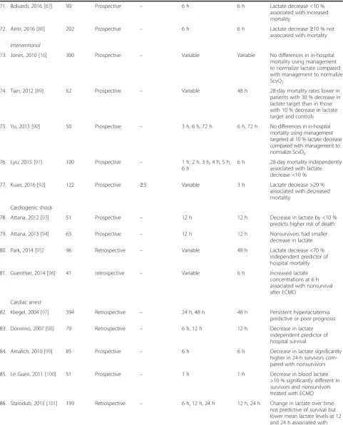
reduced elimination, or both. A dynamic evaluation of
serial lactate concentrations may thus be more
inform-ative than a single value. This concept of repeating blood
lactate concentrations over time as an indicator of
re-sponse to therapy was first proposed in 1983 [12], based
on an idea raised after a publication by Orringer et al. in
1977 [13] showing that the decrease in lactate levels
after cessation of grand mal seizures was actually quite
rapid, with a half-life of about 50 % in 1 hr. Many
stud-ies have since emphasized that changes in lactate over
the first hrs of treatment may represent a valuable
moni-toring tool. Some studies have even proposed integrating
changes in lactate concentrations as a target in
thera-peutic protocols [14
–
17] or including them as one of the
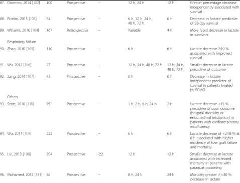
sepsis resuscitation
“
bundles
”
[18]. A number of
investi-gators have used the term
“
lactate clearance
”
to describe
decreasing lactate levels, but this is incorrect for two
reasons. The first is that the changes in lactate
concen-trations over time reflect changes in production and in
elimination. The decrease in lactate over time may
re-flect decreased (over)production more than increased
clearance by the liver and other organs [19, 20]. The
spe-cific study of lactate clearance would require intravenous
injection of radiolabeled lactate, as has been done in
sev-eral studies [21, 22]. The second reason why use of the
term is incorrect is that
“
clearance
”
or
“
elimination
”
implies
a progressive normalization of blood lactate concentrations,
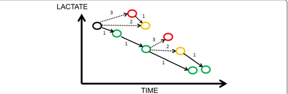
which is too simplistic. Blood lactate concentrations can
have a complex evolution and may even increase over time
(Fig. 1), a situation that one should then call
“
negative
lactate clearance
”
.
We performed a literature search on this subject to
address several questions. First, is the observation of a
better prognosis with decreasing lactate concentrations a
consistent finding in all types of critically ill patient?
Second, although some studies have suggested that
re-peated lactate measurements may be particularly useful
in sepsis, can similar observations be made in other
acute disease states or even in heterogeneous groups of
critically ill patients? Third, how fast should lactate
con-centrations decrease in optimal conditions and is there
any particular time interval that could be recommended?
Fourth, some studies in emergency medicine considered
only patients with lactate values > 4 mEq/l as an at-risk
population, but is this approach valid? In other words, is
the study of lactate kinetics more useful when lactate
concentrations exceed a given value?
Methods
We searched databases of PubMed, Science Direct, and
Embase until the end of February 2016 to identify studies
that evaluated the capacity of serial blood lactate
concen-trations to predict outcome, using the search terms
“
Lactate levels
”
OR
“
lactate clearance
”
AND
“
shock
”
OR
“
critically ill
”
AND
“
mortality
”
. We included original
prospective or retrospective clinical studies. There was no
restriction based on language. We excluded studies in
pediatric populations, experimental studies, case reports,
and studies that did not report changes over time in
lactate values or relationship of changes in lactate
concen-trations to all-cause mortality rates. We had no restriction
on the initial location in the hospital (e.g., ICU, trauma
unit, emergency room, operating room). We also checked
the reference lists of included articles to capture any
refer-ences missed during the search. We classified the different
adult populations into general ICU patients, general
surgical ICU patients, cardiac surgery patients, trauma
patients, patients with sepsis, patients with cardiogenic
shock, post-cardiac arrest patients, patients with
respira-tory failure, and others.
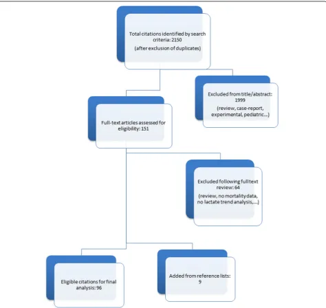
[image:2.595.61.540.538.694.2]Results
A total of 96 studies met our inclusion criteria (Fig. 2,
Table 1).
General ICU patients
Observational studies
We identified 13 observational studies in heterogeneous
critically ill populations [6, 10
–
12, 23
–
31]. All of these
studies indicated that nonsurvivors had persistently
higher lactate concentrations over time than survivors.
Only one study [26] reported that lactate reduction
dur-ing the first 24 hrs of ICU stay was useful only in septic
patients, but not in patients with hemorrhage or other
conditions.
The suggested optimal timing of lactate measurements
was not precisely defined in several of the studies that
evaluated the course of lactate concentrations over time.
The studies that did include a time interval usually
selected 6, 12 or even 24 hrs.
Interventional studies
An interventional trial of 348 patients by Jansen et al. [15]
targeted a lactate decrease of at least 20 % in 2 hrs for the
initial 8 hrs of treatment in ICU patients with an initial
lactate
≥
3 mEq/l. This strategy was associated with a
lower mortality rate in the lactate-guided therapy group
after adjustment for predefined risk factors (hazard ratio
(HR), 0.61; confidence interval (CI), 0.43
–
0.87).
[image:3.595.62.537.265.713.2]Table 1
Included studies according to population type
First author, year [reference]
Number of patients
Study design Initial minimum lactate for patient inclusion
Timing of measurements
Suggested time interval
Comments
General ICU/emergency department
Observational
1. Vincent, 1983 [12] 17 Prospective ≥4 Every 20 min during first 2 h of ICU treatment
1 h Decrease >10 % associated with survival
2. Cowan, 1984 [23] 30 Prospective – 3 h, 24 h 3 h Change in lactate predictive of
outcome but less so than simple hemodynamic variables
3. Suistomaa, 2000 [24] 100 Prospective – Every 2 h for 24 h 6 h Failure to decrease lactate at 6 h associated with higher mortality
4. Jansen, 2008 [31] 106 Prospective – Variable (at
ambulance pickup and at ER arrival)
— Decrease in lactate
independently associated with decreased hazard of death
5. Wang, 2009 [25] 101 NR ≥2 12 h, 24 h 12 h Decrease≤10 % associated
with increased mortality
6. Jansen, 2009 [26] 394 Prospective – 12 h, 24 h 12 h Decrease in lactate only of
prognostic value in patients with sepsis
7. Krishna, 2009 [27] 50 Prospective – 12 h, 24 h, 36 h 24 h, 36 h Decreasing levels associated with survival
8. Soliman, 2010 [28] 433 Prospective – 24 h, 48 h 24 h Higher lactate concentrations
at 24 and 48 h after admission associated with decreased survival
9. Nichol, 2010 [6] 7155 Retrospective – Variable 24 h Time-weighted average lactate
over 24 h independent predictor of mortality
10. Nichol, 2011 [10] 5041 Retrospective – Variable 24 h Time-weighted average lactate
and change in lactate over 24 h independent predictors of hospital mortality
11. van Beest, 2013 [29] 2251 Retrospective – Variable 6 h Normalization of lactate <6 h after ICU admission associated with better hospital survival than normalization of lactate >6 hrs
12. Zhang, 2014 [30] 6291 Retrospective >2 Variable Variable Normalization and speed of normalization related to outcome
13. Haas, 2016 [11] 400 Retrospective >10 Variable 12 h No decrease in lactate over
12 h associated with increased mortality
Interventional
14. Jansen, 2010 [15] 348 Prospective ≥3.0 2 h 8 h Objective was to decrease
lactate by 20 % or more per 2 h for the initial 8 h of ICU stay. Lactate-guided therapy was independently associated with reduced hospital mortality
Surgical ICU
15. McNelis, 2001 [32] 95 Retrospective – 8-h intervals until lactate normalized
Variable Time to lactate normalization predictive of outcome
Table 1
Included studies according to population type
(Continued)
17. Meregalli, 2004 [34] 44 Prospective – 12 h, 24 h, 48 h 48 h Blood lactate concentrations decreased with time in survivors, but remained stable in nonsurvivors
18. Cardinal Fernandez, 2009 [35]
108 Prospective >2 6 h 6 h Decrease in lactate by >40 %
associated with increased survival
19. Ibrahim, 2013 [36] 322 Prospective – 8 h, 16 h, 24 h 16 h Percent change in blood lactate at 16 h independent predictor of postoperative mortality
Cardiac surgery
20. Lindsay, 2013 [37] 1291 Retrospective – Variable Variable Longer predicted time to reach normal lactate (<1.5 mmol/l) associated with increased mortality
21. Hajjar, 2013 [38] 502 Prospective – 6 h, 12 h 6 h, 12 h Failure to decrease lactate associated with major complications, including death
22. Park, 2014 [39] 115 Retrospective – 6 h, 12 h, 24 h 6 h, 12 h,
24 h
Lack of decrease in lactate predictive of mortality
23. Lopez-Delgado, 2015 [40]
2935 Prospective – 6 h, 12 h, 24 h 24 h Later peak in lactate associated
with higher hospital and long-term mortality
24. Li, 2015 [41] 123 Retrospective – 6 h, 12 h 12 h Lactate decrease predictive of
in-hospital mortality in patients receiving ECMO
Trauma
Observational
25. Abramson, 1993 [42] 76 Prospective – 8 h, 16 h, 24 h, 36 h, 48 h
24 h Normalization of lactate by 24 h associated with 100 % survival
26. Manikis, 1995 [43] 129 Retrospective – At least three times a day
Variable Duration of hyperlactatemia correlated with the development of organ failure but not with mortality
27. Holm, 2000 [44] 21 Prospective – 12 h, 24 h, 48 h, 72 h Variable Decreasing lactate levels associated with survival
28. Cerovic, 2003 [45] 98 Prospective – Twice daily during first 2 days and once daily during next 3 days
Variable Reduced lactate levels in survivors
29. Kamolz, 2005 [46] 166 Prospective – Variable 24 h Higher mortality in patients
with initial lactate > 2 mmol/l if lactate not normalized at 24 h
30. Billeter, 2009 [47] 1032 Retrospective – Variable 24 h Delayed or absent decrease in
lactate associated with infectious complications but not mortality
31. Regnier, 2012 [48] 281 Prospective – 2 h, 4 h 2 h Early normalization of lactate
independent predictor of survival
32. Dubendorfer, 2013 [49]
724 Retrospective – Variable Variable In patients without traumatic
brain injury, decrease in lactate impaired in nonsurvivors
33. Odom, 2013 [50] 623 Retrospective ≥4 6 h 6 h Lower decrease in lactate at
Table 1
Included studies according to population type
(Continued)
34. Heinonen, 2014 [51] 610 Retrospective – Variable Variable Failure to normalize lactate associated with increased mortality
35. Freitas, 2015 [52] 117 Retrospective – 6 h 6 h No correlation between
decrease in lactate and mortality
36. Dezman, 2015 [53] 3887 Retrospective ≥3 Variable Variable No decrease in lactate independent predictor of 24-h mortality
Interventional
37. Blow, 1999 [55] 79 Retrospective – Variable 24 h Failure to decrease lactate
associated with increased mortality
38. Claridge, 2000 [56] 364 Prospective – Variable 12 h Increase in infections, length of stay, and mortality if lactate did not normalize by 12 h
Sepsis
Observational
39. Bakker, 1991 [57] 48 Prospective >2 Variable Variable Only survivors had a significant decrease in blood lactate concentrations during the course of septic shock
40. Friedman, 1995 [58] 35 Prospective >2 4 h, 24 h Variable Lactate remained high in nonsurvivors and progressively decreased in survivors
41. Bernardin, 1996 [59] 32 Prospective – 24 h 24 h Greater decrease in lactate in
survivors
42. Marecaux, 1996 [60] 38 Prospective >2 24 h, 48 h 24 h, 48 h Greater decrease in lactate in survivors
43. Bakker, 1996 [61] 87 Prospective >2 Variable Variable Duration of lactic acidosis best discriminant of survival
44. Kobayashi, 2001 [62] 22 Prospective – Every 4 hours for 4 days
Variable Decrease in lactate associated with survival
45. Nguyen, 2004 [63] 111 Prospective – 6 h 6 h Decrease in lactate≥10 %
associated with lower 60-day mortality
46. Nguyen, 2007 [64] 330 Prospective – Variable 6 h Decreased odds ratio for
mortality in patients with decreased lactate
47. Phua, 2008 [65] 72 Prospective – 24 h, 48 h 24 h Increase in lactate predictive of
mortality
48. Yang, 2009 [66] 105 Prospective – 6 h, 24 h, 72 h 6 h Decrease in lactate at 6 h
≥30 % was independent predictor of survival
49. Arnold, 2009 [67] 166 Retrospective – 6 h 6 h Lactate decrease by less than
10 % independent predictor of in-hospital death
50. Nguyen, 2010 [68] 220 Retrospective – 6 h 6 h Larger decrease in lactate
associated with decreased mortality up to 12 months
51. Nguyen, 2011 [18] 556 Prospective – 12 h 12 h Any decrease in lactate within
12 h from baseline or an initial lactate <2 mmol/l
independently associated with reduced mortality
52. Puskarich, 2012 [69] 203 Retrospective analysis of data from [16]
– 2 h, 4 h, 6 h 6 h ≥10 % decrease in lactate
Table 1
Included studies according to population type
(Continued)
53. Zanaty, 2012 [70] 53 Prospective – 6 h 6 h <15 % decrease in lactate
independent predictor of mortality
54. Puskarich, 2013 [71] 187 Retrospective analysis of data from [16]
– At least two lactate
measurements in first 6 h
6 h Lactate normalization in 6 h stronger independent predictor of survival than decrease in lactate by≥50 %
55. Walker, 2013 [72] 78 Retrospective – 6 h 6 h Decrease in lactate
independently associated with mortality, with optimal cut-off of 36 %
56. Liu, 2013 [73] 9190 Retrospective ≥2 4 h, 8 h, 12 h 12 h Reduced mortality in patients with more than 60 % lactate improvement at 12 h.
57. Marty, 2013 [74] 94 Prospective – 6 h, 12 h, 24 h 24 h Decrease in lactate at 24 h independently correlated to survival
58. Park, 2014 [75] 25 Prospective – 6 h, 12 h, 18 h, 24 h, 48 h
48 h Normalization independent predictor of survival
59. Permpikul, 2014 [76] 51 Prospective – 6 h 6 h Lactate decrease associated
with reduced 28-day mortality
60. Bao, 2015 [77] 94 Retrospective – 3 h, 6 h, 24 h 24 h 24-h lactate decrease predictive of outcome
61. Galbois, 2015 [78] 42 Prospective – 6 h, 12 h, 18 h, 24 h 6 h Lesser decrease in lactate associated with 14-day mortality
62. Lee, 2015 [79] 109 Retrospective >3.3 6 h, 24 h, 48 h 6 h, 24 h, 48 h
Decrease in lactate of <10 % in the first 6 h, 24 h, and 48 h independently associated with mortality
63. Dettmer, 2015 [17] 243 Retrospective ≥4 Variable Variable Greater reduction in lactate associated with decreased 28-day mortality
64. Lokhandwala, 2015 [80]
74 Retrospective ≥4 Variable Variable Lactate decrease < 4 mmol/l
associated with increased hospital morality
65. Wang, 2015 [81] 115 Prospective – 6 h, 12 h, 18 h, 24 h 24 h Lower lactate area score and percentage decrease in lactate associated with increased mortality
66. Bhat, 2015 [82] 207 Retrospective – Variable Variable Higher mortality in patients
with no decrease in lactate
67. Chertoff, 2016 [83] 229 Retrospective – 24-48 h 24-48 h Lower decrease in plasma
lactate 24–48 h after initiation of treatment was associated with higher 30-day mortality
68. Drumheller, 2016 [84] 411 Retrospective ≥4 Variable Variable Decrease in lactate
independently associated with decreased risk of death
69. He, 2016 [85] 84 Prospective – 8 h 8 h Patients with lactate decrease
≥10 % had lower ICU mortality than those with lactate decrease <10 %
70. Ha, 2016 [86] 208 – 6 h, 24 h 24 h Low decrease in lactate at 6
Table 1
Included studies according to population type
(Continued)
71. Bolvardi, 2016 [87] 90 Prospective – 6 h 6 h Lactate decrease <10 %
associated with increased mortality
72. Amir, 2016 [88] 202 Prospective – 6 h 6 h Lactate decrease≥10 % not
associated with mortality
Interventional
73. Jones, 2010 [16] 300 Prospective – Variable Variable No differences in in-hospital
mortality using management to normalize lactate compared with management to normalize ScvO2
74. Tian, 2012 [89] 62 Prospective – Variable 48 h 28-day mortality rates lower in
patients with 30 % decrease in lactate target than in those with 10 % decrease in lactate target and controls
75. Yu, 2013 [90] 50 Prospective – 3 h, 6 h, 72 h 6 h, 72 h No differences in in-hospital mortality using management targeted at 10 % lactate decrease compared with management to normalize ScvO2
76. Lyu, 2015 [91] 100 Prospective – 1 h, 2 h, 3 h, 4 h, 5 h, 6 h
6 h 28-day mortality independently associated with lactate decrease <10 %
77. Kuan, 2016 [92] 122 Prospective ≥3 Variable 3 h Lactate decrease >20 %
associated with decreased mortality
Cardiogenic shock
78. Attana, 2012 [93] 51 Prospective – 12 h 12 h Decrease in lactate by <10 %
predicts higher risk of death
79. Attana, 2013 [94] 63 Prospective – 12 h 12 h Nonsurvivors had smaller
decrease in lactate
80. Park, 2014 [95] 96 Retrospective – Variable 48 h Lactate decrease <70 %
independent predictor of hospital mortality
81. Guenther, 2014 [96] 41 retrospective – Variable 6 h Increased lactate
concentrations at 6 h associated with nonsurvival after ECMO
Cardiac arrest
82. Kliegel, 2004 [97] 394 Retrospective – 24 h, 48 h 48 h Persistent hyperlactatemia
predictive or poor prognosis
83. Donnino, 2007 [98] 79 Retrospective – 6 h, 12 h 12 h Decrease in lactate
independent predictor of hospital survival
84. Arnalich, 2010 [99] 85 Prospective – 6 h 6 h Decrease in lactate significantly
higher in 24-h survivors com-pared with nonsurvivors
85. Le Guen, 2011 [100] 51 Prospective – 1 h 1 h Decrease in blood lactate
>10 % significantly different in survivors and nonsurvivors treated with ECMO
Surgical patients
We identified five observational studies conducted in
general surgical ICU patients [32
–
36]. Failure of lactate
concentrations to decrease over time was associated with
worse outcomes in all studies.
After cardiac surgery
There were five observational studies in cardiac surgery
patients [37
–
41], including two studies in patients
treated with extracorporeal membrane oxygenation
(ECMO) post cardiac surgery [39, 41]. All studies
con-sistently demonstrated differences in changes in lactate
concentration between survivors and nonsurvivors.
Trauma patients
Observational studies
[image:9.595.57.536.101.466.2]We identified twelve observational studies in trauma
patients [42
–
53]. Three retrospective studies reported no
association of change in lactate levels with mortality
[43, 47, 52], although Manikis et al. [43] reported that
the duration of hyperlactatemia was associated with the
development of organ failure and Billeter et al. [47] noted
that delayed or no reduction in blood lactate was
associ-ated with increased infectious complications. Several small
studies used relatively long time intervals of 12
–
24 hrs
[45, 54]. One study reported that repeated lactate after
2 hrs could be valuable [48] and a retrospective study
pro-posed a time limit of 6 hrs [50].
Interventional studies
In a retrospective analysis of a small prospective cohort
managed according to a protocol to normalize blood
lactate levels, Blow et al. [55] reported that failure to
normalize blood lactate levels (<2.5 mmol/l) was
associ-ated with increased morbidity and mortality. In an
interventional study by Claridge et al. [56], patients were
managed according to the same protocol targeted at
re-ducing lactate levels to <2.4 mmol/l. Failure to achieve
this target was associated with increased risk of
infec-tion, increased length of stay, and increased mortality.
Table 1
Included studies according to population type
(Continued)
87. Donnino, 2014 [102] 100 Prospective – 12 h, 24 h 12 h Greater percentage decrease
independently associated with survival
88. Riveiro, 2015 [103] 54 Prospective – 6 h, 12 h, 24 h, 48 h, 72 h
6 h Decrease in lactate predictive of 28-day survival
89. Williams, 2016 [104] 167 Retrospective – Variable 4 h More rapid decrease in lactate
in survivors
Respiratory failure
90. Zhao, 2010 [105] 110 Prospective – 6 h 6 h Lactate decrease≥10 %
associated with improved survival
91. Wu, 2012 [106] 27 Prospective – 12 h, 24 h, 48 h, 72 h 12 h, 24 h, 48 h, 72 h
Smaller decrease in lactate predictive of outcome
92. Zang, 2014 [107] 43 Prospective – 6 h 6 h Decrease in lactate
independent predictor of survival in patients treated by ECMO
Others
93. Scott, 2010 [110] 95 Prospective – 1 h, 2 h, 6 h, 24 h 2 h Lactate decrease <15 % predictive of poor outcome (hospital mortality or endotracheal intubation) in patients with cardiorespiratory insufficiency
94. Wu, 2011 [109] 222 Prospective – 6 h 6 h Lactate decrease of <24.8 % at
6 h associated with higher incidence of liver graft failure and mortality
95. Lui, 2013 [108] 204 Prospective ≥2 12 h 12 h Smaller decrease in lactate
associated with increased mortality in patients with paraquat poisoning
96. Mohamed, 2014 [111] 46 Prospective – 8 h, 24 h 24 h Mortality greater if <40 %
decrease in lactate
Patients with sepsis
Observational studies
We found thirty four observational studies in patients
with sepsis [17, 18, 57
–
88]. One study reported that a
decrease in lactate levels of
≥
10 % was not associated
with mortality [88], but this study was conducted in a
low-resource setting, such that resuscitation may not
have been optimal as acknowledged by the authors.
Several studies reported that 6-hrly changes could be a
useful guide [63, 64, 66, 67, 69
–
72, 78].
Interventional studies
One interventional study by Jones et al. [16] compared
resuscitation based on lactate concentrations with a
target of obtaining a >10 % decrease from the initial
value with resuscitation based on achieving central
venous oxygen saturation (ScvO
2)
≥
70 %; there were no
differences in outcome between the two strategies. In an
analysis of patients in this trial who had simultaneous
lactate and ScvO
2measurements, Puskarich et al. [69]
concluded that failure to achieve the target lactate
de-crease was associated with a worse prognosis than
fail-ure to achieve the ScvO
2target. In a small Chinese
study [89], patients randomized to a 30 % decrease in
lactate target had better 28-day survival than those
ran-domized to a 10 % target or to control, and in another
small study [90] there were no differences in in-hospital
mortality using management targeted at a 10 % decrease
in lactate compared with management to normalize
ScvO
2. Two other Chinese studies reported that patients
randomized to lactate-directed therapy had improved
outcomes [91, 92].
Patients with cardiogenic shock
There were four studies in patients with cardiogenic
shock [93
–
96], all showing that lactate concentrations
decreased more in survivors than in nonsurvivors.
After cardiac arrest
We identified eight observational studies [97
–
104] in
post-cardiac arrest patients. All but one [101] of these
studies demonstrated differences in changes in lactate
concentration between survivors and nonsurvivors.
Patients with acute respiratory failure
We found three observational studies in patients with
acute respiratory failure [105
–
107], all showing that
decreasing lactate levels were predictive of survival.
Other conditions
Changes in lactate concentrations were also reported
following paraquat poisoning [108], after liver
trans-plantation [109], in patients with acute cardiorespiratory
failure [110], and in patients with severe
community-acquired pneumonia [111]. All studies indicated the
value of repeated lactate concentrations in these patient
populations.
Discussion
Our literature review clearly supports the value of serial
lactate measurements in the evaluation of critically ill
patients and their response to therapy. This observation
was similar across all studies and in all categories of
patients, without being restricted to those with sepsis.
We found only one study which suggested that
evaluat-ing the time course of lactate concentrations would be
useful in sepsis patients but not in other conditions
[26], and just five studies reporting no predictive
effect of decrease in lactate levels over time on mortality
[43, 47, 52, 88, 101] although two of these did suggest a
relationship with morbidity outcomes [43, 47].
Re-peated lactate concentrations can also help separate
patients with complications, such as neurological
complications after cardiac arrest [112, 113] or after
surgery [38]. A meta-analysis of these data is
compli-cated by the heterogeneity of the populations and the
different timings of the measurements, but the data
are very consistent across studies.
Increased lactate concentrations can be due to factors
other than cellular hypoxia, so the decrease in blood
lactate concentrations may not just be the result of
improvements
in
cellular
oxygen
availability.
For
example, beta-adrenergic stimulation may contribute to
increased lactate production [114]. A recent study
indi-cated the reverse phenomenon; that is, the increase in
lactate concentrations seen in patients with sepsis may
be blunted in patients previously treated with
beta-blocking agents [115]. The infusion of lactate-containing
intravenous solutions may also potentially complicate
the interpretation of blood lactate concentrations [116],
although the amount of fluid infused must be very large
to have such an effect [117]. A recent study also
reported that lactate levels decrease more slowly in
patients with a positive blood alcohol level, thus
compli-cating evaluation of blood lactate levels in these
patients [118].
with any global changes in lactate clearance,
al-though lactate half-life was prolonged. A recent
ex-perimental study indicated that liver hypoperfusion
is unlikely to contribute to increased blood lactate
concentrations [121]. In patients with
paracetamol-induced acute liver failure, higher lactate
concentra-tions were associated with more severe organ failure
and mortality [122].
Some investigators have compared lactate and ScvO
2or combined the two measures. Lactate is usually a
better prognostic marker [69]. But is it actually necessary
to choose? In an interventional study in patients with
sepsis, Jones et al. [16] reported no differences in
out-comes for patients managed according to lactate
concen-trations or to ScvO
2values, but it is difficult to evaluate
how these measurements really guided therapy because
there were no differences in administered treatments
during the first 72 hrs. In post-cardiac surgery patients,
Polonen et al. [14] reported better outcomes when
ScvO
2and lactate concentrations were targeted together
than in control patients. The most convincing evidence
in favor of lactate as a target comes from the study by
Jansen et al. [15] in which outcomes were improved in
patients treated to a target of a 20 % decrease in lactate
concentrations. Nevertheless, the relatively slow changes
in lactate make it difficult to interpret these results
—
the
trend analysis is more a marker of effective treatment
than a target in itself.
Although changes in blood lactate kinetics were clearly
significant after 6 hrs in many studies and after 12 hrs in
most, it is currently not possible to define the best time
interval between lactate measurements. The normal
re-duction in lactate concentrations when overprore-duction
of lactate abruptly ceases after grand mal seizures is
about 50 % in 1 hr [13]. Although Levraut et al. [21]
sug-gested that lactate clearance may be decreased in septic
patients, Revelly et al. [22] reported similar values in
pa-tients with sepsis and in healthy volunteers.
The rate of lactate decrease in optimal treatment
conditions is quite variable. In the best conditions,
blood lactate concentrations decreased by more than
10 % in 1 hr in patients who responded rapidly to
re-suscitation [12] or by 10
–
20 % in 2 hrs [15]. A study
by Hernandez et al. [123] suggested a >50 % decrease
in lactate concentrations during the first 6 hrs of
re-suscitation in patients with septic shock. Although
some systems now allow the quasi-continuous
meas-urement of lactate
concentrations, determinations
every 1
–
2 hrs are probably sufficient; in the
interven-tional study by Jansen et al. [15] the protocol was to
measure blood lactate every 2 hrs. Even though serial
blood lactate concentrations have been suggested to
guide therapy, our review underlines that changes in
lactate over time are relatively slow, taking place over
hrs, and this may be too slow to guide therapy. Serial
lactate concentrations should serve as a regular
con-trol, similar to how in the past a navigator would
consult a compass from time to time to ensure that
their boat was still heading in the right direction. If
lactate concentrations do not normalize over time,
the need for changes in therapy should be considered.
Conclusion
Our systematic literature review has provided the
following answers to our initial questions. First,
obser-vation of a better prognosis with decreasing lactate
concentrations is consistent throughout the literature.
Second, these observations are not specific to septic
patients, but apply to all common situations of
hyper-lactatemia and in heterogeneous patient populations.
Third, the changes are relatively slow, and it is
difficult to provide recommendations about the speed
of decrease in lactate concentrations in the best
con-ditions. Clearly repeating measurements every 12 hrs
can generally separate those who will do well from
those who are likely to die, but shorter time intervals
may be helpful. On the basis of our observations, we
would recommend checking blood lactate
concentra-tions as often as every 1
–
2 hrs in acute conditions.
Fourth, the study of lactate kinetics appears to be
valid regardless of the initial value and not only in
patients with severe hyperlactatemia.
Abbreviations
ECMO, extracorporeal membrane oxygenation; ScvO2, central venous oxygen saturation
Acknowledgements None.
Funding
Institutional funds only.
Availability of supporting data Not applicable.
Authors’contributions
AQeS and LC performed the literature search and drafted the manuscript. J-LV and FST reviewed the article for critical content. All authors read and approved the final manuscript.
Competing interests
The authors declare that they have no competing interests.
Consent for publication Not applicable.
Ethics approval and consent to participate Not applicable.
References
1. Broder G, Weil MH. Excess lactate: an index of reversibility of shock in human patients. Science. 1964;143:1457–9.
2. Weil MH, Afifi AA. Experimental and clinical studies on lactate and pyruvate as indicators of the severity of acute circulatory failure (shock). Circulation. 1970;41:989–1001.
3. Peretz DI, McGregor M, Dossetor JB. Lactic acidosis: a clinically significant aspect of shock. Can Med Assoc J. 1964;90:673–5.
4. Vincent JL, De Backer D. Circulatory shock. N Engl J Med. 2013;369:1726–34. 5. Levy B. Lactate and shock state: the metabolic view. Curr Opin Crit Care.
2006;12:315–21.
6. Nichol AD, Egi M, Pettila V, Bellomo R, French C, Hart G, et al. Relative hyperlactatemia and hospital mortality in critically ill patients: a retrospective multi-centre study. Crit Care. 2010;14:R25.
7. Singer M, Deutschman CS, Seymour CW, Shankar-Hari M, Annane D, Bauer M, et al. The Third International Consensus Definitions for Sepsis and Septic Shock (Sepsis-3). JAMA. 2016;315:801–10.
8. Kraut JA, Madias NE. Lactic acidosis. N Engl J Med. 2014;371:2309–19. 9. Cecconi M, De Backer D, Antonelli M, Beale R, Bakker J, Hofer C, et al.
Consensus on circulatory shock and hemodynamic monitoring. Task force of the European Society of Intensive Care Medicine. Intensive Care Med. 2014;40:1795–815.
10. Nichol A, Bailey M, Egi M, Pettila V, French C, Stachowski E, et al. Dynamic lactate indices as predictors of outcome in critically ill patients. Crit Care. 2011;15:R242.
11. Haas SA, Lange T, Saugel B, Petzoldt M, Fuhrmann V, Metschke M, et al. Severe hyperlactatemia, lactate clearance and mortality in unselected critically ill patients. Intensive Care Med. 2016;42:202–10.
12. Vincent JL, Dufaye P, Berre J, Leeman M, Degaute JP, Kahn RJ. Serial lactate determinations during circulatory shock. Crit Care Med. 1983;11:449–51. 13. Orringer CE, Eustace JC, Wunsch CD, Gardner LB. Natural history of lactic
acidosis after grand-mal seizures. A model for the study of an anion-gap acidosis not associated with hyperkalemia. N Engl J Med. 1977;297:796–9. 14. Polonen P, Ruokonen E, Hippelainen M, Poyhonen M, Takala J. A
prospective, randomized study of goal-oriented hemodynamic therapy in cardiac surgical patients. Anesth Analg. 2000;90:1052–9.
15. Jansen TC, van Bommel J, Schoonderbeek FJ, Sleeswijk Visser SJ, van der Klooster JM, Lima AP, et al. Early lactate-guided therapy in intensive care unit patients: a multicenter, open-label, randomized controlled trial. Am J Respir Crit Care Med. 2010;182:752–61.
16. Jones AE, Shapiro NI, Trzeciak S, Arnold RC, Claremont HA, Kline JA. Lactate clearance vs central venous oxygen saturation as goals of early sepsis therapy: a randomized clinical trial. JAMA. 2010;303:739–46.
17. Dettmer M, Holthaus CV, Fuller BM. The impact of serial lactate monitoring on emergency department resuscitation interventions and clinical outcomes in severe sepsis and septic shock: an observational cohort study. Shock. 2015;43:55–61.
18. Nguyen HB, Kuan WS, Batech M, Shrikhande P, Mahadevan M, Li CH, et al. Outcome effectiveness of the severe sepsis resuscitation bundle with addition of lactate clearance as a bundle item: a multi-national evaluation. Crit Care. 2011;15:R229.
19. Vincent JL. Serial blood lactate levels reflect both lactate production and clearance. Crit Care Med. 2015;43:e209.
20. Vincent JL. Lactic acidosis. N Engl J Med. 2015;372:1077–8.
21. Levraut J, Ciebiera JP, Chave S, Rabary O, Jambou P, Carles M, et al. Mild hyperlactatemia in stable septic patients is due to impaired lactate clearance rather than overproduction. Am J Respir Crit Care Med. 1998;157:1021–6.
22. Revelly JP, Tappy L, Martinez A, Bollmann M, Cayeux MC, Berger MM, et al. Lactate and glucose metabolism in severe sepsis and cardiogenic shock. Crit Care Med. 2005;33:2235–40.
23. Cowan BN, Burns HJ, Boyle P, Ledingham IM. The relative prognostic value of lactate and haemodynamic measurements in early shock. Anaesthesia. 1984;39:750–5.
24. Suistomaa M, Ruokonen E, Kari A, Takala J. Time-pattern of lactate and lactate to pyruvate ratio in the first 24 hrs of intensive care emergency admissions. Shock. 2000;14:8–12.
25. Wang H, Wu DW, Chen XM, Li C, Ding SF, Zhai Q, et al. Relationship between blood lactic level, lactic clearance, duration of lacticemia and prognosis of critically ill patients in intensive care unit. Zhongguo Wei Zhong Bing Ji Jiu Yi Xue. 2009;21:357–60.
26. Jansen TC, van Bommel J, Mulder PG, Lima AP, van der Hoven B, Rommes JH, et al. Prognostic value of blood lactate levels: does the clinical diagnosis at admission matter? J Trauma. 2009;66:377–85.
27. Krishna U, Joshi SP, Modh M. An evaluation of serial blood lactate measurement as an early predictor of shock and its outcome in patients of trauma or sepsis. Indian J Crit Care Med. 2009;13:66–73.
28. Soliman HM, Vincent JL. Prognostic value of admission serum lactate concentrations in intensive care unit patients. Acta Clin Belg. 2010;65:176–81.
29. van Beest PA, Brander L, Jansen SP, Rommes JH, Kuiper MA, Spronk PE. Cumulative lactate and hospital mortality in ICU patients. Ann Intensive Care. 2013;3:6.
30. Zhang Z, Chen K, Ni H, Fan H. Predictive value of lactate in unselected critically ill patients: an analysis using fractional polynomials. J Thorac Dis. 2014;6:995–1003.
31. Jansen TC, van Bommel J, Mulder PG, Rommes JH, Schieveld SJ, Bakker J. The prognostic value of blood lactate levels relative to that of vital signs in the pre-hospital setting: a pilot study. Crit Care. 2008;12:R160.
32. McNelis J, Marini CP, Jurkiewicz A, Szomstein S, Simms HH, Ritter G, et al. Prolonged lactate clearance is associated with increased mortality in the surgical intensive care unit. Am J Surg. 2001;182:481–5.
33. Husain FA, Martin MJ, Mullenix PS, Steele SR, Elliott DC. Serum lactate and base deficit as predictors of mortality and morbidity. Am J Surg. 2003;185:485–91.
34. Meregalli A, Oliveira RP, Friedman G. Occult hypoperfusion is associated with increased mortality in hemodynamically stable, high-risk, surgical patients. Crit Care. 2004;8:R60–5.
35. Cardinal Fernandez PA, Olano E, Acosta C, Bertullo H, Albornoz H, Bagnulo H. Prognostic value of lactate clearance in the first 6 hrs of intensive medicine course. Med Intensiva. 2009;33:166–70.
36. Ibrahim WA, Ahmed AS. Serial estimations of blood lactate predict postoperative outcome in cancer patients undergoing head and neck surgeries. Egyptian J Anaesth. 2013;29:149–54.
37. Lindsay AJ, Xu M, Sessler DI, Blackstone EH, Bashr CA. Lactate clearance time and concentration linked to morbidity and death in cardiac surgical patients. Ann Thorac Surg. 2013;95:486–92.
38. Hajjar LA, Almeida JP, Fukushima JT, Rhodes A, Vincent JL, Osawa EA, et al. High lactate levels are predictors of major complications after cardiac surgery. J Thorac Cardiovasc Surg. 2013;146:455–60.
39. Park SJ, Kim SP, Kim JB, Jung SH, Choo SJ, Chung CH, et al. Blood lactate level during extracorporeal life support as a surrogate marker for survival. J Thorac Cardiovasc Surg. 2014;148:714–20.
40. Lopez-Delgado JC, Esteve F, Javierre C, Torrado H, Rodriguez-Castro D, Carrio ML, et al. Evaluation of serial arterial lactate levels as a predictor of hospital and long-term mortality in patients after cardiac surgery. J Cardiothorac Vasc Anesth. 2015;29:1441–53.
41. Li CL, Wang H, Jia M, Ma N, Meng X, Hou XT. The early dynamic behavior of lactate is linked to mortality in postcardiotomy patients with extracorporeal membrane oxygenation support: A retrospective observational study. J Thorac Cardiovasc Surg. 2015;149:1445–50.
42. Abramson D, Scalea TM, Hitchcock R, Trooskin SZ, Henry SM, Greenspan J. Lactate clearance and survival following injury. J Trauma. 1993;35:584–8. 43. Manikis P, Jankowski S, Zhang H, Kahn RJ, Vincent JL. Correlation of serial
blood lactate levels to organ failure and mortality after trauma. Am J Emerg Med. 1995;13:619–22.
44. Holm C, Melcer B, Horbrand F, Worl HH, von Donnersmarck GH, Muhlbauer W. Haemodynamic and oxygen transport responses in survivors and non-survivors following thermal injury. Burns. 2000;26:25–33. 45. Cerovic O, Golubovic V, Spec-Marn A, Kremzar B, Vidmar G. Relationship
between injury severity and lactate levels in severely injured patients. Intensive Care Med. 2003;29:1300–5.
46. Kamolz LP, Andel H, Schramm W, Meissl G, Herndon DN, Frey M. Lactate: early predictor of morbidity and mortality in patients with severe burns. Burns. 2005;31:986–90.
47. Billeter A, Turina M, Seifert B, Mica L, Stocker R, Keel M. Early serum procalcitonin, interleukin-6, and 24-hr lactate clearance: useful indicators of septic infections in severely traumatized patients. World J Surg.
2009;33:558–66.
49. Dubendorfer C, Billeter AT, Seifert B, Keel M, Turina M. Serial lactate and admission SOFA scores in trauma: an analysis of predictive value in 724 patients with and without traumatic brain injury. Eur J Trauma Emerg Surg. 2013;39:25–34.
50. Odom SR, Howell MD, Silva GS, Nielsen VM, Gupta A, Shapiro NI, et al. Lactate clearance as a predictor of mortality in trauma patients. J Trauma Acute Care Surg. 2013;74:999–1004.
51. Heinonen E, Hardcastle TC, Barle H, Muckart DJ. Lactate clearance predicts outcome after major trauma. Afr J Emerg Med. 2014;4:61–5.
52. Freitas AD, Franzon O. Lactate as predictor of mortality in polytrauma. Arq Bras Cir Dig. 2015;28:163–6.
53. Dezman ZD, Comer AC, Smith GS, Narayan M, Scalea TM, Hirshon JM. Failure to clear elevated lactate predicts 24-hr mortality in trauma patients. J Trauma Acute Care Surg. 2015;79:580–5.
54. Roumen RM, Redl H, Schlag G, Sandtner W, Koller W, Goris RJ. Scoring systems and blood lactate concentrations in relation to the development of adult respiratory distress syndrome and multiple organ failure in severely traumatized patients. J Trauma. 1993;35:349–55.
55. Blow O, Magliore L, Claridge JA, Butler K, Young JS. The golden hr and the silver day: detection and correction of occult hypoperfusion within 24 hrs improves outcome from major trauma. J Trauma. 1999;47:964–9. 56. Claridge JA, Crabtree TD, Pelletier SJ, Butler K, Sawyer RG, Young JS.
Persistent occult hypoperfusion is associated with a significant increase in infection rate and mortality in major trauma patients. J Trauma. 2000;48:8–14. 57. Bakker J, Coffernils M, Leon M, Gris P, Vincent JL. Blood lactate levels are
superior to oxygen-derived variables in predicting outcome in human septic shock. Chest. 1991;99:956–62.
58. Friedman G, Berlot G, Kahn RJ, Vincent JL. Combined measurements of blood lactate concentrations and gastric intramucosal pH in patients with severe sepsis. Crit Care Med. 1995;23:1184–93.
59. Bernardin G, Pradier C, Tiger F, Deloffre P, Mattei M. Blood pressure and arterial lactate level are early indicators of short-term survival in human septic shock. Intensive Care Med. 1996;22:17–25.
60. Marecaux G, Pinsky MR, Dupont E, Kahn RJ, Vincent JL. Blood lactate levels are better prognostic indicators than TNF and IL-6 levels in patients with septic shock. Intensive Care Med. 1996;22:404–8.
61. Bakker J, Gris P, Coffernils M, Kahn RJ, Vincent JL. Serial blood lactate levels can predict the development of multiple organ failure following septic shock. Am J Surg. 1996;171:221–6.
62. Kobayashi S, Gando S, Morimoto Y, Nanzaki S, Kemmotsu O. Serial measurement of arterial lactate concentrations as a prognostic indicator in relation to the incidence of disseminated intravascular coagulation in patients with systemic inflammatory response syndrome. Surg Today. 2001;31:853–9.
63. Nguyen HB, Rivers EP, Knoblich BP, Jacobsen G, Muzzin A, Ressler JA, et al. Early lactate clearance is associated with improved outcome in severe sepsis and septic shock. Crit Care Med. 2004;32:1637–42.
64. Nguyen HB, Corbett SW, Steele R, Banta J, Clark RT, Hayes SR, et al. Implementation of a bundle of quality indicators for the early management of severe sepsis and septic shock is associated with decreased mortality. Crit Care Med. 2007;35:1105–12.
65. Phua J, Koay ES, Lee KH. Lactate, procalcitonin, and amino-terminal pro-B-type natriuretic peptide versus cytokine measurements and clinical severity scores for prognostication in septic shock. Shock. 2008;29:328–33. 66. Yang CS, Qiu HB, Huang YZ, Xie JF, Mo M, Liu SQ, et al. Prospective research
on the prognosis of septic shock based on the change of lactate concentration in arterial blood. Zhonghua Wai Ke Za Zhi. 2009;47:685–8. 67. Arnold RC, Shapiro NI, Jones AE, Schorr C, Pope J, Casner E, et al.
Multicenter study of early lactate clearance as a determinant of survival in patients with presumed sepsis. Shock. 2009;32:35–9.
68. Nguyen HB, Loomba M, Yang JJ, Jacobsen G, Shah K, Otero RM, et al. Early lactate clearance is associated with biomarkers of inflammation, coagulation, apoptosis, organ dysfunction and mortality in severe sepsis and septic shock. J Inflamm (Lond). 2010;7:6.
69. Puskarich MA, Trzeciak S, Shapiro NI, Arnold RC, Heffner AC, Kline JA, et al. Prognostic value and agreement of achieving lactate clearance or central venous oxygen saturation goals during early sepsis resuscitation. Acad Emerg Med. 2012;19:252–8.
70. Zanaty OM, Megahed M, Demerdash H, Swelem R. Delta neutrophil index versus lactate clearance: early markers for outcome prediction in septic shock patients. Alex J Med. 2012;48:327–33.
71. Puskarich MA, Trzeciak S, Shapiro NI, Albers AB, Heffner AC, Kline JA, et al. Whole blood lactate kinetics in patients undergoing quantitative resuscitation for severe sepsis and septic shock. Chest. 2013;143:1548–53. 72. Walker CA, Griffith DM, Gray AJ, Datta D, Hay AW. Early lactate clearance in
septic patients with elevated lactate levels admitted from the emergency department to intensive care: time to aim higher? J Crit Care. 2013;28:832–7. 73. Liu V, Morehouse JW, Soule J, Whippy A, Escobar GJ. Fluid volume, lactate values, and mortality in sepsis patients with intermediate lactate values. Ann Am Thorac Soc. 2013;10:466–73.
74. Marty P, Roquilly A, Vallee F, Luzi A, Ferre F, Fourcade O, et al. Lactate clearance for death prediction in severe sepsis or septic shock patients during the first 24 hrs in intensive care unit: an observational study. Ann Intensive Care. 2013;3:3.
75. Park JH, Lee J, Park YS, Lee CH, Lee SM, Yim JJ, et al. Prognostic value of central venous oxygen saturation and blood lactate levels measured simultaneously in the same patients with severe systemic inflammatory response syndrome and severe sepsis. Lung. 2014;192:435–40.
76. Permpikul C, Sringam P, Tongyoo S. Therapeutic goal achievements during severe sepsis and septic shock resuscitation and their association with patients' outcomes. J Med Assoc Thai. 2014;97 Suppl 3:S176–83.
77. Bao L, Zhang M, Yan P, Wu X, Shao J, Zheng R. Retrospective analysis of the value of arterial blood lactate level and its clearance rate on the prognosis of septic shock patients. Zhonghua Wei Zhong Bing Ji Jiu Yi Xue. 2015;27:38–42.
78. Galbois A, Bige N, Pichereau C, Boelle PY, Baudel JL, Bourcier S, et al. Exploration of skin perfusion in cirrhotic patients with septic shock. J Hepatol. 2015;62:549–55.
79. Lee SM, Kim SE, Kim EB, Jeong HJ, Son YK, An WS. Lactate clearance and vasopressor seem to be predictors for mortality in severe sepsis patients with lactic acidosis supplementing sodium bicarbonate: a retrospective analysis. PLoS One. 2015;10:e0145181.
80. Lokhandwala S, Moskowitz A, Lawniczak R, Giberson T, Cocchi MN, Donnino MW. Disease heterogeneity and risk stratification in sepsis-related occult hypoperfusion: a retrospective cohort study. J Crit Care. 2015;30:531–6. 81. Wang H, Li Z, Yin M, Chen XM, Ding SF, Li C, et al. Combination of Acute
Physiology and Chronic Health Evaluation II score, early lactate area, and N-terminal prohormone of brain natriuretic peptide levels as a predictor of mortality in geriatric patients with septic shock. J Crit Care. 2015;30:304–9. 82. Bhat SR, Swenson KE, Francis MW, Wira CR. Lactate clearance predicts
survival among patients in the emergency department with severe sepsis. West J Emerg Med. 2015;16:1118–26.
83. Chertoff J, Chisum M, Simmons L, King B, Walker M, Lascano J. Prognostic utility of plasma lactate measured between 24 and 48 h after initiation of early goal-directed therapy in the management of sepsis, severe sepsis, and septic shock. J Intensive Care. 2016;4:13.
84. Drumheller BC, Agarwal A, Mikkelsen ME, Sante SC, Weber AL, Goyal M, et al. Risk factors for mortality despite early protocolized resuscitation for severe sepsis and septic shock in the emergency department. J Crit Care. 2016;31:13–20. 85. He HW, Liu DW, Long Y, Wang XT. High central venous-to-arterial CO2
difference/arterial-central venous O2 difference ratio is associated with poor lactate clearance in septic patients after resuscitation. J Crit Care. 2016;31:76–81. 86. Ha TS, Shin TG, Jo IJ, Hwang SY, Chung CR, Suh GY et al. Lactate clearance and mortality in septic patients with hepatic dysfunction. Am J Emerg Med. 2016;34:1011–15.
87. Bolvardi E, Malmir J, Reihani H, Hashemian AM, Bahramian M, Khademhosseini P, et al. The role of lactate clearance as a predictor of organ dysfunction and mortality in patients with severe sepsis. Mater Sociomed. 2016;28:57–60.
88. Amir A, Saulters KJ, Olum S, Pitts K, Parsons A, Churchill C et al. Outcomes of patients with severe sepsis after the first 6 hrs of resuscitation at a regional referral hospital in Uganda. J Crit Care. 2016;33:78–83. 89. Tian HH, Han SS, Lv CJ, Wang T, Li Z, Hao D, et al. The effect of early goal
lactate clearance rate on the outcome of septic shock patients with severe pneumonia. Zhongguo Wei Zhong Bing Ji Jiu Yi Xue. 2012;24:42–5. 90. Yu B, Tian HY, Hu ZJ, Zhao C, Liu LX, Zhang Y, et al. Comparison of the
effect of fluid resuscitation as guided either by lactate clearance rate or by central venous oxygen saturation in patients with sepsis. Zhonghua Wei Zhong Bing Ji Jiu Yi Xue. 2013;25:578–83.
92. Kuan WS, Ibrahim I, Leong BS, Jain S, Lu Q, Cheung YB, et al. Emergency department management of sepsis patients: a randomized, goal-oriented, noninvasive sepsis trial. Ann Emerg Med. 2016;67:367–78.
93. Attana P, Lazzeri C, Chiostri M, Picariello C, Gensini GF, Valente S. Lactate clearance in cardiogenic shock following ST elevation myocardial infarction: a pilot study. Acute Card Care. 2012;14:20–6.
94. Attana P, Lazzeri C, Chiostri M, Picariello C, Gensini GF, Valente S. Strong-ion gap approach in patients with cardiogenic shock following ST-elevation myocardial infarction. Acute Card Care. 2013;15:58–62.
95. Park TK, Yang JH, Choi SH, Song YB, Hahn JY, Choi JH, et al. Clinical outcomes of patients with acute myocardial infarction complicated by severe refractory cardiogenic shock assisted with percutaneous cardiopulmonary support. Yonsei Med J. 2014;55:920–7. 96. Guenther S, Theiss HD, Fischer M, Sattler S, Peterss S, Born F, et al.
Percutaneous extracorporeal life support for patients in therapy refractory cardiogenic shock: initial results of an interdisciplinary team. Interact Cardiovasc Thorac Surg. 2014;18:283–91.
97. Kliegel A, Losert H, Sterz F, Holzer M, Zeiner A, Havel C, et al. Serial lactate determinations for prediction of outcome after cardiac arrest. Medicine (Baltimore). 2004;83:274–9.
98. Donnino MW, Miller J, Goyal N, Loomba M, Sankey SS, Dolcourt B, et al. Effective lactate clearance is associated with improved outcome in post-cardiac arrest patients. Resuscitation. 2007;75:229–34.
99. Arnalich F, Menendez M, Lagos V, Ciria E, Quesada A, Codoceo R, et al. Prognostic value of cell-free plasma DNA in patients with cardiac arrest outside the hospital: an observational cohort study. Crit Care. 2010;14:R47. 100. Le Guen M, Nicolas-Robin A, Carreira S, Raux M, Leprince P, Riou B, et al.
Extracorporeal life support following out-of-hospital refractory cardiac arrest. Crit Care. 2011;15:R29.
101. Starodub R, Abella BS, Grossestreuer AV, Shofer FS, Perman SM, Leary M, et al. Association of serum lactate and survival outcomes in patients undergoing therapeutic hypothermia after cardiac arrest. Resuscitation. 2013;84:1078–82.
102. Donnino MW, Andersen LW, Giberson T, Gaieski DF, Abella BS, Peberdy MA, et al. Initial lactate and lactate change in post-cardiac arrest: a multicenter validation study. Crit Care Med. 2014;42:1804–11.
103. Riveiro DF, de Oliveira VM, Braunner JS, Vieira SR. Evaluation of serum lactate, central venous saturation, and venous-arterial carbon dioxide difference in the prediction of mortality in postcardiac arrest syndrome. J Intensive Care Med. 2015. [Epub ahead of print]
104. Williams TA, Martin R, Celenza A, Bremner A, Fatovich D, Krause J, et al. Use of serum lactate levels to predict survival for patients with out-of-hospital cardiac arrest: a cohort study. Emerg Med Australas. 2016;28:171–8. 105. Zhao YF, Lin Y, Zhu XL. Clinical significance of early lactate clearance rate in
patients with respiratory failure. Zhonghua Jie He He Hu Xi Za Zhi. 2010;33:183–7.
106. Wu WH, Niu YY, Zhang CR, Xiao LB, Ye HS, Pan DM, et al. Combined APACH II score and arterial blood lactate clearance rate to predict the prognosis of ARDS patients. Asian Pac J Trop Med. 2012;5:656–60.
107. Zang Z, Xu H, Dong L, Gao F, Yan J. Prognostic significance of early lactate clearance rate for severe acute respiratory failure patients on extracorporeal membrane oxygenation. Zhonghua Jie He He Hu Xi Za Zhi. 2014;37:197–201. 108. Liu XW, Ma T, Qu B, Ji Y, Liu Z. Prognostic value of initial arterial lactate level
and lactate metabolic clearance rate in patients with acute paraquat poisoning. Am J Emerg Med. 2013;31:1230–5.
109. Wu JF, Wu RY, Chen J, Ou-Yang B, Chen MY, Guan XD. Early lactate clearance as a reliable predictor of initial poor graft function after orthotopic liver transplantation. Hepatobiliary Pancreat Dis Int. 2011;10:587–92. 110. Scott S, Antonaglia V, Guiotto G, Paladino F, Schiraldi F. Two-hr lactate
clearance predicts negative outcome in patients with cardiorespiratory insufficiency. Crit Care Res Pract. 2010;2010:917053.
111. Mohamed KAE, Ahmed DAE. Prognostic value of lactate clearance in severe community-acquired pneumonia. Egypt J Chest Dis Tuberc. 2014;63:1053–8. 112. Lee TR, Kang MJ, Cha WC, Shin TG, Sim MS, Jo IJ, et al. Better lactate
clearance associated with good neurologic outcome in survivors who treated with therapeutic hypothermia after out-of-hospital cardiac arrest. Crit Care. 2013;17:R260.
113. Matsumoto H, Nihei S, Endo T, Kanazawa A, Arai H, Nagata K, et al. Examination of relationship between lactate clearance and neurologic outcome in cardiac arrest induced by ventricular fibrillation. J UOEH. 2014;36:11–6.
114. Qvisth V, Hagstrom-Toft E, Enoksson S, Bolinder J. Catecholamine regulation of local lactate production in vivo in skeletal muscle and adipose tissue: role of -adrenoreceptor subtypes. J Clin Endocrinol Metab. 2008;93:240–6. 115. Contenti J, Occelli C, Corraze H, Lemoel F, Levraut J. Long-term beta-blocker
therapy decreases blood lactate concentration in severely septic patients. Crit Care Med. 2015;43:2616–22.
116. Orbegozo Cortes D, Rayo Bonor A, Vincent JL. Isotonic crystalloid solutions: a structured review of the literature. Br J Anaesth. 2014;112:968–81. 117. Didwania A, Miller J, Kassel D, Jackson Jr EV, Chernow B. Effect of
intravenous lactated Ringer's solution infusion on the circulating lactate concentration: Part 3. Results of a prospective, randomized, double-blind, placebo-controlled trial. Crit Care Med. 1997;25:1851–4.
118. Dezman ZD, Comer AC, Narayan M, Scalea TM, Hirshon JM, Smith GS. Alcohol consumption decreases lactate clearance in acutely injured patients. Injury. 2016. [Epub ahead of print]
119. Kruse JA, Zaidi SA, Carlson RW. Significance of blood lactate levels in critically ill patients with liver disease. Am J Med. 1987;83:77–82. 120. Chiolero R, Tappy L, Gillet M, Revelly JP, Roth H, Cayeux C, et al. Effect of
major hepatectomy on glucose and lactate metabolism. Ann Surg. 1999;229:505–13.
121. Tapia P, Soto D, Bruhn A, Alegria L, Jarufe N, Luengo C, et al. Impairment of exogenous lactate clearance in experimental hyperdynamic septic shock is not related to total liver hypoperfusion. Crit Care. 2015;19:188.
122. Schmidt LE, Larsen FS. Prognostic implications of hyperlactatemia, multiple organ failure, and systemic inflammatory response syndrome in patients with acetaminophen-induced acute liver failure. Crit Care Med. 2006;34:337–43. 123. Hernandez G, Luengo C, Bruhn A, Kattan E, Friedman G, Ospina-Tascon GA,
et al. When to stop septic shock resuscitation: clues from a dynamic perfusion monitoring. Ann Intensive Care. 2014;4:30.
• We accept pre-submission inquiries
• Our selector tool helps you to find the most relevant journal
• We provide round the clock customer support
• Convenient online submission
• Thorough peer review
• Inclusion in PubMed and all major indexing services
• Maximum visibility for your research
Submit your manuscript at www.biomedcentral.com/submit