S H O R T R E P O R T
Open Access
Molecular detection of
Leishmania
infantum
, filariae and
Wolbachia
spp
.
in dogs from southern Portugal
Carla Maia
1,2*, Laura Altet
3, Lorena Serrano
3, José Manuel Cristóvão
1, Maria Dolores Tabar
4, Olga Francino
3,5,
Luís Cardoso
6, Lenea Campino
1,7and Xavier Roura
8Abstract
Background:Leishmaniosis caused by the protozoanLeishmania infantumand dirofilariosis caused by the nematodesDirofilaria immitisorDirofilaria repensare vector-borne zoonoses widely present in the Mediterranean basin. In addition, some studies reported that the endosymbiontWolbachiaspp. play a role in the biology and pathogenesis of filarial parasites. The aim of this work was to evaluate the frequency of mono- and co-infections by
L. infantum, filariae andWolbachiaspp. and their association with clinical signs in dogs from the south of Portugal. Leishmanial, filarial andWolbachiaspp. DNA were evaluated by specific real-time polymerase chain reaction (qPCR) assays in blood samples from 230 dogs.
Findings:One hundred and thirty-nine (60.4 %) dogs were qPCR-positive forL. infantumand 26 (11.3 %) for filariae (24 forD. immitisonly, oneD. immitisand forAcanthocheilonema dracunculoidesand another one forAcanthocheilonema reconditumonly).Wolbachiaspp. DNA was amplified from 16 (64.0 %) out of the 25D. immitis-positive dogs. Nineteen (8.3 %) dogs were co-infected withL. infantumandD. immitis, including the one (0.4 %)A. drancunculoides-positive animal. In dogs without clinical signs consistent with leishmaniosis and/or dirofilariosis,L. infantumprevalence was 69 %, whereas in those dogs with at least one clinical manifestation compatible with any of the two parasitoses prevalence was 42.7 %.Leishmaniaprevalence was significantly higher in apparently healthy mongrels (77.2 %) and pets (76.9 %) than in defined-breed dogs (including crosses; 58.8 %) and in dogs with an aptitude other than pet (i.e. farm, guard, hunting, shepherd or stray), respectively, whereas in those dogs with at least one clinical sign, the detection ofL. infantumDNA was higher in males (53.3 %) and in those dogs not receiving insect repellents (52.8 %). Conclusions:The molecular detection of canine vector-borne disease (CVBD) agents, some of which are zoonotic, reinforces the need to implement efficient prophylactic measures, such as insect repellents and macrocyclic lactones (including compliance to administration), in the geographical areas where these agents are distributed, with the view to prevent infection and disease among mammalian hosts including humans.
Keywords:Dogs,Dirofilaria immitis,Leishmania infantum, Portugal,Wolbachiaspp
Findings
Canine vector-borne diseases (CVBD) constitute a diver-sified group of illnesses, which are caused by a multitude of pathogens transmitted by arthropod vectors [1]. In addition to their veterinary importance, dogs play a
central role in the transmission cycles of some vector-borne agents by acting as reservoirs and sentinels of human infections, thus making the control of CVBD de-sirable under the One Health umbrella.
Leishmaniosis caused by the protozoan Leishmania
infantumand heartworm disease and subcutaneous filar-iosis respectively caused by the nematodes Dirofilaria immitisandDirofilaria repensare three vector-borne zoo-noses widely present in the Mediterranean basin, with transmission of the first one by phlebotomine sand flies of * Correspondence:carlamaia@ihmt.unl.pt
1
Global Health and Tropical Medicine, Medical Parasitology Unit, Instituto de Higiene e Medicina Tropical, Universidade Nova de Lisboa, Lisbon, Portugal
2Faculty of Veterinary Medicine, Universidade Lusófona de Humanidades e
Tecnologias, Lisbon, Portugal
Full list of author information is available at the end of the article
the genusPhlebotomusand of the last two by mosquitoes mainly from the generaCulex, AedesandAnopheles[2, 3]. Other less known filarial worms endemic in Europe such asAcanthocheilonema dracunculoidesand Acanthocheilo-nema reconditum are transmitted mainly by ticks or by fleas and lice, respectively [4].
Endosymbiont bacteria Wolbachia spp. (order Rickett-siales) have been found in several filarial species, such as D. immitisandD. repens, but not inAchanthocheilonema spp. [5]. Wolbachia organisms seem to play an essential role in the biology of filarial parasites and in the pathogen-esis of infections due to these nematodes, potentially in-creasing the severity of clinical signs [6, 7].
Canine leishmaniosis is a systemic chronic condition whose clinical manifestations often include lymphadeno-megaly, cutaneous alterations, weight loss, ocular signs, epistaxis, onychogryphosis and lameness [8]. Canine diro-filariosis is associated with a dry chronic cough, exercise intolerance, dyspnoea, weakness, weight loss, epistaxis, cyanosis and even congestive heart failure [7]. Both para-sitoses are endemic in the south of Portugal.Leishmania seroprevalence in dogs has ranged from 3.8 % in randomly screened apparently healthy animals from the Algarve [9] to 40.6 % in dogs from the same region that were clinically suspect of leishmaniosis [10]. The detection ofD. immitis antigen has ranged from 2.4 % in apparently healthy dogs from Lisbon to 17.1 % in dogs from the Algarve with
clin-ical signs compatible with a CVBD [9]. Wolbachia
spp. DNA has also been detected in D. immitis infected dogs from the centre and south of Portugal [11, 12], and A. reconditum and A. drancunculoides infections have been reported in animals from the same areas [13, 14]. Furthermore, D. repens microfilariae were recently de-tected in one dog from the Algarve region, the southern-most region of continental Portugal [15].
Vector-borne pathogen co-infections may lead to an increased severity of clinical signs as previously shown in dogs from southeastern Spain with leishmaniosis and/ or filariosis [16]. On the other hand, a protective role of Wolbachialimiting the severity of leishmaniosis was also
observed in dogs co-infected with L. infantum and D.
immitis [16]. As the presence of co-infections may lead to a non-characteristic clinical outcome which will fur-ther complicate the diagnosis, treatment and prognosis, together with the zoonotic potential ofL. infantum and Dirofilariaspp., the aim of this work was to evaluate the prevalence of mono- and co-infections by L. infantum, filariae andWolbachiaand their association with clinical signs consistent with leishmaniosis or dirofilariosis in dogs from the south of Portugal.
From May 2011 to February 2014, a total of 230 dogs from veterinary medical centres and animal shelters in southern Portugal randomly selected (i.e. out of any dog present to the veterinary clinic or any dog living in the
shelter) were studied (Table 1). The dogs were from the districts of Setúbal (n= 68, including 13 dogs from the contiguous districts of Évora and Beja) and Faro (n= 162). Domestic dogs were included after informed con-sent was obtained from the owners. In the case of stray dogs, a written consent for enrolment was obtained from their legal detainer, i.e. the person in charge of the res-cue association.
Whenever available, data on gender, breed, age, life style, living conditions, prophylactic use of sand fly and/or mos-quito repellents and of macrocyclic lactones, and clinical status, i.e. absence (Table 1) or presence (Table 2) of at least one sign compatible with leishmaniosis or dirofilario-sis) were registered for each dog. Clinical signs comprised anorexia, cough, dermatological manifestations, dyspnoea, epistaxis, exercise intolerance, gastrointestinal alterations, lameness, lethargy, lymphadenopathy, muscular atrophy, onychogryphosis, ocular manifestations, pale mucous and weight loss.
This study was ethically approved by the boards of the IHMT-UNL and of the Faculty of Veterinary Medicine (ULHT) as complying with the Portuguese legislation for the protection of animals (Decree-Law no. 113/2013).
Whole blood samples were collected by cephalic or jugular venipuncture and spotted onto filter paper. Sam-ples were dried at room temperature and kept at 4 °C until DNA extraction by a commercial kit (Kit Citogene®, Citomed). Four discs of filter paper (4 mm in diameter each) were incubated with lysis buffer (150μl) and 1.5μl of proteinase K (20 mg/ml). Further DNA extraction followed the kit manufacturer’s instructions.
All the samples were submitted for real-time PCR (qPCR) forL. infantum, filariae (D. immitis, D. repens, A. dracuncu-loidesandA. reconditum) andWolbachiaspp.Leishmania quantitative PCR was performed as previously described by Francino et al. [17]. qPCRs for Spirurida (Filariidae, Oncho-cercidade and Thelaziidae) andWolbachiawere carried out in a final volume of 20μL using SYBR Select (Life Tech-nologies), 0.1μM of each primer forWolbachiaspecies (de-scribed by Tabar et al. [16]) and 0.5μM of each primer for
Spirurida (5′-CGTAATTTTARTWCTTCTTTTTATGAT
RCTA-3′; 5′
PCR-negative control. TheL. infantum load in blood was quantified according to Delta Ct, namely: low DeltaCt > 15; medium DeltaCt 5-15; high DeltaCt < 5 and very high Del-taCt negative. Spirurida qPCR positive samples were se-quenced by the Big-Dye Terminator Cycle Sequencing Ready reaction Kit (Life Technologies) using the same primers. Sequences obtained were compared with GenBank database (www.ncbi.nlm.nih.gov/BLAST).
Percentages of positivity for L. infantum, filariae and Wolbachiaspp. regarding the independent variables and categories were compared by the Chi-square or Fisher’s exact tests. A P value≤0.05 was considered as statisti-cally significant. Exact binomial 95 % confidence inter-vals (CI) were defined for the proportions. Analyses were performed with SPSS® 21 software for Windows and with StatLib.
This study was carried out with a considerable number
of dogs (n= 230), which were mainly owned and tame
ones. On the contrary, previous investigations on canine
filarial infections in southern Portugal had either been carried out exclusively with shelter dogs [13, 14] or with not so many (n= 157) domestic dogs [12].
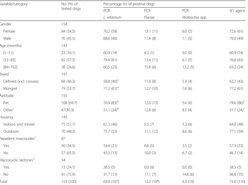
[image:3.595.58.544.109.471.2]One hundred and thirty-nine dogs were qPCR-positive toL. infantum. Out of the 97 dogs tested from November to April (Leishmanianon-transmission period) 72 (74.2 %; CI: 64.3–82.6 %) were positive, whereas from May to October (transmission period)L. infantumDNA was de-tected in 67 (50.4 %; CI: 41.6–59.2 %) of 133 canine blood samples (P< 0.001). The significantly higher (P< 0.001) Leishmaniaprevalence obtained in the present study, i.e. 69.0 % (CI: 61.1–76.2 %) in dogs with no clinical signs of leishmaniosis and/or dirofilariosis (Table 1) and 42.7 % (CI: 31.3–54.6 %) in dogs with compatible clinical signs (Table 2) compared with the 1.1 % recently obtained in dogs from the south of the country by conventional PCR [18], could be related to the higher sensitivity of the qPCR, which is able to detect less than one parasite per millilitre of blood [17]. This most recent figure of 69.0 % is more in Table 1Molecular prevalence ofL. infantum, filariae andWolbachiaamong dogs with no clinical signs compatible with
leishmaniosis or dirofilariosis (n= 155)
Variable/category No. (%) of
tested dogs
Percentage (n) of positive dogs
PCR PCR PCR ≥1 agent
L. infantum filariae Wolbachiaspp.
Gender 154
Female 84 (54.5) 70.2 (59) 13.1 (11) 6.0 (5) 72.6 (61)
Male 70 (45.5) 68.6 (48) 11.4 (8) 7.1 (5) 70.0 (49)
Age (months) 143
[1–11] 23 (16.1) 60.9 (14) 4.3 (1) 0.0 (0) 60.9 (14)
[12–83] 82 (57.3) 74.4 (61) 13.4 (11) 6.1 (5) 76.8 (63)
[84–192] 38 (26.6) 60.5 (23) 15.8 (6) 13.2 (5) 63.2 (24)
Breed 147
Defined (incl. crosses) 68 (46.3) 58.8 (40)a 11.8 (8) 5.9 (4) 63.2 (43)
Mongrel 79 (53.7) 77.2 (61)a 12.7 (10) 7.6 (6) 77.2 (61)
Aptitude 155
Pet 108 (69.7) 76.9 (83)b 12.0 (13) 5.6 (6) 79.6 (86)c
Other1 47(30.3) 51.1 (24)b 12.8 (6) 8.5 (4) 51.1 (24)c
Housing 145
Indoors and mixed 75 (51.7) 61.3 (46) 9.3 (7) 5.3 (4) 64.0 (48)
Outdoors 70 (48.3) 75.7 (53) 17.1 (12) 8.6 (6) 77.1 (54)
Repellent insecticides2 87
Yes 30 (34.5) 54.4 (31) 8.8 (5) 3.5 (2) 57.9 (33)
No 57 (65.5) 43.3 (13) 10.0 (3) 6.7 (2) 46.7 (14)
Macrocyclic lactones3 54
Yes 13 (24.1) 38.5 (5) 0.0 (0) 0.0 (0) 38.5 (5)
No 41 (75.9) 31.7 (13) 17.1 (7) 14.6 (6) 36.6 (15)
Total 155 (100) 69.0 (107) 12.3 (19*) 6.5 (10) 71.0 (110)
1
Farm, guard, hunting, shepherd or stray;2
Deltamethrin, flumethrin and/or permethrin;3
Ivermectin, mimemycin oxime or moxydectin;a
P= 0.026;b
P= 0.003;c
accordance with previously reported molecular prevalence values clearly above 50 % [16]. Nevertheless, it cannot be ruled out that positive blood results were due to transient contamination during the transmission season and may thus represent just exposure to the parasite rather than an established infection.
A negative association was found betweenLeishmania positivity and clinical signs compatible with leishmanio-sis and/or dirofilarioleishmanio-sis (69.0 % [Table 1] versus 42.7 % [Table 2];P< 0.001). The fact that more dogs considered
clinically healthy harboured Leishmania parasites in
their circulation might be due to a fact most of the en-rolled dogs were randomly selected from those attending veterinarian clinics and, therefore, probably receiving more medical care, better nutrition, and for that reason were less prone to develop disease. The prevalence of L. infantum(76.9 %; CI: 67.7–84.4 %;P= 0.003) and that of infection with at least one pathogen (≥1 agent; 79.6 %; CI: 70.8–86.8 %; P= 0.001) in dogs without compatible
clinical signs was significantly higher in pets in compari-son with dogs with other aptitudes (Table 1). The present results, therefore, disagree with those that re-vealed a higher prevalence of infection in guard dogs as compared to pets in the Madrid region, Spain [19]. These latter findings might be somewhat due to a differ-ent lifestyle of pets (i.e. urban vs. rural) in distinct
geo-graphic areas. The higher prevalence of L. infantum
DNA in clinically healthy mongrel dogs in comparison with those belonging to defined breeds or their crosses (P= 0.026) might be related with a certain level of resist-ance to disease development and progression by the mixed-breed infected dogs [20].
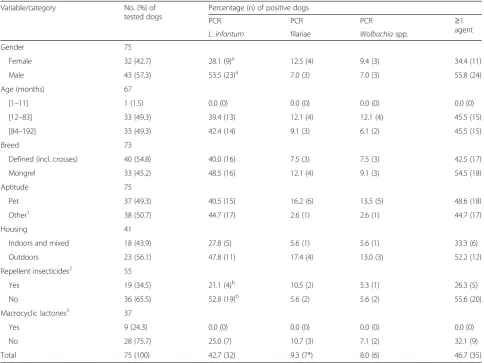
[image:4.595.56.541.109.472.2]In those dogs showing clinical signs compatible with leishmaniosis and/or dirofilariosis the detection of L. infantumDNA in the blood was significantly higher (P = 0.050; Table 2) in males (53.5 %; CI: 37.6–68.8 %). Gender predisposition to the infection has been consid-ered by some authors as a non-determinant factor for Table 2Molecular prevalence ofL. infantum, filariae andWolbachiain dogs with clinical signs compatible with leishmaniosis and/or dirofilariosis (n= 75)
Variable/category No. (%) of
tested dogs
Percentage (n) of positive dogs
PCR PCR PCR ≥1
agent
L. infantum filariae Wolbachiaspp.
Gender 75
Female 32 (42.7) 28.1 (9)a 12.5 (4) 9.4 (3) 34.4 (11)
Male 43 (57.3) 53.5 (23)a 7.0 (3) 7.0 (3) 55.8 (24)
Age (months) 67
[1–11] 1 (1.5) 0.0 (0) 0.0 (0) 0.0 (0) 0.0 (0)
[12–83] 33 (49.3) 39.4 (13) 12.1 (4) 12.1 (4) 45.5 (15)
[84–192] 33 (49.3) 42.4 (14) 9.1 (3) 6.1 (2) 45.5 (15)
Breed 73
Defined (incl. crosses) 40 (54.8) 40.0 (16) 7.5 (3) 7.5 (3) 42.5 (17)
Mongrel 33 (45.2) 48.5 (16) 12.1 (4) 9.1 (3) 54.5 (18)
Aptitude 75
Pet 37 (49.3) 40.5 (15) 16.2 (6) 13.5 (5) 48.6 (18)
Other1 38 (50.7) 44.7 (17) 2.6 (1) 2.6 (1) 44.7 (17)
Housing 41
Indoors and mixed 18 (43.9) 27.8 (5) 5.6 (1) 5.6 (1) 33.3 (6)
Outdoors 23 (56.1) 47.8 (11) 17.4 (4) 13.0 (3) 52.2 (12)
Repellent insecticides2 55
Yes 19 (34.5) 21.1 (4)b 10.5 (2) 5.3 (1) 26.3 (5)
No 36 (65.5) 52.8 (19)b 5.6 (2) 5.6 (2) 55.6 (20)
Macrocyclic lactones3 37
Yes 9 (24.3) 0.0 (0) 0.0 (0) 0.0 (0) 0.0 (0)
No 28 (75.7) 25.0 (7) 10.7 (3) 7.1 (2) 32.1 (9)
Total 75 (100) 42.7 (32) 9.3 (7*) 8.0 (6) 46.7 (35)
1
Farm, guard, hunting or stray;2
Deltamethrin or permethrin;3
Ivermectin, milbemycin oxime or moxydectin;a
P= 0.050;b
leishmaniosis [19, 20], while others have reported a higher prevalence in male dogs [21, 22]. On the other hand, the use of long-acting topical insecticides on dogs has been shown to be an effective measure in reducing the prevalence of Leishmania infection [23]. Therefore, it is not entirely surprising that the prevalence of Leish-mania was higher in those dogs not protected against insects (P= 0.048; Table 2). In fact, and although the number of effective molecules for prophylaxis against leishmaniosis has increased in the last few years, our re-sults clearly reinforce the idea that there is still a long way to go regarding the prevention and control of Leish-maniainfection.
Filarial infections were detected in 26 (11.3 %; CI: 7.5– 16.1 %) blood samples: 24 (10.4 %; CI: 6.8–15.1 %) dogs harboured D. immitis only, one (0.4 %; CI: 0.01–2.4 %) dogA. reconditumonly and another one (0.4 %; CI: 0.01– 2.4 %)A. drancunculoidesin co-infection withD. immitis. This corroborates the previous reports on the circulation of these nematodes in the south and central regions of Portugal [9, 12–14]. D. immitisprevalence in the present study (10.9 %; CI: 7.2–15.6 %) was lower (although non-significantly; P= 0.111) than the overall prevalence of 15.1 % (105/696) recently obtained in three coastal areas of central and southern-central Portugal [14]. Differences can be related to the dynamics of infection over time, which is probably linked to the distribution and abun-dance of the vectors and to the surveyed canine popula-tion. In fact, in the work performed by Alho et al. [14] all the screened animals were sheltered dogs that lived out-doors and had probably not received any prophylactic measure for heartworm infection, thus being more ex-posed to vectors and to the agents they might transmit. On the other hand, in the present study most of the tested dogs were pets living partially indoors and receiving veter-inary health care. Interestingly, and although the differ-ence was not statistically significant (P= 0.562),D. immitis DNA was not detected in any of the dogs receiving macrocyclic lactones, suggesting the importance of using this prophylactic measure in areas where dirofilariosis is endemic. As the compliance to the use of ectoparasiticides was not evaluated, it cannot be ruled out whether the rea-son for no differences between the use of insecticides/re-pellents and the presence of heartworm infection was due to irregular administration of insecticides.
The detection of Wolbachia DNA in 16 (64.0 %; CI:
42.5–82.0 %) dogs positive toD. immitiswas higher than the 52.6 % (20/38; P= 0.372) and 30.6 % (15/49; P= 0.006) recently obtained in Portuguese [11] and Spanish [16] dogs, respectively. As reported by previous
publica-tions, only D. immitis and D. repens have ever been
found infected with Wolbachia, but not all the speci-mens of these filarial parasites transport the endosymbi-ont bacteria. Taken together, these findings suggest that
in endemic areas a combined PCR for Wolbachia and
Dirofilaria should be performed for the diagnosis of heartworm, in order to avoid a high percentage of false-negative Dirofilariaresults due to the lack of testing for the endosymbiont.
Wolbachia DNA was detected in 5 (31.3 %; CI: 11.0– 58.7 %) dogs solely infected with D. immitis and in 11 (68.8 %; CI: 41.3–89.0 %;P= 0.034) co-infected dogs (11 with L. infantum-D. immitis and one with A. dracunculoides-D.immitis), reinforcing that this endo-symbiont does not seem to be present in Achanthochei-lonema spp. alone [5]. On the other hand, our results contrast with a previous study carried out in dogs with leishmaniosis and/or dirofilariosis from Spain where the prevalence of the endosymbiont was significantly lower in microfilariaemic dogs co-infected with L. infantum [16]. Interestingly, in the present study only three out of
the 11 Wolbachia, D. immitis and L. infantum
co-infected dogs presented clinical signs compatible with leishmaniosis and/or dirofilariosis, which might have been due to the stimulation of a Th1 type protective-immune response triggered by the nematode and the endosymbiont bacterium [16, 24].
In the present study, 19 (8.3 %; CI: 5.0–12.6 %) dogs were co-infected with L. infantum and D. immitis. It should be stressed that the occurrence of co-infections with these vector-borne agents, which are relatively common in dogs living in geographic areas where com-petent vectors for the different pathogens co-exist, might induce more severe and atypical clinical outcomes that will further complicate diagnosis, treatment and progno-sis [9, 16, 25]. However, this hypotheprogno-sis has not been corroborated in the present study and the reason might be the random screening of the canine population, with most dogs showing no clinical signs, even when they were positive for one or more of the studied agents.
present results further stress the need for sustained de-velopment of multi-pathogen detection methods in re-gions endemic and even non-endemic for CVBD.
Competing interests
The authors declare that they have no competing interests.
Authors’contributions
CM planned, designed and supervised the study, and wrote the manuscript; LA and LS performed qPCR assays; JMC performed DNA extraction; LuC performed data analysis and revised the manuscript; MDT, OF and LeC reviewed the manuscript; XR planned the study and reviewed the manuscript. All authors read and approved the final manuscript.
Acknowledgements
This work was partially supported by Funds to GHTM (UID/Multi/04413/ 2013). The authors thank the cooperation of veterinarians, auxiliary staff, dog owners and shelters that contributed with collection of samples. The work of C. Maia and L. Cardoso was done under the frame of EurNegVec COST Action TD1303.
Publication of this paper has been sponsored by Bayer Animal Health in the framework of the 11th CVBD World Forum Symposium.
Author details
1Global Health and Tropical Medicine, Medical Parasitology Unit, Instituto de
Higiene e Medicina Tropical, Universidade Nova de Lisboa, Lisbon, Portugal.
2Faculty of Veterinary Medicine, Universidade Lusófona de Humanidades e
Tecnologias, Lisbon, Portugal.3Vetgenomics, Parc de Recerca Universitat Autònoma de Barcelona, Bellaterra, Barcelona, Spain.4Hospital Veterinario
San Vicente, San Vicente del Raspeig, Alicante, Spain.5SVGM, Departamento
de Ciencia Animal y de los Alimentos, Facultad de Veterinària, Universitat Autònoma de Barcelona, Bellaterra, Barcelona, Spain.6Department of Veterinary Sciences, School of Agrarian and Veterinary Sciences, University of Trás-os-Montes e Alto Douro, Vila Real, Portugal.7Department of Biomedical
Sciences and Medicine, Universidade do Algarve, Faro, Portugal.8Hospital
Clínic Veterinari, Universitat Autònoma de Barcelona, Bellaterra, Barcelona, Spain.
Received: 17 January 2016 Accepted: 15 March 2016
References
1. Dantas-Torres F, Otranto D. Best Practices for preventing vector-borne diseases in dogs and humans. Trends Parasitol. 2015. doi:10.1016/j.pt.2015.09.004. 2. Ferreira CA, de Pinho MV, Novo MT, Calado MM, Gonçalves LA, Belo SM, et
al. First molecular identification of mosquito vectors ofDirofilaria immitisin continental Portugal. Parasit Vectors. 2015;8:139.
3. Maia C, Parreira R, Cristóvão JM, Freitas FB, Afonso MO, Campino L. Molecular detection ofLeishmaniaDNA and identification of blood meals in wild caught phlebotomine sand flies (Diptera: Psychodidae) from southern Portugal. Parasit Vectors. 2015;8:173.
4. Otranto D, Dantas-Torres F, Brianti E, Traversa D, PetrićD, Genchi C, et al. Vector-borne helminths of dogs and humans in Europe. Parasit Vectors. 2013;6:16. 5. Bandi C, Tress AJ, Bratting NW.Wolbachiain filarial nematodes: evolutionary
aspects and implications for the pathogenesis and treatment of filarial diseases. Vet Parasitol. 2001;98:215–38.
6. Kramer L, Grandi G, Leoni M, Passeri B, McCall J, Genchi C, et al.Wolbachia
and its influence on the pathology and immunology ofDirofilaria immitis
infection. Vet Parasitol. 2008;158:191–5.
7. McCall JW, Genchi C, Kramer LH, Guerrero J, Venco L. Heartworm disease in animals and humans. Adv Parasitol. 2008;66:193–285.
8. Campino L, Maia C. The role of reservoirs: canine leishmaniasis. In: Ponte-Sucre A, Padron-Nieves M, Diaz E, editors. Drug resistance inLeishmania parasites–consequences, molecular mechanism and possible treatments. Vienna: Springer Verlag; 2013. p. 45–64.
9. Cardoso L, Mendão C, Madeira de Carvalho L. Prevalence ofDirofilaria immitis,Ehrlichia canis,Borrelia burgdorferisensu lato,Anaplasmaspp. and
Leishmania infantumin apparently healthy and CVBD-suspect dogs in Portugal–a national serological study. Parasit Vectors. 2012;5:62. 10. Maia C, Dionísio L, Afonso MO, Neto L, Cristóvão JM, Campino L.Leishmania
infection and host-blood feeding preferences of phlebotomine sandflies
and canine leishmaniasis in an endemic European area, the Algarve Region in Portugal. Mem Inst Oswaldo Cruz. 2013;108:481–7.
11. Landum M, Ferreira CC, Calado M, Alho AM, Maurício IL, Meireles JS, et al. Detection ofWolbachiainDirofilariainfected dogs in Portugal. Vet Parasitol. 2014;204:407–10.
12. Maia C, Coimbra M, Ramos C, Cristóvão JM, Cardoso L, Campino L. Serological investigation ofLeishmania infantum.Dirofilaria immitis and Angiostrongylus vasorum in dogs from southern Portugal. Parasit Vectors. 2015;8:152.
13. Menn B, Lorentz S, Naucke TJ. Imported and travelling dogs as carriers of canine vector-borne pathogens in Germany. Parasit Vectors. 2010;3:34. 14. Alho AM, Landum M, Ferreira C, Meireles J, Gonçalves L, de Carvalho LM, et
al. Prevalence and seasonal variations of canine dirofilariosis in Portugal. Vet Parasitol. 2014;206:99–105.
15. Maia C, Lorentz S, Cardoso L, Otranto D, Naucke TJ. Detection ofDirofilaria repensmicrofilariae in a dog from Portugal. Parasitol Res. 2015;115:441–3. 16. Tabar MD, Altet L, Martínez V, Roura X.Wolbachia, filariae and
Leishmaniacoinfection in dogs from a Mediterranean area. J Small Anim Pract. 2013;54:174–8.
17. Francino O, Altet L, Sánchez-Robert E, Rodriguez A, Solano-Gallego L, Alberola J, et al. Advantages of real-time PCR assay for diagnosis and monitoring of canine leishmaniosis. Vet Parasitol. 2006;137:214–21. 18. Maia C, Almeida B, Coimbra M, Fernandes MC, Cristóvão JM, Ramos C, et al.
Bacterial and protozoal agents of canine vector-borne diseases in the blood of domestic and stray dogs from southern Portugal. Parasit Vectors. 2015;8:138. 19. Galvez R, Miró G, Descalzo MA, Nieto J, Dado D, Martín O, et al. Emerging trends in the seroprevalence of canine leishmaniasis in the Madrid region (central Spain). Vet Parasitol. 2010;169:327–34.
20. Cortes S, Vaz Y, Neves R, Maia C, Cardoso L, Campino L. Risk factors for canine leishmaniasis in an endemic Mediterranean region. Vet Parasitol. 2012;189:189–96.
21. Zivicnjak T, MartinkovićF, MarinculićA, Mrljak V, Kucer N, Matijatko V, et al. A seroepidemiologic survey of canine visceral leishmaniosis among apparently healthy dogs in Croatia. Vet Parasitol. 2005;131:35–43. 22. Miranda S, Roura X, Picado A, Ferrer L, Ramis A. Characterization of sex, age,
and breed for a population of canine leishmaniosis diseased dogs. Res Vet Sci. 2008;85:35–8.
23. Courtenay O, Kovacic V, Gomes P, Garcez L, Quinnell R. A long-lasting topical deltamethrin treatment to protect dogs against visceral leishmaniasis. Med Vet Entomol. 2009;23:245–56.
24. Oleaga A, Pérez-Sánchez R, Pagés E, Marcos-Atxutegi C, Simón F. Identification of immunoreactive proteins from the dog heartworm (Dirofilaria immitis) differentially recognized by the sera from dogs with patent or occult infections. Mol Biochem Parasitol. 2009;166:134–41. 25. De Tommasi AS, Otranto D, Dantas-Torres F, Capelli G, Breitschwerdt EB, de
Caprariis D. Are vector-borne pathogen co-infections complicating the clinical presentation in dogs? Parasit Vectors. 2013;6:97.
• We accept pre-submission inquiries
• Our selector tool helps you to find the most relevant journal
• We provide round the clock customer support
• Convenient online submission
• Thorough peer review
• Inclusion in PubMed and all major indexing services
• Maximum visibility for your research
Submit your manuscript at www.biomedcentral.com/submit