C A S E R E P O R T
Open Access
Chronic
Mycobacterium avium
skin and soft
tissue infection complicated with scalp
osteomyelitis possibly secondary to
anti-interferon-
γ
autoantibody formation
Xianli Xu
1, Xiaojie Lao
1, Chunlan Zhang
1, Cunwei Cao
2, Huarong Ding
3, Yu Pang
1, Qiuyue Ning
1, Jun Zou
4,
Ning Zang
5, Diefei Hu
1*and Maowei Chen
6*Abstract
Background:Nontuberculous mycobacterial (NTM) disease is commonly an opportunistic infection frequently found in immunocompromised individuals, but sometimes can also be found in the immunocompetent hosts, especially in East Asians. The NTM separation rate in China is increasing, which reminds us to focus on NTM infections in immunocompromised populations.
Case presentation:A 43-year-old woman with a recurrent fever for more than 8-month and a right forehead surgical wounds unhealed for more than 6-month was admitted to our hospital on February 22, 2018. On arrival, several elliptic ulcers were obvious on the right forehead with pus and fibrin exudation, and the skin around the lesions was tender, reddish, no sense of fluctuation. The result of HIV serology test was negative. CD4+ T cell count was normal and tuberculosis antibody was negative. CT of the chest and head showed bone destruction. Skin biopsy on the right forehead was performed on March 13, 2018, and pathological examination of the excisional biopsy specimen found inflammatory granuloma and suppurative inflammatory changes. Broad-spectrum
antibiotics were treated but the effect seemed discontent. Then debridement and skin grafting were performed on the right frontal ulcer under general anesthesia on April 3, 2018. The skin tissue culture that resected on March 13, 2018 found Nontuberculous mycobacteria grown after 78 days, so clarithromycin, ethambutol, protionamide, and amoxicillin clavulanate potassium were prescribed for anti-nontuberculous mycobacteria treatment beginning on May 31, 2018. In reviewing the case,Mycobacterium avium(M. avium) was identified in the skin tissue resected on April 3, 2018 by polymerase chain reaction (PCR) and the serum test of anti-interferon-γautoantibodies was positive.
Conclusions:This is a case report of“Mycobacterium aviumSSTI (skin and soft tissue infection) and OM
(osteomyelitis) with possible secondary immunodeficiency syndrome induced by anti-interferon-γautoantibody”.
Keywords:Mycobacterium avium, Infection, Skin
* Correspondence:hudiefei2007@163.com;chenmaowei2008@163.com 1
Department of infectious disease, Guangxi Medical University First Affiliated Hospital, Nanning, China
6Department of infectious disease, Wuming Hospital of Guangxi Medical
University, Nanning, China
Full list of author information is available at the end of the article
most frequent, followed by lymphadenitis in children, skin disease, and other extrapulmonary or disseminated infections in severely immunocompromised patients [4], such as malignancy or infection with human immuno-deficiency virus (HIV) [5]. Sometimes NTM infection can also be found in the immunocompetent hosts, espe-cially in East Asians [6].
MAC was one of the most identifiable mycobacterial, which often lead to disseminated infections. A study found it had an extremely high prevalence rate (97.8%) of IFN-γ autoantibodies in patients with disseminated NTM infection [5].
Here is a case of Mycobacterium aviuminfection with skin symptom as the initial manifestation which doesn’t have a predisposing condition or immunosuppression, but the testing result of IFN-γautoantibodies was positive. We aim to We aim to raise awareness of NTM infections when a patient presents with unexplained rashes, poor efficacy of medical therapy and surgery, and IFN-γ autoantibodies– associated immunodeficiency should be considered when NTM infection happens to immunocompetent hosts.
Case presentation
A 43-year-old woman was admitted to our hospital with a recurrent fever for more than 8-month and a right forehead wound disunion after the mass excision for more than 6-month. In June 2017, she incidentally found a bean-size lump over the right forehead that was grad-ually increasing in size. Her symptoms were accompan-ied with recurrent fever but body temperature was not
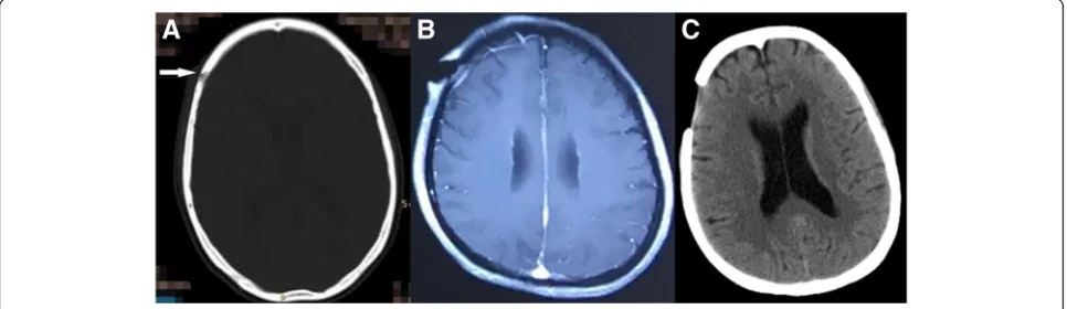
(CT) of the head showed bone loss and destruction at the corresponding place (Fig. 1a). In September 2017, she underwent a debridement on the infected scalp and destructive bone of the right forehead (Fig. 1b). The pathology showed acute suppurative osteomyelitis with a high number of inflammatory hyperplasia, pus forma-tion, and massive bone necrosis. The pathology of the right forehead mass revealed bleeding, purulent inflam-matory changes, epidermis necrosis, and negative stain-ing with PAS. The patient was impaired wound healstain-ing, and the wound oozing. Over the next few days, the pa-tient has high fever which usually persists unremittingly (up to 40 °C), and the patient may continue to have re-current rigors. Broad-spectrum antibiotics treatment seemed not effective. Then she was admitted to our de-partment in February 2018. The patient reported no sig-nificant past medical history, and she denied any exposure to contaminants or suspected water sources.
Physical examination showed low body temperature (35.7 °C), several elliptic ulcers on the right forehead with pus and fibrin exudation (2.0 cm × 1.0 cm). The skin around the lesions was tender, reddish, no sense of fluc-tuation (Fig.2j). There also had bilateral cervical lymph-adenopathy (0.7 cm × 0.8 cm). Respiratory sounds and cardiac and abdominal examination were normal.
Laboratory testing revealed a leukocyte count of 17.13 × 10⁹/L with 82.1% of neutrophils, a C-reactive pro-tein concentration of 192.00 mg/L, a procalcitonin of 0.247 ng/ml and a negative result of HIV serology test. CD4+ T cell count was normal and the levels of serum
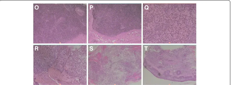
[image:2.595.57.540.555.695.2]globulins including IgG, IgA, IgM and total IgE were within the normal reference range. Tuberculosis antibody was negative, Hemoglobin 71.40 g/L and serum albumin 25.0 g/L. Other laboratory results were unremarkable. Ultrasonography revealed bilateral cervical, bilateral axil-lary and bilateral inguinal lymphadenopathy. CT of the chest revealed mild pneumonia, nodular opacity in the left upper lobe, a minimal pleural effusion on the left, and mildly enlarged lymph nodes in the mediastinal and in bi-lateral axillary (Fig.3f, g). CT of the head showed the right frontotemporal soft tissue mildly swelling (Fig. 1c). The pathology of the cervical lymph node biopsy showed re-active hyperplasia (Fig.4o, p). A bone marrow aspiration was done, and the cytology suggested mature neutrophils mostly with reactive changes with the neutrophil alkaline phosphatase (NAP) was 365. Lumbar puncture (LBP) was carried out. We found that the CSF pressure was normal and the spinal fluid examinations showed no abnormality. Skin biopsy on the right forehead was performed on March 13, 2018, and pathological examination of the exci-sional biopsy specimen found nothing but inflammatory granuloma and suppurative inflammation changes, with
PAS and acid-fast staining negative (Fig.4s, t). Blood, bone marrow, spinal fluid, wound secretion, skin and soft tissue Specimens cultures were all negative for bacteria, tubercu-losis, and fungus. Because Mycobacterium culture tech-nique was limited, the skin tissue which excised on March 13, 2018 was sent to another hospital for mycobacterial culture and antimicrobial susceptibility test. The diagnosis was as follows: 1. Right frontotemporal skin and subcuta-neous tissue infection; 2. Infectious fever. She received broad-spectrum antibiotics treatment such as Piperacillin Sodium and Tazobactam Sodium combined with amika-cin, linezolid with imipenem, moxifloxacin with teicopla-nin and voriconazole, levofloxacin with clindamycin and itraconazole. Concomitantly the patient was treated with strengthening the dressing, increasing circulation, enhan-cing immunity and other supportive therapy.
However, a new tender and reddish tubercle occurred behind the right ear, then the tubercle enlarged and eventually broke down to form an ulcer (Fig. 2k, l). On April 3, 2018, debridement and skin grafting were performed on the right frontal ulcer under general anesthesia. Postoperative anti-infective treatment and
Fig. 2j-nAre pictures of the patient’s right frontal facial lesions.jshowed the patient’s initial lesions.kandlwere taken on March 19, 2018, which showed a newly appearing nodule behind the right ear accompanied with redness and ulceration.mwas taken 1 month after anti-NTM treatment, showing the skin lesions almost healed.nwas taken on 4 months after anti-NTM treatment, showing the skin lesions healed
Fig. 3d-iAre chest computed tomography sequences.dandewere taken in August 2017, which showed no visible bone destruction in the sternum.fandgwere taken in February 2018, which showed the bones of sternal stem and bilateral clavicle sternum stalk destructed.handi
[image:3.595.58.540.87.198.2] [image:3.595.56.540.539.694.2]dressing changing were continued. The temperature grad-ually decreased but still had a low-grade temperature (the highest daily temperature was around 37.6 °C), and part of the head skin graft survived with residual wound base granulation growth. The patient was discharged from our hospital on April 14, 2018.
On May 30, 2018, the mycobacterial culture and anti-microbial susceptibility test result returned which showed that Nontuberculous mycobacteria growth was noted after 78 days of culture in a Mycobacterium Growth Indicator Tube, sensitive to ethambutol and protionamide, resistant to streptomycin, isoniazid, rifampicin, aminosalicylic acid, levofloxacin, capreomycin, and amikacin. The patient was admitted again on May 31, 2018. The test of
Mycobacter-ium tuberculosiscomplex DNA of her phlegm liquid was negative. Chest CT scan showed a few infected foci in the right middle lobe, irregular bone destruction of manu-brium sternum and the sternal end of the bilateral clavicle, and the bone changes of corpus sternum and cervical vertebra (Fig.3h, i). The infection caused by nontubercu-lous mycobacteria was confirmed, and clarithromycin, ethambutol, protionamide, and amoxicillin clavulanate potassium were prescribed for anti-nontuberculous myco-bacteria treatment. There was no abnormality when moni-toring blood routine and serum chemistry, and the effusion of wound gradually reduced. The patient was dis-charged on July 14, 2018, and she was reviewing regularly. We are still treating the patient with anti-nontuberculous mycobacteria regimen above-mentioned and change the dressing regularly.
Discussion and conclusions
An epidemiological survey showed that the rate of NTM strain isolation was up to 22.9% in 2010 in China [7]. The
clinical manifestations of NTM diseases include respiratory tract infections, disseminated infections, skin and soft tissue infections, lymphadenitis, ocular infections, and so on [8].
Skin and soft tissue infections (SSTIs) caused by NTM are underrecognized, due to their wide spectrum of clin-ical presentations and histopathologclin-ical findings that are often nonspecific [9, 10]. The manifestation of skin disease presents with nodules, subcutaneous abscesses, pustules, ulcers, or combinations thereof [4]. The patient in our case was presented with the manifestation of nod-ules, ulcers.
Multi-disciplinary collaboration is necessary for diag-nosis, including the clinician, the histopathologist, the microbiologist, and infectious disease specialists [4, 10]. In this case, the patient developed a right forehead mass with fever, and the wound did not heal after the mass re-section. The broad-spectrum antibiotic treatment was not effective. Histopathological findings showed purulent inflammation with infiltration of lymphocytes and neu-trophils. Finally, the right frontal skin biopsy tissue was cultured nontuberculous mycobacteria, so the diagnosis of the nontuberculous mycobacterial disease was estab-lished. Culture is still the gold standard, because of lim-ited sensitivity and specificity of symptoms, radiology, and direct microscopy of clinical samples [4].
Disseminated NTM diseases present as two distinct clinical syndromes [4]. The patient’s head CT showed bone destruction in the corresponding site of the lesion. Chest CT scan showed irregular bone destruction in the sternum stem and bilateral clavicle sternum, bone changes in the sternum and cervical vertebrae. Thus NTM bone disease was not excluded. The corresponding bone tissue NTM cultural examination is required. This patient could have disseminated NTM disease, but the diagnosis of dis-seminated NTM disease requires blood or bone marrow
[image:4.595.58.540.88.265.2]culture positive. Currently, positive evidence for blood or bone marrow culture was absent.
A variety of modalities including tissue culture and polymerase chain reaction (PCR) assays are crucial in order to identify the organism [10]. Reviewing the case, we re-stained the skin tissue which was excised on April 3, 2018 with acid-fast staining, and the results showed positive (see Fig.5). Then theMycobacterium avium(M.
avium) was identified by PCR. Owing to the uncertainty of manual operation and sampling, we consider that acid-fast staining of the tissue slices was related to the content of tissue samples and the thickness of slices, so there is a certain rate of missed diagnosis.
M. avium is one of the important pathogens in dis-seminated disease, whereas M. intracellular is one of the common respiratory pathogen [11]. The disseminated NTM disease mainly occurred in immunocompromised patients, especially in HIV-infected patients. The patient in our case was previously healthy and HIV negative and her CD4/CD8 lymphocyte count was normal. However, we tested the patient for IFN-γ autoantibodies and the result was positive.
Neutralizing anti–interferon-γ autoantibodies were detected in 88% of Asian adults with multiple opportunis-tic infections and were associated with an adult-onset immunodeficiency similar to advanced HIV infection [12]. High titers of IFN-γautoantibodies are mainly found in pa-tients with the disseminated non-tuberculous mycobacter-ial disease and are more common in East Asian women who have not been treated with exogenous interferon gamma [13]. IFN-γautoantibodies are evidence of acquired immunodeficiency that should be examined in cases of un-explained disseminated NTM infections in Asian-born per-sons [14]. In our case, the patient developed an NTM infection without a history of immunodeficiency and other chronic diseases, but IFN-γ autoantibodies proved to be positive. Hence acquired immunodeficiency should be con-sidered, such as adult-onset immunodeficiency syndrome.
Treatment can be challenging, as it can be dependent on multiple factors, including the causative organism, the patient’s immunological status, and the extent of disease involvement [10]. The optimal regimen against disseminated MAC is clarithromycin, ethambutol, +/− rifa-butin, whether a three-drug regimen alone in this setting would be adequate is not known. The optimal duration of treatment is also unknown, but 6 to 12 months of chemo-therapy is usually recommended [11, 15]. Specific treat-ment for IFN-γ autoantibodies associated NTM infection is not codified and required prolonged, multiple-drug reg-imens. Using immunomodulation strategies is still de-bated, and long-term suppressive treatment should be taken into account for persisting high levels of neutraliz-ing antibodies [14].
According to the culture results and drug susceptibility test, clarithromycin, ethambutol, protionamide, amoxicillin-clavulanate potassium regimen were given to treat NTM infection for our patient. One month later, the patient’s right forehead wound gradually healed and there was no new rash.
In summary, chronic Mycobacterium avium skin and soft tissue infection complicated with scalp osteomyelitis possibly secondary to anti-interferon-γ autoantibody for-mation. This case underscores the need for clinicians to be aware of the potential for NTM infection when a patient presenting with unexplained rashes, poor efficacy to med-ical therapy and surgery. When NTM infection is detected in an immunocompetent patient, IFN-γautoantibodies– as-sociated immunodeficiency should be considered.
Abbreviations
CSF:Cerebrospinal fluid; CT: Computed tomography; DNA: Deoxyribonucleic acid; HIV: Human immunodeficiency virus; IFN-γ: Interferon gamma; IgA: Immunoglobulin A; IgE: Immunoglobulin E; IgG: Immunoglobulin G; IgM: Immunoglobulin M; IHC: immunohistochemistry; LBP: Lumbar puncture; M. avium:Mycobacterium avium; MAC:Mycobacterium avium- intracellular complex; NAP: Neutrophil alkaline phosphatase; NTM: Nontuberculous mycobacterial; PAS: Periodic Acid-Schiff stain; PCR: Polymerase chain reaction; SSTIs: Skin and soft tissue infections
[image:5.595.62.540.87.243.2]The original data used in the presentation of this case report are available from the corresponding author on reasonable request.
Authors’contributions
DH, YP, QN, and HD were all directly involved in the clinical management of this case. XX drafted the manuscript under the guidance of MC, DH, and XL. XX, XL, and NZ substantively revised this manuscript. CZ participated in bibliographic research. JZ and NZ were involved in the mycobacterial culturing and antimicrobial susceptibility testing. CC was involved in anti– interferon-γautoantibodies testing. Applied for project fund support. All authors read and approved the final draft.
Ethics approval and consent to participate
Not applicable.
Consent for publication
The study participant has signed a written consent form giving her consent for publication of this case in an academic journal.
Competing interests
The authors declare that they have no competing interests.
Publisher’s Note
Springer Nature remains neutral with regard to jurisdictional claims in published maps and institutional affiliations.
Author details
1Department of infectious disease, Guangxi Medical University First Affiliated
Hospital, Nanning, China.2Department of dermatology and venereology, Guangxi Medical University First Affiliated Hospital, Nanning, China.
3Department of burns and plastic surgery, Guangxi Medical University First
Affiliated Hospital, Nanning, China.4Department of scientific education, The
fourth People’s Hospital of Nanning, Nanning, China.5Medical Scientific Research Center of Life Sciences Institute, Guangxi Medical University, Nanning, China.6Department of infectious disease, Wuming Hospital of
Guangxi Medical University, Nanning, China.
Received: 1 November 2018 Accepted: 31 January 2019
References
1. Porvaznik I, SolovičI, Mokrý J. Non-Tuberculous Mycobacteria: Classification, Diagnostics, and Therapy. Adv Exp Med Biol. 2017;944:19–25.
2. Guglielmetti L, Mougari F, Lopes A, Raskine L, Cambau E. Human infections due to nontuberculous mycobacteria: the infectious diseases and clinical microbiology specialists’point of view. Future Microbiol. 2015;10:1467–83. 3. Falkinham JO. Impact of human activities on the ecology of nontuberculous
mycobacteria. Future Microbiol. 2010;5:951–60.
4. van Ingen J. Diagnosis of nontuberculous mycobacterial infections. Semin Respir Crit Care Med. 2013;34:103–9.
5. Chi CY, Lin CH, Ho MW, et al. Clinical manifestations, course, and outcome of patients with neutralizing anti-interferon-γautoantibodies and disseminated nontuberculous mycobacterial infections. Medicine (Baltimore). 2016;95:e3927.
6. Chan JF, Yee KS, Tang BS, Cheng VC, Hung IF, Yuen KY. Adult-onset immunodeficiency due to anti-interferon-gamma autoantibody in mainland Chinese. Chin Med J. 2014;127:1189–90.
7. Yu X, Liu P, Liu G, Zhao L, Hu Y, Wei G, Luo J, Huang H. The prevalence of non-tuberculous mycobacterial infections in mainland China: systematic review and meta-analysis. J Inf Secur. 2016;73:558–67.
immunodeficiency in Thailand and Taiwan. N Engl J Med. 2012;367:725–34. 13. Patel SY, Ding L, Brown MR, et al. Anti-IFN-gamma autoantibodies in
disseminated nontuberculous mycobacterial infections. J Immunol. 2005; 175:4769–76.
14. Valour F, Perpoint T, Sénéchal A, et al. Interferon-γautoantibodies as predisposing factor for nontuberculous mycobacterial infection. Emerg Infect Dis. 2016;22:1124–6.