(
R
,
R
)-Disynephrine ether bis(hydrogen
sulfate)
William Arbuckle,aAlan R. Kennedyb* and Catriona A. Morrisonb
aSchering-Plough Research Institute, Newhouse, Motherwell ML1 5SH, Scotland,
andbDepartment of Pure & Applied Chemistry, University of Strathclyde, 295
Cathedral Street, Glasgow G1 1XL, Scotland Correspondence e-mail: a.r.kennedy@strath.ac.uk
Received 24 June 2009; accepted 30 June 2009
Key indicators: single-crystal X-ray study;T= 123 K; mean(C–C) = 0.004 A˚; Rfactor = 0.043;wRfactor = 0.111; data-to-parameter ratio = 14.9.
The asymmetric unit of the title compound [systematic name: (R,R)-2,4-bis(4-hydroxyphenyl)-N,N0
-dimethyl-3-oxapentane-1,5-diammonium bis(hydrogen sulfate)], C18H26N2O3 2+
-2HSO4
, contains one half-cation and one hydrogen sulfate anion. The cation has crystallographically imposed twofold symmetry with the rotation axis passing through the central ether O atom. Hydrogen bonds between the hydroxy group and amine H atoms of the cation to two hydrogen sulfate anions link the three ions in a ring motif. A three-dimensional network is accomplished by additional O—H O hydrogen bonds between the anions and by N—H O hydrogen bonds between the cations. Disorder with equally occupied sites affects the H-atom position in the anion.
Related literature
For the preparation and structure of the equivalent bromide salt, see: Mukhopadhyay & Dattagupta (1984, 1988). For recent examples of synephrine use, see: Blanck et al.(2007): Haller et al. (2008). For general background, see: Bruice (2007); Jacqueset al.(1981).
Experimental
Crystal data
C18H26N2O32+2HSO4
Mr= 512.54 Monoclinic,C2
a= 13.7204 (9) A˚
b= 11.5853 (5) A˚
c= 7.6579 (5) A˚
= 116.413 (8)
V= 1090.19 (13) A˚3
Z= 2
MoKradiation
= 0.31 mm1
T= 123 K
0.230.150.11 mm
Data collection
Oxford Diffraction Gemini S CCD diffractometer
Absorption correction: multi-scan (ABSPACK; Oxford Diffraction, 2007)
Tmin= 0.974,Tmax= 1.000
(expected range = 0.942–0.967) 5888 measured reflections 2401 independent reflections 2034 reflections withI> 2(I)
Rint= 0.027
Refinement
R[F2> 2(F2)] = 0.043
wR(F2) = 0.111
S= 1.03 2401 reflections 161 parameters 1 restraint
H atoms treated by a mixture of independent and constrained refinement
max= 0.57 e A˚
3 min=0.43 e A˚
3
Absolute structure: Flack (1983), 1032 Friedel pairs
[image:1.610.309.538.79.533.2]Flack parameter: 0.08 (11)
Table 1
Hydrogen-bond geometry (A˚ ,).
D—H A D—H H A D A D—H A
O1—H1 O3i
0.84 1.95 2.739 (3) 155 N1—H1N O4ii
0.83 (3) 1.93 (3) 2.700 (4) 153 (3) N1—H2N O1iii 0.82 (3) 2.27 (3) 2.999 (3) 149 (3) O5—H1S O5iv
0.91 1.65 2.502 (6) 155 O6—H2S O6i
1.05 1.60 2.493 (5) 139 Symmetry codes: (i) x;y;z; (ii) xþ1;y;zþ1; (iii) xþ1
2;y 1
2;zþ1; (iv)
x;y;zþ1.
Data collection: CrysAlis CCD (Oxford Diffraction, 2007); cell refinement:CrysAlis CCD; data reduction:CrysAlis RED (Oxford Diffraction, 2007); program(s) used to solve structure:SHELXS97 (Sheldrick, 2008); program(s) used to refine structure:SHELXL97 (Sheldrick, 2008); molecular graphics: ORTEP-3 (Farrugia, 1997); software used to prepare material for publication:SHELXL97.
The authors thank Schering-Plough for funding towards a studentship (CAM).
organic compounds
o1768
Arbuckleet al. doi:10.1107/S1600536809025288 Acta Cryst.(2009). E65, o1768–o1769Acta Crystallographica Section E Structure Reports Online
Supplementary data and figures for this paper are available from the IUCr electronic archives (Reference: WM2243).
References
Blanck, H. M., Serdula, M. K., Gillespie, C., Galuska, D. A., Sharpe, P. A., Conway, J. M., Kettel, L. & Ainsworth, B. E. (2007).J. Am. Dietetic Assoc.
107, 441–447.
Bruice, P. Y. (2007). Organic Chemistry, 5th ed., pp. 441–442. New Jersey: Pearson Prentice Hall.
Farrugia, L. J. (1997).J. Appl. Cryst.30, 565. Flack, H. D. (1983).Acta Cryst.A39, 876–881.
Haller, C. A., Duan, M. J., Jacob, P. & Benowitz, I. (2008).Br. J. Clin. Pharm.
65, 833–840.
Jacques, J., Collet, A. & Wilen, S. H. (1981).Enantiomers, Racemates and Resolutions. New York: Wiley,.
Mukhopadhyay, B. P. & Dattagupta, J. K. (1984).Acta Cryst.A40, C283. Mukhopadhyay, B. P. & Dattagupta, J. K. (1988).J. Crystallogr. Spectrosc. Res.
18, 509–516.
Oxford Diffraction (2007).CrysAlis CCD,CrysAlis REDandABSPACK. Oxford Diffraction Ltd, Abingdon, England.
Acta Cryst.
(2009). E
65
, o1768-o1769 [
doi:10.1107/S1600536809025288
]
(
R
,
R
)-Disynephrine ether bis(hydrogen sulfate)
W. Arbuckle
,
A. R. Kennedy
and
C. A. Morrison
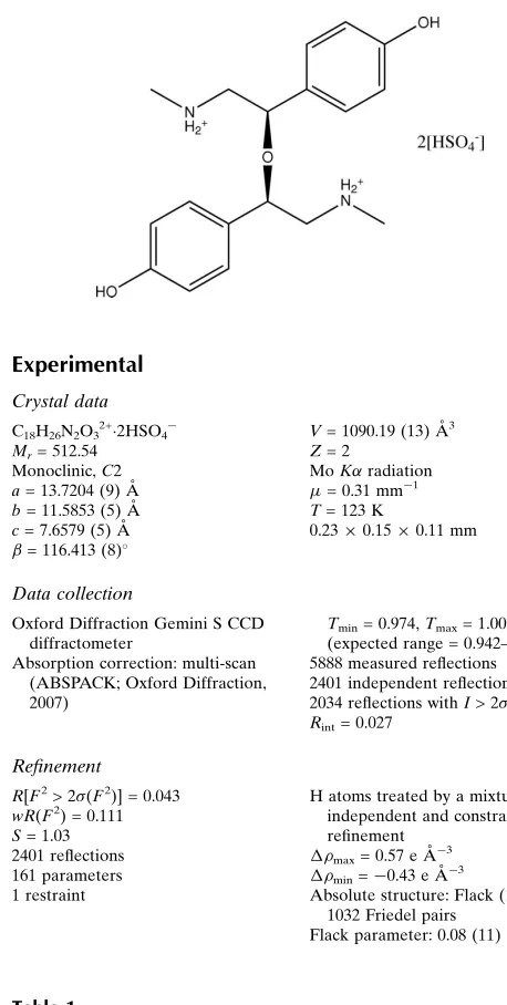
Comment
Synephrine (systematic name 2-hydroxy-2-(4-hydroxyphenyl)-
N
-methylethanamine) is a member of the phenylethylamine
drug family and it is often found in products marketed as "traditional medicines" or "weight-loss" pills (Blanck
et al.
, 2007;
Haller
et al.
, 2008). An attempt to prepare the sulfate salt of a racemic sample gave instead only the title compound,
di-syn-ephrine ether di-hydrogen sulfate, presumably
via
a variation of the well known S
N2condensation reaction (Mukhopadhyay
& Dattagupta, 1988; Bruice, 2007).
The salt crystallizes in space group
C
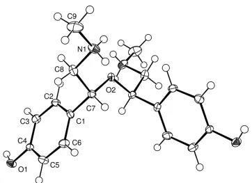
2, with half a cation and one hydrogen sulfate anion in the asymmetric unit. The
twofold rotation axis passes through O2, the etheric O atom (Fig. 1). The (
R
,
R
) conformation was assigned after refinement
of the Flack parameter (0.08 (11)). The bulk sample is presumably thus a conglomerate (Jacques
et al.
, 1981). Similar
symmetry is seen in the molecular structure of the analogous bromide salt (Mukhopadhyay & Dattagupta, 1988). Despite the
gross structural similarities imposed by the identical symmetries, the two salts do have somewhat different conformations.
This is best illustrated by the C8—C7—C7*—C8* torsion angle (-28.7 (3) ° here and -11.3 ° in the Br salt). Disorder effects
the H atom position in the [HSO4] anion, with equally occupied proton sites (50:50) associated with both O5 and O6.
Hydrogen bonds (Table 1) from the cation's hydroxy and amine H atoms to two [HSO
4] anions link the three ions (one
cation and two anions) in a ring motif (Fig. 2). Further anion to anion hydrogen bonded interactions give columns of [HSO
4]
lying along the
c
direction and complete a hydrogen bonded network in the
ac
plane (Fig. 3). A final, weaker cation to cation
interaction between the NH2 and –OH links these planes in the
b
direction.
Experimental
The title compound was obtained on treating an aqueous solution of (+/-)synephrine with dilute sulfuric acid. Single-crystals
were obtained by allowing the solvent of the reaction mixture to evaporate at 295 K.
1H NMR (DMSO-d6) 9.67 (2
H
, s
br, OH); 8.40 (4
H
, s br, NH
2); 7.07 (4
H
, d,
sp
2CH); 6.80 (4
H
, d,
sp
2CH); 4.20 (2
H
, dd, OCH); 3.32 (2
H
, m, CH
2); 2.99
(2
H
, m, CH2); 2.57 (6
H
, t, Me).
Refinement
supplementary materials
sup-2
Figures
Fig. 1. The molecular structure and atomic labelling of the cation, showing 50% probability
displacement ellipsoids. H atoms are shown as spheres of arbitrary radius.
[image:5.610.56.552.331.725.2]Fig. 2. The hydrogen-bonded ring motif between one cation and two anions in the structure of
[C18H26N2O3][SO4H]2.
Fig. 3. Packed structure viewed down the
c
-axis. S-atoms are pink and O-atoms are red.
Hy-drogen-bonding is shown as dashed lines.
(
R
,
R
)-2,4-bis(4-hydroxyphenyl)-
N
,
N
'-dimethyl- 3-oxapentane-1,5-diammonium bis(hydrogen sulfate)
Crystal data
C18H26N2O32+·2HSO4– F000 = 540
Mr = 512.54 Dx = 1.561 Mg m−3
Monoclinic, C2 Mo Kα radiation, λ = 0.71073 Å Hall symbol: C 2y Cell parameters from 3122 reflections a = 13.7204 (9) Å θ = 2.4–29.1º
b = 11.5853 (5) Å µ = 0.31 mm−1
c = 7.6579 (5) Å T = 123 K β = 116.413 (8)º Prism, colourless V = 1090.19 (13) Å3 0.23 × 0.15 × 0.11 mm Z = 2
Data collection
Oxford Diffraction Gemini S CCD
diffractometer 2401 independent reflections Radiation source: fine-focus sealed tube 2034 reflections with I > 2σ(I) Monochromator: graphite Rint = 0.027
T = 123 K θmax = 28.0º ω scans θmin = 2.4º Absorption correction: multi-scan
Refinement
Refinement on F2 Hydrogen site location: inferred from neighbouring sites
Least-squares matrix: full H atoms treated by a mixture ofindependent and constrained refinement
R[F2 > 2σ(F2)] = 0.043 w = 1/[σ 2(F
o2) + (0.071P)2] where P = (Fo2 + 2Fc2)/3
wR(F2) = 0.111 (Δ/σ)max < 0.001
S = 1.03 Δρmax = 0.57 e Å−3 2401 reflections Δρmin = −0.43 e Å−3 161 parameters Extinction correction: none
1 restraint Absolute structure: Flack (1983), 1032 Friedel pairs Primary atom site location: structure-invariant direct
methods Flack parameter: 0.08 (11) Secondary atom site location: difference Fourier map
Special details
Geometry. All e.s.d.'s (except the e.s.d. in the dihedral angle between two l.s. planes) are estimated using the full covariance mat-rix. The cell e.s.d.'s are taken into account individually in the estimation of e.s.d.'s in distances, angles and torsion angles; correlations between e.s.d.'s in cell parameters are only used when they are defined by crystal symmetry. An approximate (isotropic) treatment of cell e.s.d.'s is used for estimating e.s.d.'s involving l.s. planes.
Refinement. Refinement of F2 against ALL reflections. The weighted R-factor wR and goodness of fit S are based on F2, convention-al R-factors R are based on F, with F set to zero for negative F2. The threshold expression of F2 > σ(F2) is used only for calculating R -factors(gt) etc. and is not relevant to the choice of reflections for refinement. R-factors based on F2 are statistically about twice as large as those based on F, and R- factors based on ALL data will be even larger.
Fractional atomic coordinates and isotropic or equivalent isotropic displacement parameters (Å
2)
x y z Uiso*/Ueq Occ. (<1) S1 0.08291 (6) 0.20896 (7) 0.33257 (10) 0.0289 (2)
O1 0.17446 (14) 0.40319 (17) −0.2357 (3) 0.0181 (4) H1 0.1185 0.3616 −0.2796 0.027* O2 0.5000 0.1062 (2) 0.5000 0.0128 (5) O3 0.02688 (19) 0.31541 (19) 0.3192 (4) 0.0304 (6) O4 0.19286 (16) 0.2108 (3) 0.4864 (3) 0.0395 (6) O5 0.0193 (2) 0.1104 (2) 0.3550 (4) 0.0456 (7)
H1S 0.0204 0.1255 0.4724 0.068* 0.50
O6 0.0896 (2) 0.1834 (3) 0.1477 (4) 0.0519 (8)
H2S 0.0295 0.2160 0.0149 0.078* 0.50
N1 0.6712 (2) 0.0279 (2) 0.4186 (4) 0.0191 (5) C1 0.4315 (2) 0.2368 (2) 0.2158 (3) 0.0135 (6) C2 0.3329 (2) 0.1795 (2) 0.1126 (4) 0.0156 (6)
H2 0.3247 0.1025 0.1470 0.019*
supplementary materials
sup-4
H3 0.1807 0.1927 −0.1078 0.020* C4 0.2581 (2) 0.3458 (2) −0.0891 (4) 0.0139 (5) C5 0.3554 (2) 0.4041 (3) 0.0124 (4) 0.0182 (6) H5 0.3633 0.4812 −0.0217 0.022* C6 0.4414 (2) 0.3491 (2) 0.1646 (4) 0.0173 (6)
H6 0.5079 0.3893 0.2346 0.021*
C7 0.5277 (2) 0.1742 (2) 0.3714 (4) 0.0136 (5)
H7 0.5851 0.2315 0.4495 0.016*
C8 0.5734 (2) 0.0900 (3) 0.2748 (4) 0.0172 (6) H8A 0.5165 0.0329 0.1989 0.021* H8B 0.5928 0.1326 0.1827 0.021* C9 0.7267 (3) −0.0381 (3) 0.3240 (5) 0.0302 (7) H9A 0.7837 −0.0864 0.4212 0.045* H9B 0.7595 0.0155 0.2662 0.045* H9C 0.6738 −0.0873 0.2216 0.045* H1N 0.711 (2) 0.082 (3) 0.482 (4) 0.013 (8)* H2N 0.648 (3) −0.011 (3) 0.480 (4) 0.012 (8)*
Atomic displacement parameters (Å
2)
U11 U22 U33 U12 U13 U23
S1 0.0228 (4) 0.0289 (4) 0.0312 (4) 0.0004 (4) 0.0086 (3) −0.0031 (4) O1 0.0142 (9) 0.0149 (9) 0.0191 (10) 0.0002 (8) 0.0019 (8) 0.0039 (8) O2 0.0139 (12) 0.0143 (13) 0.0101 (11) 0.000 0.0053 (10) 0.000 O3 0.0196 (11) 0.0272 (12) 0.0420 (13) 0.0024 (9) 0.0117 (10) −0.0016 (10) O4 0.0275 (11) 0.0363 (12) 0.0388 (12) 0.0082 (14) 0.0005 (9) 0.0003 (14) O5 0.0485 (16) 0.0330 (14) 0.0552 (17) −0.0104 (13) 0.0231 (14) −0.0060 (13) O6 0.0427 (15) 0.072 (2) 0.0398 (14) 0.0081 (15) 0.0172 (13) −0.0090 (13) N1 0.0160 (12) 0.0187 (13) 0.0244 (12) 0.0036 (10) 0.0106 (11) 0.0065 (11) C1 0.0139 (13) 0.0157 (15) 0.0108 (11) 0.0032 (9) 0.0054 (10) 0.0027 (9) C2 0.0190 (13) 0.0106 (13) 0.0178 (12) −0.0004 (10) 0.0088 (11) 0.0009 (9) C3 0.0134 (13) 0.0172 (16) 0.0178 (12) −0.0026 (10) 0.0057 (10) −0.0019 (10) C4 0.0164 (13) 0.0118 (12) 0.0125 (12) 0.0040 (10) 0.0055 (11) 0.0010 (10) C5 0.0184 (14) 0.0138 (12) 0.0217 (14) 0.0003 (12) 0.0082 (12) 0.0044 (11) C6 0.0135 (13) 0.0140 (13) 0.0224 (13) −0.0030 (11) 0.0063 (11) −0.0010 (11) C7 0.0143 (12) 0.0120 (12) 0.0133 (12) −0.0016 (9) 0.0053 (10) 0.0011 (9) C8 0.0145 (13) 0.0239 (15) 0.0148 (12) 0.0049 (11) 0.0079 (10) 0.0031 (11) C9 0.0292 (18) 0.0236 (17) 0.047 (2) 0.0087 (13) 0.0251 (16) 0.0051 (14)
Geometric parameters (Å, °)
S1—O3 1.433 (2) C1—C7 1.513 (3)
S1—O4 1.443 (2) C2—C3 1.375 (4)
S1—O6 1.488 (3) C2—H2 0.9500
S1—O5 1.492 (3) C3—C4 1.392 (4)
O1—C4 1.368 (3) C3—H3 0.9500
O1—H1 0.8400 C4—C5 1.386 (4)
O2—C7i 1.438 (3) C5—H5 0.9500
O5—H1S 0.9095 C6—H6 0.9500
O6—H2S 1.0540 C7—C8 1.517 (4)
N1—C9 1.477 (4) C7—H7 1.0000
N1—C8 1.489 (3) C8—H8A 0.9900
N1—H1N 0.83 (3) C8—H8B 0.9900
N1—H2N 0.82 (3) C9—H9A 0.9800
C1—C6 1.383 (4) C9—H9B 0.9800
C1—C2 1.395 (4) C9—H9C 0.9800
O3—S1—O4 112.25 (15) O1—C4—C3 122.1 (2) O3—S1—O6 111.20 (16) C5—C4—C3 119.8 (2) O4—S1—O6 107.16 (15) C4—C5—C6 119.6 (3)
O3—S1—O5 110.11 (14) C4—C5—H5 120.2
O4—S1—O5 111.70 (16) C6—C5—H5 120.2
O6—S1—O5 104.10 (17) C1—C6—C5 121.0 (2)
C4—O1—H1 109.5 C1—C6—H6 119.5
C7—O2—C7i 113.6 (3) C5—C6—H6 119.5
S1—O5—H1S 102.3 O2—C7—C1 113.45 (19)
S1—O6—H2S 119.6 O2—C7—C8 105.9 (2)
C9—N1—C8 112.3 (2) C1—C7—C8 109.2 (2)
C9—N1—H1N 109 (2) O2—C7—H7 109.4
C8—N1—H1N 102 (2) C1—C7—H7 109.4
C9—N1—H2N 114 (2) C8—C7—H7 109.4
C8—N1—H2N 104 (2) N1—C8—C7 112.5 (2)
H1N—N1—H2N 114 (3) N1—C8—H8A 109.1
C6—C1—C2 118.7 (2) C7—C8—H8A 109.1
C6—C1—C7 120.9 (2) N1—C8—H8B 109.1
C2—C1—C7 120.3 (2) C7—C8—H8B 109.1
C3—C2—C1 120.8 (2) H8A—C8—H8B 107.8
C3—C2—H2 119.6 N1—C9—H9A 109.5
C1—C2—H2 119.6 N1—C9—H9B 109.5
C2—C3—C4 120.1 (2) H9A—C9—H9B 109.5
C2—C3—H3 120.0 N1—C9—H9C 109.5
C4—C3—H3 120.0 H9A—C9—H9C 109.5
O1—C4—C5 118.1 (2) H9B—C9—H9C 109.5
C6—C1—C2—C3 0.6 (4) C7i—O2—C7—C1 −70.07 (17) C7—C1—C2—C3 −175.4 (2) C7i—O2—C7—C8 170.1 (2) C1—C2—C3—C4 −0.4 (4) C6—C1—C7—O2 139.0 (2) C2—C3—C4—O1 −178.7 (2) C2—C1—C7—O2 −45.1 (3) C2—C3—C4—C5 0.2 (4) C6—C1—C7—C8 −103.1 (3) O1—C4—C5—C6 178.8 (2) C2—C1—C7—C8 72.8 (3) C3—C4—C5—C6 −0.1 (4) C9—N1—C8—C7 −169.9 (2) C2—C1—C6—C5 −0.5 (4) O2—C7—C8—N1 −59.6 (3) C7—C1—C6—C5 175.4 (2) C1—C7—C8—N1 177.9 (2) C4—C5—C6—C1 0.3 (4)
supplementary materials
sup-6
Hydrogen-bond geometry (Å, °)
D—H···A D—H H···A D···A D—H···A
O1—H1···O3ii 0.84 1.95 2.739 (3) 155